In Vitro Heme Coordination of a Dye-Decolorizing Peroxidase- The Interplay of Key Amino Acids, pH, Buffer and Glycerol
←
→
Page content transcription
If your browser does not render page correctly, please read the page content below
Article

In Vitro Heme Coordination of a Dye-Decolorizing Peroxidase—
The Interplay of Key Amino Acids, pH, Buffer and Glycerol
Kevin Nys 1,†, Vera Pfanzagl 2,†, Jeroen Roefs 1, Christian Obinger 2 and Sabine Van Doorslaer 1,*

                                           1 BIMEF Laboratory, Department of Chemistry, University of Antwerp, 2610 Antwerp, Belgium;
                                             kevin.nys@hotmail.com (K.N.); jeroen@roefs.be (J.R.)
                                           2 Division of Biochemistry, Department of Chemistry, BOKU—University of Natural Resources and Life

                                             Sciences, 1190 Vienna, Austria; vera.pfanzagl@boku.ac.at (V.P.); Christian.obinger@boku.ac.at (C.O.)
                                           * Correspondence: sabine.vandoorslaer@uantwerpen.be; Tel.: +32-3-265-2461
                                           † These authors contributed equally to this work.

                                           Abstract: Dye-decolorizing peroxidases (DyPs) have gained interest for their ability to oxidize an-
                                           thraquinone-derived dyes and lignin model compounds. Spectroscopic techniques, such as electron
                                           paramagnetic resonance and optical absorption spectroscopy, provide main tools to study how the
                                           enzymatic function is linked to the heme-pocket architecture, provided the experimental conditions
                                           are carefully chosen. Here, these techniques are used to investigate the effect of active site perturba-
                                           tions on the structure of ferric P-class DyP from Klebsiella pneumoniae (KpDyP) and three variants of
                                           the main distal residues (D143A, R232A and D143A/R232A). Arg-232 is found to be important for
                                           maintaining the heme distal architecture and essential to facilitate an alkaline transition. The latter
                                           is promoted in absence of Asp-143. Furthermore, the non-innocent effect of the buffer choice and
                                           addition of the cryoprotectant glycerol is shown. However, while unavoidable or indiscriminate
                                           experimental conditions are pitfalls, careful comparison of the effects of different exogenous mole-
Citation: Nys, K.; Pfanzagl, V.; Roefs,    cules on the electronic structure and spin state of the heme iron contains information about the
J.; Obinger, C.; Van Doorslaer, S.         inherent flexibility of the heme pocket. The interplay between structural flexibility, key amino acids,
In Vitro Heme Coordination of a            pH, temperature, buffer and glycerol during in vitro spectroscopic studies is discussed with respect
Dye-Decolorizing Peroxidase—The            to the poor peroxidase activity of bacterial P-class DyPs.
Interplay of Key Amino Acids, pH,
Buffer and Glycerol. Int. J. Mol. Sci.     Keywords: heme peroxidases; electron paramagnetic resonance; active site structure; UV-vis spec-
2021, 22, 9849. https://doi.org/10.3390/
                                           troscopy; alkaline transition; ligand binding; glassing agents
ijms22189849

Academic Editor: Yasushi Sugano

Received: 30 June 2021
                                           1. Introduction
Accepted: 3 September 2021                       Heme-containing proteins are widespread among all kingdoms of life. Their function
Published: 12 September 2021               is largely determined by the folding architecture of the peptide chain, modulating the
                                           possible redox properties of the heme pocket as well as its solvent and substrate accessi-
Publisher’s Note: MDPI stays neu-          bility [1,2]. Perhaps not surprisingly, this results in an intriguingly high level of functional
tral with regard to jurisdictional         versatility. It has become a major goal in heme-protein research to unravel how the enzy-
claims in published maps and institu-
                                           matic functions are governed by the local environment [2]. To achieve this, a wide variety
tional affiliations.
                                           of complementary techniques has been developed for structural and electronic character-
                                           ization [3]. While X-ray crystallography or NMR spectroscopy provide insight into the
                                           protein structure, electron paramagnetic resonance (EPR) allows probing (transient) par-
                                           amagnetic intermediates of the active site, thus exploring key mechanistic steps in the
Copyright: © 2021 by the authors. Li-
                                           enzymatic cycle or protein function [4,5]. When performing EPR spectroscopy on heme
censee MDPI, Basel, Switzerland.
                                           proteins, one should, however, keep in mind that external factors possibly influence the
This article is an open access article
distributed under the terms and con-
                                           observations of the active site and thus induce illegitimate conclusions. In this context,
ditions of the Creative Commons At-        awareness was raised by Svistunenko et al. in a low-temperature continuous-wave (CW)
tribution (CC BY) license (http://crea-    EPR study on the ferric heme forms of Mycobacterium tuberculosis catalase-peroxidase
tivecommons.org/licenses/by/4.0/).         (MtKatG) [6]. It was shown that not only pH, but also buffer type could alter the electronic

Int. J. Mol. Sci. 2021, 22, 9849. https://doi.org/10.3390/ijms22189849                                                    www.mdpi.com/journal/ijms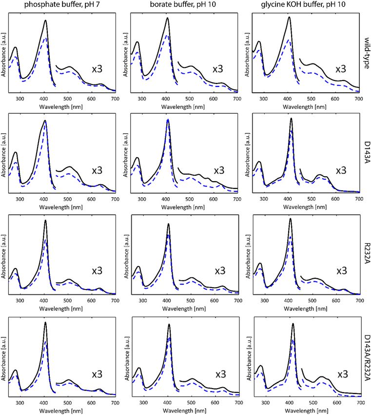
Int. J. Mol. Sci. 2021, 22, 9849                                                                                         2 of 19

                                   architecture of the heme cavity. Earlier studies report on freezing-induced distortions of
                                   the heme cavity and stabilizing effects of commonly used glassing agents, such as glycerol
                                   [7–10]. Here, we use multi-frequency EPR to understand pH-dependent changes in the
                                   heme pocket of KpDyP, a dye-decolorizing peroxidase from the human pathogen Klebsiella
                                   pneumoniae.
                                         Dye-decolorizing peroxidases or DyPs are hydrogen-peroxide dependent oxidore-
                                   ductases of predominantly bacterial origin, which are further classified in three distinct
                                   classes (P, I and V with KpDyP belonging to the P-class DyPs (formerly B-class)) [11]. Bac-
                                   terial P-class DyPs are very poor peroxidases with unknown biological function. Hydro-
                                   gen peroxide efficiently mediates the rapid formation of Compound I, which is remarka-
                                   bly stable and shows only modest reactivity towards organic and inorganic electron do-
                                   nors [12,13]. KpDyP has been biochemically [12] and structurally [13,14] well character-
                                   ized and serves as a good model for this protein family, which has gained significant in-
                                   terest in recent years in regard to their biotechnological potential [15]. The conserved core
                                   fold of DyPs (four beta sheets connected by alpha helices) classifies them within the di-
                                   meric α + β-structural superfamily together with the phylogenetically related enzymes
                                   chlorite dismutases (Clds) and coproheme decarboxylases (ChdCs) [16–18]. All three pro-
                                   tein families have a characteristic loop, which forms the outer wall of the active site and
                                   dictates accessibility and shape of the cavity [12,13,19–21]. In DyPs this loop furthermore
                                   contains one of the main catalytic residues of the distal side, Asp-143 in KpDyP, which
                                   was shown to be required for heterolytic cleavage of hydrogen peroxide [12]. The equally
                                   conserved distal arginine, Arg-232 in KpDyP, is highly important to maintain the integrity
                                   of the active site. The crystal structure of wild-type (WT) KpDyP in Figure 1 shows the
                                   extensive hydrogen bonding network connecting Arg-232 and Asp-143 with propionate p6
                                   and active site waters. Limited access to the heme iron through the main channel (Figure 1,
                                   green) whose bottleneck is formed by the catalytic Asp-Arg pair together with Phe-248
                                   and Leu-246 was proposed to be the main reason for the observed low catalytic activity of
                                   this enzyme [12]. This channel is proposed to be the main entry route for hydrogen per-
                                   oxide required for Compound I formation. Previous studies showed that the overall struc-
                                   ture of the active site is maintained in a D143A variant but that this mutation increases the
                                   bottleneck radius [12,13]. Exchange of Arg-232 for alanine (R232A); however, has severe
                                   structural consequences, leading to a rearrangement of the loop and collapse of the active
                                   site. This effectively removes the main heme iron access route as seen in the crystal struc-
                                   ture and molecular dynamics simulations. Taking the crystal structure of the
                                   D143A/R232A double variant into account, where the WT-like loop conformation is solely
                                   due to a coordinating glycerol molecule, it is reasonable to expect an increased structural
                                   susceptibility of these mutants to external factors, such as temperature and buffer compo-
                                   nents [12].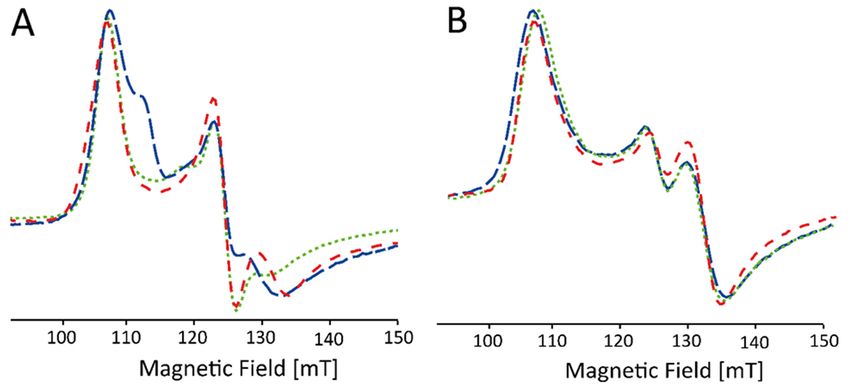
Int. J. Mol. Sci. 2021, 22, 9849                                                                                                 3 of 19

                                   Figure 1. Overall crystal structure of the P-class dye-decolorizing peroxidase of wild-type Klebsiella
                                   pneumoniae (KpDyP) and active site structures of WT KpDyP (PDB entry 6FKS) and three variants:
                                   D143A (6FL2), R232A (6FKT) and D143A/R232A (6FIY). (A) Cartoon representation of the crystal
                                   structure of dimeric WT KpDyP. (B) Active site structure of WT KpDyP displaying key amino acids
                                   (H215, D143, R232, L246 and F248) of the heme cavity, as well as a glycerol molecule (designated G)
                                   and possible hydrogen bonds between water molecules (green). (C) Active site structures of WT
                                   KpDyP and three variants, displaying the heme access channels, as determined by CAVER 3.0 [22].

                                         In order to further investigate the role of the conserved Asp-143/Arg-232 pair in the
                                   restriction of the substrate accessibility and ligand binding to the heme iron, we used con-
                                   tinuous-wave (CW) and pulsed EPR to explore the active site of WT KpDyP and D143A,
                                   R232A and D143A/R232A variants in different buffers at neutral and high alkaline pH, as
                                   ligand binding is often enhanced in the alkaline region due to deprotonation. The buffers
                                   were chosen based on their widespread use and potential to influence the heme active
                                   site. We purposefully omitted the acidic pH range as no specific structural changes are
                                   expected other than pH induced unfolding starting from pH 5 [12]. We show the im-
                                   portance of the conserved distal arginine for accommodating a hydroxo ligand at alkaline
                                   pH. Furthermore, our results challenge the non-innocent role of glycerol and buffer mol-
                                   ecules on heme proteins with an accessible active site. While the glassing agent glycerol is
                                   commonly added to prevent freezing artefacts at the low temperatures required for the
                                   EPR experiment [23], it inhibits certain ligation states in the KpDyP variants under study
Int. J. Mol. Sci. 2021, 22, 9849 4 of 19
and with related spectral changes in the EPR and optical absorption spectra. Moreover,
we highlight a strong effect of the type of buffer on the KpDyP variants. An alkaline tran-
sition is not consistently maintained for different buffers and use of a glycine-containing
buffer at high pH induced ligation of the ferric heme iron with glycine. Although slight
effects of buffer molecules on the EPR spectra of heme proteins have been reported before
[6,24], this is the first time that glycine binding to heme iron is clearly identified at low
temperatures (EPR) and room temperature (optical absorption). We discuss how the dif-
fering ligand accessibility in WT KpDyP and its variants links to the proposed reaction
mechanism of the wild-type protein.
2. Results
2.1. Optical Absorption Spectroscopy
Figure 2 (solid lines) depicts the optical absorption spectra of WT KpDyP and the
D143A, R232A and D143A/R232A variants in phosphate buffer (pH 7.0), borate buffer (pH
10.0) and glycine-KOH buffer (pH 10.0) taken at room temperature. The phosphate buffer
was chosen in accordance with previous studies on WT KpDyP [12]. As reported earlier
[12], at pH 7 WT KpDyP exhibits a Soret peak at 406 nm with a broad shoulder at the lower
wavelength side, Q-bands at 508 and 540 nm and a charge transfer (CT) band at 641 nm,
in accordance with a high-spin (HS) Fe(III) state of the heme iron. The same spectrum is
found for the D143A variant, with a narrower Soret peak at 406 nm and slightly blue-
shifted CT band (630 nm) for the R232A and D143A/R232A variants. While the latter spec-
tral features agree with an S = 5/2 Fe(III) state, the absorption spectra of WT KpDyP and
D143A may indicate some degree of quantum mixing of the S = 5/2 state with an interme-
diate S = 3/2 state [25]. Addition of 25% glycerol (Figure 2, dashed lines) induces a nar-
rowing of the Soret peak with concomitant shift of the CT peak for the D143A variant,
while it does not affect the absorption spectra of the other variants (Figure 2).
In borate buffer (pH 10) without glycerol, WT, R232A and D143A/R232A KpDyP ex-
hibit the same optical absorption spectra as at pH 7, but a marked change is observed for
the D143A variant indicating the presence of HS and low-spin (LS, S = 1/2) ferric heme
states. LS ferric states occur when a strong base, such as a hydroxo-anion or imidazole, is
ligating the heme iron. Previous work has shown that an alkaline transition to a hydroxo-
ligated heme species takes place for this variant, with the absorption spectrum of this spe-
cies exhibiting a Soret peak at 410 nm and additional bands at 540, 576 and 610 nm [12].
The presence of this LS species is inhibited by the addition of glycerol.
The non-innocent effect of the buffer is noticed when a glycine-KOH buffer is used
(Figure 2). While WT and R232A KpDyP maintain the HS-state, a significant change in the
optical absorption spectrum is noticed for the variants where Asp-143 is exchanged for an
alanine. Here, a new LS state is observed with D143A showing a red-shifted Soret peak at
412 nm and Q-bands at 532 and 561 nm (double variant: 413, 532 and 562 nm). The CT
peak is no longer visible demonstrating the absence of a HS state. Addition of glycerol
does not alter the UV-visible spectral signatures in this case.Int. J. Mol. Sci. 2021, 22, 9849 5 of 19
Figure 2. Optical absorption spectra of WT KpDyP and variants in three different buffer systems
without (black, full) and with glycerol (blue, dashed).
2.2. Influence of Buffer and Glycerol on CW EPR Spectra
The optical absorption analysis reveals a worrying non-innocent effect of glycerol
and the type of buffer molecules on the heme ligation in the KpDyP variants under study.
To understand this further and to relate this to the protein’s heme-pocket architecture and
behavior, we performed X-band continuous-wave (CW) EPR. Moreover, glycerol is com-
monly used in EPR as a cryoprotectant and is in some cases essential to avoid strong di-
polar interactions between the paramagnetic centers [23].
Figure 3A shows the X-band CW EPR spectra of WT KpDyP and variants in phos-
phate buffer (pH 7.0) without glycerol. Figure 3B shows the comparative results with glyc-
erol in accordance with our earlier reported data [12]. It is clear that the site-directed mu-
tations as well as the cryoprotectant cause changes to the electronic structure of the active
site that go beyond the minor modifications that are expected because of the change in
dielectric constant. As can be derived from the EPR parameters given in Table S1 (Supple-
mentary Material), differences include a variation in abundancy of specific species, a
change in rhombicity of the zero-field tensor (E/D) and the appearance of extra HS species.
The double variant exhibits an even more striking change, since it reveals formation of an
LS species (~24%) that disappears with the addition of glycerol (see inset Figure 3A, LS1Int. J. Mol. Sci. 2021, 22, 9849 6 of 19
in Table S1). The origin of this contribution is unknown so far and cannot be deduced
solely from the g-values. However, this LS species was not observed in the optical absorp-
tion spectra (Figure 2) and is thus due to a ligation induced by the low temperatures. The
appearance of HS species with E/D values ≥ 0.017 seems to agree with the observation of
a broader Soret band and higher wavelength and broadening of the CT band in the optical
absorption spectra (Figure 2). This correlation is not unexpected, since the increase of the
E/D parameter and the concomitant decrease of =( + )/2 has been linked to
an increased admixture of the S = 5/2 and S = 3/2 states. According to the concepts outlined
by Maltempo [26], the g12 values associated with the largest E/D values found here (g12 =
5.83 to 5.685) can be related to 10–15% of the S = 3/2 state.
Figure 3. Experimental (black) and simulated (red) X-band CW EPR spectra of frozen solutions of
WT KpDyP and variants (c ≈ 500 µM) in phosphate buffer (pH 7.0) without (A) and with (B) glycerol.
EPR simulation parameters can be found in Table S1 and the different contributions are displayed
in Figure S1 (Supplementary material). An asterisk (*) indicates the position of a stable organic
radical identified earlier in the resting-state of the protein [12]. The spectra are shown normalized
to allow a facile comparison. An inset in (A) highlights the presence of a LS1 species in the double
variant (5× amplified).
Furthermore, without addition of glycerol, the spectral shape of the EPR signal of the HS
components in WT KpDyP is found to be strongly batch-dependent (Figure 4A). Only after
addition of the glassing agent, all batches exhibit the same spectral signature (Figure 4B).
Note that the stable organic radical, observed earlier in the resting state of WT KpDyP
(Figure 3, asterisk [12]), is not affected by the addition of glycerol.
At alkaline pH, several heme proteins are known to display a distal coordination of
a hydroxo ligand [27–29]. For KpDyP variants, it appears this is only the case under spe-
cific conditions as was illustrated by the optical absorption spectroscopy (Figure 2). The
CW EPR spectra of KpDyP and variants in borate buffer at pH 10.0 (Figure 5) mostly agree
with the optical absorption data. In general, the HS Fe(III) state is maintained in all frozen
solutions both with and without glycerol, although the specific type and weight of the HS
contributions again depends on whether glycerol is added or not (Figure 5, Table S2, Fig-
ure S2, Supplementary Material). Moreover, the change of buffer and pH influences the
electronic structure of the paramagnetic center. A careful inspection of the EPR parame-
ters obtained from the spectra in Figure 5 (Table S2) and Figure 3 (Table S1) reveals that a
more alkaline pH provokes a decrease in abundancy of the HS components with higherInt. J. Mol. Sci. 2021, 22, 9849 7 of 19
rhombicity (E/D ≥ 0.017). This is in line with optical absorption spectra, where a broadened
Soret band is only found for WT KpDyP (Figure 2).
Figure 4. Low-field part of X-band CW EPR spectra of frozen solutions of WT KpDyP (c ≈ 500 µM)
in phosphate buffer (pH 7.0). The different traces (indicated by different colors) represent different
batches before (A) and after (B) the addition of glycerol. The spectra are shown normalized to allow
facile comparison.
Figure 5. Experimental (black) and simulated (red) X-band CW EPR spectra of frozen solutions of
WT KpDyP and variants in borate buffer (pH 10.0) without (A) and with (B) glycerol. EPR simulation
parameters can be found in Table S2 and the different contributions are displayed in Figure S2
(Supplementary Material). An asterisk (*) indicates the position of a stable organic radical identified
earlier in the resting-state of the protein [12]. The spectra are shown normalized to allow facile
comparison. The position of the gz-component of the OH1’ (dashed-dotted), OH2/OH2’ (dashed)
and the OH3’ (dotted) species is indicated, Figure S3 (supplementary material) shows an
enlargement of the spectra in this magnetic field area to allow visual control of the presence of these
species.
Figure S3 (Supplementary Material) shows an enlargement of the EPR spectra of Figure
5 in the (180–400)mT range, revealing the additional presence of contributions of different
LS species. For both WT KpDyP and the D143A variant (without glycerol, Figure S3A) we
find contributions of hydroxo-ligated ferric heme species. In the former this only accounts
for 4% of all the ferric heme contributions, but the EPR spectrum of D143A displays threeInt. J. Mol. Sci. 2021, 22, 9849 8 of 19
different contributions of hydroxo-ligated heme species that together account for ~30%
(Table S2). This agrees with the contribution of a hydroxo-ligated ferric form in the optical
absorption spectrum of D143A (Figure 2). The g-values of the different hydroxo-ligated
heme species are summarized in Table 1. While the g-tensor of species OH1’ is very similar
to the one found for horseradish peroxidase (HRP) [30], the parameters of OH2/OH2’ and
OH3’ are more common for alkaline forms of cytochrome c peroxidase and mammalian
myoglobin [24,29,31]. A high diversity of hydroxo-ligated species was found in hemoglo-
bin of Thermobifida fusca [32] and the globin domain of the GLB-33 globin of C. elegans [33].
The Arg-232 variants (both single and double variant) do not exhibit hydroxo-ligated spe-
cies. However, all but the D143A variant show a contribution (labeled LS2) with g-values
ascribed to binding of a glutathione or a buffer molecule in other heme proteins [34,35].
In addition, the EPR spectrum of the double variant reveals the same LS form of unknown
nature (LS1) observed at pH 7. Addition of glycerol removes all LS contributions, except
for the LS2 form in the R232A variant. Addition of 30% glycerol reduces the pH of borate
buffer to pH 7 [36], where all variants are predominantly HS. The LS2 form, retained in
R232A, is thus likely a buffer molecule, as it is not observed in phosphate buffer.
Table 1. Experimental principal g-tensor values of the low spin species in frozen solutions from WT
KpDyP and the D143A variant in pH 10 borate buffer without glassing agent (experimental error ±
0.02 for gz and ± 0.05 for gx,y) and a representative selection of heme proteins with a distal hydroxo
ligation at alkaline pH. HRP, horseradish peroxidase; CcP, cytochrome c peroxidase; swMb, sperm
whale myoglobin; CeGLB-33: GLB-33 from C. elegans; TfHb, hemoglobin from Thermobifida fusca.
gz gy gx Ref.
WT KpDyP
OH2 2.77 2.17 1.77 this work
D143A KpDyP
OH1’ 2.89 2.12 1.78 this work
OH2’ 2.728 2.15 1.772 this work
OH3’ 2.67 2.214 1.82 this work
HRP 2.94 2.08 1.63 [30]
CcP 2.74 2.22 1.74 [31]
swMb 2.55 2.17 1.85 [29]
2.62 2.20 1.815
CeGLB-33 [33]
2.845 2.12 1.69
2.73 2.19 1.76
TfHb 2.66 2.19 1.81 [32]
2.82 2.32 1.60
The optical absorption spectra (Figure 2) showed a drastic effect of the use of a gly-
cine-KOH buffer at pH 10. Both D134A and the double variant show maxima at 533 nm
and 574 nm, which are distinct from the absorption maxima typical for OH- ligation ob-
served for D134A in borate buffer (540, 574 and 610 nm). This is confirmed in the corre-
sponding EPR spectra (Figure 6). Without addition of glycerol, only the EPR spectrum of
the WT enzyme shows an appreciable contribution of HS ferric heme forms (~27%) (Figure
6A). The majority of the EPR signal of WT KpDyP stems from a LS Fe(III) species with
maximum g-value 3.27 (species Gly2 in Table S3, supplementary material, Figure S5). Sim-
ilar contributions are also present in the R232A and D143A variants (Figure 6A, Table S3).
In addition, LS species with gz ≈ 3.11–3.14 and gy ≈ 2.09–2.11 are observed in all single
variants, which we indicate as Gly1-type signals (Table S3). For the double variant, the
contribution of a single LS species with g-values in between those of Gly1 and Gly2 is
observed (Gly2’ in Table S3). Figure S5 (Supplementary Material) shows a detailed com-
parison of the LS species.Int. J. Mol. Sci. 2021, 22, 9849 9 of 19
Figure 6. Experimental (black) and simulated (red) X-band CW EPR spectra of frozen solutions of
WT KpDyP and variants in glycine-KOH buffer (pH 10.0) without (A) and with (B) glycerol. EPR
simulation parameters can be found in Table S3 and the different contributions are displayed in
Figure S4 (Supplementary Material). An asterisk (*) indicates the position of a stable organic radical
identified earlier in the resting-state of the protein [12] and the gz-component of the Gly1 (dashed-
dotted) and Gly2 (dashed) species is indicated as well. The spectra are shown normalized to allow
facile comparison. An inset in (B) highlights the presence of a Gly2 species in the R232A variant (5×
amplified).
Upon addition of glycerol, the Gly2 contribution disappears fully from the EPR spec-
trum of WT KpDyP and the HS ferric heme forms again dominate the spectrum (Figure
6B and S4, Table S3). Similarly, the EPR spectrum of R232A KpDyP is governed by HS
forms, while only the Gly2 LS contribution remains present in the EPR spectrum (Table
S3). The D143A variant exhibits a less appreciable influence of glycerol addition with both
LS features only shifting in relative amount and no appearance of a HS contribution (Table
S3). Finally, the EPR spectrum of the double variant remains essentially the same upon
addition of the cryoprotectant, with small shifts in the g-values that can be attributed to
the change in the dielectric constant. The g-values point to a Gly2-type, rather than Gly1-
type contribution (Table S3). Note that the optical absorption spectra revealed a dominant
HS form for WT and R232A KpDyP in this buffer, while for the variants in which the Asp-
143 is mutated only the LS heme form is detected independently of the presence of glyc-
erol (Figure 2). This highlights that the glycerol-dependent LS↔HS conversion observed
by EPR for WT and R232A KpDyP is to some extent also induced by the low temperatures.
W-band electron-spin-echo (ESE) detected EPR experiments were performed in an
attempt to facilitate the interpretation of the X-band CW EPR shown in Figure 6B (see
Supplementary Material, Figure S6). Although the experiments essentially confirmed the
above analysis of the X-band EPR data, only minor additional information could unfortu-
nately be obtained due to the low spin-echo intensity and the presence of contaminants.
2.3. Subtle Changes in the Heme Pocket Revealed by Pulsed EPR
While optical spectroscopy revealed that the heme of each described variant is in a
HS Fe(III) state at pH 7 (Figure 2), CW EPR spectroscopy revealed clear differences in the
g and zero-field parameters of these HS forms (Table S1, Figure 3), which can be related
to subtle changes in the electronic structure of the heme pocket. Mutation of the distal
Asp-143 and/or Arg-232 in KpDyP formation of HS species with lower rhombicity of theInt. J. Mol. Sci. 2021, 22, 9849 10 of 19
zero-field parameters (smaller E/D) is more favored, especially in the presence of glycerol
(Table S1).
Here, we will deploy X-band hyperfine sublevel correlation (HYSCORE) spectros-
copy to further evaluate the implications of the distal changes on the electronic structure
of the cofactor. This 2D pulsed EPR experiment enables the determination of hyperfine
and nuclear quadrupole values of the neighboring magnetic nuclei. The latter interaction
is present for nuclei with nuclear spin I > ½ such as 14N.
In HS ferric forms of globins, such as mammalian myoglobin, low E/D-values are
observed when a water molecule is ligating the heme iron on the distal side [4]. In order
to probe whether water ligation induces the observed reduction in the rhombicity of the
zero-field values in the KpDyP variants, 1H HYSCORE is measured of WT, D143A and
R232A KpDyP in glycerol-containing frozen solutions at pH 7 (Figure 7, bottom spectra).
If distal water ligation occurs, characteristic cross peaks are found in the 1H HYSCORE
spectra measured at the high-field magnetic-field position (gzeff) [37,38]. The region in
which these cross peaks are expected is indicated in the spectra in Figure 7. It is clear that
the HYSCORE spectra of all three KpDyP variants lack this feature and that distal water
ligation therefore does not occur in the three proteins under these conditions.
Figure 7. X-band HYSCORE of frozen solutions of WT, D143A and R232 KpDyP variants in phos-
phate buffer (pH 7.0) and 25% glycerol, taken at 4 K and B0 = 348 mT (magnetic field setting corre-
sponding with gzeff). The (-, +) quadrant (top panel) shows cross peaks stemming from the 14N heme
nuclei and the (+, +) quadrant in the (10–20, 10–20) MHz region (lower panel) indicates interaction
between the electron spin and nearby protons. The experimental spectrum (black) and the simula-
tion (red) in the top panel are obtained by averaging over four τ-values (96 ns, 104 ns, 114 ns and
128 ns). Essential simulation parameters are highlighted in Table 2. The red circles (lower panel)
indicate the position where the cross peaks due to distal water protons should appear in the case of
an aquomet form [37].
Figure 7 (top panel) shows the (−, +) quadrant of the HYSCORE spectra for WT,
D143A and R232A KpDyP variants taken at a magnetic-field setting corresponding to gzeff.
The observed cross peaks correspond to the interaction between the electron spin and the
14N heme nuclei, in accordance with observations for other HS heme proteins, such as
globins [37,39] and peroxidases [38] (Table 2). The interpretation of the individual cross
peaks in the HYSCORE spectra is given in the supplementary information (Figure S7).
Because of the orientation selection at this magnetic-field setting, the spectral simulationInt. J. Mol. Sci. 2021, 22, 9849 11 of 19
of the cross peaks only reveals the z-component (i.e., component along the heme normal)
of the hyperfine and quadrupole tensor of the porphyrin 14N nuclei (Table 2). While ferric
WT and D143A KpDyP show identical 14N hyperfine values, a marked change is observed
for the R232A variant.
A previous in-depth study of aquometmyoglobin showed that the cross peaks related
to the interaction of the electron spin with the Nε of the proximal histidine are usually
weak or suppressed in the standard HYSCORE spectra, as is also observed here. At other
magnetic-field settings, the matched 1H HYSCORE spectrum reveals the characteristic
cross-peaks due to the protons at the Cε and Cδ positions of the proximal His (Figure S8)
[40].
The (0–7, 0–7) MHz region of the HYSCORE spectra at g = gzeff reveals signals from
the interaction of the electron spin with weakly coupled 14N and 13C nuclei, the latter in
natural abundance (Figure S9) [37]. Again, marked shifts are found in these couplings
upon mutation of the distal Arg-232, while this is not the case for the mutation of the distal
Asp-143 (see supplementary material for more details).
Table 2. Comparison between the z-component of the hyperfine (experimental error ± 0.03 MHz)
and nuclear quadrupole values (experimental error ± 0.05 MHz) of the porphyrin 14N nuclei in WT,
D143A and R232A KpDyP variants and those of other HS heme proteins. The z-direction is along
the heme normal. n.r., not reported.
|Azeff| [MHz] |Qz| [MHz]
KpDyP (this work)
WT 6.90 0.25
14Nporf,1
14Nporf,2 6.70 0.28
D143A
14Nporf,1
6.90 0.25
14Nporf,2 6.70 0.28
R232A
14Nporf,1
7.07 0.32
14Nporf,2 6.95 0.28
metMb [39]
14Nporf,1
7.42 0.33
14Nporf,2 7.10 0.23
NGB [37]
14Nporf
7.25 0.23
Dehaloperoxidase [38]
14Nporf 7.50 n.r.
CcmE chaperone [41]
14Nporf
8.01 0.25
3. Discussion
3.1. Heme Cavity Heterogeneity and the Influence of Glycerol
At neutral pH, the heme in the homodimeric dye-decolorizing peroxidase of K. pneu-
moniae exhibits a high-spin Fe(III) state both at room temperature and in frozen solution.
Mutation of the catalytically important residues Asp-143 and Arg-232 to alanine or the
addition of a glassing agent, i.e., glycerol, preserves the oxidation and high-spin state.
While this observation may be derived from both optical absorption (Figure 2) and low-
temperature EPR spectroscopy (Figure 3), significant differences are observable in the
electronic architecture. WT KpDyP and variants display a heterogeneous heme pocket de-Int. J. Mol. Sci. 2021, 22, 9849 12 of 19
picted by the presence of contributions of different HS states in the EPR spectra, charac-
terized by a changing rhombicity of the zero-field splitting parameter (Figure 3). An in-
crease in this rhombicity (larger E/D-value), which translates in a splitting of the EPR fea-
ture at g ~ 6, reflects a departure from the tetragonal symmetry that is distinctive for the
porphyrin and that has been related to an increased admixture with the intermediate S =
3/2 state [25,26]. This lowering of the symmetry is governed by the physical environment,
i.e., the ligands of the heme and its incorporation in the protein matrix. The HS metal ion
is thus able to sense conformational changes of the protein moiety that impact the geom-
etry of the heme cavity [42,43].
For many HS ferric heme proteins, small E/D-values are related to the binding of a
water molecule at the distal site, as is known to be the case for aquometmyoglobin [3,39].
However, while the EPR spectra of both the Asp-143 and Arg-232 variants of KpDyP con-
tain species with small E/D-values (HS3, Table S1), water binding is contradicted by the
HYSCORE data (Figure 7). Our earlier study revealed that WT KpDyP and variants exhibit
a pronounced hydrogen-bonding network [12]. The reported presence of multiple water
molecules in the heme pocket [12], stabilized by the catalytically important residues and
a heme propionate (Figure 1), suggest that different arrangements in the distal heme area
are possible and may explain the heterogeneity in the heme cavity observed by EPR. Ear-
lier EPR studies already pointed out that heme peroxidases commonly display a certain
variability in their active site [6,10,42,44,45]. Moreover, measurements of in situ horserad-
ish peroxidase (HRP) showed that the HS Fe(III) signals changed their spectral shape upon
transfer from a test tube to an EPR tube [42]. And studies on yeast cytochrome c peroxi-
dase indicated that an identical preparation of the enzyme gave rise to EPR spectra with
a different ratio in LS and HS species [46]. A similar observation was made here with
different batches of WT KpDyP (Figure 4A). Cao et al. showed that the freezing and thaw-
ing rate in potassium phospate buffers influenced the denaturarion of different proteins
[47]. Both surface denaturation and pH shift due to precipitation of the buffer salt are
indicated as likely reasons of protein damage during freezing. Note, however, that the
observation of high fractions of HS heme species with E/D ≥ 0.017 by EPR at low temper-
ature for ferric WT and D143A KpDyP (Table S1) correlate with the observation of a broad-
ened Soret peak and red shift of the CT band at room temperature optical absorption spec-
tra (Figure 2), indicating that the observed EPR-spectral differences between the variants
is not merely an effect of the low temperature.
The variability in the electronic structure of the heme pocket observed by low-tem-
perature EPR can be prevented by the addition of glycerol (Figure 4B). It has been reported
before that the use of a glassing agent not only affects the glassing temperature of the
solvent but also limits the co-existence of different conformational sub-states of the pro-
tein [48]. Resonance Raman spectroscopy of ferric cytochrome c peroxidase revealed that
glycerol induces a stabilization of the protein structure, accompanied by a reduced inter-
action of the iron with its distal ligand [49]. Furthermore, glycerol-induced changes have
been reported in the EPR spectra of ferric CcmE heme chaperone [41]. In the crystal struc-
ture of ferrous as well as ferric KpDyP [12,14] a glycerol molecule was found to be coordi-
nated in the upper region (i.e., above the bottle neck) of the main access channel. Addi-
tionally, although KpDyP displays a penta-coordination both with and without glycerol,
it is highly likely that the glycerol molecule is coordinated in the active site channel and
thus influences the active site hydrogen bonding network, its dynamics and variability in
solution. Indeed, addition of glycerol forces the different heterogeneous HS Fe(III) states
in different batches into an identical set of sub-states (Figure 4B). Moreover, the crystal
structure of the D143A/R232A variant reveals the presence of a glycerol molecule in the
heme pocket [12]. In fact, crystals of this double variant could only be obtained with glyc-
erol as part of the crystallization solution and glycerol was believed to be essential to sta-
bilize the heme region in a WT-like manner in place of Arg-232 [12].Int. J. Mol. Sci. 2021, 22, 9849 13 of 19
3.2. The Non-Innocent Effect of Buffer Molecules
The comparison of the optical absorption and EPR spectra of WT KpDyP and variants
at pH 10 in two different buffers reveals the non-innocent effect of buffer molecules on the
electronic spin state of the heme iron. In a glycine-KOH buffer, the optical absorption
spectra show a full conversion to the low-spin state for D143A and D143A/R232A KpDyP
but not for WT KpDyP, while this is not observed in a borate buffer at the same pH (Figure
2). EPR reveals that this is due to ligation of a strong base to the heme iron (Figure 6A,
Table S3). The large gz values (≥3) of species Gly1 and Gly2 (Table S3) are typical of a
highly anisotropic LS heme species [50], and are in line with parameters observed for en-
dogenous and exogenous amine ligands [4]. The difference in EPR parameters between
Gly1, Gly2 and Gly2’ probably relate to a different orientation and stabilization of the
glycine molecule in the heme cavity. Although addition of glycerol slightly changes the
EPR parameters, in line with the earlier discussed ability of glycerol to enter the heme
access channel or heme pocket, glycine remains bound to the heme iron in the variants
involving exchange of Asp-143 for an alanine (Figure 6B, Table S3). At low temperature,
dominant ligation of glycine to the heme is also found for R232A KpDyP and to a minor
extent for WT KpDyP. This ligation is, however, far less resistant to glycerol addition (Fig-
ure 6B, Table S3). Earlier studies of other heme proteins already hinted that the buffer can
affect the electronic structure of the active site and even its catalytic activity [6,49]. Inter-
estingly the observed glycine ligation and the effect of glycerol addition seems to be
unique to Dye-decolorizing peroxidases, as it was not observed in an in-depth study on
myoglobin [36]. Moreover, the glycine ligation appears to be stronger in D143A/R232A
than in the R232A. As this cannot be solely due to accessibility of the active site, it suggests
that Asp-143 selectively blocks substrate binding.
3.3. Alkaline Transition in KpDyP Variants
From the above, it is clear that a well-considered choice of buffer is key to studying
alkaline transitions in dye-decolorizing peroxidases. Alkaline pH does not necessarily im-
ply ligation of a hydroxo ligand to the heme iron, especially if there is no aquomet state
present in neutral pH conditions. In borate buffer, an alkaline transition is observed for
D143A KpDyP, both at room temperature (Figure 2) and at 10 K (Figure 5A). Moreover,
WT KpDyP exhibits a small fraction of hydroxo-ligated species in frozen solution (Figure
5A, Table S2). If R232 is missing, no hydroxo-ligation can be observed at all, suggesting
that R232 is required to stabilize this ligand. It is possible that in the case of WT KpDyP
the hydroxo-ligated fraction is either too low at room temperature to be detected in the
optical spectrum or that the OH− ligation observed at low temperature originates from
packing forces on the heme induced by freezing as is the case for the alkaline state at neu-
tral pH in HRP isozyme A2 [28,30]. Moreover, the pH of buffer solutions is known to be
slightly changed upon freezing with the effect being buffer-dependent [51]. Interestingly,
no hydroxo ligation is displayed in the variants of KpDyP where Arg-232 is replaced by
an alanine. This agrees with the general observation that binding of OH- in heme peroxi-
dases is accommodated by a distal arginine forming a hydrogen bond [52–54]. The pres-
ence of hydrogen bonding can be deduced from the gz component that reflects the strength
of the stabilization [33]. The most dominant contribution in D143A KpDyP, OH1’, shows
g values very similar to the values found for HRP isozyme A2 and lignin peroxidase iso-
zyme H2 [27,28,30] and is indicative of strong hydrogen bonds between the hydroxo lig-
and and the distal Arg-232. On the other hand, there is a small contribution of OH3’ with
a significantly lower gmax in the range that is common for globins [29]. A third component
is observed in both the WT and D143A variant and appears to be a species where the
hydroxo ligand experiences a stabilization with intermediate strength.
Both optical absorption and EPR spectroscopy reveal no alkaline transitions in the
KpDyP variants in the presence of glycerol. It is reported, however, that the acidity of aInt. J. Mol. Sci. 2021, 22, 9849 14 of 19
boric acid solution is lowered upon addition of glycerol, possibly explaining the inhibition
of an alkaline transition [55,56].
Comparison of Tables S1 and S2 shows that the nature of the HS heme species is also
affected by the pH. In general, when the HS Fe(III) state is maintained in borate buffer,
the rhombicity of the co-existing species changes (see Table S4). A study on the ferric heme
forms of the catalase peroxidase (KatG) of Mycobacterium tuberculosis reported an increase
in rhombicity with increasing pH [6]. For KpDyP, we generally observe the opposite be-
havior, showing that these trends are not uniform across various peroxidase structural
families.
3.4. The Distal Heme Side in KpDyP
In the previous section, we highlighted the importance of Arg-232 for stabilizing a
distal hydroxo ligand at high pH, as well as its general importance in maintaining the WT-
like loop conformation (Figure 1). In the WT-like conformation the heme iron is accessible
through a main access channel (Figure 1, green channel), proposed as the main entry route
for hydrogen peroxide and located perpendicular to the heme plane [12]. The channel’s
bottleneck is formed by Asp-143, Arg-232 together with Phe-248 and Leu-246 and blocks
larger molecules such as glycerol from reaching the heme iron. Molecular dynamics sim-
ulations have shown that the D143A and D143A/R232A variants display a broader access
channel and thus an increased solvent and substrate accessibility [13]. Fittingly only in
these variants a glycine molecule is able to enter and occupy the sixth binding position of
the Fe(III) ion at room temperature. Although glycine binding is also observed for WT and
R232A KpDyP at low temperature, this is most likely mediated by freezing-induced dis-
tortions of the heme cavity/entrance channels as already seen for other heme proteins [7–
10]. Notably, this is not a common feature of all heme proteins, since ferric myoglobin is
not binding glycine at room or low temperatures [36].
The heme architecture of WT, D143A and R232A KpDyP was further analyzed at pH
7 in the presence of glycerol using the HYSCORE method. Glycerol was needed in this
case as glassing agent to increase the electronic relaxation times and allow HYSCORE ex-
periments. The overall HYSCORE features of the different KpDyP variants are qualita-
tively in agreement with what was observed for other high-spin heme systems [37–42].
While the HYSCORE results are essentially the same for WT and D143A KpDyP, subtle
but significant difference were observed for R232A. Not surprisingly, a change was ob-
served in the spectral region where contribution of remote 14N nuclei, such as the nitrogen
of the distal Arg-232 are expected (Figure S9). However, the mutation of Arg-232 to Ala
also influences the hyperfine interactions with the porphyrin 14N (Table 2). This does not
seem to be correlated with the difference in E/D values, which are more alike for the two
variants than for WT KpDyP (Table S1). X-ray crystallography has, however, shown that
the heme cavity architecture is lost in the R232A variant, including displacement of a loop
that holds Asp-143, thus altering the active site access channels [12]. The structural reor-
ganization is also accompanied by the loss of a salt bridge involving a modified orienta-
tion of the heme propionate p7. It is thus interesting to note that the spin-density distri-
bution in the heme plane is sensitive to these changes at the periphery of the heme moiety.
So far, high-spin heme proteins are still scarcely studied by hyperfine spectroscopy allow-
ing only limited comparison. Overall, the hyperfine values of the heme 14N nuclei of the
KpDyP are somewhat lower than those observed for other heme proteins (Table 2). All the
investigated variants of KpDyP show magnetically inequivalent heme nitrogens, in line
with an earlier observation for metMb [56] [39]. Quantum-chemical computations are
needed to link the observed differences to the electronic structure of the ferric heme center,
but state-of-the-art techniques, such as DFT, still fail to accurately reproduce the EPR pa-
rameters of these high-spin systems.Int. J. Mol. Sci. 2021, 22, 9849 15 of 19
4. Materials and Methods
4.1. Protein Preparation
WT KpDyP, and three variants (D143A, R232A and D143A/R232A) were recombi-
nantly expressed and purified following the procedure described in [12]. Buffer exchange
from a 50 mM phosphate buffer (pH 7.0) to a 50 mM borate buffer (pH 10.0) or a 50 mM
glycine KOH buffer (pH 10.0) was done by dissolving the protein in a 10-fold excess of
the new buffer solution and subsequent centrifugation in 0.5 mL Amicon centrifugation
units (30 kDa cut-off). This was repeated three times, with an effective dilution of the ini-
tial buffer of 1 to 10,000 (UV-vis) or 1:400 (EPR) including the initial dilution to reach the
desired protein concentration.
4.2. Optical Absorption Spectroscopy
Absorption spectroscopy in the UV and visible region was performed using a Varian
Cary 5E UV-Vis-NIR spectrometer combined with 10 mm quartz cells (Hellma Analytics,
Kruibeke, Belgium). The spectra of all samples (protein concentration ≈ 20 µM) were rec-
orded at room temperature for wavelengths ranging from 250 to 700 nm. In some cases,
glycerol was added up to 25% of the total volume. All results were corrected with a base-
line of the buffer (and glycerol) solution.
4.3. EPR Spectroscopy
EPR was conducted on frozen protein solutions (enzyme concentration ≈ 500 µM) in
the absence and presence of glycerol (25%). All samples were inserted in quartz EPR tubes
(o.d = 4 mm for X-band and 0.6 mm for W-band) and flash frozen in liquid N2.
• X-band continuous-wave (CW) EPR experiments were performed on a Bruker ESP300E
spectrometer (Bruker Biospin, Rheinstetten, Germany) operating at a microwave fre-
quency of ca. 9.44 GHz equipped with a liquid-helium cryostat (Oxford Inc., Oxford,
UK) to enable temperatures from 2.5 K up to room temperature. Calibration of the
magnetic field was done using a Bruker ER035M NMR Gaussmeter. The EPR tubes
were vacuum-pumped to 1 mbar prior to and during the experiments to remove ex-
cess of paramagnetic dioxygen.
All spectra of the ferric proteins were recorded at 10 K under non-saturating condi-
tions at 1 mW microwave power, 0.5 mT modulation amplitude and 100 kHz modulation
frequency.
• X-band HYSCORE (hyperfine sublevel correlation spectroscopy) experiments [57]
were carried out on a Bruker E580 Elexsys spectrometer (Bruker Biospin, Rheinstet-
ten, Germany) (microwave frequency ≈ 9.74 GHz) equipped with an Oxford Instru-
ments gas-flow cryogenic system to obtain an operating temperature of 4 K. The
pulse sequence π/2-τ- π/2-t1-π-t2- π/2-τ-echo was performed using tπ/2 = 16 ns, tπ = 32
ns and was repeated for four different τ-values (96, 104, 114 and 128 ns) at a magnetic
field corresponding to gzeff. Matched HYSCORE [58] was performed at a magnetic
field corresponding to gyeff, using tπ/2 = 16 ns, tπ = 32 ns, tHTA = 24 ns and was repeated
for two different τ-values (144 and 164 ns). t1 and t2 were varied in time steps of 16
ns, starting from 96 ns to 4896 ns. The HYSCORE spectra are baseline corrected using
a third-order polynomial, apodized with a Hamming window and zero-filled. After
Fourier transformation, the absolute value spectrum was calculated. HYSCORE
measurements recorded with different τ-values were added together as indicated in
the figure captions.
• W-band (93.98 GHz) electron spin echo (ESE)-detected EPR were performed on a
Bruker Elexsys E680 spectrometer (Bruker Biospin, Rheinstetten, Germany)
equipped with a standard single-mode cylindrical resonator from Bruker and a con-
tinuous-flow cryostat and superconducting magnet from Oxford Instruments. Using
a π/2-τ-π-τ-echo sequence, the low-field part (Int. J. Mol. Sci. 2021, 22, 9849 16 of 19
pulse lengths of 60 (120) ns and τ = 200 ns. The high-field part, representative for the
low-spin signals, required π/2 (π) pulse lengths of 80 (160) ns and τ = 400 ns.
Simulation of the experimental spectra was done using EasySpin [59], a toolbox de-
veloped for EPR simulations using MATLAB (MathWorks, Natick, MA, USA).
5. Conclusions
The spectroscopic study on WT KpDyP and variants presented here draws attention
to the impact of experimental conditions on the coordination state of heme proteins in
general and more specifically of dye-decolorizing peroxidases. Buffer choice is often based
on habit rather than reasoning. The case of the TRIS buffer exemplifies this; it is one of the
most commonly used buffers, despite the fact that it strongly interacts with the peptide
backbone [60]. Notably, other buffers such as HEPES have been shown to form radicals,
rendering them unsuitable for studying redox active enzymes. Here, we have shown that
a well-considered choice of buffer is important since buffer components are not only able
to perturb the electronic architecture of the active site but can also be directly involved in
binding. We have demonstrated the non-innocent role of buffer components by compar-
ing WT KpDyP and the D143A, R232A and D143A/R232A variants in widely used borate
and glycine-KOH buffer at pH 10. The buffer molecule glycine is found to bind at high
pH to the heme iron at the distal side of the heme when the bottle neck of the access chan-
nels is widened by an exchange of Asp-143 to Ala. When studying alkaline transitions in
dye-decolorizing peroxidases (and other heme proteins), a careful choice of the buffer is
thus needed. Indeed, hydroxo ligation at room temperature only occurs for the D143A
variant at pH 10. Low-temperature EPR shows that the distal hydroxo ligand can be sta-
bilized in this protein in three ways associated with different hydrogen bond strengths.
Less frequently used buffers CHES and CAPS may provide good alternatives, but will
require thorough investigation [61].
In line with earlier observations for other peroxidases, EPR reveals a large heteroge-
neity in the observed heme centers of WT KpDyP and variants at different pH values. This
heterogeneity is due to a combination of freezing effects, (undesired) ligation of exoge-
nous molecules, pH and overall flexibility of the heme cavity. The freezing-induced arti-
facts can be prevented by a glassing agent, such as glycerol. Glassing agents are indispen-
sable when performing pulsed EPR spectroscopy to reveal changes in the hyperfine val-
ues of the surrounding magnetic nuclei. However, our data show that glycerol may also
influence the outcome of some of the experiments in a structure-dependent manner. Since
glycerol can be accommodated in the access channel to the active site or in the heme pocket
when the bottle neck radius of the channel allows so, it may prevent distal heme ligation,
as observed for some of the variants at high pH. Conversely, as the effect of glycerol and
buffer molecules is protein-variant specific, it may, when analyzed with care, be used to
gain information on the accessibility of the heme region.
Both Asp-143 and Arg-232 are fully conserved in DyPs. The present work supports
the role of this amino-acid pair in restricting the access to the heme cavity. Additionally,
the observed strong ligation of glycine to all variants lacking Asp-143 suggests that is se-
lectively inhibits substrate/ligand binding. With the exception of hydrogen peroxide no
organic or inorganic substrates can enter the heme cavity nor approach the heme periph-
ery for electron delivery. The ferric high-spin WT KpDyP efficiently reacts with hydrogen
peroxide, thereby forming a stable and unreactive Compound I with Asp-143 acting as
proton acceptor in the heterolytic cleavage of hydrogen peroxide [12]. The role of Arg-232
in Compound I formation could not be evaluated so far because the distal heme cavity
collapsed upon its exchange [12]. We previously proposed that Arg-232 supports Com-
pound I formation by H-bonding of the hydroperoxyl anion (i.e., Compound 0 state) and
heterolysis of the O–O bond electrostatically. That a hydroxo ligation is only possible in
the presence of Arg-232, which presumably forms a hydrogen bond with the hydroxo-
anion, strongly supports this hypothesis.Int. J. Mol. Sci. 2021, 22, 9849 17 of 19
Supplementary Materials: The following are available online at www.mdpi.com/arti-
cle/10.3390/ijms22189849/s1.
Author Contributions: Conceptualization, S.V.D., K.N. and V.P.; formal analysis and investigation,
K.N., J.R., V.P. and S.V.D.; data curation, K.N. and J.R.; validation, writing—original draft prepara-
tion, K.N.; writing—review and editing, S.V.D., K.N., C.O., J.R. and V.P.; visualization, K.N. and
V.P.; supervision, S.V.D.; funding acquisition, S.V.D. and C.O. All authors have read and agreed to
the published version of the manuscript.
Funding: This research was funded by the Research Foundation—Flanders (FWO) and Austrian
Science Fund (FWF) through the FWO/FWF lead agency Grant G005416N.
Data Availability Statement: The data presented in this study are openly available in Open Science
Framework (OSF) at doi:10.17605/OSF.IO/6B9WE.
Conflicts of Interest: The authors declare no conflicts of interest. The funders had no role in the
design of the study; in the collection, analyses, or interpretation of data; in the writing of the manu-
script, or in the decision to publish the results.
References
1. Mathews, C.K.; van Holde, K.E.; Ahern, K.G. Biochemistry, 3rd ed.; Benjamin Cummings: London, UK, 2000.
2. Paoli, M.; Marles-Wright, J.; Smith, A. Structure-function relationships in heme-proteins. DNA Cell Biol. 2002, 21, 271–280,
doi:10.1089/104454902753759690.
3. Walker, F.A. Magnetic spectroscopic (EPR, ESEEM, Mössbauer, MCD and NMR) studies of low-spin ferriheme centers and their
corresponding heme proteins. Coord. Chem. Rev. 1999, 185–186, 471–534, doi:10.1016/S0010-8545(99)00029-6.
4. Van Doorslaer, S.; Cuypers, B. Electron paramagnetic resonance of globin proteins–a successful match between spectroscopic
development and protein research. Mol. Phys. 2018, 116, 287–309.
5. Van Doorslaer, S. Probing the structure–function relationship of heme proteins using multifrequency pulse EPR techniques. In
High Resolution EPR; Springer: Berlin/Heidelberg, Germany, 2009; pp. 397–417.
6. Svistunenko, D.A.; Worrall, J.A.; Chugh, S.B.; Haigh, S.C.; Ghiladi, R.A.; Nicholls, P. Ferric haem forms of Mycobacterium tu-
berculosis catalase-peroxidase probed by EPR spectroscopy: Their stability and interplay with pH. Biochimie 2012, 94, 1274–
1280, doi:10.1016/j.biochi.2012.02.021.
7. Aasa, R.; Ellfolk, N.; Rönnberg, M.; Vänngård, T. Electron paramagnetic resonance studies of Pseudomonas cytochrome c pe-
roxidase. Biochim. Biophys. Acta (BBA)-Protein Struct. 1981, 670, 170–175, doi:10.1016/0005-2795(81)90005-2.
8. Hollenberg, P.; Hager, L.; Blumberg, W.; Peisach, J. An electron paramagnetic resonance study of the high and low spin forms
of chloroperoxidase. J. Biol. Chem. 1980, 255, 4801–4807.
9. Yonetani, T.; Wilson, D.F.; Seamonds, B. Studies on cytochrome c peroxidase: VIII. The effect of temperature on light absorptions
of the enzyme and its derivatives. J. Biol. Chem. 1966, 241, 5347–5352.
10. Yonetani, T.; Anni, H. Yeast cytochrome c peroxidase. Coordination and spin states of heme prosthetic group. J. Biol. Chem.
1987, 262, 9547–9554.
11. Yoshida, T.; Sugano, Y. A structural and functional perspective of DyP-type peroxidase family. Arch. Biochem. Biophys. 2015,
574, 49–55, doi:10.1016/j.abb.2015.01.022.
12. Pfanzagl, V.; Nys, K.; Bellei, M.; Michlits, H.; Mlynek, G.; Battistuzzi, G.; Djinovic-Carugo, K.; Van Doorslaer, S.; Furtmüller,
P.G.; Hofbauer, S. Roles of distal aspartate and arginine of B-class dye-decolorizing peroxidase in heterolytic hydrogen peroxide
cleavage. J. Biol. Chem. 2018, 293, 14823–14838, doi:10.1074/jbc.RA118.004773.
13. Pfanzagl, V.; Bellei, M.; Hofbauer, S.; Laurent, C.V.; Furtmüller, P.G.; Oostenbrink, C.; Battistuzzi, G.; Obinger, C. Redox ther-
modynamics of B-class dye-decolorizing peroxidases. J. Inorg. Biochem. 2019, 199, 110761, doi:10.1016/j.jinorgbio.2019.110761.
14. Pfanzagl, V.; Beale, J.H.; Michlits, H.; Schmidt, D.; Gabler, T.; Obinger, C.; Djinović-Carugo, K.; Hofbauer, S. X-ray–induced
photoreduction of heme metal centers rapidly induces active-site perturbations in a protein-independent manner. J. Biol. Chem.
2020, 295, 13488–13501, doi:10.1074/jbc.RA120.014087.
15. Sugano, Y.; Yoshida, T. DyP-Type Peroxidases: Recent Advances and Perspectives. Int. J. Mol. Sci. 2021, 22, 5556.
16. Zámocký, M.; Hofbauer, S.; Schaffner, I.; Gasselhuber, B.; Nicolussi, A.; Soudi, M.; Pirker, K.F.; Furtmüller, P.G.; Obinger, C.
Independent evolution of four heme peroxidase superfamilies. Arch. Biochem. Biophys. 2015, 574, 108–119,
doi:10.1016/j.abb.2014.12.025.
17. Hofbauer, S.; Schaffner, I.; Furtmüller, P.G.; Obinger, C. Chlorite dismutases–a heme enzyme family for use in bioremediation
and generation of molecular oxygen. Biotechnol. J. 2014, 9, 461–473, doi:10.1002/biot.201300210.
18. Goblirsch, B.; Kurker, R.C.; Streit, B.R.; Wilmot, C.M.; DuBois, J.L. Chlorite dismutases, DyPs, and EfeB: 3 microbial heme en-
zyme families comprise the CDE structural superfamily. J. Mol. Biol. 2011, 408, 379–398, doi:10.1016/j.jmb.2011.02.047.
19. Sündermann, A.; Reif, M.M.; Hofbauer, S.; Obinger, C.; Oostenbrink, C. Investigation of ion binding in chlorite dismutases by
means of molecular dynamics simulations. Biochemistry 2014, 53, 4869–4879, doi:10.1021/bi500467h.You can also read