Radial extracorporeal shockwave promotes subchondral bone stem/progenitor cell self-renewal by activating YAP/TAZ and facilitates cartilage ...
←
→
Page content transcription
If your browser does not render page correctly, please read the page content below
Zhao et al. Stem Cell Research & Therapy   (2021) 12:19
https://doi.org/10.1186/s13287-020-02076-w

 RESEARCH                                                                                                                                             Open Access

Radial extracorporeal shockwave promotes
subchondral bone stem/progenitor cell self-
renewal by activating YAP/TAZ and
facilitates cartilage repair in vivo
Zhidong Zhao1,2†, Yuxing Wang1,2†, Qian Wang1,2†, Jiawu Liang1,2, Wei Hu1,2, Sen Zhao1,2, Peilin Li2,
Heng Zhu2,3* and Zhongli Li1*

  Abstract
  Background: Radial extracorporeal shockwave (r-ESW), an innovative and noninvasive technique, is gaining
  increasing attention in regenerative medicine due to its mechanobiological effects. Subchondral bone stem/
  progenitor cells (SCB-SPCs), originating from the pivotal zone of the osteochondral unit, have been shown to have
  multipotency and self-renewal properties. However, thus far, little information is available regarding the influences
  of r-ESW on the biological properties of SCB-SPCs and their therapeutic effects in tissue regeneration.
  Methods: SCB-SPCs were isolated from human knee plateau osteochondral specimens and treated with gradient
  doses of r-ESW in a suspension stimulation system. The optimized parameters for SCB-SPC self-renewal were
  screened out by colony-forming unit fibroblast assay (CFU-F). Then, the effects of r-ESW on the proliferation,
  apoptosis, and multipotency of SCB-SPCs were evaluated. Moreover, the repair efficiency of radial shockwave-
  preconditioned SCB-SPCs was evaluated in vivo via an osteochondral defect model. Potential mechanisms were
  explored by western blotting, confocal laser scanning, and high-throughput sequencing.
  (Continued on next page)

* Correspondence: zhudingdingabc@163.com; lizhongli@263.net
†
 Zhidong Zhao, Yuxing Wang and Qian Wang contributed equally to this
work.
2
 Beijing Institute of Radiation Medicine, No. 27 Taiping Road, Haidian District,
Beijing 100850, China
1
 Chinese People’s Liberation Army (PLA) General Hospital, Chinese PLA
Medical School, No. 28 Fuxing Road, Haidian District, Beijing 100853, China
Full list of author information is available at the end of the article

                                          © The Author(s). 2021 Open Access This article is licensed under a Creative Commons Attribution 4.0 International License,
                                          which permits use, sharing, adaptation, distribution and reproduction in any medium or format, as long as you give
                                          appropriate credit to the original author(s) and the source, provide a link to the Creative Commons licence, and indicate if
                                          changes were made. The images or other third party material in this article are included in the article's Creative Commons
                                          licence, unless indicated otherwise in a credit line to the material. If material is not included in the article's Creative Commons
                                          licence and your intended use is not permitted by statutory regulation or exceeds the permitted use, you will need to obtain
                                          permission directly from the copyright holder. To view a copy of this licence, visit http://creativecommons.org/licenses/by/4.0/.
                                          The Creative Commons Public Domain Dedication waiver (http://creativecommons.org/publicdomain/zero/1.0/) applies to the
                                          data made available in this article, unless otherwise stated in a credit line to the data.
Zhao et al. Stem Cell Research & Therapy   (2021) 12:19                                                            Page 2 of 15

 (Continued from previous page)
 Results: The CFU-F data indicate that r-ESW could augment the self-renewal of SCB-SPCs in a dose-dependent
 manner. The CCK-8 and flow cytometry results showed that the optimized shockwave markedly promoted SCB-SPC
 proliferation but had no significant influence on cell apoptosis. Radial shockwave exerted no significant influence
 on osteogenic capacity but strongly suppressed adipogenic ability in the current study. For chondrogenic
 potentiality, the treated SCB-SPCs were mildly enhanced, while the change was not significant. Importantly, the
 macroscopic scores and further histological analysis strongly demonstrated that the in vivo therapeutic effects of
 SCB-SPCs were markedly improved post r-ESW treatment. Further analysis showed that the cartilage-related markers
 collagen II and proteoglycan were expressed at higher levels compared to their counterpart group. Mechanistic
 studies suggested that r-ESW treatment strongly increased the expression of YAP and promoted YAP nuclear
 translocation in SCB-SPCs. More importantly, self-renewal was partially blocked by the YAP-specific inhibitor
 verteporfin. Moreover, the high-throughput sequencing data indicated that other self-renewal-associated pathways
 may also be involved in this process.
 Conclusion: We found that r-ESW is capable of promoting the self-renewal of SCB-SPCs in vitro by targeting YAP
 activity and strengthening its repair efficiency in vivo, indicating promising application prospects.
 Keywords: Extracorporeal shockwave, Stem/progenitor cells, Self-renewal, Cartilage regeneration, YAP/TAZ

Introduction                                                      addition, we previously repaired cartilage injury by combin-
Radial extracorporeal shockwave (r-ESW), an innovative            ing microfracture with extracorporeal shockwave and
and noninvasive therapy technique, has been widely used           achieved remarkable improvement, but the mechanisms
in treating various musculoskeletal diseases [1–3]. More          were still unknown [23]. Moreover, our recent work target-
encouragingly, as a specific mechanical stimulus form, r-         ing bone marrow mesenchymal stem cells suggests that ra-
ESW has gained increasing attention in regenerative               dial shockwave may promote its therapeutic effects [4].
medicine due to its mechanobiological effects on stem             Therefore, based on our previous findings and the literature,
cells [4–6]. Nevertheless, the molecular mechanisms of            along with the pivotal role of SCB-SPCs, we hypothesize that
r-ESW remain largely indistinct [7, 8].                           r-ESW may enhance the properties of SCB-SPCs and that
  Subchondral bone [9], consisting of subchondral bone            primed SCB-SPCs may yield more promising repair results.
plate and cancellous bone, has been demonstrated to                 In the current study, we harvested SCB-SPCs from hu-
play a vital role in the pathogenesis of related diseases,        man knee osteochondral specimens. Then, the SCB-
such as cartilage/osteochondral injury, osteoarthritis,           SPCs were treated with gradient doses of r-ESW, and
and osteochondritis dissecans [10–12]. It has become a            the cells from the optimized group were transplanted
potential therapeutic target of various therapeutic               into the osteochondral injury model via the cell-PLGA
methods [13, 14]. However, as one predilection site for           construct in vivo. In addition, mechanosensitive YAP
clinical diseases, including traumatic injury, inflamma-          signals were selected to explore the underlying mecha-
tion, or even tumors, the mechanisms of injury and re-            nisms that control the therapeutic effect.
pair in subchondral bone are extremely complicated.
  With the rapid development of regenerative medicine, an         Methods and materials
increasing number of endogenous stem/progenitor cells, in-        Ethics
cluding synovium-derived MSCs, synovial fluid-derived             The human knee plateau osteochondral samples used in
MSCs, intraarticular fat pad-derived stem cells, and cartilage-   this study were approved by the institutional ethical re-
derived stem/progenitor cells, have been identified, applied,     view board of the Chinese People’s Liberation Army Gen-
and targeted to repair cartilage injury [15–17]. However, few     eral Hospital (rapid review and approval of scientific
studies are available concerning the repair potential of stem/    research projects for use of discarded biological materials),
progenitor cells originating from the native subchondral          and informed consent was obtained from all donors.
bone region [18]. Previous studies have shown the multipo-
tency and self-renewal properties of SCB-SPCs [19, 20]. Our       Isolation, cultivation, and characterization of stem/
previous study suggested that SCB-SPCs displayed superior         progenitor cells from human knee tibial plateau
self-renewal capacity than their bone marrow counterparts in      subchondral bone
rabbits [21]. Lian et al. [22] suggested that SCB-SPCs showed     Human SCB-SPCs were isolated from the subchondral
increased expression of stem cell makers and stronger prolif-     bone of the knee lateral tibial plateau (K-L, 1-2 level).
eration capacity. Hence, the repair role of SCB-SPCs closely      All osteochondral specimens used in our study were col-
involved in osteochondral regeneration must be explored. In       lected from patients diagnosed with primary knee
Zhao et al. Stem Cell Research & Therapy   (2021) 12:19                                                        Page 3 of 15

osteoarthritis who received total knee arthroplasty           for approximately 2–3 weeks. After induction, the cells
(Table S1). After the osteochondral specimen was rinsed       were processed for histological staining and quantitative
with 0.1 mol/L phosphate-buffered saline (PBS), the sub-      real-time PCR (RT-PCR) analysis.
chondral bone was cut into small fragments (2 mm × 2            After osteogenic differentiation, the cells were stained
mm × 2 mm) and digested for 2 h at 37 °C using 0.1%           with an ALP assay kit (Sigma-Aldrich) at 2 weeks and a
collagenase II (Sigma). Then, the bone chips were neu-        Von Kossa kit (Sigma-Aldrich) at 4 weeks. After adipo-
tralized with alpha-minimal essential medium (α-MEM)          genic differentiation, the cells were stained with Oil Red O
supplemented with 10% fetal bovine serum (FBS) (Invi-         (Sigma-Aldrich, USA) for 30 min to investigate the intra-
trogen Life Technologies), centrifuged at 500g for 10         cellular accumulation of adipocyte lipids. After chondro-
min at 4 °C, and washed twice with PBS successively to        genic differentiation, the microspheres were fixed with 4%
obliterate residual tissue, after which the fragments were    formaldehyde and then paraffinized, sectioned and stained
placed in 25 cm2 cell culture flasks containing α-MEM         with HE, safranin O, toluidine blue, and immunohisto-
supplemented with 10% FBS and 100 U/ml penicillin             chemical staining of Sox-9 and collagen II.
and incubated at 37 °C in an atmosphere of 5% CO2.
Medium exchange was performed every 3 days. After ap-         Preparation of SCB-SPCs and direct r-ESW stimulation in a
proximately 7–10 days, migrated cells outgrown from           suspension system
the subchondral bone chips could be observed. When            To reduce the influence of transmission media such as
the cells reached approximately 80–90% confluence, they       cushions or gels in the process of energy transmission as
were subcultivated using trypsin (0.25%, Invitrogen) and      much as possible, a floating culture system was used.
replated in 75 cm2 cell culture flasks. SCB-SPCs at pas-      Briefly, 4 × 10⁶ SCB-SPCs were harvested and resus-
sages 3–5 were used for the subsequent experiments un-        pended in 20 ml of culture medium in 100-mm cell cul-
less otherwise stated.                                        ture dishes. The shockwave applicator was maintained
  Flow cytometry and trilineage induction were per-           just below the surface of the liquid level to treat the dis-
formed according to previous protocols [24]. Briefly,         tributing SCB-SPCs. Radial shockwaves were generated
SCB-SPCs were suspended in PBS containing 5% bovine           by a Swiss DolorClast Master (Electro Medical Systems
serum albumin (Sigma Aldrich, USA) at a concentration         SA, Switzerland). The treatment parameters applied
of 5 × 10⁵ cells/1 mL and incubated with phycoerythrin        were based on previous protocols and our preliminary
(PE)-conjugated monoclonal antibodies against human           experiments: continuous pulse, frequency 5 Hz, with
CD29, CD44, CD73, CD166, fluorescein isothiocyanate           300, 600, 900, 1200 impulses, combined with 1, 2, and 3
(FITC)-conjugated monoclonal antibodies against hu-           bar; 0 bar served as the control group. Corresponding to
man CD45, CD90, CD271, and allophycocyanin (APC)-             the numbers of stimulations, the durations of r-ESW
conjugated antibodies against CD31 and CD105 at 4 °C          stimulation were 1, 2, 3, and 4 min, respectively. The op-
for 1 h. All antibodies were purchased from eBio-             timized parameters selected by CFU-F were used for the
Science. Then, the cells were washed three times using        following in vitro and in vivo experiments.
PBS and incubated with fluorescein isothiocyanate
(FITC)-labeled secondary anti-mouse antibody (1:200 di-       Colony-forming unit fibroblast formation assay (CFU-F)
lution, Invitrogen) at 4 °C for 30 min. The appropriate       Passage 3 SCB-SPCs were trypsinized, resuspended, har-
rabbit isotype antibodies were used as controls. Samples      vested, and prepared in 100-mm culture dishes. After gra-
were processed using a FACS Canto II flow cytometer           dient doses of shockwave treatment, aliquots of cell
(BD Biosciences, USA) and analyzed with FlowJo 7.6.           suspensions were allocated to six-well culture plates at a
  To verify the multipotency of SCB-SPCs, trilineage in-      concentration of 2 × 10³ cells/well and cultured for ap-
duction experiments were conducted. For osteogenic dif-       proximately 10 days (three replications per group). Crystal
ferentiation, passage 3 SCB-SPCs were cultured in 48-well     violet was used to stain the colonies, and the number was
plates at a density of 3 × 10³ cells/well supplemented with   recorded and analyzed using microscopic investigation (≥
osteogenic induction medium (Cyagen, HUXMA-90021).            50 cells). Gross appearances were imaged vertically by
For chondrogenic differentiation, 4 × 10⁵ SCB-SPCs were       digital photography. Based on the comprehensive analysis
harvested in a 15-ml centrifuge tube followed by centrifu-    results of the colony, an optimized energy parameter was
gation to form the microsphere. Then, the pellets were        chosen for the subsequent experimental studies.
cultured in chondrogenic induction medium (Cyagen,
HUXMA-9004) for approximately 4 weeks. For adipogenic         The effects of optimized r-ESW on SCB-SPC proliferation,
differentiation, passage 3 SCB-SPCs were cultivated in 48-    apoptosis, and trilineage differentiation
well plates at a density of 5 × 10⁴ cells/well supplemented   The Cell Counting Kit-8 (CCK8, Dojindo) was applied
with adipogenic induction medium (Cyagen, HUXMA-              to investigate the influence of radial shockwave on SCB-
90031). The induction medium was replaced every 3 days        SPC proliferation. SCB-SPCs were seeded at a density of
Zhao et al. Stem Cell Research & Therapy   (2021) 12:19                                                         Page 4 of 15

2 × 10³ cells/well (five wells in each group) in 96-well       (CST, USA) antibodies at 4 °C overnight. After washing
plates and then measured at individual points in time.         three times using Tris-buffered saline containing
Ten microliters of reagents were added to each well,           Tween-20 (TBST), horseradish peroxidase (HRP) sec-
followed by incubation at 37 °C for 1 h. Then, the ab-         ondary antibody was diluted to 1:1000 in 5% nonfat dry
sorbance value at 450 mm was measured by a microplate          milk in TBST solution and then incubated with the
reader.                                                        membrane at room temperature for 1 h. Residual sec-
   Apoptosis tests were performed according to the man-        ondary antibody was rinsed off from the membrane with
ufacturer’s instructions. Briefly, 1 × 10⁵ cells/sample were   TBST, and a chemiluminescent signal was generated by
prepared in 100 μl of binding buffer in a labeled tube.        using the detecting reagents (ECL western blotting Sub-
Then, Annexin V and propidium iodide (PI) (Sigma-Al-           strate, 32,106, Thermo Scientific, USA) according to the
drich, 5 μl each) were successively added to the tube          manufacturer’s protocols. The western blotting assay
with vortexing and incubation. PBS was then added and          was performed at least 3 times independently, and repre-
gently vortexed before the samples were analyzed by            sentative results are shown below. The intensities of the
flow cytometry.                                                immunoreactive proteins were measured by computer-
   To further investigate the effect of radial shockwave       ized image analysis and normalized to GAPDH levels.
on the differentiation ability of SCB-SPCs, we subjected       Experiments were repeated at least 3 times.
SCB-SPCs to r-ESW stimulation prior to multidifferen-
tiation induction.                                             Immunofluorescence staining
                                                               According to the intervention factor, the control, r-ESW
Real-time quantitative PCR analysis (RT-PCR)                   and r-ESW + verteporfin groups were set. The vertepor-
Total RNA was extracted by using TRIzol reagent (Invi-         fin concentration was screened out by safety evaluation
trogen, USA) from both r-ESW-treated and untreated             (Fig. S1). A total of 4 × 10⁴ SCB-SPCs were cultured on
SCB-SPCs. The RNA was then reverse-transcribed into            glass coverslips in 12-well plates in every group (N = 5).
cDNA by a DNA synthesis kit (TaKaRa, Shiga, Japan).            After approximately 24 h of cultivation, cells were fixed
When assessing gene changes related to differentiation         at room temperature with 4% paraformaldehyde for 20
and self-renewal, the SCB-SPCs were cultured in osteo-         min followed by permeabilization with 0.2% Triton X-
genic, adipogenic, and chondrogenic induction medium           100 for 15 min. After washing with PBS 3 times, samples
for 7 days before they were harvested. Human Nanog,            were incubated with 1% BSA-PBS for 30 min to increase
Sox-2, Runx-2, OCN, CEBPα, PPARγ, Sox-9, and colla-            specific binding. Then, the samples were incubated over-
gen type II (Col-II) cDNAs were amplified by real-time         night with primary antibodies against YAP (CST), Nanog
PCR using a SYBR PCR Master Mix Kit (Sigma-Aldrich).           (Invitrogen), and Sox2 (Abcam) at 4 °C. Samples were
Real-time PCR was performed with a real-time PCR de-           rewarmed and incubated with secondary antibodies at
tection system (ABI, Foster City, USA). The 2−ΔΔCT             room temperature for 1 h. Then, DAPI was used to stain
method was used to analyze the relative gene expression        the nuclei. After dyeing was completed, the samples
levels. The primer sequences used in this study are listed     were washed with PBS, and the images were captured by
in the Table S2.                                               laser confocal microscopy (Leica TCS SP8 Scan).

Western blot assay                                             YAP localization qualification
On the basis of the CFU-F results, several gradient pa-        Referring to previous studies [25], the relative
rameters were selected (0 bar as the control; 1 bar 300        localization proportion of YAP was evaluated with the
times; 2 bar 600 times; and 3 bar 1200 times as the ex-        following status: prior nuclear localization (N), indicating
perimental groups) to explore the possible mechanisms          that YAP almost overlapped with the nucleus; homoge-
of r-ESW. Passage 3 SCB-SPCs were cultured in a 75             neous distribution (N/C), indicating that the protein was
cm2 culture flask and starved in serum-free α-MEM              located in both the cytoplasm and nucleus; and prior
medium for at least 12 h before radial shockwave stimu-        cytoplasmic distribution, indicating that the protein was
lation. After shockwave treatment with the aforemen-           mainly localized in the cytoplasm and barely observed in
tioned energy parameters, the protein samples of SCB-          the nucleus (C). Percentages were used to present the
SPCs were collected using protein lysis buffer (Bio-Rad,       data. Values are means ± SD (n = 3).
Hercules, CA, USA). The extracted cellular proteins
were separated by 10% SDS-PAGE-denaturing gels. After          The in vivo repair efficiency of SCB-SPCs pretreated by
electrophoresis, proteins were transferred/electroblotted      optimized radial shockwave
onto a polyvinylidene difluoride membrane and blocked          Animals
in 5% wt/vol nonfat dry milk. The membranes were in-           Fifty healthy New Zealand white rabbits aged 120 days
cubated with anti-YAP, anti-TAZ, and anti-GAPDH                with body weights ranging from 2.5 to 3 kg were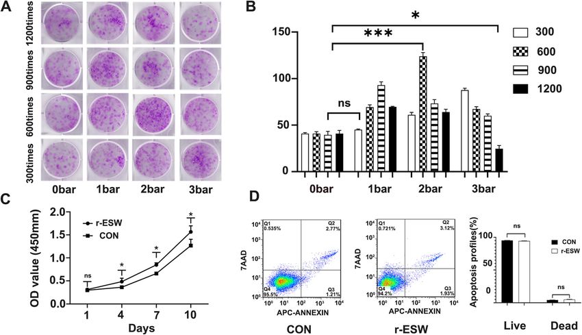
Zhao et al. Stem Cell Research & Therapy   (2021) 12:19                                                        Page 5 of 15

incorporated into this 6- and 12-week study. Rabbits          in 10% neutral buffered formalin for approximately 24–
were kept in a standardized feeding environment. An-          48 h, after which the specimens were decalcified in 10%
imals were supplied by the Experimental Animals               ethylenediaminetetraacetic acid (EDTA) for 30–45 days.
Center of the Chinese People’s Liberation Army                Then, the samples were dissected sagittally perpendicu-
(PLA) General Hospital. Experimental protocols in             lar to the surface of the lesion.
our operation procedure were in compliance with the              The regenerated tissues in paraffin blocks were sec-
Animal Welfare Act and were approved by the Ani-              tioned into 5-μm sections and stained with HE staining.
mal Care and Use Committee of the Laboratory Ani-             Cartilaginous matrix distribution was evaluated by safra-
mal Research Center at the PLA General Hospital               nin O and toluidine blue staining and immunohisto-
(Reference number: 2019-X15-57).                              chemical staining of collagen type II. The regenerated
   To evaluate the in vivo repair efficiency of SCB-SPCs      tissue was graded and analyzed semiquantitatively using
pretreated by optimized shockwave stimulation,                a modified ICRS histological scale by 3 independent
polylactic-co-glycolic acid (PLGA) was prepared by hole       blinded observers.
puncher (4.5 mm in diameter and 4 mm in thickness)
and was used as cell carrier scaffolds following our pre-     High-throughput transcriptome analysis
vious protocol [26]. The constructs were then implanted       Equal amounts of RNA were isolated from radial
into the osteochondral defects model in rabbits.              shockwave-treated SCB-SPCs and the untreated group
Shockwave-treated SCB-SPCs were seeded onto the pre-          using TRIzol reagent. High-throughput transcriptome
pared PLGA films and cultured in medium for 12 h. The         analysis was performed by Genewiz (Suzhou, China).
osteochondral defect model of the rabbit knee was im-         Briefly, after base calling the original sequences by
plemented following our previous protocol. In brief,          Bcl2fastq (v2.17.1.14), quality control was conducted by
anesthesia was administered by peritoneal injection of        FastQC (v0.10.1). Then, the original data were filtered by
ketamine/xylazine/buprenorphine. A medial parapatellar        Cutadapt (version 1.9.1). Clean data were compared with
incision was created after shaving and depilating, and        the reference genome using Hisat2 (v2.0.1). New tran-
then the patella was everted for sufficient exposure of       script prediction was performed using StringTie (v1.3.3b)
the trochlear femur and distal femur. A cylindrical           and Cuffcompare (v2.2.1). SNV and InDel analyses were
osteochondral defect of 4.5 mm in diameter and 5 mm           conducted using samtools (v0.1.19) and annovar
in depth was created in the trochlear femur using a ster-     (v2013.02.11). Gene expression analysis was conducted
ile trephine. Then, the models were treated with the re-      using Htseq (v.0.6.1). Differentially expressed genes were
spective interventions. All rabbits were randomly             screened by threshold values of ≥ 2-fold change and P
allocated into 5 different groups at 6 and 12 weeks.          value ≤ 0.05. Gene Ontology enrichment analysis was per-
Group A, osteochondral defects without treatment (con-        formed according to the categories molecular function,
trol group). Group B, defects with PLGA scaffolds only.       cellular component, and biological process. KEGG (Kyoto
Group C, defects with constructs composed of unpro-           Encyclopedia of Genes and Genomes) analysis was also
cessed SCB-SPCs and PLGA scaffolds. Group D, defects          performed. The bioinformatics analyses of differentially
with constructs composed of radial-shockwave treated          expressed genes were performed following the manufac-
SCB-SPCs and PLGA scaffolds and Sham group (Table             turers’ instructions. The genes correlated with cell prolif-
S3). Among the two latter groups, 1 × 10⁶ SCB-SPCs            eration or self-renewal and mechanotransduction-
were incorporated onto the PLGA films before they were        associated components such as the cytoskeleton were
implanted into osteochondral defects. At the end of the       screened out and selectively validated by RT-PCR.
surgery, the patella was returned to its original position,
and then the capsule and skin were closed using resorb-       Statistical analysis
able sutures and nylon wire, respectively. All animals        All data are presented as the mean values with standard
were allowed to move freely in cages after anesthesia.        deviations. Independent t test or one-way analysis of
Intramuscular penicillin injections were given to each        variance (ANOVA) followed by post hoc comparisons
rabbit to prevent infection.                                  were employed for normally distributed quantitative
                                                              data. P < 0.05 was considered statistically significant. All
Pathological analysis of repaired tissues                     tests were analyzed using IBM SPSS Version 20.0.
At 6 and 12 weeks after the operation, the animals were
euthanized for investigations. The trochlear femur was        Results
harvested and photographed by a digital camera for            Stem/progenitor cells remain present in human
macroscopic evaluation following the guidelines of the        subchondral bones
International Cartilage Repair Society (ICRS) scoring         The isolation protocols were based on previous studies
system. After gross examination, the samples were fixed       with minor modifications [27]. After approximately 7–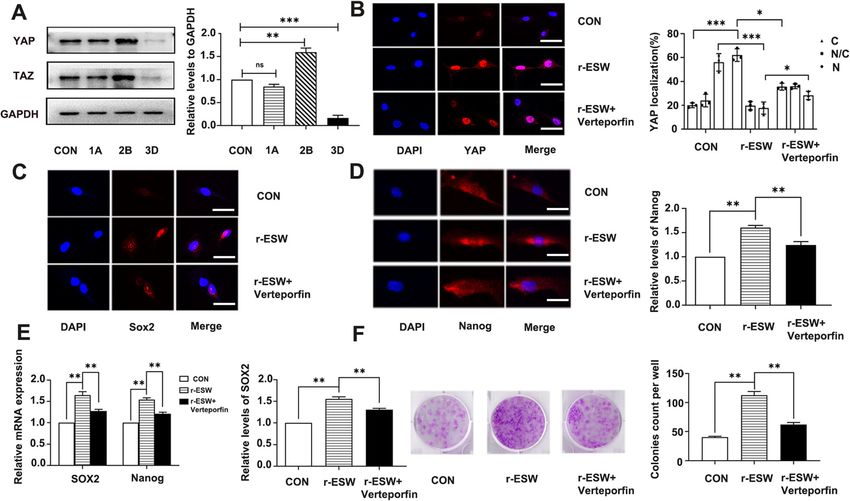
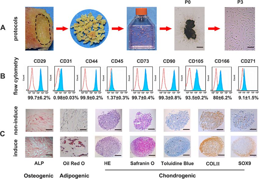
Zhao et al. Stem Cell Research & Therapy       (2021) 12:19                                                                              Page 6 of 15

10 days of initial culture, fibroblast-like cells could be                   staining of Sox9 and collagen II after culturing in a three-
observed migrating around the subchondral bone chips,                        dimensional pellet media system. The induced group
and the cells exhibited an adherent growth state. As time                    showed spacious cartilage matrix and significant increases
increased, an increasing number of cells migrated from                       in cartilage-associated markers, including proteoglycan,
the chips (Figs. 1a, and 2a). Flow cytometry analysis re-                    aggrecan, collagen II, and Sox9 (Fig. 2c, Fig. S2).
vealed that SCB-SPCs were positive for the expression of
mesenchymal stem cells with regard to CD29 (99.7 ±                           r-ESW promoted self-renewal of SCB-SPCs in a dose-
6.2%), CD44 (99.9 ± 0.2%), CD73 (99.7 ± 0.4%), CD166                         dependent manner
(80 ± 6.2%), and the stemness-related markers CD90                           A colony-forming unit fibroblast (CFU-F) formation
(99.3 ± 0.8%) and CD105 (93.5 ± 0.2%). Meanwhile, SCB-                       assay was performed after the SCB-SPCs were pretreated
SPCs were negative for the expression of the endothelial                     with gradient doses of radial shockwave in the floating
or hematopoietic markers CD31 (0.98 ± 0.03%) and                             system (Fig. 1b). The crystal violet staining results
CD45 (1.37 ± 0.3%), further supporting the characteris-                      showed that the radial shockwave-treated group exhib-
tics of stem/progenitor cells (Fig. 2b).                                     ited larger and denser colonies than the untreated group.
   Regarding multipotency, ALP activity, concomitant                         However, the positive effects may decrease and even re-
with significant increased expression levels of osteogenic                   verse to negative when the energy exceeds a certain
markers Runx2 and osteocalcin after inducing culture                         range. According to the staining and qualitative analysis
reveal the osteogenic capacity of SCB-SPCs. The SCB-                         results of colonies under microscopy, we found that
SPCs also exhibited visible intracytoplasmic lipid droplet                   when the pressure was 1 bar, with the increase in stimu-
accumulation through Oil Red O staining, associated                          lation number from 300 to 900 times, self-renewal ex-
with corresponding elevated gene expression levels of                        hibited a growing trend, after which self-renewal tended
adipogenic markers such as CEBPα and PPARγ. The                              to decrease. At 2 bar, the progressive increase effects of
chondrogenic capacity was evaluated by HE, toluidine                         r-ESW only existed from 300 to 600 stimulations. Simi-
blue, and safranin O staining and immunohistochemical                        larly, when the pressure was 3 bar, as the treatment

 Fig. 1 Isolation, cultivation and characterization of SCB-SPCs. a Representative images showing the protocols. The marked subchondral bone
 region of the lateral tibia plateau was cut into bone pieces, rinsed, mildly digested, and then incubated in a culture flask. SCB-SPCs outgrew from
 bone chips after approximately 10 days of cultivation. The P0 cells were passaged successively to P3 (n = 10 donors). b Stem/progenitor cell
 surface immunophenotype expression (CD29, CD31, CD44, CD45, CD73, CD90, CD105, CD166, and CD271) was identified via flow cytometric
 analysis (n = 5). c The multiple differentiation potency of SCB-SPCs was examined by induction assay and staining. HE, safranin O, toluidine blue
 staining, and collagen II and Sox9 immunohistochemical staining to detect chondrogenesis after micropellet culture. ALP staining for
 osteogenesis. Oil Red O staining for adipogenesis. Scale bars represent 500 μm (ALP staining), 100 μm (Oil Red O staining), and 200 μm
 (chondrogenic-related staining). SCB-SPCs, subchondral bone stem/progenitor cells
Zhao et al. Stem Cell Research & Therapy (2021) 12:19 Page 7 of 15
Fig. 2 General flow diagram of the experiment. a Isolation, cultivation, and characterization of SCB-SPCs. b Optimization of r-ESW parameters for
the self-renewal of SCB-SPCs. c The influences of the optimized parameters on the proliferation, apoptosis, and multidifferentiation of SCB-SPCs. d
The in vivo therapeutic effects of SCB-SPCs pretreated by r-ESW via an osteochondral lesion model. e The underlying mechanisms involved in the
mechanotransduction process mediated by r-ESW were explored. SCB-SPCs, subchondral bone stem/progenitor cells. r-ESW, radial
extracorporeal shockwave
numbers increased, the self-renewal gradually decreased. between the two groups. Regarding adipogenic potential,
The optimal parameter (2 bar, 600 times) was screened few Oil-Red-O-positive intracytoplasmic lipid droplet ac-
out for the following experiments (Fig. 3a, b). cumulations were discovered in radial shockwave-
The CCK8 results showed that compared to the con- treated SCB-SPCs, while more lipid droplets formed in
trol group, the r-ESW group significantly accelerated the the control group. The mRNA expression levels of
proliferation of SCB-SPCs (Fig. 3c). Meanwhile, r-ESW CEBPα and PPARγ further support the above staining
treatment exerted no significant influence on apoptosis outcomes. In terms of chondrogenic potency, the radial
(Fig. 3d). shockwave-treated group exhibited richer aggrecan accu-
mulation in the extracellular matrix through safranin O
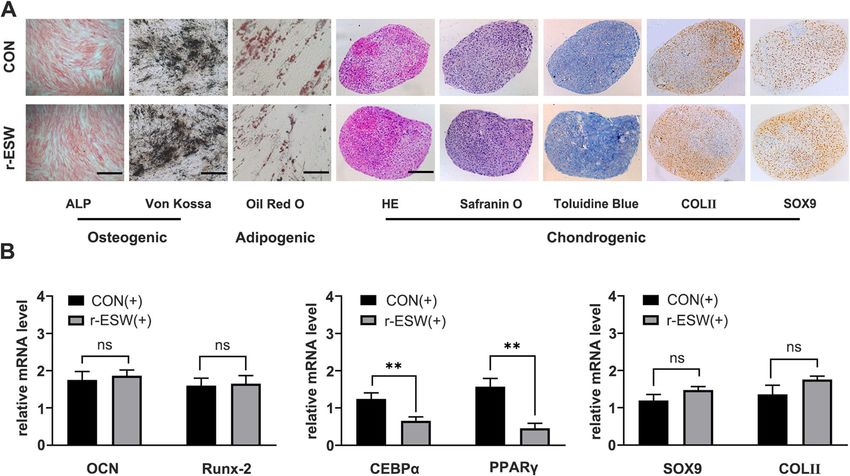
r-ESW differentially influences tridifferentiation of SCB- and toluidine blue staining. Moreover, collagen type II
SPCs and Sox9 were more abundant in the experimental
To further explore the influence of optimized radial group. The mRNA expression levels of Sox9 and Col II
shockwave stimulation on SCB-SPC multipotency, in- also increased slightly. However, the change was not sig-
duction experiments were performed. In terms of osteo- nificant (Fig. 4).
genic potential, no significant difference was detected
concerning ALP activity and mineralized matrix accu- r-ESW pretreatment markedly enhances SCB-SPC-
mulation between the r-ESW group and the nontreated mediated cartilage repair in vivo
group. Consistent with the staining results, the mRNA Trochlear osteochondral samples were harvested at 6
expression levels of osteogenic genes, including Runx2 and 12 weeks after surgery for macroscopic and histo-
and OCN, also revealed no significant differences logic evaluation. No infection was discovered in any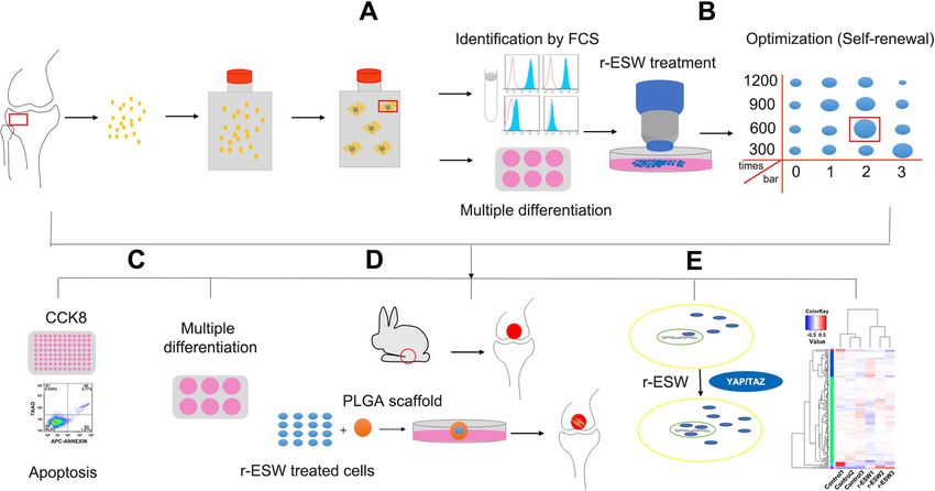
Zhao et al. Stem Cell Research & Therapy      (2021) 12:19                                                                            Page 8 of 15

 Fig. 3 Optimization of radial shockwave parameters. a Representative images of CFU-F stained by crystal violet after gradient doses of radial
 shockwave stimulation. b Quantitative comparison of colony numbers in each group based on CFU-F results. The combined analysis suggests
 that radial shockwave promotes the self-renewal of SCB-SPCs in a dose-dependent manner (n = 5, with each 3 technical repeats). c The optimized
 parameters of radial shockwave significantly enhance the proliferation of SCB-SPCs (n = 5, with each 3 technical repeats). d Compared with the
 control group, the optimized radial shockwave stimulation had no significant influence on the apoptosis of SCB-SPCs (n = 5, with each 3 technical
 repeats). *p < 0.05, ***p < 0.001, ns, not significant. SCB-SPCs, subchondral bone stem/progenitor cells

rabbits. At 6 weeks, only a few visually visible fibrous                   however, there was still an obvious line between the re-
films filled the bottom of the osteochondral defects, and                  generated and native tissue. The osteochondral defects
the defects remained clear in the model group. In the                      in the scaffold combined with SCB-SPCs were almost
scaffold only group, approximately 80% of the osteo-                       80% covered with hyaline cartilage-like tissue and were
chondral defect area was filled with fibrous-like repair                   filled with obscure demarcation from surrounding cartil-
tissue. When the PLGA scaffold was loaded with un-                         age. Encouragingly, in the scaffold- and radial
treated SCB-SPCs, newly formed tissue covered ap-                          shockwave-pretreated SCB-SPC groups, the regenerated
proximately 90% of the defect area; however, the neo-                      tissues were all glossy white and integrated with the sur-
tissue was bumpy and irregular. The osteochondral                          rounding normal cartilage, and the surface was smoother
defects in the PLGA scaffold loaded with the radial                        and phenotypically much closer to normal cartilage
shockwave-primed SCB-SPCs group were nearly cov-                           (Fig. 6a). Relative quantitative evaluation based on the
ered with newly formed tissue that had a relatively                        ICRS scoring system further supports gross observation.
smooth surface and obscure demarcation from sur-                           The ICRS score was highest in the scaffold- and radial
rounding cartilage (Fig. 5a).                                              shockwave-pretreated SCB-SPC groups (Fig. 5b, Fig. 6b).
   Macroscopic evaluation revealed that in all groups, the                    HE staining was implemented to identify the general
repair rate of defect depth, demarcating border, and sur-                  characteristics of the repaired tissues. Toluidine blue,
face smoothness increased with time. At 12 weeks, the                      safranin O, and collagen type II stainings were employed
regenerated tissue in the control group was glossy white,                  to investigate the cartilage matrix accumulation level. At
indicating the formation of fibrous cartilage; however,                    6 weeks after implantation, both images showed distinct
the repair area was largely limited to the periphery of                    borders between regenerated and surrounding tissue,
the lesion, and there was still a significant defect in the                and the neocartilage matrix was shallow compared with
center. In the scaffold only group, white fibrous-like re-                 the native tissue in all groups. At 12 weeks, the defects
pair tissue covered approximately 90% of the defect area;                  remained concave, although with a smoother surface.
Zhao et al. Stem Cell Research & Therapy       (2021) 12:19                                                                               Page 9 of 15

 Fig. 4 The influences of radial shockwave on the multidifferentiation of SCB-SPCs. a ALP and Von Kossa staining indicated that radial shockwave
 had no significant influence on the osteogenic capacity of SCB-SPCs. Oil Red O staining showed that few positive intracytoplasmic lipid droplets
 accumulated in radial shockwave-treated SCB-SPCs, and adipogenesis was suppressed by radial shockwave. For chondrogenic potentiality, the
 treated SCB-SPCs mildly increased, while the change was not significant (n = 5, with each 3 technical repeats). Scale bars represent 500 μm (ALP
 and von Kossa staining), 100 μm (Oil Red O staining), and 200 μm (chondrogenic-related staining). b The influences of r-ESW on SCB-SPC
 tridifferentiation were further validated by the mRNA expression level. Runx2 and OCN for osteogenesis, CEBPα and PPARγ for adipogenesis, Sox9
 and Col II for chondrogenesis (n = 5, with each 3 technical repeats). The results were consistent with the staining outcomes. *p < 0.05, **p < 0.01,
 ns, not significant

The pathological analysis discriminated the difference in                    YAP activation and nuclear translocation contributed to r-
the repair effects between individual groups and further                     ESW-mediated self-renewal enhancement of SCB-SPCs
validated the macroscopic evaluation results.                                To explore the possible mechanisms of radial-shockwave
   HE staining of the untreated group showed that the                        potentiated self-renewal, three gradient doses of radial-
osteochondral defect remained concave with only a few                        shockwave on the basis of CFU-F evaluation outcomes
fibrous tissue and inflammatory cells at the bottom and                      were selected (Fig. 3a): a blank control group, a 1A
periphery. Toluidine blue and safranin O staining and                        group (1 bar, 300 times), a 2B group (2 bar, 600 times),
immunochemistry of Col II showed few positive staining                       and a 3D group (3 bar, 1200 times). The protein samples
areas. In the scaffold group, the regenerated tissue was                     were collected after radial shockwave stimulation at the
larger than that in the control group; however, most of                      respective doses. The mechanically sensitive effector
the reparative tissues were nonfunctional fibrous scars                      protein YAP was chosen to explore the potential mech-
with a border between repair tissue and normal cartilage.                    anism. The western blot results showed that the expres-
Special and immunological staining further complemen-                        sion level of YAP/TAZ was closely related to CFU-F
ted the above results with sporadically positive histo-                      outcomes. Specifically, compared to the basic expression
chemical staining. In contrast, the defects were filled                      level of the blank control group, the 1A group had no
with hyaline-like cartilage with PLGA scaffold/SCB-SPC                       significant variation, the 2B group had a marked in-
constructs. More importantly, the scaffold/SCB-SPCs                          crease, and the 3D group had a conspicuous decline
pretreated with r-ESW exhibited a more desirable effect                      (Fig. 7a). The YAP expression levels in the respective
with good integration into normal cartilage, although                        groups showed change tendencies consistent with the
the content of cartilaginous matrix was slightly less                        corresponding CFU-F phenomenon (Fig. 3a, b). To fur-
abundant than that of normal cartilage (Fig. 6a). Modi-                      ther confirm the critical role of YAP/TAZ in SCB-SPC
fied ICRS pathological scores further validated the above                    self-renewal, the optimal group 2B was selected for sub-
results (Fig. 6c).                                                           sequent research. The YAP/TAZ pathway-specific
Zhao et al. Stem Cell Research & Therapy       (2021) 12:19                                                                              Page 10 of 15

 Fig. 5 Macroscopic and histologic assessment of cartilage repair effects of SCB-SPCs pretreated by radial shockwave in vivo at 6 weeks. a
 Macroscopic observation of cartilage repair in the femoral trochlea at 6 weeks after surgery. Representative HE, safranin O, toluidine blue, and
 immunohistochemical staining of Col II. Scale bars represent 1 mm. b ICRS rating scores of the regenerated tissue in each group (n = 5). c
 Modified ICRS histological scores of cartilage repair in each group (n = 5). *p < 0.05, **p < 0.01, ***p < 0.001

 Fig. 6 Macroscopic and histologic assessment of cartilage repair effects of SCB-SPCs pretreated by radial shockwave in vivo at 12 weeks. a
 Macroscopic observation of cartilage repair in the femoral trochlea at 12 weeks after surgery. Representative HE, safranin O, toluidine blue, and
 immunohistochemical staining of Col II. Scale bars represent 1 mm. b ICRS scores of the regenerated tissue in each group (n = 5). c Modified ICRS
 histological scores of cartilage repair in each group (n = 5). *p < 0.05, **p < 0.01, ***p < 0.001
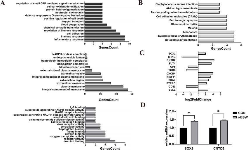
Zhao et al. Stem Cell Research & Therapy       (2021) 12:19                                                                            Page 11 of 15

 Fig. 7 YAP activation and nuclear translocation contributed to radial shockwave-mediated promotion of self-renewal of SCB-SPCs. a The YAP/TAZ
 expression level exhibited a consistent tendency with CFU-F under the respective doses of r-ESW stimulation: control group, 1A (1 bar, 300 times),
 2B (2 bar, 600 times), and 3D (3 bar, 1200 times) (n = 5, with each 3 technical repeats). b Immunofluorescence of YAP localization and
 semiqualitative evaluation of YAP distribution in SCB-SPCs among the control, r-ESW, and r-ESW + verteporfin groups (n = 5, with each 3 technical
 repeats). c Immunofluorescence of Sox2 expression and semiqualitative evaluation in SCB-SPCs among the control, r-ESW, and r-ESW + verteporfin
 groups (n = 5, with each 3 technical repeats). d Immunofluorescence of Nanog expression and semiqualitative evaluation in SCB-SPCs among the
 control, r-ESW, and r-ESW + verteporfin groups (n = 5, with each 3 technical repeats). e The relative expression levels of self-renewal-associated
 genes in SCB-SPCs among the control, r-ESW, and r-ESW + verteporfin groups (n = 5, with each 3 technical repeats). f The general staining and
 qualitative analysis of CFU-F among the control, r-ESW, and r-ESW + verteporfin groups (n = 5, with each 3 technical repeats). Scale bars represent
 25 μm. *p < 0.05, **p < 0.01, ***p < 0.001

inhibitor verteporfin was used to verify the role YAP/                      Nanog and Sox2 exhibited consistent change tendencies
TAZ plays in the self-renewal process mediated by radial                    with the above results (Fig. 7e).
shockwave. Before this validation, the SCB-SPCs were
cultured in medium supplemented with gradient con-
centrations of verteporfin. Then, the secure dose was se-                   Mechanisms besides YAP signaling were involved in r-
lected by microscopic observation and CFU-F evaluation                      ESW-mediated regulation of SCB-SPC self-renewal
(Supplementary Fig. S1). Compared to the radial-                            Compared with the untreated group, a total of 600
shockwave group, self-renewal of SCB-SPCs significantly                     differentially expressed genes were identified in radial
decreased in the inhibitor group (Fig. 7f). Further con-                    shock wave-treated SCB-SPCs. Three hundred of the
focal microscopy outcomes suggested that compared                           genes were upregulated, and 300 were downregulated.
with the control group, the YAP nuclear localization                        Gene ontology (GO) enrichment suggested that the
proportion significantly increased in the r-ESW group.                      differentially expressed genes were involved in various
Moreover, compared with the control group, Nanog and                        biological processes, including cell proliferation, adhe-
Sox2 contents exhibited consistent tendencies with YAP                      sion, migration, cytoskeletal transformation, and other
expression level and status. Importantly, the YAP nu-                       biological processes (Fig. 8a, b). Significantly changed
clear location proportion and Nanog and Sox2 expres-                        genes related to self-renewal, cell adhesion, cell actin
sion levels significantly declined in the r-ESW +                           cytoskeleton, and ECM-receptor interaction are listed
verteporfin group (Fig. 7b–d). Meanwhile, the mRNA                          (Fig. 8c). Sox2 and CNTD2 were validated by RT-
expression levels of the self-renewal-associated genes                      PCR (Fig. 8d).
Zhao et al. Stem Cell Research & Therapy       (2021) 12:19                                                                            Page 12 of 15

 Fig. 8 mRNA sequence analysis of SCB-SPCs before and after r-ESW treatment. a Classification of differentially expressed genes according to
 biological process, cell components, and molecular function. b KEGG analysis of significantly changed genes after r-ESW treatment. c Significantly
 changed genes related to self-renewal and components that may be involved in the mechanotransduction process are listed. d Validation of
 SOX2 and CNTD2 expression levels by RT-PCR (n = 5, with each 3 technical repeats). *p < 0.05

Discussion                                                                  radial shockwave and other noninvasive physical means
In the present study, we demonstrated that radial shock-                    are receiving increasing attention [6, 32–34].
wave is capable of enhancing the self-renewal capacity of                     r-ESW has been demonstrated to clinically accelerate
SCB-SPCs in vitro, and we screened out the optimized                        the healing of meniscus tears and muscle injuries and
parameters in our experimental system. In addition,                         has chondroprotective effects [7, 35]. Intriguingly, r-
primed SCB-SPCs exhibit stronger repair in an osteo-                        ESW can even strengthen the stemness of stem cells
chondral injury model. Moreover, we identified that the                     through its unique mechanical characteristics [4, 36]. As
activation of YAP and translocation to the nucleus con-                     one specificity of stem cells, self-replication or self-
tributed to this promising effect mediated by radial                        renewal controls the stem cell pool and determines the
shockwave.                                                                  repair efficiency. Therefore, in our research, we first op-
   As a critical factor in regenerative medicine, increasing                timized the parameters of radial shockwave on SCB-
numbers of tissue-specific stem/progenitor cells have                       SPCs via CFU-F evaluation. Our results show that radial
been identified and studied [28–30]. In this study, we                      shockwave influences the self-renewal of SCB-SPCs in a
chose SCB-SPCs due to their pivotal location and poten-                     dose-dependent manner. Moreover, we combined shock-
tial therapeutic role in osteochondral repair [13, 31]. Nu-                 wave intensity (bar) with stimulation numbers in our
merous previous studies have indicated the multipotency                     protocols. The CFU-F data suggest that the intensity and
of SCB-SPCs [19]. Krüger et al. [20] proposed SCB-SPCs                      stimulation times collectively influence the self-renewal
as an alternative source for cartilage regeneration. How-                   of SCB-SPCs. Based on the data, we speculate that a par-
ever, the in vivo repair efficiency still needs to be eluci-                ameter characterizing the total energy may be able to
dated. Meanwhile, as the repair efficiency correlates                       comprehensively represent the compositive effects of in-
closely with the vitality of stem cells, enhancing the                      tensity and stimulation times. Our results further verify
tissue-regenerative properties of seed cells is of great sig-               the critical role of radial shockwave energy on biological
nificance for clinical therapy. In addition to the com-                     effects, and the results provide a reference for clinical
monly used chemical means or gene modifications,                            therapy. Following the optimized parameters based on
Zhao et al. Stem Cell Research & Therapy   (2021) 12:19                                                         Page 13 of 15

the CFU-F results, we further explored the biological ef-       composed of compression, tension, and shear forces, are
fect of radial shockwave on multidifferentiation.               transient pressure disturbances that propagate rapidly in
   Accumulating evidence suggests that shockwave                three-dimensional space. However, whether YAP activity
stimulation could regulate the differentiation potential of     is involved in radial shockwave-mediated biological ef-
stem cells [37–39]; however, the effects are still contro-      fects is still unclear. Therefore, we explored its activity
versial. In the current study, we found that the opti-          in our study. The results demonstrated that the activity
mized radial shockwave screened out based on CFU-F              of YAP also contributes to the radial shockwave-
has no significant influence on the osteogenic capacity         mediated process of enhancing the self-renewal of SCB-
of SCB-SPCs. The outcomes were different from the ma-           SPCs. To exclude side effects, the YAP-specific inhibitor
jority of previous studies in that the osteogenic capacity      verteporfin was used to further validate this effect. Previ-
of mesenchymal stem cells was enhanced after radial             ous studies suggested that Sox2 and Nanog are binding
shockwave stimulation [37, 38]. This phenomenon may             targets of YAP/TAZ by genome-wide analysis [46]. We
result from the stimulation regimen selected based on           found that with the activation and nuclear translocation
CFU-F results, which may not be optimal for differenti-         of YAP after radial shockwave stimulation, the mRNA
ation induction. Because the effect of radial shockwave         and protein expression of Sox2 and Nanog also in-
on SCB-SPC differentiation was first reported, the out-         creased. Importantly, after the YAP-specific inhibitor
comes may still need further validation. Chondrogenic           verteporfin was used, the expression of Sox2 and Nanog
capacity was mildly increased, while the change was not         significantly decreased. The change tendency of YAP
significant. Consistent with our previous work and the          status and the corresponding expression of Sox2 and
literature, the adipogenic potential was significantly sup-     Nanog are consistent with the self-renewal of SCB-SPCs
pressed. However, we are aware that different stem cell         characterized by CFU-F. The comprehensive analysis of
populations, shockwave stimulation systems, and proto-          the results suggests that YAP/TAZ and downstream
cols may contribute to the discrepancy effects. More-           self-renewal-associated genes are involved in the bio-
over, protein-based assay would be helpful to further           logical process of radial shockwave-mediated promotion
validate our results.                                           of SCB-SPC self-renewal. The results are consistent with
   To further investigate the in vivo repair efficiency of      previous studies that demonstrated that YAP correlated
shockwave-treated SCB-SPCs, the classical and stable            closely with the self-renewal of ES cells because elevated
model of osteochondral defects was used. Porous PLGA            YAP maintains the potency of ES cells [46, 47]. Mean-
scaffolds were applied as cell carriers based on our previ-     while, others reported that YAP/TAZ are dispensable for
ous study [26]. The gross observations characterized by         ES cell fate [48]. Hence, the cell-specific functional style
ICRS scores along with histological analysis following          of YAP/TAZ still needs further exploration. Moreover,
modified ICRS scores of neo-tissues at 2 different points       the detailed signal activity events between the receptor
demonstrated that SCB-SPCs pretreated by r-ESW                  and ultimate YAP effector still need further investiga-
yielded better repair results. The in vivo repair effect val-   tion. In view of the results that CFU-F in the r-ESW +
idation further strengthens the translational application       verteporfin group was stronger than that in the control
value of radial shockwave. However, the concrete mech-          group, we speculate that other pathways may also be in-
anisms of r-ESW in vivo still need to be explored.              volved in radial shockwave-mediated self-renewal
   YAP/TAZ, the effector of the Hippo pathway, has been         prompting, except the YAP pathway. Hence, high-
demonstrated to play pivotal roles in mechano-                  throughput sequences were implemented. The results
meditated stem cell plasticity and stemness regulation          revealed that self-renewal-related genes increased after
during tissue regeneration [40, 41]. Moreover, numerous         radial shockwave and supported the idea that other self-
studies have suggested that YAP protein activity could          renewal-associated pathways may also contribute to this
be regulated directly by mechanical cues from both the          effect.
microenvironment, such as extracellular matrix stiffness,         There are still several limitations in the present study.
and other extraneous stimulation independent of the             Although a series of gradient parameters have been de-
Hippo pathway [42, 43]. Notably, mechanical stimuli             signed to explore the biological effect of radial shock-
could influence YAP nuclear-cytoplasmic distribution            wave, the quantitative evaluation parameters and
and in turn modulate cell biological function [40, 44].         corresponding biological effects still need to be explored
DuPont et al. identified YAP/TAZ as sensors and ef-             owing to the complexity of radial shockwaves. In
fector proteins of mechanical cues perceived from extra-        addition, the optimized parameters in the current study
cellular rigidity [45]. Additionally, YAP/TAZ also              were screened out by CFU-F, which may not be optimal
responds to different kinds of mechanical stresses, in-         for the differentiation capacity of SCB-SPCS. Further
cluding shearing, compressing, or pulling [40]. Extracor-       precise output of radial shockwave energy will facilitate
poreal shockwaves, a unique physical stimulus                   the optimization of different characteristics of stem cells
Zhao et al. Stem Cell Research & Therapy           (2021) 12:19                                                                                 Page 14 of 15

and accelerate their clinical application. Meanwhile, the                      Availability of data and materials
SCB-SPCs were cultured in vitro, which may not com-                            The datasets used and/or analyzed during the current study are available
                                                                               from the corresponding author on reasonable request.
prehensively represent the in vivo status. Moreover, the
mechanisms of in vivo experiments still need further ex-                       Ethics approval and consent to participate
ploration and validation.                                                      The human knee plateau osteochondral samples used in this study were
                                                                               approved by the institutional ethical review board of the People’s Liberation
                                                                               Army General Hospital (rapid review and approval of scientific research
Conclusion                                                                     projects for use of discarded biological materials), and informed consent was
We found that radial extracorporeal shockwave is cap-                          obtained from all donors. Animals were supplied by the Experimental
                                                                               Animals Center of the Chinese People’s Liberation Army (PLA) General
able of promoting the self-renewal of SCB-SPCs in vitro                        Hospital. Experimental protocols in our operation procedure were in
by targeting YAP activity and strengthening its repair ef-                     compliance with the Animal Welfare Act and were approved by the Animal
ficiency in vivo, indicating promising application                             Care and Use Committee of the Laboratory Animal Research Center at the
                                                                               PLA General Hospital (Reference number: 2019-X15-57).
prospects.
                                                                               Consent for publication
                                                                               Not applicable
Supplementary Information
The online version contains supplementary material available at https://doi.
org/10.1186/s13287-020-02076-w.                                                Competing interests
                                                                               The authors declare no competing financial interests.
 Additional file 1: Table S1. Demographic, clinical, and imaging
                                                                               Author details
 characteristics of the donors. Table S2. Primer sequences for RT-qPCR.        1
                                                                                Chinese People’s Liberation Army (PLA) General Hospital, Chinese PLA
 Table S3. Experiment groups of in vivo study (6 weeks and 12 weeks).
                                                                               Medical School, No. 28 Fuxing Road, Haidian District, Beijing 100853, China.
 Additional file 2: Figure S1. The concentration screening experiment          2
                                                                                Beijing Institute of Radiation Medicine, No. 27 Taiping Road, Haidian District,
 of YAP specific inhibitor verteporfin. (a) primary concentration screening    Beijing 100850, China. 3Graduate School of Anhui Medical University, No. 81
 by photomicrographs and CFU-F evaluation under a series of verteporfin        Meishan Road, Shu Shan District, Hefei 230032, Anhui Province, China.
 concentration; (b) secondary screening on the basis of primary results.
 The selected concentration was 0.05uM.                                        Received: 17 October 2020 Accepted: 7 December 2020
 Additional file 3: Figure S2. The comparison of tridifferentiation
 associated genes expression level between non-induced and induced
 group. The relative mRNA expression level of osteogenic related markers       References
 (OCN, Runx-2), adipogenic markers (CEBPα and PPARγ), chondrogenic as-         1. Moya D, Ramón S, Schaden W, Wang CJ, Guiloff L, Cheng JH. The role of
 sociated markers (Collagen II and Sox9) were significantly higher in in-          extracorporeal shockwave treatment in musculoskeletal disorders. J Bone
 duced group. *p < 0.05, **p < 0.01, ***p < 0.001.                                 Joint Surg Am. 2018;100(3):251–63.
 Additional file 4.                                                            2. Hsu CC, Cheng JH, Wang CJ, Ko JY, Hsu SL, Hsu TC. Shockwave therapy
                                                                                   combined with autologous adipose-derived mesenchymal stem cells is
 Additional file 5.
                                                                                   better than with human umbilical cord Wharton’s jelly-derived
                                                                                   mesenchymal stem cells on knee osteoarthritis. Int J Mol Sci. 2020;21(4):
                                                                                   1217.
Abbreviations
                                                                               3. Hashimoto S, Ichinose T, Ohsawa T, Koibuchi N, Chikuda H. Extracorporeal
α-MEM: Alpha-minimal essential medium; CCK8: Cell Counting Kit-8; CEBP/
                                                                                   shockwave therapy accelerates the healing of a meniscal tear in the
α: CCAAT/enhancer binding protein alpha; CFU-F: Colony-forming unit
                                                                                   avascular region in a rat model. Am J Sports Med. 2019;47(12):2937–44.
fibroblast assay; Col-II: Type II collagen; EDTA: Ethylenediaminetetraacetic
                                                                               4. Zhang H, Li ZL, Yang F, Zhang Q, Su XZ, Li J, et al. Radial shockwave
acid; FBS: Fetal bovine serum; GAPDH: Glyceraldehyde-3-phosphate
                                                                                   treatment promotes human mesenchymal stem cell self-renewal and
dehydrogenase; GO: Gene ontology; ICRS: International Cartilage Repair
                                                                                   enhances cartilage healing. Stem Cell Res Ther. 2018;9(1):54.
Society; MSCs: Mesenchymal stem cells; OCN: Osteocalcin; K-L: Kellgren-
                                                                               5. Suhr F, Delhasse Y, Bungartz G, Schmidt A, Pfannkuche K, Bloch W. Cell
Lawrence; KEGG: Kyoto Encyclopedia of Genes and Genomes;
                                                                                   biological effects of mechanical stimulations generated by focused
PBS: Phosphate-buffered saline; PI: Propidium iodide; PPARγ: Peroxisome
                                                                                   extracorporeal shock wave applications on cultured human bone marrow
proliferator-activated receptor gamma; PLGA: Polylactic-co-glycolic acid; r-
                                                                                   stromal cells. Stem Cell Res. 2013;11(2):951–64.
ESW: Radial extracorporeal shockwave; RT-qPCR: Real-time quantitative
                                                                               6. Ireland RG, Simmons CA. Human pluripotent stem cell mechanobiology:
polymerase chain reaction; Runx-2: Runt-related transcription factor 2; SCB-
                                                                                   manipulating the biophysical microenvironment for regenerative medicine
SPCs: Subchondral bone stem/progenitor cells; Sox-9: Sex determining
                                                                                   and tissue engineering applications. Stem Cells. 2015;33(11):3187–96.
region Y-box 9
                                                                               7. Yu J, Chen Z, Yan F. Advances in mechanism studies on ultrasonic gene
                                                                                   delivery at cellular level. Prog Biophys Mol Biol. 2019;142:1–9.
Acknowledgements                                                               8. Liu T, Shindel AW, Lin G, Lue TF. Cellular signaling pathways modulated by
Not applicable                                                                     low-intensity extracorporeal shock wave therapy. Int J Impot Res. 2019;31(3):
                                                                                   170–6.
                                                                               9. Madry H, van Dijk CN, Mueller-Gerbl M. The basic science of the
Authors’ contributions
                                                                                   subchondral bone. Knee Surg Sports Traumatol Arthrosc. 2010;18(4):419–33.
ZDZ, YXW, and QW conceived of this study, performed the experiments, and
                                                                               10. Pape D, Filardo G, Kon E, van Dijk CN, Madry H. Disease-specific clinical
wrote the manuscript supervised by ZLL and HZ who revised the
                                                                                   problems associated with the subchondral bone. Knee Surg Sports
manuscript. JWL, WH, SZ, and PLL participated in the data acquisition and
                                                                                   Traumatol Arthrosc. 2010;18(4):448–62.
interpretation. All authors read and approved the final manuscript.
                                                                               11. Saltzman BM, Riboh JC. Subchondral bone and the osteochondral unit:
                                                                                   basic science and clinical implications in sports medicine. Sports Health.
Funding                                                                            2018;10(5):412–8.
This work was supported by National Natural Sciences Grants China              12. Goldring SR, Goldring MB. Changes in the osteochondral unit during
(No.81871771, 81572159) and the Beijing Natural Sciences Foundation                osteoarthritis: structure, function and cartilage-bone crosstalk. Nat Rev
(No.7182123).                                                                      Rheumatol. 2016;12(11):632–44.
You can also read