Rabbit Vomeronasal Organ-Derivered Cells Have Mesenchymal Profile and Neuronal Commitment
←
→
Page content transcription
If your browser does not render page correctly, please read the page content below
Int. J. Morphol.,
38(5):1463-1472, 2020.

                   Rabbit Vomeronasal Organ-Derivered Cells Have
                   Mesenchymal Profile and Neuronal Commitment

                               Las Células Derivadas del Órgano Vomeronasal del Conejo
                                  tienen Perfil Mesenquimatoso y Compromiso Neuronal

           Rodrigo S. N. Barreto1; Franceliusa Delys de Oliveira1; Gustavo de Sá Schiavo Matias1; Marcio N. Rodrigues2;
                    Rafael C. Carvalho3; André Luis R. Franciolli1; Paula Fratini1 & Maria Angelica Miglino1

BARRETO, R. S. N.; DE OLIVEIRA, F. D.; MATIAS, G. S. S.; RODRIGUES, M. N.; CARVALHO, R. C.; FRANCIOLLI,
A. L. R.; FRATINI, P. & MIGLINO, M. A. Rabbit vomeronasal organ-derivered cells have mesenchymal profile and neuronal
commitment. Int. J. Morphol., 38(5):1463-1472, 2020.

         SUMMARY: The vomeronasal organ (VNO) is an accessory organ involved on the olfactory pathway, that detects pheromones
and emits signals in order to modulate social and reproductive behavior. The VNO stem cells replace neurons throughout life. The aim of
this study was to isolate and characterize cells derived from the vomeronasal organ from New Zealand rabbits. Five male rabbits with
120 days were used for cell isolation and culture. Results: VNO-derived cells presented labelling for proliferation (PCNA), undifferentiated
profile (Nanog), neuronal (GFAP), mesenchymal stem cells (CD73, CD90 and CD105 and Stro-1). Also, presence of cytoskeletal (Vimentin,
b-tubulin and CK-18) and absence of hematopoietic markers (CD34, CD117 and CD45) both by immunofluorescence and flow cytometry.
By PCR it was possible to verify the expression of some undifferentiated profile (Oct-4), neuronal (Nestin) and mesenchymal (CD73,
CD105 and Vimentin) genes. Functionally, VNO-derived cells differentiate in vitro into adipocytes, osteocytes and chondrocytes, and
presented no tumorigenic potential when injected to Balb/c nu/nu mice. In conclusion, the rabbit VNO-derived cells have a profile that
could be supportive to VNO olfactory/neuroreceptor epithelium by delivering factors to epithelial turnover or even by differentiation
into epithelial cells to replacement of commissural epithelium.

          KEY WORDS: Olfactory System; Olfactory epithelium; Olfactory bulb; Neuronal stem cells.

INTRODUCTION

        Most vertebrates have a vomeronasal system                         et al., 2018). Presents on the lamina propria, the vomeronasal
consisting in vomeronasal organ (VNO), accessory olfactory                 glands secrete mucous substances into the luminal surface of
bulb (AOB), vomeronasal amygdala and nerves that connect                   sensory epithelia, responsible for pheromones detection
these structures. The VNO is related to be a social and                    (Tomiyasu et al., 2017).
reproductive structure, involved in communication by
molecules, as pheromones, detection and transmission of                            In general, replacement of sensorial cells occurs on
the signals to central nervous system (Park et al., 2014). It              sensitive epithelium by vertical migration of cells derived
is composed by a pair of tubular structures, situated along                from basal to the apical and sensory cells layer (Monti
the anterior portion of the nasal septum. Medial wall and ven-             Graziadei et al., 1980; Moulton et al., 2008). However, the
tral and dorsal commissures is covered by a sensory                        continuous renewal of VNO sensitive cells (even in
neuroreceptor/olfactory epithelium composed by scarce basal                adulthood) occurs from the ventral and dorsal commissures
cells, receptor cells with apical processes and sustentacular              to the medial wall, and can be regulated by environmental
cells. Laterally is covered by a non-sensory typical respiratory           factors (Wilson & Raisman, 1980; Brann & Firestein, 2014;
epithelium classified as ciliated pseudostratified (Villamayor             Villamayor et al.).

1
    Department of Surgery, School of Veterinary Medicine and Animal Science, University of Sao Paulo, Cidade Universitária, Av. Prof. Dr. Orlando
    Marques de Paiva, 87, São Paulo, SP 05508-270, Brazil.
2
    Escola Superior Batista do Amazonas (ESBAM) – Manaus – Amazonas – Brazil.
3
    Federal University of Maranhão (UFMA) – Center of Agricultural and Environmental Sciences. BR 222, Km 04, Boa Vista – Chapadinha – Maranhão
    – Brazil. 65500-000.

                                                                                                                                         1463
BARRETO, R. S. N.; DE OLIVEIRA, F. D.; MATIAS, G. S. S.; RODRIGUES, M. N.; CARVALHO, R. C.; FRANCIOLLI, A. L. R.; FRATINI, P. & MIGLINO, M. A. Rabbit vomeronasal
                                organ-derivered cells have mesenchymal profile and neuronal commitment. Int. J. Morphol., 38(5):1463-1472, 2020.

         The adult olfactory bulb in one of rare areas that                        using the three culture medium conditions, with medium
receive neogenerated neurons, even in adulthood, from                              replacement each 2-3 days for10 days. Then, adherent cells
subventricular zone (SVZ) of lateral ventricules by migration                      were fixed with paraformaldehyde 4 % (PFA) (Sigma) and
though the rostral migratory stream. Together with SVZ, the                        stained with crystal violet (Sigma) for 30 min. All experi-
subgranular zone (SGZ) of the hippocampus produce                                  mental procedures were performed in triplicate.
neurons that integrate the dentate gyrus granular layer (Pino
et al., 2017; Shohayeb et al., 2018). In rodents, near to 20,000                   MTT and Trypan blue assays. The colorimetric [3-(4,5-
new neuronal progenitors reach, maturate and integrate per                         dimethylthiazol-2-yl) – 2,5-diphenyltetrazolium bromide
day in the adult olfactory bulb, and around 95 % differentiate                     (MTT), #M6494, Invitrogen] assay was performed
in granule cells and others in periglomerular neurons (Hardy                       according to the protocol previously established
& Saghatelyan, 2017). This constant neuronal cell supply                           (Carmichael et al., 1987). In order to verify cellular
guarantees the environmental odor adaptations and the                              proliferation, using all medium conditions, 5 × 10³ VNO-
plasticity of olfactory bulb as part of the VNO. Even the                          derived cells were plated on 96 wells plate and the analysis
morphological characteristics of VNO was studied in several                        was performed at 48, 96, 144, 192, 240 and 288 hours.
species as rabbit, rat, mice, cats, ruminants and pigs (Park et                    Cells were washed with PBS and 100 µl of MTT solution
al.; Villamayor et al.), few studies explored your potential                       incubated at 37 °C for 3 hours. Thereafter, was added 50 µl
as stem or neuronal precursor cells. Then, this manuscript                         of dymethyl sulfoxide (DMSO, #D12345, Invitrogen) and
aimed to isolate and characterize putative neuronal                                quantified in a spectrophotometer.
precursors in rabbit VNO parenchyma.
                                                                                          To analyze cells viability, VNO-derived cells
                                                                                   cultured in High glucose DMEM were harvested with 0.25
MATERIAL AND METHOD                                                                % trypsin solution each 3 days for 33 days. The results
                                                                                   were plotted on a graph using the GraphPad Software.

Animals: The vomeronasal organ (VNO) was collected from                            Immunophenotyping. VNO-derived cells were cultured
five 120-day old New Zealand male rabbits (Oryctolagus                             for 48 hours over coverslips, washed twice with tris-
cuniculus) with approximately 3 kg. All animals were treated                       buffered saline solution (TBS) and fixed for 24 hours in
equally. For tumor formation assay, were used two 60-day                           PFA 4 %. After blocking with 5 % bovine serum albumin,
old male Balb/c nu/nu mice (Mus musculus). These                                   cells were incubated for 1 h at room temperature with
experiments were approved by the Ethics Committee of the                           primary antibodies: mouse anti-PCNA monoclonal (#SC-
School of Veterinary Medicine and Animal Science of São                            56, Santa Cruz Biotechnology (SCBT)), goat anti-Nanog
Paulo University (Protocol number 2337/2011).                                      polyclonal (#SC-30331, SCBT), anti-GFAP polyclonal
                                                                                   (#SC-33673 SCBT), mouse anti-vimentin monoclonal
Isolation and cell culture. For VNO collection, rabbits                            (#SC-73259, SCBT), mouse anti-STRO-1 monoclonal
were anesthetized with intramuscular injection of 50 mg/                           (#SC-47733, SCBT), human anti-b-tubulin monoclonal
kg of ketamine and 5 mg/kg of xylazine, and then                                   (#SC-47751, SCBT), mouse anti-cytokeratin 18
euthanatized by intracardiac potassium chloride. Then VNO                          monoclonal (#SC-32329, SCBT), goat polyclonal CD73
were isolated and washed five times with phosphate                                 (#SC-14684, SCBT), anti-mouse CD105 monoclonal (#SC-
buffered saline (PBS) containing 3 % penicillin-                                   71042, SCBT), anti-human CD34 monoclonal (#SC-19621,
streptomycin (#15140163, Gibco (*)) to remove                                      SCBT), anti-mouse CD90/Thy-1 monoclonal (#14-0901-
contaminants. Then, VNO samples were fragmented in                                 81, Invitrogen) and anti-mouse-CD45 monoclonal (#F10-
explants and then cultured in DMEM/F12 (#113200338                                 89-4, Invitrogen). After washing thrice in TBS, goat anti-
(*)), High glucose DMEM (#41965062 (*)) or MEM alpha                               mouse (#A-32723, Invitrogen), donkey anti-goat (#A-
(#12571063 (*)) both supplemented 15 % Fetal Bovine                                11055, Invitrogen) or goat anti-human (#A-11013,
Serum (FBS, #16000044 (*)), 1 % L-Glutamine                                        Invitrogen) secondary antibody Alexa Fluor 488 conjugated
(#25030081 (*)), 1 % Penicillin-Streptomycin, 1 % non-                             were incubated for 40 min at room temperature. Then
essential amino acids (#11140050 (*)) and 10-4 M b-                                coverslips were mounted on microscope slides with Vector
mercaptoethanol (#21985023 (*)).                                                   shield mounting medium and evaluated under
                                                                                   epifluorescence Eclipse 800 microscope (Nikon).
Fibroblast colony-forming unit assay. Was performed as
established before (Friedenstein et al., 1976; Owen et al.,                        Flow cytometry. Flow cytometry was performed using 106
1987; Sekiya et al., 2002), the first passage, 103 VNO-                            VNO-derived cells, in solution, for each primary antibody
derived cells were plated on 90 mm petri dishes (Corning)                          (same as used for immunofluorescence). Cells were
1464
BARRETO, R. S. N.; DE OLIVEIRA, F. D.; MATIAS, G. S. S.; RODRIGUES, M. N.; CARVALHO, R. C.; FRANCIOLLI, A. L. R.; FRATINI, P. & MIGLINO, M. A. Rabbit vomeronasal
                                organ-derivered cells have mesenchymal profile and neuronal commitment. Int. J. Morphol., 38(5):1463-1472, 2020.

incubated with primary antibody for two hours at 4 ºC,                             at 94 ºC for denaturation, 1 min for annealing (temperatures
washed and followed by incubation with goat anti-mouse                             above) and 5 min at 72 ºC for extension and final extension
(#A-32723, Invitrogen), donkey anti-goat (#A-11055,                                at 72 ºC for 10 min. PCRs products were separated by 1.5
Invitrogen) or goat anti-human (#A-11013, Invitrogen)                              % agarose gel electrophoresis, stained with ethidium
secondary antibody Alexa Fluor 488 conjugated. Flow-                               bromide and visualized by UV transluminator. The
cytometric analysis was performed on a fluorescence-                               following primers were used: CD73 (SH3) S-
activated cell sorter.                                                             ACACGGCATTAGCTGTTATT                         and        AS-
                                                                                   AAGTATTTGTTCTTTGGGCA (56 ºC); CD105(SH2) S-
Osteogenic, adipogenic and chondrogenic                                            TCTGGACCACTGGAGAATAC                         and        AS-
differentiation assays. To promote osteogenic                                      GAGGCATGAAGTGAGACAAT (56 ºC); Vimentin S-
differentiation, 104 VNO-derived cells were cultured using                         AAGCAGGAGTCCACTGAGTACC                          and     AS-
osteogenic medium, composed of high glucose DMEM                                   GAAGGTGACGAGCCATTTCC (60 ºC); OCT-4 S-
supplemented with and 2 % FBS, 50 mM ascorbate-2-                                  GACAGGGGGAGGGGAGGAGCTAGG and AS-
phosphate (#113170-55-1, Sigma) and 0.1 mM                                         CTTCCCTCCAACCAGTTGCCCCAAAC (60 ºC); Nestin
dexamethasone (#D4902, Sigma). After 10 days, osteogenic                           S-CTCTGACCTGTCAGAAGAAT                         and      AS-
medium was supplemented with 10 mM b-                                              GACGCTGACACTTACAGAAT (60 ºC), and GAPDH S-
glycerolphosphate (#50020, Sigma). At day 21, cells were                           ACCACAGTCCATGCCATCAC                         and        AS-
washed twice with PBS, fixed for 24 hours in PFA 4 % and                           TCCACCACCCTGTTGCTGTA (60 ºC).
stained with Von Kossa, for calcium deposition. Adipogenic
differentiation was performed using 104 VNO-derived cells                          Tumorigenic formation in nude mice assay. VNO-derived
plated in 6-well plate using adipogenic differentiation                            cells (106) were re-suspended in PBS and intramuscular
medium, composed of high glucose DMEM supplemented                                 injected into the left limb of 2 Balb/c nu/nu mice. Every
with 2 % FBS, 0.1 mM dexamethasone, 100 mM                                         week, for 8 weeks, the animals were clinically examined to
indomethacin (#I7378, Sigma), 0.5 mM 1-methyl-3-                                   identify possible tumor formation. Then, the animals were
isobutyxanthine (#I7018, Sigma), 10 mg/mL insulin                                  euthanized following the principles of the Ethical Committee
(#I2643, Sigma) and 1 % antibiotic. After 10 days, cells                           of the School of Veterinary Medicine and Animal Science
were washed and fixed as described above and stained for                           (intramuscular injection of 50 mg/kg of ketamine and 5 mg/
Oil Red and Scarlet Sudan to detect intracellular lipid                            kg of xylazine, and then euthanatized by intracardiac
accumulation. Chondrogenic differentiation was carried out                         potassium chloride) and samples from the biceps, liver, lung,
using a cell pellet formed with 2 x 10⁶ VNO-derived cells,                         kidney, and cardiac muscle tissue were collected and fixed
and cultured with chondrogenic differentiation medium,                             in PFA 4 %. Tissues for histopathology were embedded in
composed of high glucose DMEM supplemented with 1x                                 paraffin and sectioned at 5 µm, transferred to glass slides,
ITS universal cell cultured supplement premix (#354351,                            stained for hematoxylin and eosin, and analyzed at Eclipse
BD), 5.33 mg/mL linoleic (#L1376, Sigma), 0.1 mM                                   E-800 microscope (Nikon).
dexamethasone, 1mM sodium pyruvate (#P2256, Sigma),
50 mg/mL ascorbate-2-phosphate and with transforming
growth factor b1 (TGFb1, #240-B-002, LGC                                           RESULTS
Biotechnology). After 21 days, cell aggregates were
embedded in paraffin, sectioned, transferred to glass slides
and stained for Masson’s Trichrome and Picrosirius.                                VNO-derived cell morphology. Until day five of culture,
                                                                                   the release of cells from explant was discrete, however was
RNA extraction and conventional PCR. Total RNA was                                 possible to see cells attached to the plate (Fig. 1 A-C),
extracted from 2 x 10⁶ VNO-derived cells using TRIzol                              independently of the used media. The presence of small
reagent (#15596026, Invitrogen), DNA digestion was                                 colonies of adherent cells was observed after 10 days of
performed with DNAse I amplification grade (#18068-015,                            culture (Fig. 1 D-F). After day 15 of culture a great number
Life) and cDNA conversion with High Capacity cDNA                                  of cells were evident (Fig. 1 G-I) and after day 20 acquired
Reverse Transcription Kit (#4374967, Life); all following                          fibroblastoid shape, prevailing the homogeneity of the
the manufacturer instructions. For PCR reactions, 1µL of                           colonies (Fig. 1 J-L). At day 25, only small fibroblastoid
cDNA, 0.2 µM of each primer, 0.2 µm of dNTPs                                       cells were observed reaching plate confluence and the first
(#18427088, Invitrogen), 1 unit of Taq DNA Polymerase,                             cells passage was performed.
1.5 mM of magnesium chloride and buffer Taq DNA
Polymerase (#10342020, Invitrogen). PCR conditions were:                           Colony forming units-fibroblastic assay (CFU), activity
initial denaturation at 94 ºC for 4 min, followed 35 cycles                        and cellular viability. Independent of the media, the VNO-
                                                                                                                                                          1465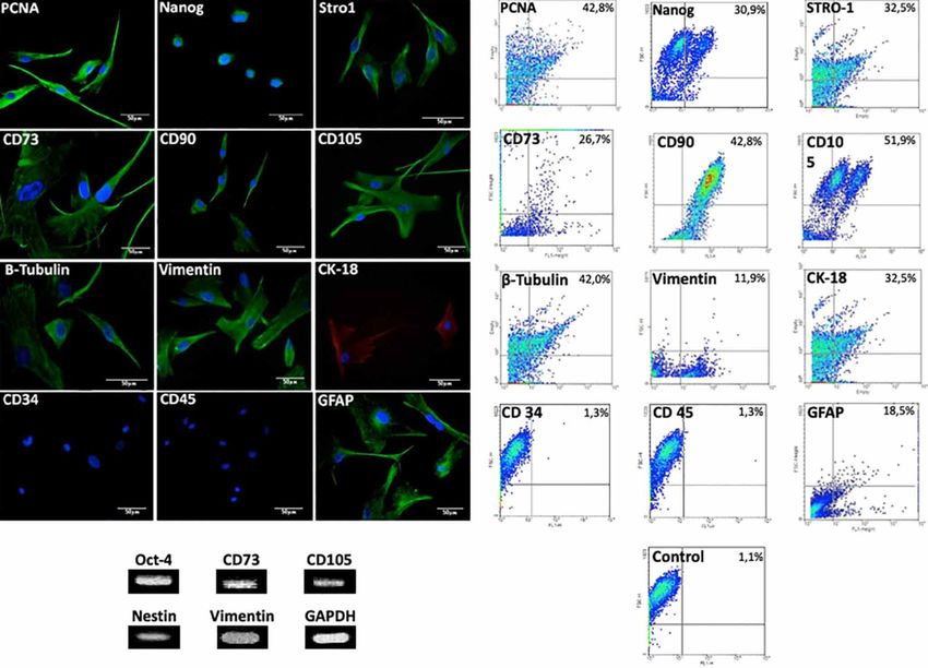
BARRETO, R. S. N.; DE OLIVEIRA, F. D.; MATIAS, G. S. S.; RODRIGUES, M. N.; CARVALHO, R. C.; FRANCIOLLI, A. L. R.; FRATINI, P. & MIGLINO, M. A. Rabbit vomeronasal
                                organ-derivered cells have mesenchymal profile and neuronal commitment. Int. J. Morphol., 38(5):1463-1472, 2020.

Fig. 1. Morphology of VNO-derived cell cultured with different media and periods. At day 5, discrete cell release from explant (A-C).
Beginning of colonies formation at day 10 (D-F). Colonies with large number of cells at day 15 (G-I). Cells acquired fibroblastoid shape
(J-L).

derived cells were able to generate around 46 colonies per                         during the evaluation period of 12 days. DMEM/F12 medium
103 plated cells (Fig. 2 A-B). In MTT assay, it was found that                     decreased cells growth around the eighth day and after this
High Glucose DMEM was the better medium for cells growth                           increased again during the next days (Fig. 2C). In Alpha-MEM

1466
BARRETO, R. S. N.; DE OLIVEIRA, F. D.; MATIAS, G. S. S.; RODRIGUES, M. N.; CARVALHO, R. C.; FRANCIOLLI, A. L. R.; FRATINI, P. & MIGLINO, M. A. Rabbit vomeronasal
organ-derivered cells have mesenchymal profile and neuronal commitment. Int. J. Morphol., 38(5):1463-1472, 2020.
medium cells grew between the first and second analysis, experiments. In the viability analysis with trypan blue, it
decline between the second and third analysis and, increase was verified that the number of living cells increased over
again gradually during successive stages (Fig. 2C). Then High time. It was possible to verify exponential growth of these
Glucose DMEM was selected as the medium to all other cells during the observed period (Fig. 2D).
Fig. 2. Colony forming units-fibroblastic assay (CFU), activity and cellular viability of VNO-derived cells. (A) colony formation delimited
by dashed circle and stained with crystal violet. (B) magnification of colony showed in A. (C) MTT assay for different media culture
conditions, evidencing exponential growth in high glucose DMEM. (D) Live and dead cell number stained by trypan blue in high glucose
DMEM cultured VNO-derived cells.
Presence of mesenchymal and neuronal markers. By Differentiation and tumorigenic formation in nude mice
immunofluorescence and flow cytometry, were observed assay. VNO-derived cells were inducted to adipogenic,
presence of markers for proliferation (PCNA), chondrogenic and osteogenic differentiation. After 21 days
undifferentiated profile (NANOG), neuronal lineages of adipogenic differentiation was possible to observed
(GFAP), mesenchymal stem cells (CD73, CD90, CD105 and VNO-derived cells differentiated into adipocyte lineage,
STRO-1) and cytoskeletal (Vimentin, b-tubulin and CK-18). by lipid cytoplasmic vacuoles stained in red by Oil Red
In addition, hematopoietic markers (CD34 and CD45) were and Scarlet Sudan techniques (Fig. 4A). Furthermore, after
absent in the VNO-derived cells (Fig. 3). Some proteins were 21 days of osteogenic differentiation was possible to ob-
selected to have their gene expression qualitatively analyzed, serve VNO-derived cells acquiring polygonal morphology
then CD73, CD105, vimentin, OCT4, nestin detection were with large cytoplasm filled with vacuoles instead of
in accordance with protein presence (Fig. 3). fibroblastoid shape, also bone extracellular matrix
1467BARRETO, R. S. N.; DE OLIVEIRA, F. D.; MATIAS, G. S. S.; RODRIGUES, M. N.; CARVALHO, R. C.; FRANCIOLLI, A. L. R.; FRATINI, P. & MIGLINO, M. A. Rabbit vomeronasal
organ-derivered cells have mesenchymal profile and neuronal commitment. Int. J. Morphol., 38(5):1463-1472, 2020.
Fig. 3. Presence of mesenchymal and neuronal markers by immunofluorescence, flow citometry and PCR. Labeling of markers for
proliferation (PCNA), undifferentiated profile (Nanog), mesenchymal stem cells (CD73, CD90, CD105 and STRO-1), cytoskeletal
(Vimentin, b-tubulin and CK-18) and neuronal lineages (GFAP). And absence of hematopoietic markers (CD34, CD117 and CD45).
formation was observed by calcium granules staining by and culture, and have some neuronal markers, such as nestin
Von Kossa technique (Fig. 4B). For chondrogenic and GFAP. The VNO luminal surface is covered by two
differentiation, by histology, were possible to detect that different epithelia, in the lateral part is the respiratory
VNO-derived cell changed their shape to be chondrocyte- epithelia, and the medial part and commissures are covered
like with large amount of collagen fibers interposed in- by the neuroreceptor or olfactory epithelium (Villamayor
between, evidenced by Masson’s Trichrome and Picrosirius et al.). The rabbit VNO-derived cells have a homogeneous
staining (Fig. 4C). For tumorigenic formation in the Balb/ fibroblastoid morphology with capability to form colonies,
c nu/nu mice, biceps, liver, lung, kidney, and heart were as similarity as described by others for murine (Friedenstein
histopathologicaly analyzed after 60 days of VNO-derived et al.; Murrell et al., 2009; Wetzig et al., 2011) or rabbit
cells injection, and no tumor formation was observed in (Ercolin et al., 2016) olfactory epithelium.
any organ analyzed (Fig. 5).
The PCNA labelling indicates a proliferative status
of those cells, and after 5 days of culture the cells increase
DISCUSSION their metabolism and the number of live cells, when cultured
in high glucose DMEM. Those parameters enforce that
rabbit VNO-derived cells are easy to obtain and to maintain
Herein we showed that rabbit VNO could be a fa- in culture, some favorable aspects to be used as cell therapy
vorable source of mesenchymal cells that is easy to isolate (Ercolin et al.; Borghesi et al.,2017).
1468BARRETO, R. S. N.; DE OLIVEIRA, F. D.; MATIAS, G. S. S.; RODRIGUES, M. N.; CARVALHO, R. C.; FRANCIOLLI, A. L. R.; FRATINI, P. & MIGLINO, M. A. Rabbit vomeronasal
organ-derivered cells have mesenchymal profile and neuronal commitment. Int. J. Morphol., 38(5):1463-1472, 2020.
Fig. 4. Differentiation and tumorigenic formation in nude mice assay. Adipogenic differentiation stained with Oil Red (A) and Sudam
Scarlet (B), note the deposition of lipid droplets (arrows). In (C) osteogenic differentiation stained with Von Kossa observe deposition of
calcium colored in black (arrows). In (D) magnification of C. In (E-F) Chondrogenic differentiation stained by Masson's Trichrome and
Picrosirus, note the collagen fibers deposition (arrows). In the upper right edge of the image B, D and F are the controls.
As stated by Parolini et al. (2008), mesenchymal stem as CD45 and CD34); and also, be able to differentiate to
cells need to be adherent to plastic, express CD105, CD73 osteoblasts, adipocytes and chondroblasts. As our VNO-
and CD90; lack expression of hematopoietic markers (such derived cells attended those parameters, we can suggest that
1469BARRETO, R. S. N.; DE OLIVEIRA, F. D.; MATIAS, G. S. S.; RODRIGUES, M. N.; CARVALHO, R. C.; FRANCIOLLI, A. L. R.; FRATINI, P. & MIGLINO, M. A. Rabbit vomeronasal
organ-derivered cells have mesenchymal profile and neuronal commitment. Int. J. Morphol., 38(5):1463-1472, 2020.
they have mesenchymal profile. Moreover, those cells are mice, been saved to cell therapy use[U1] (Tropel et al.,
unable to be tumorigenic when injected in immunodeficient 2004).
Fig. 5. Tumorigenic potential of the VNO cells in immunocompromised nude mice. No alterations were found in hind limb inoculation
(arrow, A), skeletal muscle (B), cardiac muscle (C), liver (D), lung (E) and kidney (F).
1470BARRETO, R. S. N.; DE OLIVEIRA, F. D.; MATIAS, G. S. S.; RODRIGUES, M. N.; CARVALHO, R. C.; FRANCIOLLI, A. L. R.; FRATINI, P. & MIGLINO, M. A. Rabbit vomeronasal
organ-derivered cells have mesenchymal profile and neuronal commitment. Int. J. Morphol., 38(5):1463-1472, 2020.
Another characteristic of the rabbit VNO-derived RESUMEN: El órgano vomeronasal (OVN) es un órgano
cells is the presence of some pluripotent markers at mRNA accesorio de la vía olfatoria, que detecta feromonas y emite seña-
and protein levels (OCT4 and NANOG), indicating a partial les que afectan la modulación del comportamiento social y
undifferentiated profile of those cells, because complete reproductivo. Las células madre OVN reemplazan las neuronas
durante toda la vida. El objetivo de este estudio fue aislar y carac-
undifferentiated profile is regulated by OCT4, SOX2, KLF4
terizar células derivadas del órgano vomeronasal de conejos raza
e C-MYC co-expression (Táncos et al., 2015). Also, Nueva Zelanda. Para el aislamiento y el cultivo celular se utiliza-
NANOG activates the OCT4 promoter region, then they co- ron cinco conejos machos con una edad de 120 días. Las células
expression given a less restrictive differentiation potential del OVN presentaron etiquetado para la proliferación (PCNA), un
to mesenchymal cells (Pei et al., 2018; Yang et al., 2018). perfil indiferenciado (Nanog), neuronal (GFAP), células madre
mesenquimales (CD73, CD90 y CD105 y Stro-1). Además, se ob-
Those cells have higher expression of CK-18 than servó presencia de citoesqueleto (Vimentina, β-tubulina y CK-18)
vimentin, that imbalance of cytoskeletal markers could y ausencia de marcadores hematopoyéticos (CD34, CD117 y CD45)
indicate mesenchymal-epithelial transition (MET). The tanto por inmunofluorescencia como por citometría de flujo. Me-
diante PCR fue posible verificar la expresión de algunos genes de
epithelial-mesenchymal/mesenchymal-epithelial transition
perfil indiferenciado (Oct-4), neuronal (Nestin) y mesenquimatoso
(EMT/MET) are events that occur physiologically in (CD73, CD105 y Vimentin). Las células derivadas del OVN se
reparative and/or proliferative tissues and in association with diferencian in vitro en adipocitos, osteocitos y condrocitos, y no
stem cells, such as mesenchymal ones (Li et al., 2011). Also, presentan un potencial tumorigénico al ser infiltrados en ratones
those events on mesenchymal cells are necessary to permit Balb / c nu / nu. En conclusión, las células derivadas de OVN de
proliferation, migration and differentiation (Li et al.). In the conejo tienen un perfil que podría ser compatible con el epitelio
VNO there is a constant turnover of epithelial cells from olfatorio / neurorreceptor de OVN transmitiendo factores al re-
commissures to medial wall, requiring a continuous cambio epitelial o incluso mediante la diferenciación en células
replacement of commissural cells (Villamayor et al.), then epiteliales para reemplazar el epitelio comisural.
VNO-derived cell, as mesenchymal profile and possibly in
PALABRAS CLAVE: Sistema olfativo; Epitelio
MET, could support the VNO olfactory epithelium. olfativo; Bulbo olfatorio; Células madre neuronales.
On VNO medial parenchyma, the connective tissue
surrounds the unmyelinated nerves and supports the sen-
sorial epithelium, then shares some markers with those two REFERENCES
tissues, such as neuroreceptors (Gai2, GaO) and olfactory
marker protein (Villamayor et al.). Furthermore, in our
Borghesi, J.; Mario, L. C.; Carreira, A. C. O.; Miglino, M. A. & Favaron, P.
results, the VNO-derived cells that have parenchymal O. Phenotype and multipotency of rabbit (Oryctolagus cuniculus)
characteristics also presented neuronal markers, such as amniotic stem cells. Stem Cell Res. Ther., 8(1):27, 2017.
Nestin and GFAP, that also indicate that those cells could Brann, J. H. & Firestein, S. J. A lifetime of neurogenesis in the olfactory
derivate from a neuronal lineage and be supportive to system. Front. Neurosci., 8:182, 2014.
Carmichael, J.; DeGraff, W. G.; Gazdar, A. F.; Minna, J. D. & Mitchell, J.
olfactory/neuroreceptor epithelium. In conclusion, the B. Evaluation of a tetrazolium-based semiautomated colorimetric assay:
rabbit VNO-derived cells have a marker profile that could assessment of radiosensitivity. Cancer Res., 47(4):943-6, 1987.
be supportive to VNO olfactory/neuroreceptor epithelium Ercolin, A. C. M.; Roballo, K. C. S.; Casals, J. B.; Pieri, N. C. G.; Souza,
by delivering factors to epithelial turnover, or even by A. F.; Barreto, R. S. N.; Bressan, F. F:; Feitosa, M. L. T.; Miglino, M.
A.; Meirelles, F. V.; et al. Rabbit olfactory stem cells. Isolation protocol
differentiation into epithelial cells to replacement of and characterization. Acta Cir. Bras., 31(1):59-66, 2016.
commissural epithelium. Friedenstein, A. J.; Gorskaja, J. F. & Kulagina, N. N. Fibroblast precursors
in normal and irradiated mouse hematopoietic organs. Exp. Hematol.,
4(5):267-74, 1976.
Hardy, D. & Saghatelyan, A. Different forms of structural plasticity in the
ACKNOWLEDGEMENTS. This work was supported by adult olfactory bulb. Neurogenesis (Austin), 4(1):e1301850, 2017.
The São Paulo Research Foundation [FAPESP, grant number Li, B.; Zheng, Y. W.; Sano, Y. & Taniguchi, H. Evidence for mesenchymal-
epithelial transition associated with mouse hepatic stem cell
12/11560-4].
differentiation. PLoS One, 6(2):e17092, 2011.
Monti Graziadei, G. A.; Karlan, M. S.; Bernstein, J. J. & Graziadei, P. P.
Reinnervation of the olfactory bulb after section of the olfactory nerve
BARRETO, R. S. N.; DE OLIVEIRA, F. D.; MATIAS, G. S. in monkey (Saimiri sciureus). Brain Res., 189(2):343-54, 1980.
S.; RODRIGUES, M. N.; CARVALHO, R. C.; FRANCIOLLI, Moulton, D. G.; Çelebi, G. & Fink, R. P. Olfaction in Mammals—Two
A. L. R.; FRATINI, P. & MIGLINO, M. A. Las células deriva- Aspects: Proliferation of Cells in the Olfactory Epithelium and
Sensitivity to Odours. In: Wolstenholme, G. E. W. & Knight, J. (Eds.).
das del órgano vomeronasal del conejo tienen perfil
Ciba Foundation Symposium - Internal Secretions of the Pancreas
mesenquimatoso y compromiso neuronal. Int. J. Morphol., (Colloquia on Endocrinology). Novartis Foundation Symposia, 2008.
38(5):1463-1472, 2020. pp.227-50.
1471BARRETO, R. S. N.; DE OLIVEIRA, F. D.; MATIAS, G. S. S.; RODRIGUES, M. N.; CARVALHO, R. C.; FRANCIOLLI, A. L. R.; FRATINI, P. & MIGLINO, M. A. Rabbit vomeronasal
organ-derivered cells have mesenchymal profile and neuronal commitment. Int. J. Morphol., 38(5):1463-1472, 2020.
Murrell, W.; Sanford, E.; Anderberg, L.; Cavanagh, B. & Mackay-Sim, A. Corresponding author:
Olfactory stem cells can be induced to express chondrogenic phenotype Maria Angelica Miglino
in a rat intervertebral disc injury model. Spine J., 9(7):585-94, 2009. Av. Prof. Orlando Marques de Paiva, 87
Owen, M. E.; Cavé, J. & Joyner, C. J. Clonal analysis in vitro of osteogenic
CEP: 05508-270
differentiation of marrow CFU-F. J. Cell Sci., 87(Pt. 5):731-8, 1987.
Park, C.; Ahn, M.; Lee, J. Y.; Lee, S.; Yun, Y.; Lim, Y. K.; Taniguchi, K. &
Cidade Universitária
Shin, T. A morphological study of the vomeronasal organ and the Butantã
accessory olfactory bulb in the Korean roe deer, Capreolus pygargus. São Paulo, SP
Acta Histochem., 116(1):258-64, 2014. BRAZIL
Parolini, O.; Alviano, F.; Bagnara, G. P.; Bilic, G.; Bühring, H. J.; Evange-
lista, M.; Hennerbichler, S.; Liu, B.; Magatti, M.; Mao, N.; et al. Concise
review: isolation and characterization of cells from human term Email: miglino@usp.br
placenta: outcome of the First International Workshop on Placenta
Derived Stem Cells. Stem Cells, 26(2):300-11, 2008.
Pei, W.; Lu, T.; Wang, K.; Ji, M.; Zhang, S.; Chen, F.; Li, L.; Li, X. &
Guan, W. Biological characterization and pluripotent identification of Received: 27-03-2020
ovine amniotic fluid stem cells. Cytotechnology, 70(3):1009-21, 2018. Accepted: 02-05-2020
Pino, A.; Fumagalli, G.; Bifari, F. & Decimo, I. New neurons in adult brain:
distribution, molecular mechanisms and therapies. Biochem.
Pharmacol., 141:4-22, 2017.
Sekiya, I.; Larson, B. B. L.; Smith, J. J. R.; Pochampally, R.; Cui, J. G. &
Prockop, D. J. Expansion of human adult stem cells from bone marrow
stroma: conditions that maximize the yields of early progenitors and
evaluate their quality. Stem Cells, 20(6):530-41, 2002.
Shohayeb, B.; Diab, M.; Ahmed, M. & Ng, D. C. H. Factors that influence
adult neurogenesis as potential therapy. Transl. Neurodegener., 7:4,
2018.
Táncos, Z.; Bock, I.; Nemes, C.; Kobolák, J. & Dinnyés, A. Cloning and
characterization of rabbit POU5F1, SOX2, KLF4, C-MYC and
NANOG pluripotency-associated genes. Gene, 566(2):148-57, 2015.
Tomiyasu, J.; Kondoh, D.; Sakamoto, H.; Matsumoto, N.; Sasaki, M.;
Kitamura, N.; Haneda, S. & Matsui, M. Morphological and histological
features of the vomeronasal organ in the brown bear. J. Anat.,
231(5):749-57, 2017.
Tropel, P.; Noël, D.; Platet, N.; Legrand, P.; Benabid, A. L. & Berger, F.
Isolation and characterisation of mesenchymal stem cells from adult
mouse bone marrow. Exp. Cell Res., 295(2):395-406, 2004.
Villamayor, P. R.; Cifuentes, J. M.; Fdz-de-Troconiz, P. & Sanchez-
Quinteiro, P. Morphological and immunohistochemical study of the
rabbit vomeronasal organ. J. Anat., 233(6):814-27, 2018.
Wetzig, A.; Mackay-Sim, A. & Murrell, W. Characterization of olfactory
stem cells. Cell Transplant., 20(11-12):1673-91, 2011.
Wilson, K. C. & Raisman, G. Age-related changes in the neurosensory
epithelium of the mouse vomeronasal organ: extended period of
postnatal growth in size and evidence for rapid cell turnover in the
adult. Brain Res., 185(1):103-13, 1980.
Yang, P. J.; Yuan, W. X.; Liu, J.; Li, J. Y.; Tan, B.; Qiu, C.; Zhu, X. L.; Lai,
D. M.; Guo, L. H. & Yu, L. Y. Biological characterization of human
amniotic epithelial cells in a serum-free system and their safety
evaluation. Acta Pharmacol. Sinica, 39:1305-16, 2018.
1472You can also read