Pulsed versus continuous wave low-level light therapy on osteoarticular signs and symptoms in limited scleroderma (CREST syndrome): a case report
←
→
Page content transcription
If your browser does not render page correctly, please read the page content below
Pulsed versus continuous wave low-
                 level light therapy on osteoarticular
                 signs and symptoms in limited
                 scleroderma (CREST syndrome): a
                 case report

                 Daniel Barolet

Downloaded From: https://www.spiedigitallibrary.org/journals/Journal-of-Biomedical-Optics on 25 Jun 2022
Terms of Use: https://www.spiedigitallibrary.org/terms-of-use
Journal of Biomedical Optics 19(11), 118001 (November 2014)

                Pulsed versus continuous wave low-level light
                therapy on osteoarticular signs and symptoms in
                limited scleroderma (CREST syndrome): a case report
                Daniel Baroleta,b,*
                a
                 RoseLab Skin Optics Laboratory, 3333 100th Avenue, Suite 200, Laval, Quebec, H7T 0G3, Canada
                b
                 McGill University, Department of Medicine, Division of Dermatology, Montreal, Quebec, H3A 1A1, Canada

                              Abstract. Limited cutaneous systemic sclerosis (lcSSc) was formerly known as CREST syndrome in reference
                              to the associated clinical features: calcinosis, Raynaud’s phenomenon, esophageal dysfunction, sclerodactyly,
                              and telangiectasias. The transforming growth factor beta has been identified as a major player in the pathogenic
                              process, where low-level light therapy (LLLT) has been shown to modulate this cytokine superfamily. This case
                              study was conducted to assess the efficacy of 940 nm using millisecond pulsing and continuous wave (CW)
                              modes on osteoarticular signs and symptoms associated with lcSSc. The patient was treated two to three times a
                              week for 13 weeks using a sequential pulsing mode on one elbow and a CW mode on the other. Efficacy assess-
                              ments included inflammation, symptoms, pain, health scales, patient satisfaction, clinical global impression,
                              and adverse effects monitoring. Considerable functional and morphologic improvements were observed
                              after LLLT, with the best results seen with the pulsing mode. No adverse effects were noted. Pulsed LLLT
                              represents a treatment alternative for osteoarticular signs and symptoms in limited scleroderma (CREST
                              syndrome). © 2014 Society of Photo-Optical Instrumentation Engineers (SPIE) [DOI: 10.1117/1.JBO.19.11.118001]
                              Keywords: limited cutaneous systemic sclerosis; low-level light therapy; CREST syndrome; transforming growth factor beta; pulse
                              structure; continuous wave mode; photobiomodulation; osteoarticular symptoms and signs.
                              Paper 140030PR received Jan. 29, 2014; accepted for publication Oct. 23, 2014; published online Nov. 13, 2014.

                1     Introduction                                                                 increases collagen synthesis by fibroblasts and downregulates
                Scleroderma, or systemic sclerosis (SSc), is a lifelong condition                  extracellular matrix degradation. Evidence comes from past
                characterized by vasculopathy, fibrosis of skin and various inter-                 studies reporting, for instance, a TGF-β upregulation, an
                nal organs, and inflammation or autoimmunity.1,2 Systemic                          increase in the expression of TGF-β receptors, as well as the
                scleroderma is a rare disorder, with an annual incidence in                        observation that the blockade of endogenous TGF-β signaling
                the United States of about 20 cases per 1 million adults, and                      prevents upregulated collagen synthesis in scleroderma fibro-
                a prevalence of 100 to 300 per 1 million population.3,4 It is                      blasts.13–15 TGF-β is also known to be involved in immunomo-
                more common among women than men, and in certain groups                            dulatory activities. Thus, TGF-β appears to be a sound target for
                such as Native Americans.3,4                                                       therapeutic intervention.16,17
                    Limited cutaneous SSc (lcSSc) is part of the heterogeneous                         Interestingly, low-level light therapy (LLLT) with red to
                group of sclerodermas. LcSSc was formerly known as CREST                           near-infrared (NIR) wavelengths has been shown to trigger natu-
                                                                                                   ral intracellular photobiochemical reactions including TGF-β
                syndrome in reference to the associated clinical features: calci-
                                                                                                   modulation.18–21 Red to NIR light is thought to be absorbed
                nosis, Raynaud’s phenomenon, esophageal dysfunction, sclero-
                                                                                                   by mitochondrial respiratory chain components.22,23 Absorbed
                dactyly, and telangiectasias. This connective tissue disease
                                                                                                   light converted to chemical kinetic energy results in the increase
                typically has gradual onset and disease expressions are restricted
                                                                                                   of reactive oxygen species and adenosine triphosphate, initiating
                to certain areas of the skin.5 In patients with lcSSc, the core man-
                                                                                                   a signaling cascade which can modulate the expression of
                ifestations of the disease, including skin calcifications, are
                                                                                                   growth factors and cytokines.24–27 Hence, LLLT might be help-
                mostly confined to the fingers, hands, and forearms distal to the
                                                                                                   ful in the treatment of symptoms associated with lcSSc.
                elbows, with or without tightening of the skin of the lower
                                                                                                       This case study was conducted to assess the efficacy of NIR
                extremities distal to the knees. Cutaneous telangiectasias on the
                                                                                                   (940 nm) LLLT using millisecond (ms) pulsing and CW on
                face are also seen, along with varying degrees of internal organ
                                                                                                   osteoarticular signs and symptoms associated with lcSSc.
                involvement. Proximal extremities and the trunk are not involved.
                LcSSc may be debilitating and influences a person’s ability to
                participate in activities of daily life in different ways.6–8                      2       Materials and Methods
                    Although the pathogenesis of this condition is unclear, a
                                                                                                   2.1      Case Description
                number of studies have suggested that the transforming growth
                factor beta (TGF-β) is an important candidate in the pathogenic                    The patient was a Caucasian 34-year-old female with Fitzpatrick
                process.9–12 TGF-β is a prototypic profibrotic cytokine that                       phototype II. She had a 13-year history of symptoms and pre-
                                                                                                   sented with the following features of the disease: generalized

                *Address all correspondence to: Daniel Barolet, E-mail: daniel.barolet@mcgill
                 .ca                                                                               0091-3286/2014/$25.00 © 2014 SPIE

                Journal of Biomedical Optics                                               118001-1                                     November 2014   •   Vol. 19(11)

Downloaded From: https://www.spiedigitallibrary.org/journals/Journal-of-Biomedical-Optics on 25 Jun 2022
Terms of Use: https://www.spiedigitallibrary.org/terms-of-use
Barolet: Pulsed versus continuous wave low-level light therapy on osteoarticular signs. . .
calcinosis, Raynaud’s phenomenon, sclerodactyly, and telan-
giectasia. There was no history of esophageal dysmotility. The
extent of her calcinosis affected her forearms, chin, face, and
buttocks. She underwent a surgical procedure to remove calci-
fications from her buttocks. The patient presented with elbow
mobility restrictions. She had a history of juvenile dermatomyo-
sitis (quiescent). Her medication included Coumadin and
Adalat. She failed to respond to a variety of pharmacological
treatments including methotrexate. The patient was initially
referred to our clinic by her rheumatologist in December 2010.
She initially received a series of LLLT treatments (940 nm)
three times a week on her face and chin over a 6-month period,
using Lumiphase technology (OPUSMED Inc., Montreal,
Canada).
Fig. 2 Hypodermic needle probe inserted at 1- and 3-mm depth in the
2.2 Study Procedures dermis during LLLT treatments.
This was a single-blind within subject case study, where the left
forearm was randomly assigned to receive LLLT using sequen- throughout the LLLT session (Type-T thermocouple, Omega,
tial pulsing mode and the right forearm assigned to LLLT using Montreal, Canada; Fig. 2).
a CW mode.
The patient was treated two to three times a weeks for 13
2.3 Patient Assessments
weeks with 940 nm, using LumiPhase™ technology
(OPUSMED Inc.). For the sequential pulsing mode, the power Efficacy assessments included the examination of inflammation,
density was of 60 mW∕cm2 for a total fluence of 81 J∕cm2 pain, and other signs and symptoms associated with the patient’s
(30 min). The pulsing patterns and time on-and-time-off sequen- condition, a clinical evaluation, and a patient satisfaction ques-
ces were as follows (see Fig. 1): Pulse duration (time on) 500 μs, tionnaire. Treatment safety was examined by adverse effects
pulse interval (time off) 150 μs, 4 pulses per pulse train, and a monitoring. Assessments were conducted at baseline and
pulse train interval of 1550 μs. For the CW mode, an irradiance after 13 weeks of treatment (endpoint). The clinical rater and
of 60 mW∕cm2 was used for a total fluence of 81 J∕cm2 the patient were blinded to which forearm received the pulsed
(15 min). The size of the treatment areas were 20 cm × 10 cm, or CW LLLT treatment.
and the treatment distance was 4 0.4 cm.
2.3.1 Symptoms scale
2.2.1 Digital photographs
For each forearm, the degree of morning stiffness, flexibility,
Photographs (Canon Dual Flash EOS 10D, Canon, Tokyo, elbow amplitude (flexion/extension), strength, ability to lift
Japan with EX SIGMA 50 mm 1:2.8 macrolens, Sigma, heavy objects (10 lb and more), mobility (rotation), calcium
Aizu, Japan) were taken before and at the follow-up visit. deposits (visually), ulceration, and skin thickness were rated.
Each photograph was taken maintaining as much as possible The percent improvement from baseline was recorded at endpoint.
the identical ambient lighting, pose, and camera angles.
2.3.2 Inflammation scale
2.2.2 Skin temperature monitoring
The degree of swelling, tenderness, or warmth was rated using a
NIR radiation typically induces molecular vibrations and rota- 3-point scale (0 ¼ none, 1 ¼ moderate, 2 ¼ severe) at endpoint.
tions and by so doing increases skin temperature.1 Papillary der-
mis temperature was monitored at a depth of 1 and 3 mm with
needle probes placed on the interior face of the left forearm 2.3.3 Clinical assessment
The clinical global impression of change was rated at
endpoint using a range of responses from 0 ¼ none; 1 ¼ mild;
2 ¼ moderate; 3 ¼ good; 4 ¼ excellent.
2.3.4 Patient satisfaction
A series of 12 questions were asked to evaluate the extent
to which the treatments received on each forearm met the
patients’ needs and expectations. Aside from yes/no questions,
these were rated on a scale 1 to 7 (1 ¼ worse outcome to
7 ¼ best outcome). The list of questions is presented in Table 1.
Fig. 1 Schematization of the sequential pulsing mode used on the left 2.3.5 Treatment-related adverse effects
forearm. Pulse duration [time on (PD)] 500 μs, pulse interval [time off
(PI)] 150 μs, 4 pulses per pulse train (PPT), and a pulse train interval The presence of discomfort, erythema, edema, and pain associ-
(PTI) of 1550 μs. ated with the treatment was recorded.
Journal of Biomedical Optics 118001-2 November 2014 • Vol. 19(11)
Downloaded From: https://www.spiedigitallibrary.org/journals/Journal-of-Biomedical-Optics on 25 Jun 2022
Terms of Use: https://www.spiedigitallibrary.org/terms-of-useBarolet: Pulsed versus continuous wave low-level light therapy on osteoarticular signs. . .
Table 1 Patient satisfaction questionnaire.
Question Pulse-Treated CW-Treated
side side
1. How satisfied or dissatisfied are you with the way the treatment relieved your symptoms? 7 5
2. How satisfied or dissatisfied are you with the ability of the treatment to prevent worsening of your condition? 7 7
3. How satisfied or dissatisfied are you with the amount of time it took for the treatment to start working? 7 7
4. How convenient or inconvenient was the amount of time necessary to administer the treatment? 3 5
5. Did you have any side-effects from the treatment? No No
6. How bothersome were the side effects of the treatment? N/A N/A
7. To what extent did the side effects interfere with your physical health and ability to function (i.e., pain)? N/A N/A
8. To what degree do the side effects affect your overall satisfaction with the treatment? N/A N/A
9. Overall, how confident are you that using this treatment was a good thing for you? 7 7
10. How certain are you that the good things about the treatment outweigh the bad things? 7 7
11. Taking all things into account, how satisfied or dissatisfied are you with this treatment? 7 7
3 Results downside and was overall very satisfied with the treatment
Figure 3 depicts the photographs of the patient’s forearms at (Q.9 to Q.10). The patient stated that she would recommend
baseline and after 13 weeks of LLLT treatment using pulsed this treatment to other patients with a similar condition
or CW delivery mode. Efficacy assessments revealed that both (Q.12). Responses for each forearm on the satisfaction question-
forearms improved after LLLT treatment. However, some naire are presented in Table 1.
differences, mostly in favor of the pulse-treated side, were Both the pulse and CW LLLTs were well tolerated. Other
than slight erythema noted on the left forearm, no significant
seen in clinical outcomes.
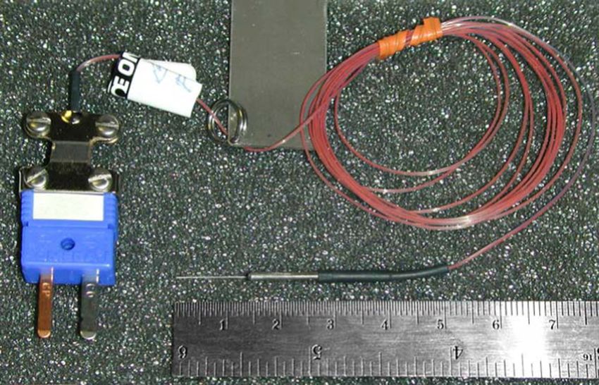
The percent improvement from baseline was recorded at end-
point for symptoms associated with the patient’s condition. As
can be appreciated in Fig. 4, the degree of improvement was
greater for most symptoms on the pulse-treated side in compari-
son to the CW-treated side, with the greatest difference seen for
calcium deposits (40% for the pulse side versus 4% for the CW
side). A small improvement (5%) was seen in favor of the CW-
treated area for strength/ability to lift heavy objects. No differ-
ence between treatment sides was observed for ulceration and
skin thickness. Symptoms assessment also revealed that only
moderate tenderness was noted on both forearms, as docu-
mented on the inflammation scale conducted at the end of
the treatment period; no swelling or warmth was observed.
The clinical assessment was rated at endpoint using a range
of responses from 0 (none) through 4 (excellent). The CW-
treated forearm was rated as moderately improved, whereas
the change seen on the pulse-treated side was deemed excellent.
The pattern of results was similar from the patient’s perspective.
At the end of the study period, the patient reported being very
satisfied with various aspects of both treatments including the
ability of the treatment to prevent worsening of symptoms
and with the amount of time it took for the treatment to start
working; the degree of satisfaction with symptom relief was,
however, deemed superior on the pulse-treated side (Q.1 to Q.3).
The amount of time necessary to administer the treatment was
judged to be somewhat more convenient for the CW-treated
forearm in comparison with the pulse-treated forearm (Q.4).
The patient also reported no side effects from treatment (Q.5 Fig. 3 Patient forearms before (upper panel) and after (lower panel)
to Q.8) on either forearm. The patient also judged that the treat- LLLT treatments. The left forearm receives LLLT using a sequential
ment on both forearms was satisfactory outweighing the pulsing mode and the right forearm receives LLLT using a CW mode.
Journal of Biomedical Optics 118001-3 November 2014 • Vol. 19(11)
Downloaded From: https://www.spiedigitallibrary.org/journals/Journal-of-Biomedical-Optics on 25 Jun 2022
Terms of Use: https://www.spiedigitallibrary.org/terms-of-useBarolet: Pulsed versus continuous wave low-level light therapy on osteoarticular signs. . .
Fig. 4 Percent improvement from baseline at endpoint in the degree of symptoms for each forearm.
treatment-related adverse effects were noted during the entire LLLT significantly improved the appearance and severity of
study period including the presence of discomfort, edema, lesions. Benefits to the patient were also noted from the patient’s
and pain. perspective. Furthermore, no treatment-emergent adverse effects
In the present study, skin temperature was monitored. were observed. Overall, significant functional and morphologic
Temperature variations were registered by thermocouple hypo- improvements following LLLT treatment were observed with
dermic probes rigorously placed with adhesive tape and were the best results seen with the pulsing mode. One perceived ad-
never greater than 39.8°C at a depth of 1 mm and 38.3°C at vantage of the CW over the pulse delivery was the treatment
3 mm (typical treatment session temperature curves shown in duration; however, given the added benefits of the pulsed
Fig. 5). Monitoring attested that the skin temperature during mode, this does not appear to be a significant drawback.
LLLT application increased without reaching the skin injury LLLT therapy appears to bring relief to patients affected by
threshold level (>42°C). this debilitating disorder in a noninvasive manner. LLLT therapy
potentially has two mechanisms of action: thermal and nonther-
4 Discussion mal. NIR wavelengths can raise skin temperature to 45°C—
Results from this case study suggest that 940-nm LLLT was effi- although the thermal effects do not create tissue injury—so
cacious in alleviating signs and symptoms associated with as to provide inside-out heating possibly vasodilating capillaries
lcSSc. Data from the clinical assessment revealed that the which in turn increases catabolic processes leading to a
Fig. 5 Papillary dermis temperature monitoring with 1- and 3-mm needle probes throughout the LLLT
session.
Journal of Biomedical Optics 118001-4 November 2014 • Vol. 19(11)
Downloaded From: https://www.spiedigitallibrary.org/journals/Journal-of-Biomedical-Optics on 25 Jun 2022
Terms of Use: https://www.spiedigitallibrary.org/terms-of-useBarolet: Pulsed versus continuous wave low-level light therapy on osteoarticular signs. . .
reduction of in situ calcinosis.28 Second, nonthermal effects also 11. V. Falanga and J. M. Julien, “Observations in the potential role of trans-
take place presumably resulting in a cascade of cellular reactions forming growth factor-beta in cutaneous fibrosis. Systemic sclerosis,”
Ann. N. Y. Acad. Sci. 593, 161–171 (1990).
including the modulation of growth factors and inflammatory
12. H. Ihn, “Autocrine TGF-beta signaling in the pathogenesis of systemic
mediators. It has been suggested that the LLLT anti-inflamma- sclerosis,” J. Dermatol. Sci. 49, 103–113 (2008).
tory effects are mediated via the activation of the TGF-β 13. T. Kawakami et al., “Increased expression of TGF-beta receptors by
complex.18,19 In this mechanism, LLLT-emitted photons must scleroderma fibroblasts: evidence for contribution of autocrine TGF-
be absorbed by a molecular chromophore. A growing body of beta signaling to scleroderma phenotype,” J. Invest. Dermatol. 110,
evidence suggests that the photobiomodulation mechanisms 47–51 (1998).
are ascribed to the activation of mitochondrial cytochrome c 14. A. C. Gilliam, “Scleroderma,” Curr. Dir. Autoimmun. 10, 258–279
(2008).
oxidase.29 Respiration in the mitochondria can be inhibited 15. H. Ihn et al., “Blockade of endogenous transforming growth factor β
by nitric oxide (NO) binding to cytochrome c oxidase which signaling prevents up-regulated collagen synthesis in scleroderma fibro-
competitively displaces oxygen and affects cell metabolism. blasts: association with increased expression of transforming growth
Excess NO binding is associated with inflammatory processes, factor β receptors,” Arthritis Rheum. 44, 474–480 (2001).
cell damage, and apoptosis. Light absorption dissociates NO, 16. R. W. Simms and J. H. Korn, “Cytokine directed therapy in sclero-
allowing cellular respiration to resume and normalization of cell derma: rationale, current status, and the future,” Curr. Opin.
Rheumatol. 14, 717–722 (2002).
activity, ultimately triggering biomolecular processes. Pulsed 17. C. Charles, P. Clements, and D. E. Furst, “Systemic sclerosis:
light delivery, as opposed to a CW mode, might favorably hypothesis-driven treatment strategies,” Lancet 367(9523), 1683–1691
enhance this cellular strategy. Short and intermittent light emis- (2006).
sions might enhance NO dissociation, therefore augmenting 18. P. R. Arany et al., “Activation of latent TGF-beta1 by low-power laser in
mitochondrial energy production and cellular activity. vitro correlates with increased TGF-beta1 levels in laser-enhanced oral
Overall, these preliminary results suggest a beneficial effect wound healing,” Wound Repair Regener. 15, 866–874 (2007).
19. H. Toyokawa et al., “Promotive effects of far-infrared ray on full thick-
on the alleviation and progression of symptoms. While these
ness skin wound healing in rats,” Exp. Biol. Med. (Maywood) 228,
findings are encouraging, additional research in larger samples 724–729 (2003).
of patients is needed to further evaluate this promising therapy. 20. M. Yu, J. O. Naim, and R. J. Lanzafame, “The effects of photoirradia-
Future studies should include long-term assessments to docu- tion on the secretion of TGF and PDGF from fibroblasts in vitro,” Lasers
ment maintenance of benefits over time. Further trials are also Med. Sci. 6, 8 (1994).
necessary to identify the cellular processes underlying the mech- 21. X. Gao and D. A. Xing, “Molecular mechanisms of cell proliferation
anisms at play in the therapeutic effect. In the future, LLLT may induced by low power laser irradiation,” J. Biomed. Sci. 16, 4
(2009).
well become a new treatment option to provide enhanced daily 22. T. I. Karu, L. V. Pyatibrat, and N. I. Afanasyeva, “A novel mitochondrial
relief to patients with this incapacitating condition. This novel signalling pathway activated by visible-to-near infrared radiation,”
therapeutic modality may broaden the currently restricted thera- Photochem. Photobiol. 80, 366–372 (2004).
peutic armamentarium of the disease. 23. M. R. Hamblin and T. N. Demidova, “Mechanisms of low level light
therapy,” Proc. SPIE 6140, 1–12 (2006).
24. T. Karu, “Primary and secondary mechanisms of action of visible to
References near-IR radiation on cells,” J. Photochem. Photobiol. B 49, 1–17
(1999).
1. D. E. Furst and P. J. Clements, Systemic Sclerosis, pp. 275–286, 25. A. Amat et al., “Modification of the intrinsic fluorescence and the bio-
Williams and Wilkins Publishers, Baltimore, MD (1996). chemical behavior of ATP after irradiation with visible and near-infrared
2. V. D. Steen et al., “Factors predicting development of renal involvement laser light,” J. Photochem. Photobiol. B 81, 26–32 (2005).
in progressive systemic sclerosis,” Am. J. Med. 76, 779–86 (1984). 26. S. Nemoto et al., “Role for mitochondrial oxidants as regulators of cel-
3. M. D. Mayes et al., “Prevalence, incidence, survival, and disease char- lular metabolism,” Mol. Cell Biol. 20, 7311–7318 (2000).
acteristics of systemic sclerosis in a large US population,” Arthritis 27. S. M. Schieke, P. Schroeder, and J. Krutmann, “Cutaneous effects of
Rheum. 48, 2246–2255 (2003). infrared radiation: from clinical observations to molecular response
4. M. Mayes, “Scleroderma epidemiology,” Rheum. Dis. Clin. North Am. mechanisms,” Photodermatol. Photoimmunol. Photomed. 19, 228–234
29, 239–254 (2003). (2003).
5. E. C. LeRoy et al., “Scleroderma (systemic sclerosis): classification, 28. J. S. Dover, T. J. Phillips, and K. A. Arndt, “Cutaneous effects and thera-
subsets and pathogenesis,” J. Rheumatol. 15, 202–205 (1988). peutic uses of heat with emphasis on infrared radiation,” J. Am. Acad.
6. P. A. Merkel et al. for the Scleroderma Clinical Trials Consortium, Dermatol. 20, 278–286 (1989).
“Measuring disease activity and functional status in patients with sclero- 29. T. I. Karu, “Mitochondrial signaling in mammalian cells activated by
derma and Raynaud’s phenomenon,” Arthritis Rheum. 46, 2410–2420 red and near-IR radiation,” Photochem. Photobiol. 84, 1091–1099
(2002). (2008).
7. G. Sandqvist et al., “Daily activities and hand function in women with
scleroderma,” Scand. J. Rheumatol. 32, 1–7 (2004).
Daniel Barolet is a dermatologist who has been specializing in laser
8. G. Sandqvist, A. Åkesson, and M. Eklund, “Daily occupations and well- therapy since 1991. A frontrunner in laser applications for the treat-
being in women with limited cutaneous systemic sclerosis,” Am. J. ment of vascular lesions and an innovator in the field of laser hair
Occup. Ther. 59, 390–397 (2005). removal, he is also a leading researcher in the area of photobiomo-
9. E. C. Leroy et al., “A strategy for determining the pathogenesis of sys- dulation. He is also adjunct professor of dermatology at McGill
temic sclerosis. Is transforming growth factor beta the answer?” University (Montreal, Quebec, Canada). For more than 15 years, he
Arthritis Rheum. 32(7), 817–825 (1989). has been active in research and development through his skin optics
10. T. Kawakami et al., “Immunohistochemical expression of transforming research laboratory, RoseLab. This research has led to the develop-
growth factor beta3 in calcinosis in a patient with systemic sclerosis and ment of numerous technological innovations, patents, and new treat-
CREST syndrome,” Br. J. Dermatol. 143, 1098–1100 (2000). ment methods.
Journal of Biomedical Optics 118001-5 November 2014 • Vol. 19(11)
Downloaded From: https://www.spiedigitallibrary.org/journals/Journal-of-Biomedical-Optics on 25 Jun 2022
Terms of Use: https://www.spiedigitallibrary.org/terms-of-useYou can also read