Protein Signature of Human Skin Fibroblasts Allows the Study of the Molecular Etiology of Rare Neurological Diseases
←
→
Page content transcription
If your browser does not render page correctly, please read the page content below
Protein Signature of Human Skin Fibroblasts Allows
the Study of the Molecular Etiology of Rare
Neurological Diseases
Andreas Hentschel
Leibniz-Institut fur Analytische Wissenschaften - ISAS eV
Artur Czech
Leibniz-Institut fur Analytische Wissenschaften - ISAS eV
Ute Münchberg
Leibniz-Institut fur Analytische Wissenschaften - ISAS eV
Erik Freier
Leibniz-Institut fur Analytische Wissenschaften - ISAS eV
Ulrike Schara-Schmidt
Universitat Duisburg-Essen Medizinische Fakultat
Albert Sickmann
Leibniz-Institut fur Analytische Wissenschaften - ISAS eV
Jens Reimann
Universitatsklinikum Bonn
Andreas Roos ( andreas.roos@uk-essen.de )
Leibniz-Institut fur Analytische Wissenschaften - ISAS eV https://orcid.org/0000-0003-2050-2115
Research
Keywords: Allgrove syndrome, Aladin, AAAS, triple-A syndrome, Myopodin/Synaptopodin-2, Ataxin-2
DOI: https://doi.org/10.21203/rs.3.rs-48014/v1
License: This work is licensed under a Creative Commons Attribution 4.0 International License.
Read Full License
Page 1/30
Abstract
Background: The elucidation of pathomechanisms leading to the manifestation of rare (genetically
caused) neurological diseases including neuromuscular diseases (NMD) represents an important step
toward the understanding of the genesis of the respective disease and might help to de ne starting
points for (new) therapeutic intervention concepts. However, these “discovery studies” are often limited by
the availability of human biomaterial. Moreover, given that results of next-generation-sequencing
approaches frequently result in the identi cation of ambiguous variants, testing of their pathogenicity is
crucial but also depending on patient-derived material.

Results: To systematically address the question if human skin broblasts might serve as valuable
biomaterial for (molecular) studies of NMD, using proteomic pro ling, we generated a protein library by
decreasing protein complexity via pH8-based sample fractionation: cataloguing of 8280 proteins revealed
the expression of a variety of such linked to genetic forms of motoneuron diseases, congenital
myasthenic syndromes, neuropathies and muscle disorders. In silico-based pathway analyses revealed
expression of a variety of proteins involved in muscle contraction and such decisive for neuronal function
and maintenance suggesting the suitability of human skin broblasts to study the etiology of NMD.
Based on these ndings, next we aimed to further demonstrate the suitability of this in vitro model to
study NMD by a use case: utilizing a data independent acquisition approach, the proteomic signature of
whole protein extracts of broblasts derived from an Allgrove-patient was studied. Paradigmatic
dysregulated proteins were con rmed in muscle biopsy of the patient and protein-functions could be
linked to neurological symptoms known for this disease. Moreover, protoemic investigation of nuclear
protein composition allowed the identi cation of protein-dysregulations according with structural
perturbations observed in the muscle biopsy. As proteomic data suggested a perturbed lipid homeostasis,
BODIPY-staining was performed on broblasts and coherent anti-stokes Raman scattering microscopy on
muscle biopsy. Results of both investigations suggest altered lipid storage as part of the underlying
disease-etiology.

Conclusions: our combined data reveal that human broblasts may serve as an in vitro system to study
the molecular etiology of rare neurological diseases exempli ed on Allgrove syndrome in an unbiased
fashion.

Introduction
A considerable number of patients suffer from diseases affecting the proper function of the nervous
system or skeletal muscle or even both. Although these diseases are individually considered as rare, they
nevertheless cause a signi cant burden for the patients and their families. Along the neuronal axis,
prominent examples of these disorders are genetically-caused forms of brain malformations (as a very
heterogeneous disease entity), motoneuron diseases such as 5q-associated spinal muscular atrophy and
familiar amyotrophic lateral sclerosis, neuropathies/ peripheral nerve diseases and disorders affecting
the nerve terminals (congenital myasthenic syndromes; CMS) as well as myopathic disorders. Often,

                                                 Page 2/30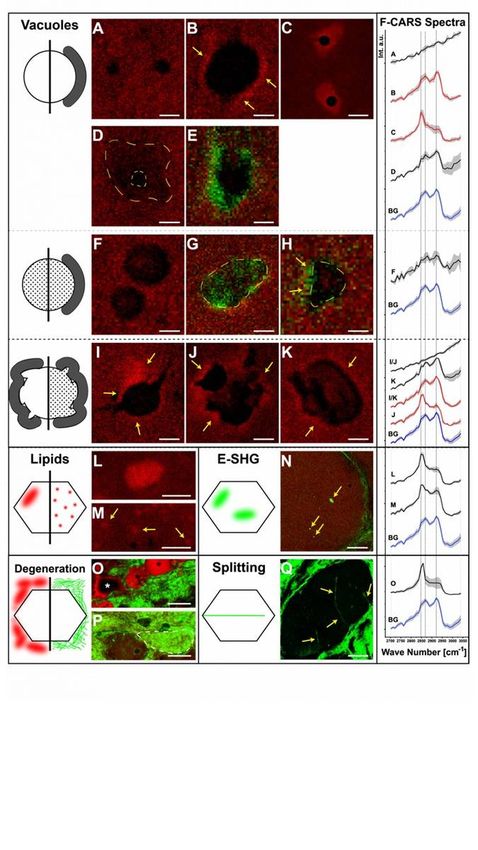
these diseases have both a wide clinical phenotype and partially overlapping manifestations suggesting
the presence of common pathophysiological cascades. Together, this group of diseases affects a large
fraction (approximately 3–5%) of all children, usually at early age and often leading to premature death
or chronic disability. Despite the discovery of many genes – especially in the next-generation-sequencing
era – causative for different forms of neurological diseases, in recent years, limited progress has been
achieved in the therapeutic intervention of these diseases which is not based on gene-replacement. In
addition, also for NMDs treatable by gene replacement, additional therapeutic concepts targeting
pathological alterations which are not reversed by gene/protein-restoration (such as altered signaling
modules) are demanded (1). One strategy to overcome this problem might be the classi cation of
disease groups based on the main underlying pathomechanisms leading toward the de nition of
common treatment-concepts (2). However, doing so, a profound understanding of these mechanisms is
needed. Clinical proteomics has gained enormous popularity over the last decade nding increasing
application in the elucidation of neurological diseases (3), albeit with a main focus on common
neurological disorders such as dementia and Parkinson’s disease (4) as these represent a real challenge
for the health care system and aging. Often, biochemical and functional studies of neurological disorders
are ”limited“ to animal models. This is caused by a lack of availability of human material, especially in
vitro models. However, different clinical proteomic studies toward a better understanding of the
underlying pathophysiology of rare forms of inherited neurological diseases have been carried out on
patient-derived skin broblasts. Results of these studies provided signi cant insights into cellular
mechanisms leading to phenotypical manifestation (5, 6) suggesting that broblasts might serve as a
valuable in vitro model for molecular studies of these diseases. To systematically address this
hypothesis, in this study, a protein/ spectral library of human skin broblasts was generated and in silico-
based pathway analyses were performed to highlight the abundance of proteins crucial of neuronal
development, maintenance and function. Moreover, an alignment of our library with proteins known to be
responsible for genetic forms of NMD revealed the expression of a magnitude of such linked to
motoneuron diseases, congenital myasthenic syndromes, neuropathies/ peripheral nervous system
disease and muscle disorders. Using this library as the basis for data independent acquisition
approaches, an exemplary rare inherited neurological disease, Allgrove syndrome (MIM: 231550), was
studied. Proteomic ndings and results of validation studies provided signi cant molecular insights into
underlying molecular etiology and hereby added perturbed lipid homeostasis to the list of
pathophysiological processes. Hence, our combined data support the concept of broblasts serving as a
valuable model to study the molecular genesis of neurological diseases as exempli ed here for Allgrove
syndrome.

Results
Protein cataloguing in human broblasts
To investigate if human skin broblasts may serve as suitable in vitro models to study the molecular
etiology of neurological diseases such as neuromuscular disorders and may thus help to overcome a

                                                  Page 3/30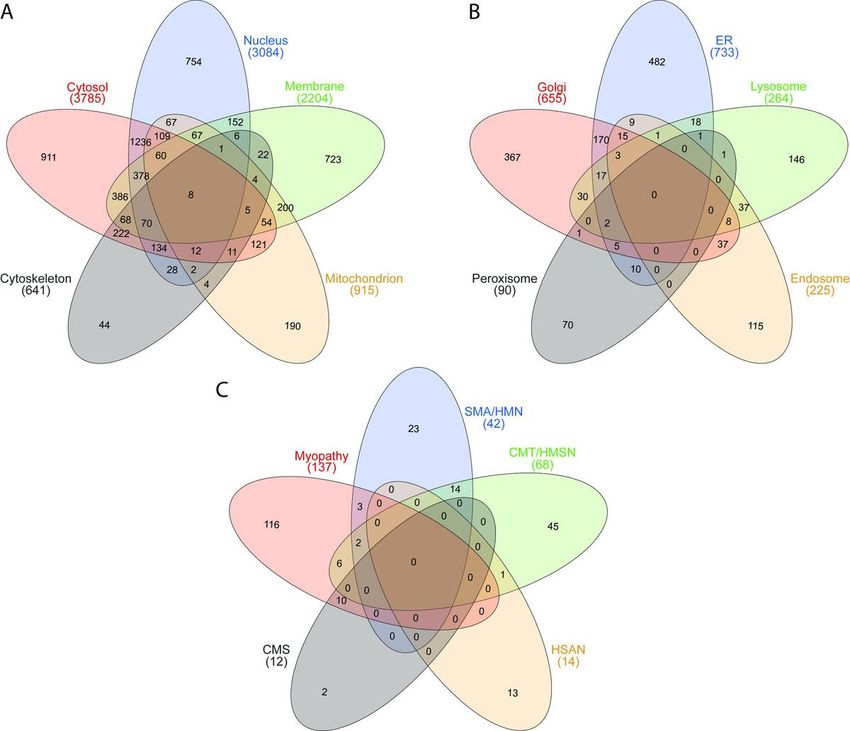
limitation of human material enabling these studies, we created and analysed a protein/spectral library:
after pH8-based fractionation of tryptic peptides obtained from whole protein extracts of broblasts
followed by LC-MS/MS-based protein identi cation, we generated a database of 8280 proteins (referring
to a total number of 96,512 peptides) covering a total range of 5 orders of magnitude. Hereby, Vimentin
(P08670; a class-III intermediate lament attached to the nucleus, endoplasmic reticulum and
mitochondria) was identi ed as the highest abundant and Kallikrein-7 (P49862; degradation-catalyst of
intercellular cohesive structures in the corni ed skin layer) as the lowest abundant protein (Suppl.
Table 1). These 8280 proteins cover all subcellular compartments (Fig. 1A & B). For the cytosol and the
nucleus (including the nucleoplasm) 911 and 754 proteins are annotated to be exclusively present in
these subcellular structures, respectively (Fig. 1A). The number of identi ed membrane proteins also
include such localizing to the endoplasmic reticulum membrane, mitochondrial membrane, Golgi
membrane and nuclear membrane, with 723 proteins only localized to one subcellular compartment. In
addition, our library covers 190 proteins known to be only present in mitochondria and 44 proteins
exclusively belonging to the cytoskeleton (Fig. 1A).

To evaluate the proteomic signature of human skin broblasts for potential analyses of genetically
caused NMD, catalogued proteins were analysed: we ltered for proteins where defects in the
corresponding genes were linked to the manifestation of diseases along the neuromuscular axis
including motoneuron and peripheral nervous system diseases, defects in neuromuscular transmission
and muscle disorders (https://neuromuscular.wustl.edu/; (7). This approach revealed the expression of a
total of 257 from 385 proteins (66.8%) linked to the manifestation of a respective disorder: for hereditary
motoneuron disorders (consisting of spinal muscular atrophy (SMA), distal SMA and hereditary
motoneuropathies (HMN), 42 out of a total of 53 registered proteins (80.8%) were identi ed. For
hereditary motor sensory neuropathies (HMSN), comprising a list of 91 known genes/ proteins, 68
(74.7%) were identi ed. In addition, 14 out of 26 known genes/ proteins (46.2%) causative for hereditary
sensory and autonomic neuropathies (HSAN) were detected in skin broblasts. For congenital
myasthenic syndrome (CMS) 12 of the currently known causative 35 genes/ proteins (34.3%) were
identi ed in our protein/ spectral library. In addition, from a further list of 351 proteins related to muscle
diseases, comprising not only such based on the above-mentioned sources but also candidates based on
experimental ndings for different muscle diseases, 137 proteins (39.0%) could be identi ed. These
proteins are listed in supplementary table 2 and the distribution is illustrated in Fig. 1C. The spectral
library data have been deposited to the ProteomeXchange Consortium via the PRIDE partner repository
with the dataset identi er PXD019060.
Global proteomic pro ling of Allgrove-patient derived
 broblasts
After demonstration of the expression of a variety of proteins of neuromuscular signi cance in human
skin broblasts, we next aimed to systematically address the suitability of these cells to study the
molecular genesis of neuromuscular diseases and thus investigated the proteomic signature of
 broblasts derived from a patient suffering from Allgrove syndrome (see above) with pronounced

                                                  Page 4/30
neuromuscular symptoms (8). Our approach allowed the quanti cation of 4817 proteins out of which
228 (4.73%) were signi cantly increased and 156 (3.24%) signi cantly decreased (Suppl. Table 3). The
proteomic pro ling data have been deposited to the ProteomeXchange Consortium via the PRIDE partner
repository with the dataset identi er PXD019201. To unravel underlying pathomechanisms, in silico-
based pathway analyses were performed including GO-term analysis as well as proteomaps considering
the increased and decreased proteins separately:

Cellular processes in uenced by proteins with increased
abundance
Our GO-term based analysis focussing on biological processes, molecular functions and cellular
compartments revealed that proteins increased in the patient-derived cells are among others affecting cell
adhesion, transcript production and processing, protein folding, fatty acid oxidation, signal transduction
and cytoskeleton (relevant for muscle bre contraction) (Fig. 2A). Moreover, a vulnerability of the
functional ER-Golgi network crucial for protein processing can be deduced from our proteomic data
(Suppl. Table 3), a molecular observation which accords with the known sub-cellular localization of the
ALADIN protein within the endoplasmic/ sarcoplasmic reticulum (9). A proteomaps-based pathway
analysis revealed altered regulation of signalling processes (including Ras-signalling), transcript-
production, -processing and -translation (the latter is indicated by altered abundance of several ribosomal
proteins) as well as metabolic processes including glycan-, purine- and lipid-metabolism (Fig. 2C i-ii).
Moreover, vulnerability of muscle cell contraction as well as of regular protein clearance is indicated by
the results of the proteomaps-based pathway analysis focussing on proteins presenting increased
abundance in Allgrove-patient derived broblasts (Fig. 2C i-ii). Focussing on proteins of neurological/
neuromuscular relevance it is important to note that a variety of such causative for or involved in the
manifestation of neurological/neuromuscular disorders are increased in abundance. Among others, these
proteins include Collagen alpha-1(XII) chain (MIM: 616471), Neprilysin (MIM: 617017 & 617018),
Periostin (10), Synaptopodin-2 (11), Lysosomal alpha-glucosidase (MIM: 232300), Ataxin-2 (MIM:
183090), Fragile X mental retardation syndrome-related protein 2 (12), Dystroglycan (MIM: 613818),
Sphingomyelin phosphodiesterase 4 (13), Tropomyosin beta chain (MIM: 609285 & 108120), Atlastin-1
(MIM: 613708 & 182600), Alpha-crystallin B chain (MIM: 608810 & 613869), Dynein light chain 1 (14),
Neuronal calcium sensor 1 (15) and Dolichol-phosphate mannosyltransferase subunit 3 (MIM: 612937)
(Suppl. Table 3).

Cellular processes in uenced by proteins with decreased
abundance
Focussing on proteins presenting with decreased abundances in patient-derived broblasts, GO-term
analysis revealed - among others - vulnerability of proteins involved in aging, structural molecule activity,
protein binding & homodimerization (including kinases and cadherins) intermediate lament and also
organization of the contractile apparatus (Fig. 2B). Proteomaps-based studies of cellular processes

                                                  Page 5/30
altered by decreased protein abundances also revealed altered signal transduction processes including
Ca2+-, FoxO- and MAP-kinase signalling as well as again altered transcript-production, -processing and -
translation. Moreover, vulnerability of the protein clearance machinery in addition to perturbed metabolic
processes including glycolysis, amino acid turnover and lipid-metabolism is (in accordance with the
indicated cellular vulnerabilities re ected by the increased proteins) indicated upon loss of the functional
ALADIN in broblasts (Fig. 2D i-ii). Focussing on proteins of neurological/ neuromuscular relevance it is
worth noting that a variety of such causative for or involved in the manifestation of neurological/
neuromuscular disorders are decreased in the patient-derived broblasts. Among others, these proteins
include Atlastin-3 (MIM: 615632), Huntingtin-interacting protein 1 (16), Dynamin-1 (MIM: 616346), Heat
shock protein HSP 90-beta (17, 18) and Glycogenin-1 (MIM: 613507 & 616199) (Suppl. Table 3).
Nuclear proteomic pro ling of Allgrove-patient derived
broblasts
Prompted by (i) the results of our global proteomic pro ling revealing the differential expression of a
variety of nuclear-resident proteins as well as by (ii) the known function of ALADIN as nucleoporin along
with (iii) the myonuclear abnormalities observed in the muscle biopsy of the patient (8), we further aimed
to study the impact of loss of functional ALADIN on nuclear protein composition. After enrichment of
nuclei derived from patient and control broblasts and subsequent mass spectrometric analysis, ten
proteins were found to be increased whereas nine were decreased in the nuclear fractions of patient-
derived cells (Suppl. Table 4) con rming the concept of nuclear vulnerability upon loss of functional
ALADIN. Next, proteins statistically signi cantly altered in both, the global and the nuclear proteome were
selected to identify potential mis-targeting events. Indeed, this strategy resulted in the delineation of three
proteins out of which two presented with increased abundance in both pro les (Periostin & Collagen
alpha-1(XII) chain), whereas one (Synaptopodin-2) was reduced in the nuclear but increased in the global
proteomic pro le (Suppl. Tables 2 & 3; Fig. 2).
Validation of proteomic ndings and further cellular studies
Lipid staining in broblasts
Prompted by the known crucial role of the nuclear envelope/ endoplasmic reticulum in lipid biosynthesis
(19) as well as by altered expression of proteins involved in lipid maintenance and metabolism such as
Perilipin-2, we hypothesized that loss of functional ALADIN impacts on proper cellular lipid homeostasis.
To address this assumption, BODIPY-staining was performed in control and Allgrove-patient derived
broblasts. Results of microscopic inspection revealed a statistically signi cant increase of BODIPY-
uorescence-intensity in patient-derived cells compared to controls (Fig. 3). This result not only accords
with our proteomic ndings and the regular subcellular localization of ALADIN but also suggests that
altered lipid turnover might be part of the pathophysiological cascades leading to clinical manifestation
of Allgrove syndrome.
CARS-microscopic studies on muscle biopsy
Page 6/30To further elucidate the nding of altered lipid homeostasis in broblasts, using CARS, we next analysed
the biochemical composition of muscle biopsies without any staining, labelling or pre-treatment. CARS
microscopy and subsequent data evaluation on muscle biopsies of an Allgrove syndrome patient
revealed an increased variability in bre size (patient: 60.02 µm ± 35.72 µm (based on 103 investigated
bres); control 1: 39.38 µm ± 7.23 µm (based on 318 investigated bres); control 2: 43.48 µm ± 10.49 µm
(based on 184 investigated bres); control 3: 36.94 µm ± 6.07 µm (based on 89 investigated bres)) and
con rmed some of the feature ndings reported by (8) including the presence of rimmed and non-rimmed
vacuoles. However, here moreover sorted 11 vacuole variants into 3 groups (Fig. 4A-K): ve variants were
assigned to the group of "empty vacuoles” with and without a rim. This type of vacuole is characterized
by such a low intensity inside that it appears empty compared to the surrounding area. Within this group,
the vacuoles have either no rim (Fig. 4A), a protein rim (Fig. 4B) or a lipid rim (Fig. 4C). Furthermore, some
"empty vacuoles” present with broad fuzzy boundaries and just a small “empty” core (Fig. 4D). Other
"empty vacuoles” show rims with an E-SHG signal (Fig. 4E) indicating a highly organized substance. The
second group comprises “ lled vacuoles” with and without rim. The substances within the vacuoles have
either an F-CARS signal (Fig. 4F & 4H) or an E-SHG signal (Fig. 4G). In addition, F-CARS “ lled vacuoles”
may present with a rim characterized by an E-SHG signal (Fig. 4H). Vacuoles of the third group are
characterized by their amorphous form: there can appear both as "empty” (Fig. 4I & 4J) and “ lled”
(Fig. 4K) amorphous vacuoles. The "empty amorphous vacuoles” can have a protein rim (Fig. 4I) or a lipid
rim (Fig. 4J). In contrast, the “ lled amorphous vacuoles” show only protein rims (Fig. 4K). No
“amorphous vacuole” presented with an E-SHG signal.
In addition to the vacuoles, our CARS-microscopic studies revealed changes in lipid distribution: “cloud-
like” (Fig. 4L) and “dot-like” (Fig. 4M) lipid accumulations in bres were detected. Another type of “cloud-
like” accumulations shows only a pure E-SHG signal (Fig. 4N). Two forms of degeneration were also
identi ed: lipid- lled areas with bre-like shape (Fig. 4O, asterisk) are attributed to fatty degeneration (20).
Increased amounts of connective tissue around muscle tissue were detected by E-SHG signals (Fig. 4P).
Moreover, connective tissue surrounding muscle bres was also detected (Fig. 4P, dashed line). Both
ndings indicate brous degeneration. Further degenerative processes are indicated by the identi ed bre
splitting (Fig. 4Q).
The F-CARS raw spectra, employed to study these features, were rst normalized. From these, the mean
spectra with standard deviations were calculated (Fig. 4, F-CARS spectra). We identi ed that the standard
protein peak at 2932 cm− 1 (21, 22) is shifted to 2921 cm− 1. Additionally, the standard peak of ordered
lipid (23) at 2889 cm− 1 seems shifted to 2868 cm− 1. We consider the vibrational bands at 2932 cm− 1
and 2921 cm− 1 as a single vibrational band (24). The peak of disordered lipids is at 2847 cm− 1 (23). The
ratio2921/2847 represents the proportion of protein to lipid (Table 1). All spectra are compared to the bre
background (Fig. 4B & 4G), except the ones representing the lack of content in “empty vacuoles” (steady
slope without any discernible peaks). The substance in “ lled vacuoles” (Fig. 4F) shows a slightly
reduced protein proportion compared to lipid. The substance in the “ lled amorphous vacuoles” (Fig. 4K)
shows an increased protein share. The ratio of the fuzzy border vacuoles (Fig. 4D) is like the substance
Page 7/30from the “amorphous lled vacuoles”. The F-CARS raw spectra of the content of the “ lled vacuoles” and
the broad borders of the “fuzzy vacuole” have less signal intensity (factor 1.4–2) than the bre
background raw spectra (not visible in Fig. 4 due to normalisation). The protein rims show an increased
protein to lipid ratio. The ratio of “amorphous vacuoles” protein rims (Fig. 4I & 4K) is greater than the one
of "empty vacuoles” protein rims (Fig. 4B). Lipid rims are characterised by a protein to lipid ratio < 1
(Fig. 4C & 4J). The rims of “empty vacuoles” have a highest lipid ratio among the presented features. We
are not aware of any previous reports for this feature.
Of note, two types of lipid accumulations were also identi ed in Allgrove syndrome patient-derived
muscle bres (Fig. 4L & 4M). The “cloud-like” lipid accumulations show a high lipid ratio. The “dot-like”
lipid accumulations show a higher protein proportion than the lipid clouds, but still have a higher lipid
than protein signal. In addition to the lipid rims, we also detected areas of fatty degeneration (Fig. 4O). As
expected, these areas show a high lipid amount.
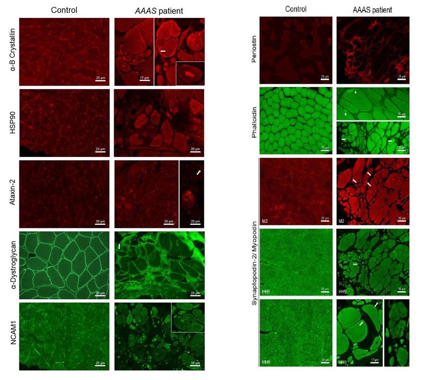
Immuno uorescence on muscle biopsy
Results of our global proteomic pro ling revealed the altered expression of a variety of proteins known to
be causative for or involved in the molecular genesis of neurological diseases. Therefore, vulnerability of
paradigmatic ones with known relevance for the development of muscular diseases was con rmed in a
muscle biopsy derived from the same Allgrove-patient exhibiting a profound myopathology as described
previously (8). Results of our immuno uorescence-based validation studies revealed an extracellular
increase of Periostin. Moreover, uneven sarcolemmal immunoreactivity including focal increases for α-
Dystroglycan and focal sarcoplasmic dots/ accumulations immunoreactive for α-B-Crystallin and for
HSP90 were identi ed in addition to subsarcolemmal increase of α-B-Crystallin (white arrow) (Fig. 5). In
addition, increased abundance of sarcoplasmic dots immunoreactive for Ataxin-2 and perinuclear
enrichment of Ataxin-2 (white arrow) was detected. Moreover, sarcoplasmic increase of NCAM1 was
identi ed (Fig. 5). Prompted by the altered expression of proteins involved in actin-cytoskeleton as well as
signi cant increase of actin itself in Allgrove-patient derived skin broblasts, FITC-Phalloidin-staining was
carried out to study actin-distribution in patient-derived muscle biopsy specimen. Results of these studies
showed some focal subsarcolemmal sarcoplasmic increases (white stars) in addition to perinuclear
enrichment (white arrows) of actin (Fig. 5). Given that ultra-structural studies of the Allgrove-patient
derived muscle biopsy revealed ne lamentous structures in subsarcolemmal location and brillary
nuclear bodies, immuno uorescence-based analysis of Synaptopodin-2/ Myopodin (displaying actin-
bundling activity) as a protein altered in both, the global and the nuclear proteome was performed using
two different antibodies (M2 and HH9). Results of these studies revealed increased immunoreactivity at
and below the sarcolemma (white arrows) as well as focal enrichment within the sarcoplasm (white star)
as also observed for actin (by FITC-Phalloidin staining) (Fig. 5).
Discussion
Page 8/30Protein cataloguing in human broblasts reveals expression
of a variety of proteins of neurological relevance
Although collection of muscle and/ or nerve biopsies represented a standard procedure in the diagnostic
work-up of patients suffering from neuromuscular diseases in the past, this approach became less
essential especially in the light of the rapidly developing omics approaches toward the identi cation of
the underlying genetic defect (25). Although this is less invasive and stressful for the patients, in turn it
causes a lack of biomaterial needed for protein studies aiming to address the pathogenicity of identi ed
genetic variants and/ or to elucidate pathophysiological cascades – a prerequisite for the de nition of
new therapeutic intervention concepts. Given that broblasts have already often been used for in vitro
studies with a focus on pathophysiological processes taking place in the development and manifestation
of neurological diseases (e.g. (5, 6), we aimed to generate a comprehensive catalogue of proteins
expressed in broblasts and analyzed same one for the abundance of proteins relevant for processes
with particular relevance for the function and maintenance of muscle and neuronal cells. Indeed, by
cataloguing the most abundant 8280 proteins and subsequent in silico based pathway analyses, we
could demonstrate that in human broblasts a variety of proteins are expressed which control
neurological processes such as muscle cell contraction or neuronal survival (see supplemental table 1).
This in turn shows that broblasts represent a cellular system suitable to study these processes in vitro.
This is further highlighted by the expression of 257 proteins out of a list of 385 (66.8%) associated with
the manifestation of genetically caused rare forms of neurological disorders. The nding that the
coverage of proteins encoded by genes responsible for motoneuron and peripheral nervous system
diseases is much higher than the one for muscle disorders (Suppl. Table 2) might be caused by the fact
that broblasts and neuronal cells derive from the same embryotic layer, the ectoderm bearing the
neuroectoderm. In contrast, muscle cells are derived from mesoderm.
Focusing on individual proteins, it is worth noting that Dystroglycan has been identi ed to be expressed
in broblasts (Suppl. Table 1). This might open new avenues in (i) the diagnostic work-up of patients with
the clinical suspicion of a so-called “dystroglycanopathy“ making the collection of a muscle biopsy less
essential as well as in (ii) the study of the precise molecular nature of this disease group and (iii) to test
new therapeutic concepts pre-clinically by having the chance to make use of human material with
different genetic defects leading to ”dystroglycanopathies“ (26). This postulate is supported by the
results of our proteomic and immuno uorescence studies on skin broblasts and muscle cells derived
from a patient with Allgrove syndrome (see below).
Proteomic studies on a use case allowed insights into the
molecular etiology of the disease
Relevance of proteomic- and BODIPY- ndings for
neurological signs of Allgrove syndrome
Page 9/30The Allgrove syndrome also known as triple A syndrome (AAAS) is caused by biallelic mutations in the
AAAS gene, encoding a 546 amino acid-sized nuclear pore complex protein known as ALADIN (alacrima
achalasia adrenal insu ciency neurologic disorder). ALADIN belongs to the WD-repeat family of
regulatory proteins. The broad neurological manifestations of the disease occur later in the course of the
disease and Allgrove syndrome patients show a wide variability (27). Neurodegeneration can affect
spinal motoneurons, Purkinje cells, striatal neurons and the autonomic system and the precise molecular
mechanisms leading to neuronal loss are still unclear (28). A recent functional study on central nervous
system tissues and broblasts of a novel Allgrove syndrome patient (homozygous AAAS mutation
c.464G > A; p.R155H) presenting with a motor neuron disease, cerebellar ataxia (accompanied by severe
loss of motor neurons and Purkinje cells) and autonomic dysfunction revealed signi cant reduction in the
perinuclear expression of p.R155H-mutant ALADIN in the patient's brain correlating with a signi cant
reduction of the AAAS-1 transcript, while the AAAS-2 transcript was upregulated in broblasts (28). Taken
together, this neuropathological study demonstrated the effect of loss of functional ALADIN in the human
central nervous system. Of note, results of our proteomic study indicated altered expression of a variety
of proteins decisive for proper neuronal function. This molecular nding might not only provide rst
insights into the biochemical origin of the broad neurological involvement of Allgrove syndrome but also
con rms the suitability of broblasts to identify pathophysiological processes of neuronal relevance:
Ataxin-2, a polyglutamine protein causative for spinocerebellar ataxia type 2 upon repeat expansion > 34,
was shown to function as modi er of Amyotrophic Lateral Sclerosis (ALS) pathology (as a rapidly
progressing neurodegenerative disease) with considerable negative impact of Ataxin-2 expression on
pathophysiology (29, 30). Therefore, we hypothesize that increased Ataxin-2 expression as identi ed by
our unbiased proteomic pro ling contributes to the pathophysiology of Allgrove syndrome. This
assumption is supported by the identi cation of focal sarcoplasmic and perinuclear enrichment of
Ataxin-2 in Allgrove-patient derived muscle cells. Interestingly, a trigger of these pathophysiological
processes was associated with elevated stress of the ER, the subcellular compartment to which ALADIN
localizes. In the same context, overexpression of human recombinant Sphingomyelin phosphodiesterase
4 (SMPD4; increased in broblasts of our patient) shows localization to the outer nuclear envelope and
the ER and additionally revealed interactions with several nuclear pore complex proteins as identi ed by
proteomics analysis (13). In addition, broblasts derived from SMPD4 patients suffering from a
developmental disorder characterized by microcephaly and congenital arthrogryposis show ER
abnormalities and are more prone to apoptosis under stress conditions. Based on their ndings, Magini
and co-workers postulated that SMPD4 links homeostasis of membrane sphingolipids to cell fate by
regulating the crosstalk between the ER and the outer nuclear envelope (13). Thus, increased level of
SMPD4 in cells lacking functional ALADIN accompanied by the identi cation of altered lipid homeostasis
(dysregulated lipid metabolism has already been linked to a variety of neurodegenerative disease, e.g.
(31) as veri ed by the results of our BODIPY-staining support the concept of a crosstalk between lipid
homeostasis and nuclear envelope integrity. Moreover, these combined ndings indicate that perturbed
lipid homeostasis may be part of the molecular etiology of Allgrove syndrome.
Page 10/30Relevance of proteomic and CARS-microscopic ndings for
myopathology of Allgrove syndrome
Ultra-structural studies of a muscle biopsy derived from our Allgrove-patient revealed foci of disorganized
myo brils, occasionally vacuole-associated, dilatations of sarcoplasmic reticulum, (containing a ne
granular structure), abnormal mitochondria, myonuclei with peripheral enriched heterochromatin and
irregular invaginations in addition to the rare nding of intra-myonuclear large diameter tubulo-
lamentous inclusions (8). Interestingly, results of our unbiased proteomic pro ling and subsequent
veri cation studies revealed vulnerability of proteins which accords with these pathomorphological
ndings: altered expression of a variety of mitochondrial proteins – presumably as a biochemical
correlate to initiated apoptosis – is in line with perturbed mitochondrial integrity. Increase of chaperones
re ecting perturbed protein processing as identi ed by proteomic pro ling of skin broblasts could be
veri ed in muscle cells of the same patient by showing a profound increase of sarcoplasmic dots
immunoreactive for α-B Crystallin and HSP90. This nding accords with the previous nding of vacuole-
associated dilatations of sarcoplasmic reticulum (8) a pathomorphological nding indicative for ER-
stress. Results of our CARS-microscopic studies allowed a more precise molecular strati cation of these
vacuoles and revealed that apart from protein content, these are also characterized by irregular lipid-
distribution. In accordance with of BODIPY- ndings obtained in patient-derived broblasts (see above),
results of our CARS-microscopic studies furthermore led to the identi cation of lipid-aggregates
suggesting that altered lipid-homeostasis is part of the underlying pathophysiology across different
cellular populations. Given that bulk membrane lipid biogenesis in primary cells largely occurs in the
endomembrane compartment, which includes the domains of the ER (32) and the ER is known to be
important for proper lipid transportation to other organelles such as mitochondria (33), one might
assume that loss of functional ALADIN perturbs proper lipid transportation leading to irregular cellular
build-up. The above-mentioned pathological ndings in mitochondria might support this assumption.
The previous identi cation of disorganized myo brils (8) might correlate with the focal sarcoplasmic
enrichment of Actin and Synaptopodin-2 (as a Actin-bundling protein). This nding hints toward a
vulnerability of this subcellular structure to which wildtype ALADIN localizes. Focal subsarcolemmal and
sarcoplasmic increase of Synaptopodin-2 not only indicates an impact of this protein (which is also
involved in the modulation of autophagy; (34) in the molecular myopathology of Allgrove syndrome but
also accords with our proteomic ndings obtained in both proteomic studies on skin broblasts derived
from the same patient. Given that an adverse effect of elevated Ataxin-2 expression in the neuronal
pathophysiology of ALS is known (29, 30), the nding of Ataxin-2 increase in the muscle biopsy derived
from the Allgrove patient suggests an impact of elevated Ataxin-2 expression also in myopathological
processes. Changes in the level of NCAM1 expression and localization in muscle cells correspond to key
morphogenetic events during muscle differentiation and repair (35). Our immunological ndings on α-
Dystroglycan (verifying the proteomic ndings obtained in broblasts) highlight a secondary vulnerability
of this protein in the molecular etiology of Allgrove syndrome and thus add this multisystemic disease to
the growing list of so-called secondary dystroglycanopathies.
Page 11/30Conclusions
Intersection of a spectral/ protein library obtained from cultured human skin broblasts with a list of
proteins known to be causative for the manifestation of neurological diseases shows that approximately
80% of proteins causative for motoneuron diseases or disease of the peripheral nervous system are
abundant in these cells. Moreover, approximately 40% of proteins encoded by genes causative for
diseases of the neuromuscular junction or of the skeletal muscle are present in our spectral/ protein
library covering the 8280 most abundant proteins. Proteomic pro ling of broblasts derived from a
patient suffering from Allgrove syndrome was carried out as an case example and ndings allowed novel
insights into the underlying pathophysiology also by adding perturbed lipid homoeostasis to the list of
pathobiochemical ndings known for this rare neurological disorder. In general, veri cation of protein-
dysregulations as identi ed in skin broblasts in muscle cells derived from the same patient in turn
demonstrates the suitability of skin broblasts to identify and further study processes relevant for the
manifestation of neurological diseases.
Patient, Materials & Methods
Allgrove-patient derived and control skin broblasts and
muscle biopsy specimen
The muscle biopsy derived from a genetically con rmed Allgrove patient (c.762delC mutation in the
nucleoporin gene AAAS; (8)) was collected for diagnostic purpose including histological and electron
microscopic investigations. In the same procedure, a skin biopsy was collected and used to set-up a
broblast culture now available for further functional studies focusing on the cellular impact of loss of
functional ALADIN. Primary human broblasts from the Allgrove-patient and controls (n = 3; obtained
from the MRC Centre Neuromuscular Biobank Newcastle (36)) were cultured in minimum essential media
supplemented with 10% fetal bovine serum, 1% penicillin/streptomycin, 2 mM L-glutamine, 1x non‐
essential amino acids, 1x minimum essential medium vitamins, 1 mM sodium pyruvate, 50 µg/mL
uridine (ThermoFisher Scienti c), at 37 °C, in a humidi ed 5% CO2 atmosphere. Cells were grown to a
con uency of 70% prior collection for proteomic studies: cells were collected in a 1.5 ml tube, washed
twice with ice-cold PBS, snap-frozen in liquid nitrogen and stored at -80 °C until protein extraction for
subsequent proteomic studies (see below).
Sample preparation of broblasts for spectral library
generation
Snap-frozen broblasts were lysed in a buffer containing 1% SDS, 50 mM Tris, 150 mM NaCl, pH 7.8 and
cOmplete™ ULTRA protease inhibitor using the Bioruptor® (Diagenode) for 10 minutes (30 seconds on, 30
seconds off, 10 cycles) at 4 °C. Afterwards, 20 µl of each sample was taken and diluted 1:4 with 10 mM
ammoniumbicarbonate buffer, pH 7.8 (ABC) to conduct a BCA-based determination of protein
concentration according to the manufacturer’s instructions (Pierce BCA protein assay kit). Reduction and
Page 12/30carbamidomethylation of the remaining samples were carried out using 10 mM Tris-(2-carboxyethyl)-
phosphin (TCEP) for 30 min at 37 C followed by application of 15 mM Iodacetamide (IAA) for further
30 min at room temperature (RT).
Samples were further processed using the S-Trap™ (Proti ) sample preparation procedure: after acidifying
the samples by adding 12% aqueous phosphoric acid, they were diluted with S-Trap binding buffer (90%
methanol (MeOH), triethylammonium bicarbonate (TEAB) 100 mM, pH 7.1). Protein-loading to S-Trap
columns including centrifugation steps was performed according to the manufacturer’s instructions.
Filter-based tryptic digestion was carried out for 2 h at 47 °C using a trypsin to protein ratio of 1:20.
Afterwards, peptides were eluted by applying multiple eluting steps starting with 10 mM ABC followed by
elution with 0.1% formic acid (FA) and last with 80% acetonitrile (ACN). Drying of the eluted peptides was
performed in a vacuum concentrator followed by dissolving of peptides in 0.1% TFA for subsequent LC-
MS/MS analysis or 10 mM ammonium acetate with 0.4 mM FA (pH 8.0) for subsequent pH8 reversed
phase fractionation.
All proteolytic digests were analyzed using monolithic column separation system (PepSwift monolithic
PS-DVB PL-CAP200-PM, Dionex) on an inert Ultimate 3000 HPLC (Dionex) by direct injection of 1 µg
sample to proof effectiveness of tryptic digestion. A binary gradient (solvent A: 0.1% TFA, solvent B:
0.08% TFA, 84% ACN) ranging from 5–12% B in 5 min and then from 12–50% B in 15 min at a ow rate
of 2.2 µl/min and at 60 °C, was applied. UV traces were acquired at 214 nm (37).
PH8-based sample fractionation
Each of the digested and desalted samples selected for the following generation of a spectral library was
rst dried using a vacuum concentrator. The peptides were then dissolved in a buffer containing 10 mM
ammonium acetate and 0.4 mM formiate (pH 8.0) (concentration 50 µg/µl) and separated on a C18 RP
chromatography column (loading amount 50 µg). Doing so, peptides were loaded onto the column with
solvent A (10 mM ammonium acetate, 0.4 mM formiate, pH 8.0) at a ow rate of 12.5 µl/min. Separation
and fractionation were performed with the following gradient with solvent B (84% acetonitrile in 10 mM
ammonium acetate, 0.4 mM formiate, pH 8.0): 3–10% in 10 min, 10–25% for 35 min, 25–40% for 20 min,
40–95% for 10 min, 95% for 5 min and 20 min equilibration at 3%. The individual fractions were collected
in an interval of 60 s, with each sample divided into 15 fractions. Collection was performed in the time
interval of 10 to 75 min of the gradient. The fractions were collected in combined form, so that after
reaching the last fraction, the sample was collected again for one minute in fraction 1. After fractionation,
the individual samples were dried in a vacuum concentrator and dissolved in 0.1% TFA prior subsequent
nano LC-MS/MS analysis (1 µg/µl).
Spectral library generation
Given that setting up a spectral library is a prerequisite to perform a data independent LC-MS/MS-based
sample analysis, all fractions derived from the pH8 fractionation mentioned above were analyzed by
nano LC-MS/MS using 1 µg respectively: samples were loaded on an Ultimate 3000 Rapid Separation
Liquid chromatography (RSLC) nano system with a ProFlow ow control device coupled to a Fusion
Page 13/30Lumos Tribrid mass spectrometer (both from Thermo Scienti c). After initial loading, peptides were
concentrated on a trapping column (Acclaim C18 PepMap100, 100 µm, 2 cm) using 0.1% TFA at a
owrate of 10 µl/min. Following sample separation was accomplished on a reversed phase column
(Acclaim C18 PepMap100, 75 µm 50 cm) using a binary gradient: 3% solvent B (84% ACN with 0.1% TFA)
for 10 min, a linear increase of solvent B to 35% for 120 min, a linear increase of solvent B to 95% for
10 min followed by a linear decrease of solvent B to 3% for 5 min. MS survey scans were acquired on the
Fusion Lumos using settings as follows: mass spectrometer was operated in data dependent acquisition
mode (DDA) with full MS scans from 300 to 1500 m/z at a resolution of 120,000 (Orbitrap) using the
polysiloxane ion at 445.12002 m/z as lock mass. The automatic gain control (AGC) was set to 2E5 and
the maximum injection time to 50 milliseconds. The most intense ions above a threshold ion count of
5E3 were selected for fragmentation at a normalized collision energy (nCE) of 30% (HCD) in each cycle of
the acquisition analysis, following each survey scan. The dynamic exclusion time was set to 15 seconds.
The number of selected precursor ions for fragmentation was determined by the “rapid” acquisition
algorithm. Fragment ions were acquired in the linear ion trap with an AGC of 1E4 and a maximum
injection time of 35 milliseconds.
The acquired data were imported into the software Spectronaut (Biognosys). As proteome background
the human proteome data was selected from UniProt (www.uniprot.org) containing 20,374 entries. The
processing settings were set as following: enzyme was trypsin, the minimum and maximum peptide
length was set to 7 and 52 respectively, missed cleavages was set to 2. Carbamidomethyl for cystein was
set as xed modi cation and acetyl (Protein N-term) and oxidation of methionine was set as variable
modi cations. All settings regarding the library generation including tolerances, identi cation, lters, iRT
calibration and work ow were set to factory defaults. For the relative quanti cation, the option Top N
max 3 was taken meaning that for each protein the average of the 3 most intense identi ed peptides are
taken to give the protein the quantitative value.
DIA-LC-MS/MS analysis
For the data independent acquisition (DIA) approach, the same nano LC-MS/MS setup as for the DDA
acquisition was used. Each sample analyzed was mixed with an appropriate amount of iRT standard
(Biognosys) peptides and 1 µg of each sample was subjected to the analysis. Full MS scans were
acquired from 300–1100 m/z at a resolution of 60,000 (Orbitrap) using the polysiloxane ion at
445.12002 m/z as lock mass. The automatic gain control (AGC) was set to 5E5 and the maximum
injection time to 20 milliseconds. Full MS scans were followed by 30 DIA windows acquired at a
resolution 30,000 (Orbitrap) with an AGC set to 1E6, a maximum injection time of 60 milliseconds and
nCE of 32 (HCD).
Analysis of DIA data
For the analysis of the samples acquired with nano-LC-MS/MS in DIA mode, the data was introduced to
the Spectronaut software and analyzed with a library-based search. As library, the above created spectral
Page 14/30library was used. Search and extraction settings were kept as standard (BGS Factory settings). As
background proteome, same data were used as selected for the establishment of the spectral library.
Preparation of nuclear protein fraction from broblasts
Snap-frozen broblasts (3 controls and 3 patient samples) were processed utilizing the “Qproteome Cell
Compartment Kit” (Qiagen; Cat No./ID: 37502): frozen cell pellets were dissolved with 1 ml of extraction
buffer CE1 (+ protease inhibitor) and incubated for 10 min at 4 °C on a Thermomixer (Eppendorf). After
centrifugation (1000 g for 10 min at 4 °C), supernatant was collected and pellets were dissolved in ice
cold extraction buffer CE2 (+ protease inhibitor), followed by an incubation for 30 min at 4 °C (on a
Thermomixer). After centrifugation (6000 g for 10 at 4 °C), the supernatant was collected in a new
reaction tube and Benzonase was added to the pellets followed by an incubation for 15 min at RT.
Afterwards 500 µl ice cold extraction buffer CE3 (+ protease inhibitor) was added and samples were
incubated for 10 min at 4 °C. Insoluble material was pelleted by centrifugation with 6800 g for 10 min at
4 °C and the supernatant containing the nuclear proteins was collected and stored at -80 °C until further
processing.
After thawing nuclear proteins fractions on ice, samples were mixed with ice cold acetone and incubated
at -80 °C for 12 hours. After centrifugation (20.000 g for 20 min at 4 °C), supernatant was discarded, and
the precipitated protein pellet was dried under a ow hood. Afterwards, 50 µl of 8 M Urea was added to
dissolve the protein pellet at RT. After diluting the solution to 2 M urea with 10 mM ammonium
bicarbonate buffer, pH 7.8 (ABC), a BCA-based determination of protein concentration was conducted
according to the manufacturer’s instructions (Pierce BCA protein assay kit). Reduction and
carbamidomethylation of the samples were carried out using 10 mM tris-(2-carboxyethyl)-phosphine
(TCEP) for 30 min at 37 °C followed by application of 15 mM iodacetamide (IAA) for another 30 min at
room temperature (RT). Samples were further diluted to 1 M urea using 10 mM ABC buffer adding trypsin
(ratio 1:100) to warrant protein hydrolyzation over night at 37 °C. Next, the tryptic-digestion was
terminated by adding 3 µl of 99% FA to decrease the pH to 2. Afterwards, samples were desalted using
solid phase extraction with C18 lter cartridges, washed with 0.1% TFA and eluted with 80% ACN. Cleaned
samples were dried by using a vacuum concentrator. Concentration was adjusted to 1 µg/µl with 0.1%
TFA.
Microscopic studies
Fibroblasts
Fibroblasts derived from the Allgrove patient as well as from respective sex and age matched controls
were grown on coverslips, washed three times with 1xPBS, xed with 4% formaldehyde solution for
20 min at room temperature and afterwards washed thrice with PBS. After 200 µL BODIPY 493/503
(ThermoFisher Scienti c, diluted 1:1000 in PBS) was added to each coverslip, samples were incubated in
a climate chamber for 1 h at room temperature in the dark. The staining solution was removed, samples
were washed thrice in 1xPBS and mounted onto a slide utilizing a drop of mounting medium (Prolong
Page 15/30Gold Antifade reagent with DAPI, Invitrogen). After solidi cation of the mounting medium, samples were
sealed with nail polish and stored at 6 °C in the dark until further microscopic investigation.
Fluorescence measurements were performed on a modi ed Leica TCS SP8 CARS laser scanning
microscope equipped with a 25x water immersion objective (Fluotar VISIR 25x/0.95 WATER). BODIPY
uorescence was excited at 488 nm and detected with a hybrid detector (Leica HyD, speci cations can be
found on the manufacturer’s website) in the range 495–600 nm. DAPI uorescence was excited at
405 nm and detected with a photomultiplier tube ranging from 415 to 475 nm. For each image, both
uorescence measurements were performed sequentially. Fluorescence data were acquired as small 3D
stacks with a resolution of 2048 × 2048 pixel (pixel size 227 × 227 nm) in X→Y direction and 9–12 layers
in Z direction with a step size of 570 nm.
Data processing was performed using Matlab R2015a. For better comparability, measurements were
preprocessed as following: for each image stack, the mean intensity was calculated in Z direction. For the
resulting 2D image, background noise was reduced by setting all data points with intensities lower than
1% to this background threshold. To account for cosmic spikes, an upper intensity threshold was
determined. Consequently, less than 0.1% of all data points above background threshold showed an
intensity value higher than this threshold. Intensities of data points exceeding this upper intensity
threshold were set to this value. Afterwards, the image was rescaled to full range (8 bit) between the two
threshold values.
From the processed mean images, single cell images were selected using a manually de ned irregular
octagon. For each of these single cell images, an intensity histogram analysis was generated whereby
histogram values were normalized to percentage values (to account for the different sizes of the single
cell images). The total fraction of data points showing 50% or more of the maximum intensity value was
calculated for each single cell. At least 30 individual cells were evaluated per sample.
Muscle biopsy specimen
Cryosections for immuno uorescence studies were cut at 7 µm thickness, xed in acetone for 5 min at −
20 °C and air dried either to be stained directly or stored at − 20 °C for subsequent staining, except of
sections for anti-α-Dystroglycan staining, which were xed for 1 minute at -20 °C in an 1:1 mixture of
ethanol/ glacial acetic acid and then washed in PBS, not dried.
Following primary antibodies were used: anti-α-B-Cyrstallin (polyclonal rabbit, 1:200; Stressgen, Canada),
anti-α-Dystroglycan (monoclonal mouse, 1:75; VIA4-1; Merck Millipore, Germany), anti-NCAM1
(monoclonal mouse, 1:100; Abcam), anti-HSP90 (polyclonal rabbit, 1:100; Genetex), anti-Ataxin 2
(polyclonal rabbit, 1:50; Abcam), anti-Periostin (polycloncal rabbit, 1:100; Abcam), anti-Synpatopodin-2
(M2 polyconal rabbit, 1:300 and HH9 monoclonal mouse, 1:2; both kindly provided by Peter F.M. van der
Ven/ Dieter O. Fürst). Primary antibodies were diluted in PBS and applied overnight at 4 °C. Furthermore,
uorescence staining of Actin laments was performed using the Phalloidin-iFluor 488 reagent (Abcam).
Page 16/30Secondary antibodies used were Alexa 488 conjugated donkey anti-mouse IgG (1:400) and Alexa 555
conjugated donkey anti-rabbit IgG (1:800) (Molecular Probes, Invitrogen, Thermo Fisher Scienti c,
Germany). The secondary antibodies were diluted in 1x PBS and applied for one hour at room
temperature. The sections were nally mounted in a Mowiol 4–88 (Calbiochem, Merck Chemicals,
Germany) and glycerol mix in pH 8.5 Tris buffer with 0.1% DABCO (1,4-Diazabicyclo(2, 2, 2)octane;
Sigma-Aldrich). Images were acquired with an AxioScope.A1 microscope using an Axiocam 503 color
camera and ZEN 2.3 (blue edition) software (all Carl Zeiss Microscopy Ltd., Germany). Further image
processing was performed with Adobe Photoshop CS6 Extended (Adobe Systems Inc., CA, USA).
Coherent anti-Stokes Raman scattering (CARS) and second harmonic generation (SHG) measurements
were performed on a modi ed Leica TCS SP8 CARS with an APE picoEmerald laser system. Ten
micrometer thick sections were cut from cryo tissue blocks. The samples were stored at -80 °C and
thoroughly dried before measurements at room temperature. No further sample preparation was applied.
The statistical evaluation is based on the 10 CARS images. A total of 103 muscle bres were collected for
the evaluation. For further details see supplemental document 1.
Declarations
Ethics approval and consent to participate
Informed consent was obtained from all patients and the ethics committee of University Medicine Essen
(19-9011-BO) had granted ethical approval.
Consent for publication
N/A
Availability of data and materials
The spectral library data have been deposited to the ProteomeXchange Consortium via the PRIDE partner
repository with the dataset identi er PXD019060. The proteomic pro ling data have been deposited to the
ProteomeXchange Consortium via the PRIDE partner repository with the dataset identi er PXD019201.
Competing interests
The authors declare not to have any competing con ict of interest.
Funding
Page 17/30Financial support by the Ministerium für Innovation, Wissenschaft und Forschung des Landes Nordrhein-
Westfalen, the Senatsverwaltung für Wirtschaft, Technologie und Forschung des Landes Berlin and the
Bundesministerium für Bildung und Forschung, also in the context of the Leibniz-Research cluster (#
031A360E) is gratefully acknowledged. Parts of this study were nanced in the framework of the NME-
GPS project (www.nmd-gps.net) by the European Regional Development Fund (ERDF). This work was
also supported by a grant from the AFM (to A.R.; #21644).
Authors' contributions
AR, EF, USS, AS and JR designed the study. AH performed the proteomic experiments. UM performed the
BODIPY-studies. AC and EF performed the CARS-microscopic studies. AR, AH, EF, AC and JR drafted the
manuscript.
Acknowledgements
We thank Mrs. Karin Kappes-Horn for expert technical assistance.
References
1. Hensel N, Kubinski S, Claus P. The Need for SMN-Independent Treatments of Spinal Muscular
Atrophy (SMA) to Complement SMN-Enhancing Drugs. Front Neurol. 2020;11:45.
2. Thompson R, Bonne G, Missier P, Lochmuller H. Targeted therapies for congenital myasthenic
syndromes: systematic review and steps towards a treatabolome. Emerg Top Life Sci. 2019;3(1):19-
37.
3. Roos A, Thompson R, Horvath R, Lochmuller H, Sickmann A. Intersection of Proteomics and
Genomics to "Solve the Unsolved" in Rare Disorders such as Neurodegenerative and Neuromuscular
Diseases. Proteomics Clin Appl. 2018;12(2).
4. Bayes A, Grant SG. Neuroproteomics: understanding the molecular organization and complexity of
the brain. Nat Rev Neurosci. 2009;10(9):635-46.
5. Bansagi B, Phan V, Baker MR, O'Sullivan J, Jennings MJ, Whittaker RG, et al. Multifocal
demyelinating motor neuropathy and hamartoma syndrome associated with a de novo PTEN
mutation. Neurology. 2018;90(21):e1842-e8.
6. Mingirulli N, Pyle A, Hathazi D, Alston CL, Kohlschmidt N, O'Grady G, et al. Clinical presentation and
proteomic signature of patients with TANGO2 mutations. J Inherit Metab Dis. 2020;43(2):297-308.
7. Benarroch L, Bonne G, Rivier F, Hamroun D. The 2020 version of the gene table of neuromuscular
disorders (nuclear genome). Neuromuscul Disord. 2019;29(12):980-1018.
8. Reimann J, Kohlschmidt N, Tolksdorf K, Weis J, Kuchelmeister K, Roos A. Muscle Pathology as a
Diagnostic Clue to Allgrove Syndrome. J Neuropathol Exp Neurol. 2017;76(5):337-41.
Page 18/309. Kind B, Koehler K, Lorenz M, Huebner A. The nuclear pore complex protein ALADIN is anchored via
NDC1 but not via POM121 and GP210 in the nuclear envelope. Biochem Biophys Res Commun.
2009;390(2):205-10.
10. Lorts A, Schwanekamp JA, Baudino TA, McNally EM, Molkentin JD. Deletion of periostin reduces
muscular dystrophy and brosis in mice by modulating the transforming growth factor-beta
pathway. Proc Natl Acad Sci U S A. 2012;109(27):10978-83.
11. Claeys KG, van der Ven PF, Behin A, Stojkovic T, Eymard B, Dubourg O, et al. Differential involvement
of sarcomeric proteins in myo brillar myopathies: a morphological and immunohistochemical study.
Acta Neuropathol. 2009;117(3):293-307.
12. Guo W, Zhang L, Christopher DM, Teng ZQ, Fausett SR, Liu C, et al. RNA-binding protein FXR2
regulates adult hippocampal neurogenesis by reducing Noggin expression. Neuron. 2011;70(5):924-
38.
13. Magini P, Smits DJ, Vandervore L, Schot R, Columbaro M, Kasteleijn E, et al. Loss of SMPD4 Causes
a Developmental Disorder Characterized by Microcephaly and Congenital Arthrogryposis. Am J Hum
Genet. 2019;105(4):689-705.
14. Chen YM, Gerwin C, Sheng ZH. Dynein light chain LC8 regulates syntaphilin-mediated mitochondrial
docking in axons. J Neurosci. 2009;29(30):9429-38.
15. Rockle I, Hildebrandt H. De cits of olfactory interneurons in polysialyltransferase- and NCAM-
de cient mice. Dev Neurobiol. 2016;76(4):421-33.
16. Choi SA, Kim SJ, Chung KC. Huntingtin-interacting protein 1-mediated neuronal cell death occurs
through intrinsic apoptotic pathways and mitochondrial alterations. FEBS Lett. 2006;580(22):5275-
82.
17. Erekat N, Al-Khatib A, Al-Jarrah M. Heat shock protein 90 is a potential therapeutic target for
ameliorating skeletal muscle abnormalities in Parkinson's disease. Neural Regen Res. 2014;9(6):616-
21.
18. Paepe BD, Creus KK, Weis J, Bleecker JL. Heat shock protein families 70 and 90 in Duchenne
muscular dystrophy and in ammatory myopathy: balancing muscle protection and destruction.
Neuromuscul Disord. 2012;22(1):26-33.
19. Jacquemyn J, Cascalho A, Goodchild RE. The ins and outs of endoplasmic reticulum-controlled lipid
biosynthesis. EMBO Rep. 2017;18(11):1905-21.
20. Natarajan A, Lemos DR, Rossi FM. Fibro/adipogenic progenitors: a double-edged sword in skeletal
muscle regeneration. Cell Cycle. 2010;9(11):2045-6.
21. Gonzalez Coraspe JA, Weis J, Anderson ME, Munchberg U, Lorenz K, Buchkremer S, et al.
Biochemical and pathological changes result from mutated Caveolin-3 in muscle. Skelet Muscle.
2018;8(1):28.
22. Cheng JX, Xie XS. Vibrational spectroscopic imaging of living systems: An emerging platform for
biology and medicine. Science. 2015;350(6264):aaa8870.
Page 19/30You can also read