INSIGHTS IN CHADOX1 NCOV-19 VACCINE-INDUCED IMMUNE THROMBOTIC THROMBOCYTOPENIA (VITT)
←
→
Page content transcription
If your browser does not render page correctly, please read the page content below
American Society of Hematology
                          2021 L Street NW, Suite 900,
                          Washington, DC 20036
                          Phone: 202-776-0544 | Fax 202-776-0545
                          editorial@hematology.org

Insights in ChAdOx1 nCov-19 Vaccine-induced Immune Thrombotic Thrombocytopenia (VITT)
Tracking no: BLD-2021-013231R1

Andreas Greinacher (University Medicine Greifswald, Germany) Kathleen Selleng (University Medicine

                                                                                                           Downloaded from http://ashpublications.org/blood/article-pdf/doi/10.1182/blood.2021013231/1824689/blood.2021013231.pdf by guest on 03 November 2021
Greifswald, Institute of Immunology and Transfusion Medicine, Germany) Jan Wesche (Universitätsmedizin
Greifswald, Germany) Raghavendra Palankar (University Medicine Greifswald, Germany) Stefan Handtke
(University Medicine Greifswald, Germany) Martina Wolff (University Medicine Greifswald, Germany)
Konstanze Aurich (Universitätsmedizin Greifswald, Germany) Michael Lalk (University of Greifswald,
Germany) Karen Methling (University Greifswald, Germany) Uwe Volker (4. DZHK (German Centre for
Cardiovascular Research), Partner Site Greifswald, Germany) Christian Hentschker (University Medicine
Greifswald, Germany) Stephan Michalik (University Medicine Greifswald, Germany) Leif Steil (University
Medicine Greifswald, Germany) Alexander Reder (University Medicine Greifswald, Germany) Linda Schönborn
(University Medicine Greifswald, Germany) Martin Beer (Friedrich-Loeffler Institut, Greifswald-Insel
Riems, Germany) Kati Franzke (Friedrich-Loeffler Institut, Greifswald-Insel Riems, Germany) Andreas
Büttner (Rostock University Medical Center, Germany) Boris Fehse (9. German Center for Infection
Research (DZIF), Partner Site Hamburg-Lübeck-Borstel-Riems, Germany) Evi Stavrou (Case Western Reserve
University School of Medicine, United States) Chandini Rangaswamy (University Medical Center Hamburg
(UKE), Germany) Reiner Mailer (University Medical Center Hamburg-Eppendorf (UKE), Germany) Hanna Englert
(University Medical Center Hamburg (UKE), Germany) Maike Frye (University Medical Center Hamburg (UKE),
Germany) Thomas Thiele (Institut für Immunologie und Transfusionsmedizin, Germany) Stefan Kochanek
(University of Ulm, Germany) Lea Krutzke (University of Ulm, Germany) Florian Siegerist (University
Greifswald, Germany) Nicole Endlich (University Medicine Greifswald, Germany) Theodore Warkentin
(McMaster University, Canada) Thomas Renné (University Medical Center Hamburg (UKE), Germany)

Abstract:
SARS-CoV-2 vaccine ChAdOx1 nCov-19 (AstraZeneca) causes a thromboembolic complication termed vaccine-
induced immune thrombotic thrombocytopenia (VITT). Using biophysical techniques, mouse models and
analysis of VITT patient samples we identified determinants of this vaccine-induced adverse reaction.
Super-resolution microscopy visualized vaccine components forming antigenic complexes with platelet
factor 4 (PF4) on platelet surfaces to which anti-PF4 antibodies obtained from VITT patients bound.
PF4/vaccine complex formation was charge-driven and increased by addition of DNA. Proteomics identified
substantial amounts of virus production-derived T-REx HEK293 proteins in the EDTA-containing vaccine.
Injected vaccine increased vascular leakage in mice leading to systemic dissemination of vaccine
components known to stimulate immune responses. Together, PF4/vaccine complex formation and the vaccine-
stimulated proinflammatory milieu trigger a pronounced B cell response that results in the formation of
high-avidity anti-PF4 antibodies in VITT patients. The resulting high-titer anti-PF4 antibodies potently
activated platelets in the presence of PF4 or DNA and polyphosphate polyanions. Anti-PF4 VITT patient
antibodies also stimulated neutrophils to release NETs in a platelet PF4-dependent manner. Biomarkers of
procoagulant NETs were elevated in VITT patient serum, and NETs were visualized in abundance by
immunohistochemistry in cerebral vein thrombi obtained from VITT patients. Together, vaccine-induced
PF4/adenovirus aggregates and proinflammatory reactions stimulate pathologic anti-PF4 antibody
production that drive thrombosis in VITT. The data support a two-step mechanism underlying VITT that
resembles the pathogenesis of (autoimmune) heparin-induced thrombocytopenia.

Conflict of interest: COI declared - see note

COI notes: We acknowledge all colleagues for helpful discussions, especially Drs. Sabine Eichinger-
Hasenauer, Brigitte Keller-Stanislawski, Dirk Mentzer, Klaus Cichutek and Axel Karger. We thank Drs.
Elke Hammer, Manuela Gesell-Salazar and Sascha Blankenburg and Katrin Schoknecht for supporting
proteomics and immunoproteomics analyses, Dr. Martina Wurster and Simone Seefeldt for help with
metabolomics analyses, Dr. Konrad for provided VITT patient material, and Dr. Frédéric Ebstein for the
25-mer DNA. We are grateful to Ulrike Strobel, Carmen Freyer, Katrin Stein, Ines Warnig, Ricarda
Raschke, Julia Klauke, Jessica Fuhrmann, Petra Meyer, Mandy Jörn, and Anita Badbaran for excellent
technical support. The study has been funded by the Deutsche Forschungsgemeinschaft (DFG, German
Research Foundation) grants: 374031971 - TRR240, 398967434 -SFB/TR261, 25440785 - SFB877, P6 - KFO306,
80750187 - SFB841, DFG-FR4239/1-1 and INST 2026/13-1 FUGG, European Union (Marie Sklodowska-Curie grant
agreement No 840189), the German Heart Foundation (F/34/18), the Ministerium für Wirtschaft, Arbeit und
Gesundheit Mecklenburg Vorpommern (project COVIDPROTECT), Leibniz WissenschaftsCampus - ComBioCat -
W10/2018, the Südmeyer fund for kidney and vascular research ("Südmeyer-Stiftung für Nieren- und
Gefäßforschung"), and the Dr. Gerhard Büchtemann fund. The contents are solely the responsibility of the
authors and do not necessarily represent the official views of the NIH, the U.S. Department of Veterans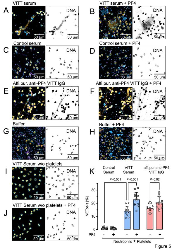
Affairs or the United States Government.

Preprint server: Yes; Research Square 10.21203/rs.3.rs-440461/v1

Author contributions and disclosures: A.G. developed the concept, designed experiments, and wrote the
first draft of the manuscript; J.W., S.H., R.P., K.A., M.W. performed the platelet, granulocyte, DLS,
ELISA, and some confocal microscopy experiments; M.L., K.M. performed the NMR studies; U.V., C.H., S.M.,
L.St., A.R. performed the proteome studies; LS, KS, TT characterized patients, contributed to the
concept and helped write the manuscript; K.F. performed the electron microscopy studies; M.B. analyzed
data, discussed the concept, and helped write the manuscript; S.K., L.K. analyzed the vaccine, and
contributed to the study concept; F.S., N.E. performed the 3D-super-resolution microscopy; T.E.W.
contributed to the concept, discussed results, helped write the manuscript; R.K.M., C.R., T.R. performed
the DNase experiments, NETs analyses, mouse studies, discussed data and helped write the manuscript.
B.F. designed the dPCR. M.F., H.E., E.X.S. performed and analyzed the immunohistochemistry analyses.
A.B. provided autopsy material. All authors have critically revised and approved the final version of
the manuscript. A.G., K.S., T.E.W., T.R have accessed and verified the underlying data

                                                                                                           Downloaded from http://ashpublications.org/blood/article-pdf/doi/10.1182/blood.2021013231/1824689/blood.2021013231.pdf by guest on 03 November 2021
Non-author contributions and disclosures: No;

Agreement to Share Publication-Related Data and Data Sharing Statement: For original data, please
contact andreas.greinacher@med.uni-greifswald.de or thomas@renne.net

Clinical trial registration information (if any):
Insights  in   ChAdOx1            nCov-19    Vaccine-induced       Immune       Thrombotic
Thrombocytopenia (VITT)

Short title: Mechanisms of VITT

Andreas Greinacher1,*, Kathleen Selleng1, Raghavendra Palankar1, Jan Wesche1, Stefan
Handtke1, Martina Wolff1, Konstanze Aurich1, Michael Lalk2, Karen Methling2, Uwe Völker3,4,
Christian Hentschker3, Stephan Michalik³, Leif Steil³, Alexander Reder3, Linda Schönborn1,
Martin Beer5, Kati Franzke6, Andreas Büttner7, Boris Fehse8,9, Evi X. Stavrou10,11, Chandini

                                                                                                Downloaded from http://ashpublications.org/blood/article-pdf/doi/10.1182/blood.2021013231/1824689/blood.2021013231.pdf by guest on 03 November 2021
Rangaswamy12, Reiner K. Mailer12, Hanna Englert12, Maike Frye12, Thomas Thiele1, Stefan
Kochanek13, Lea Krutzke13, Florian Siegerist14, Nicole Endlich14,15, Theodore E. Warkentin16,
Thomas Renné12,17 *

Affiliations:
  1. Institute of Immunology and Transfusion Medicine, Department of Transfusion
       Medicine, University Medicine Greifswald, Greifswald, Germany
  2. Institute of Biochemistry, University of Greifswald, Greifswald, Germany
  3. Interfaculty Institute of Genetics and Functional Genomics, Department Functional
       Genomics, University Medicine Greifswald, Greifswald, Germany
  4. German Centre for Cardiovascular Research (DZHK), Partner Site Greifswald,
       Greifswald, Germany
  5. Institute of Diagnostic Virology, Friedrich-Loeffler Institute, Greifswald-Insel Riems,
       Germany
  6. Institute of Infectious Diseases, Friedrich-Loeffler Institute, Greifswald-Insel Riems,
       Germany
  7. Institute of Forensic Medicine, Rostock University Medical Center, Rostock, Germany
  8. Research Dept. Cell & Gene Therapy, Dept. of Stem Cell Transplantation, University
       Medical Center Hamburg-Eppendorf, Hamburg Germany
 9. German Center for Infection Research (DZIF), Partner Site Hamburg-Lübeck-Borstel-
      Riems, Germany
 10. Department of Medicine, Hematology and Oncology Division, CWRU School of
      Medicine, Cleveland, OH, USA
 11. Department of Medicine, Section of Hematology-Oncology, Louis Stokes Veterans
      Administration Medical Center, Cleveland, OH, USA
 12. Institute of Clinical Chemistry and Laboratory Medicine, University Medical Center
      Hamburg-Eppendorf (UKE), Hamburg, Germany
 13. Department of Gene Therapy, Ulm University, Ulm, Germany
 14. Institute of Anatomy and Cell Biology, University Medicine Greifswald, Greifswald,
      Germany
 15. NIPOKA GmbH, Greifswald, Germany
 16. Department of Pathology and Molecular Medicine, and Department of Medicine,
      McMaster University, Hamilton, Ontario, Canada
 17. Center for Thrombosis and Hemostasis (CTH), Johannes Gutenberg University Medical
      Center, Mainz, Germany
Mechanism of VITT                                                       Greinacher et al.

*
 Corresponding authors:
Andreas Greinacher, MD
Institut für Immunologie und Transfusionsmedizin,
Abteilung Transfusionsmedizin
Sauerbruchstr.
D-17487 Greifswald, Germany
andreas.greinacher@med.uni-greifswald.de
or

                                                                                            Downloaded from http://ashpublications.org/blood/article-pdf/doi/10.1182/blood.2021013231/1824689/blood.2021013231.pdf by guest on 03 November 2021
Thomas Renné, MD, PhD.
Institute for Clinical Chemistry and Laboratory Medicine
University Medical Center Hamburg-Eppendorf (UKE)
Martinistrasse 52
D-20246 Hamburg, Germany
thomas@renne.net

Abstract word count: 222
Text word count: 4842
Figure Count: 7
Supplemental Figure count: 5
Table Count: 0
Supplemental Table count: 3
Reference count: 61
MESH Keywords: Thrombocytopenia, Vaccine, Thrombosis, COVID-19, ChadOx1 nCoV-19,
Platelet factor 4, Polyphosphate, VITT

Key points
    1. ChAdOx1 nCov-19 vaccine contains human TRex HEK293 cell-derived proteins and
       EDTA.
    2. Vaccine components and PF4 form complexes on platelet surfaces to which VITT
       patient antibodies bind.

                                                                                       2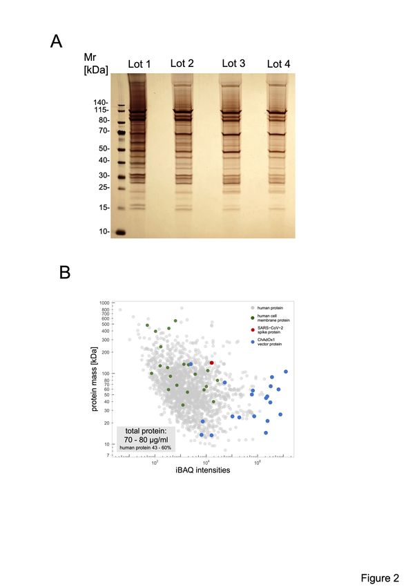
Mechanism of VITT                                                                 Greinacher et al.

ABSTRACT

SARS-CoV-2 vaccine ChAdOx1 nCov-19 (AstraZeneca) causes a thromboembolic
complication termed vaccine-induced immune thrombotic thrombocytopenia (VITT). Using
biophysical techniques, mouse models and analysis of VITT patient samples we identified
determinants of this vaccine-induced adverse reaction. Super-resolution microscopy
visualized vaccine components forming antigenic complexes with platelet factor 4 (PF4) on
platelet surfaces to which anti-PF4 antibodies obtained from VITT patients bound.

                                                                                                      Downloaded from http://ashpublications.org/blood/article-pdf/doi/10.1182/blood.2021013231/1824689/blood.2021013231.pdf by guest on 03 November 2021
PF4/vaccine complex formation was charge-driven and increased by addition of DNA.
Proteomics identified substantial amounts of virus production-derived T-REx HEK293
proteins in the EDTA-containing vaccine. Injected vaccine increased vascular leakage in
mice leading to systemic dissemination of vaccine components known to stimulate immune
responses.      Together,   PF4/vaccine   complex   formation   and   the   vaccine-stimulated
proinflammatory milieu trigger a pronounced B cell response that results in the formation of
high-avidity anti-PF4 antibodies in VITT patients. The resulting high-titer anti-PF4 antibodies
potently activated platelets in the presence of PF4 or DNA and polyphosphate polyanions.
Anti-PF4 VITT patient antibodies also stimulated neutrophils to release NETs in a platelet
PF4-dependent manner. Biomarkers of procoagulant NETs were elevated in VITT patient
serum, and NETs were visualized in abundance by immunohistochemistry in cerebral vein
thrombi obtained from VITT patients. Together, vaccine-induced PF4/adenovirus aggregates
and proinflammatory reactions stimulate pathologic anti-PF4 antibody production that drive
thrombosis in VITT. The data support a two-step mechanism underlying VITT that resembles
the pathogenesis of (autoimmune) heparin-induced thrombocytopenia.

                                                                                                 3
Mechanism of VITT                                                                   Greinacher et al.

INTRODUCTION

Vaccination against severe acute respiratory syndrome coronavirus-2 (SARS-CoV-2) is an
important countermeasure to fight the ongoing Covid-19 pandemic. The European Medicines
Agency (EMA) has approved two adenoviral vector-based vaccines, recombinant
chimpanzee adenoviral [ChAdOx1-S] vector encoding the spike glycoprotein of SARS-CoV-
2, COVID-19 Vaccine AstraZeneca [ChAdOx1 nCov-19; Vaxzevria]; and recombinant human
adenovirus type 26 vector encoding SARS-CoV-2 spike glycoprotein, Covid-19 Vaccine

                                                                                                        Downloaded from http://ashpublications.org/blood/article-pdf/doi/10.1182/blood.2021013231/1824689/blood.2021013231.pdf by guest on 03 November 2021
Janssen. ChAdOx1-S is propagated using T-REx HEK293 cells, a transformed human
embryonic kidney cell line.1

Beginning in March 2021, cerebral venous sinus thrombosis (CVST), splanchnic vein
thrombosis,     and   other    unusual   severe   thrombotic   events    in    combination     with
thrombocytopenia, were reported in otherwise healthy individuals beginning 5 to 20 days
following ChAdOx1 nCov-19 vaccination. This novel disorder, “vaccine-induced immune
thrombotic     thrombocytopenia”    (VITT;   synonym,   thrombosis      with   thrombocytopenia
syndrome, TTS), has been associated with high-titer immunoglobulin G (IgG) class
antibodies directed against the cationic platelet chemokine, platelet factor 4 (PF4).2 Anti-PF4
antibodies potently activate platelets with platelet activation greatly enhanced by PF4.2,3
Pathologic anti-PF4 antibodies were infrequently found in CVST patients prior to VITT,
suggesting that the vaccine-induced antibodies drive these thrombotic complications.4

PF4 opsonizes negatively charged surfaces of microbial pathogens, facilitating binding of
anti-PF4 antibodies.5 This is likely an evolutionary ancient immune defense mechanism.6-8
However, a misdirected strong anti-PF4 antibody response underlies the thromboembolic
disorder immune heparin-induced thrombocytopenia (HIT; caused by anti-PF4/heparin
antibodies) and its most severe presentation, autoimmune HIT.9-12 This latter subtype of HIT
is characterized by the formation of high-avidity platelet-activating anti-PF4 antibodies that
are reactive even in the absence of heparin.13

HIT proceeds by a two-step mechanism: initially, PF4/heparin complexes expose a
neoantigen on PF4 that stimulate B cells to produce high-affinity anti-PF4/heparin antibodies
in the presence of proinflammatory co-stimuli. Five to 10 days following heparin exposure,
sufficient quantities of these antibodies are present to activate cellular Fcγ receptors on
platelets, monocytes and granulocytes, culminating in life-threatening thrombosis. Major risk
factors for forming anti-PF4 antibodies and for developing HIT, are inflammation and tissue
trauma. Both disease states provide immunologic “danger signals” that increase the
likelihood and intensity of forming an anti-PF4 immune response.14,15 Marginal zone B-cells
mediate anti-PF4/heparin antibody production in HIT and their activation depends on Notch-2
signalling.16-18 Although anti-PF4/heparin antibodies develop commonly after heparin
                                                                                                   4
Mechanism of VITT Greinacher et al.
exposure, only a small subset of heparin-sensitized patients develop thrombocytopenia and
thrombosis. Despite decades of research, comprehensive insight into the factors that
predispose to adverse heparin-induced immune thrombotic events has remained enigmatic.19
VITT closely mimics autoimmune HIT both clinically and serologically;20 however the nature
of neoantigens that trigger pathologic anti-PF4 antibodies, the “danger signal(s)” that prime
for adverse immune reactions and prothrombotic mechanisms remain to be established in
VITT. Here, we identify key components of VITT immunopathogenesis. The data suggest
that VITT proceeds via a two-step mechanism: (i) vaccine components including the
Downloaded from http://ashpublications.org/blood/article-pdf/doi/10.1182/blood.2021013231/1824689/blood.2021013231.pdf by guest on 03 November 2021
adenovirus hexon protein form complexes with PF4 leading to neoantigen exposure on PF4.
Vaccine components also have the capability to trigger proinflammatory responses that are
“danger signals” known to amplify anti-PF4 antibody production in autoimmune HIT; (ii)
between days 5-20 post vaccination, anti-PF4 antibodies from VITT patients activate
platelets in a PF4- and polyanion-dependent manner. Additionally, anti-PF4 antibodies
activate granulocytes in the presence of platelets to release procoagulant neutrophil
extracellular traps (NETs) that are found in abundance in CVSTs of VITT patients. Together,
the data highlight similarities in VITT and HIT pathogenesis and identify strategies to interfere
with VITT-driven thrombotic events.
5Mechanism of VITT Greinacher et al.
METHODS
Detailed description of antibodies, reagents, and additional methods can be found in the
Supplemental Methods section (available on the Blood website).
3D-Super Resolution Microscopy
For 3D-Super Resolution Microscopy imaging of platelets and immobilized ChAdOx1 nCoV-
Downloaded from http://ashpublications.org/blood/article-pdf/doi/10.1182/blood.2021013231/1824689/blood.2021013231.pdf by guest on 03 November 2021
19 vaccine, coverslips were mounted inversely in Mowiol (Carl Roth, Karlsruhe, Germany) or
Everspark buffer (Idylle Labs, Paris, France) for dSTORM localization microscopy. 3D-Super
Resolution Microscopy imaging was either performed at NIPOKA GmbH, Greifswald,
Germany on a Nikon N-SIM E system (Nikon Instruments, Japan) equipped with 488 and
640 nm laser lines and a 100x 1.35NA silicon immersion objective, or on a Zeiss Elyra PS.1
super-resolution system equipped with a 63x 1.4NA oil-immersion objective. Raw image
stacks with 3 rotations and 5 shifts of the illumination grating were acquired and
reconstructed as super-resolved 3D SIM datasets, as described.21 Reconstructed data were
saved as .nd or .czi files and imported to FIJI22 using the BioFormats importer. Chromatic
aberration was corrected using analogous prepared reference slides coated with 100 nm
Tetraspek beads diluted 1:2,000 in ultrapure water. Data were analyzed using customized
FIJI and NIS scripts. Full dSTORM super-resolution imaging methods are given in the
supplemental methods.
Dynamic Light Scattering
All dynamic light scattering (DLS) measurements were performed in a fixed scattering angle
Zetasizer Nano-S system (Malvern Instruments Ltd., Malvern, UK). The hydrodynamic
diameter (nm) was measured at 25°C, and light scattering was detected at 173° using three
repeating measurements consisting of 12 runs. Experimental data were collected from four
independent experimental replicates. For all DLS measurements, ChAdOx1 n-CoV-19 was
diluted at a ratio of 1:10 in sterile-filtered 0.9% NaCl supplemented with 4 mg/mL D (+)
saccharose (RNase/DNase free; Cat. No. 9097.1, Carl Roth GmbH, Germany). Assessment
of changes in the hydrodynamic diameter of ChAdOx1 n-CoV-19 vector in the presence of
PF4 was performed by incubating 10 µg/mL of human PF4 (Chromatec, Greifswald,
Germany) with ChAdOx1 n-CoV-19 vector at RT for 2 min before DLS measurements. Anti-
PF4 mouse monoclonal IgG (Clone RTO, Cat. No. MA5-17639, Invitrogen) affinity-purified
anti-PF4 IgG antibodies from VITT sera, and control human IgG from a healthy individual,
were purified by Protein G affinity purification and were used at 5 µg/mL final concentration.
Double-stranded annealed DNA 25-mer was used at 0.5 µg/mL. Dissociation of complexes
6Mechanism of VITT Greinacher et al.
formed between ChAdOx1 n-CoV-19 vector and added components was achieved by 100
IU/mL unfractionated heparin (UFH, Ratiopharm GmbH, Ulm, Germany).
Transmission Electron Microscopy (TEM)
For transmission electron microscopy (TEM), the vaccine was incubated with biotinylated
PF4 (10 ng/mL in PBS; PF4-biotin) for at least 1 h at RT. The sample was transferred to
formvar-coated TEM grids (400 mesh, Plano GmbH, Germany), washed with PBS, and
Downloaded from http://ashpublications.org/blood/article-pdf/doi/10.1182/blood.2021013231/1824689/blood.2021013231.pdf by guest on 03 November 2021
blocked with BSA in PBS. Samples were labeled with an anti-adenovirus mAb (B025/AD51,
Abcam, USA; ab7428, 1:500) for 1 h at RT and detected with an anti-mouse gold conjugate
secondary antibody (BBI Solutions, GMHL10, 10 nm, 1:50). Alternatively, for PF4-biotin
staining, the same samples were labeled with a streptavidin-gold conjugate (Sigma, 10 nm,
1:10) for 45 min at RT. All grids were stained with 1% phosphotungstic acid at pH 7.4 and
analyzed with a Tecnai-Spirit (FEI, Eindhoven, NL) transmission electron microscope at an
accelerating voltage of 80 kV. The same procedure was used for preparing control samples
of vaccine alone and PF4-biotin. The vaccine was also incubated with heparin (100 IU/mL)
for 30 min. After that, samples were transferred to formvar-coated TEM grids (400 mesh,
Plano GmbH) and processed as above.
Composition of ChAdOx1 CoV-19 vaccine
ChAdOx1 CoV-19 vaccine was analyzed by mass spectrometry, 1H-nuclear magnetic
resonance (NMR) spectroscopy, and 1D-SDS PAGE (detailed methods are given in the
supplemental methods).
Platelet activation assay with washed platelets (PIPA test)
Platelet activation and aggregation by VITT patient serum were tested as previously
described.2 In brief, washed platelets of at least three healthy donors were incubated in the
absence or presence of either PF4 (10 mg/mL), DNA (ds 25-mer, 1 µg/mL) or EDTA (0.2 µM)
and serum from VITT patients under stirring conditions. Therefore, 75 µL platelet suspension
was mixed with 20 µL serum prior to stirring. The time to aggregation was measured up to 45
min. The test was determined to be positive if platelets aggregated within 30 min.
Immunofluorescence staining and confocal imaging of cerebral sinus vein thrombus
For visualization of NETs within cerebral sinus vein thrombi, 5 µm paraffin sections were
deparaffinized and rehydrated prior to antigen retrieval using sodium citrate. Slides were
blocked in 2% BSA, 0.1% Triton X-100 in PBS for 45 min at RT, followed by incubation with
7Mechanism of VITT Greinacher et al.
primary antibodies overnight at 4°C. After washing three times with PBS, sections were
incubated with secondary antibodies for 60 min at RT, before further washing and quenching
autofluorescence with 0.1% Sudan Black B in 70% EtOH for 25 min. Sections were washed
again and covered with Dako fluorescence mounting medium containing DAPI.
For visualization of NETs, 100 µm transverse vibratome sections of a cerebral sinus vein
thrombus from a VITT patient and a non-vaccinated control patient, were permeabilized
using 0.5% Triton X-100 in PBS for 20 min at RT, followed by blocking with 3% BSA in
PBSTx for 2 h. Primary antibodies were incubated overnight at 4°C, washed three times with
Downloaded from http://ashpublications.org/blood/article-pdf/doi/10.1182/blood.2021013231/1824689/blood.2021013231.pdf by guest on 03 November 2021
PBSTx and subsequently incubated with secondary antibodies overnight at 4°C, before
further washing and mounting in Dako fluorescence mounting medium.
The following primary antibodies were used: anti-neutrophil elastase (NE; 1:100, ab68672,
Abcam), anti-chromatin (anti-histone H2A/H2B/DNA-complex, 5 µg/mL, Davids
Biotechnologie GmbH), anti-vWF (1:100, A0082, Dako) and anti-MPO (1:25, ab9535,
Abcam). Secondary antibodies conjugated to AlexaFluor-488 and -594 were obtained from
Jackson ImmunoResearch (all donkey, used in 1:200 dilution). Confocal tissue images
represent maximum intensity projections of Z-stacks that were acquired using a Leica SP8
inverted confocal microscope with 10x HC PL APO CS and 63x HC PL APO Oil CS2
objectives and Leica LAS-X software.
8Mechanism of VITT Greinacher et al.
RESULTS
ChAdOx1 nCov-19 vaccine constituents and PF4 form immunogenic complexes recognized
by VITT patient anti-PF4 IgG.
ChAdOx1 nCoV-19 vaccination is associated with delayed local and systemic reactions after
the first administration indicating immunogenic reactions triggered by vaccine components.23
We analyzed interactions of vaccine constituents with blood by biophysical techniques. 3D-
super-resolution immunofluorescence microscopy visualized complexes formed between
Downloaded from http://ashpublications.org/blood/article-pdf/doi/10.1182/blood.2021013231/1824689/blood.2021013231.pdf by guest on 03 November 2021
adenovirus-derived hexon protein and platelets (Figure 1A, upper left panel), as well as
between adenovirus particles and PF4 (Figure 1A, lower left panel). These interactions led
to complexes comprised of vaccine particles and PF4 to which VITT patient-derived anti-PF4
IgG bound on platelet surfaces (Figure 1A, right panel). Quantification of the components of
the particles revealed that 45% contained ternary complexes of PF4, adenoviral hexon
proteins and anti-PF4 antibodies while approximately 50% were positive for PF4/adenoviral
hexon protein or PF4/anti-PF4 antibody complexes only (Figure 1B). Dynamic light
scattering (DLS; Figure 1C) confirmed formation of PF4/vaccine aggregates that were
24
recognized by VITT patient antibodies: Addition of PF4 (diameter 5 nm ) to ChAdOx1
nCoV-19 vaccine increased the size of adenovirus particles from 105.5 ± 2.8 nm to 185.2 ±
44.5 nm (mean diameter ± SD; p1 IU/mL)
dissociated 98.5% of complexes. As an indication of charge-driven complex formation of
vaccine components with PF4, surface zeta potential of the vaccine particles was negative (-
12 ± 4.2 mV), and addition of PF4, but not heparin, neutralized their negative charge (-5.2 ±
2.1 and -13 ± 6.8 mV, respectively; Figure 1D). Consistent with DLS, transmission electron
microscopy (TEM) visualized adenovirus particles and small amorphous structures contained
in the vaccine (Figures 1E). Larger aggregates formed upon incubation of the vaccine with
PF4. The aggregates stained positive for adenovirus hexon polypeptide and PF4 (Figures
1F, 1G and Supplemental Figure S1). Both patient-purified and hybridoma-cell derived anti-
PF4 antibodies, specifically detected PF4 alone or PF4 in a mixture with the vaccine;
however, there was no detectable signal with ChAdOx1 nCov-19 vaccine alone. Similarly,
anti-AV hexon protein antibodies stained the ChAdOx1 nCov-19 vaccine alone or in mixture
with PF4, however they did not bind to PF4 alone (Supplemental Figure S2). The data
reveal charge-driven complex formation of PF4 and adenovirus hexon proteins, to which
VITT patient anti-PF4 IgG bound.
9Mechanism of VITT Greinacher et al.
ChAdOx1 nCov-19 vaccination-induced inflammatory reactions in mice and humans
We hypothesized that ChAdOx1 nCoV-19 vaccine induces a proinflammatory “danger signal”
that promotes pathologic anti-PF4 antibody production in autoimmune HIT and possibly in
VITT.25,26 Accordingly, we sought to characterize the vaccine composition. Unexpectedly, we
found 70-80 µg protein/mL in four independent ChAdOx1 nCoV-19 vaccine lots analyzed.
Silver staining of ChAdOx1 nCoV-19 vaccine separated on SDS-PAGE showed numerous
Downloaded from http://ashpublications.org/blood/article-pdf/doi/10.1182/blood.2021013231/1824689/blood.2021013231.pdf by guest on 03 November 2021
protein bands (Figure 2A). Proteomics identified adenoviral vector proteins (blue dots), virus
production-derived T-REx HEK293 (human) proteins (grey dots)–some constituting
membrane proteins (green dots)–and the SARS-CoV-2 spike protein (red dot), in the
ChAdOx1 nCov-19 vaccine. Approximately 43-60% of the protein content of the vaccine (15-
24 µg per dose) was assigned to T-REx HEK293 cell protein origin (Figure 2B and
Supplemental Table S1).
1
We further analyzed the ChAdOx1 nCov-19 vaccine for small molecules by H-NMR
spectrometry and identified EDTA (~100 µM; Figure 3A). Leaky vessels are a hallmark of
inflammation and the Ca2+ chelator EDTA increases vascular leakage by VE-cadherin
endothelial junctional disassembly.27 We therefore analyzed ChAdOx1 nCoV-19 vaccine for
inducing proinflammatory reactions in mice using the Miles edema model (Figure 3B).
Intradermally injected vaccine triggered leakage in dermal vessels that was quantified by
Evans Blue tracer extravasation. Increase of vascular permeability appeared mostly
mediated by EDTA in the vaccine, as intradermal injection of EDTA alone (100 µM)
stimulated vascular leakage to a similar extent as the vaccine. Moreover, reconstituting the
ChAdOx1 nCoV-19 vaccine and EDTA with Ca2+ (100 µM) prior to intradermal injection
prevented vaccine- and EDTA-triggered increases in permeability and vascular leakage
(Supplemental Figure S3). We assessed consequences of EDTA-induced local vascular
leakage for dissemination of vaccine components in challenged mice. The vast majority of
the intradermally-injected adenovirus DNA remained at the injection site (>99.99%, at 30 min
post injection, Supplemental Figure S4). However, digital PCR detected ChAdOx1 nCoV-19
specific sequences in multiple tissues including the brain, as well as the spleen, where B-
cells are enriched in the splenic marginal zones; 28,29 (Figure 3C).
Consistent with the dissemination of vaccine constituents in the mouse model,
ChAdOx1nCoV-19 vaccination triggered systemic inflammatory reactions in humans. Out of
22 healthy vaccinated healthcare workers, 12 reported adverse effects including fever, chills,
and joint pain starting 6-12 hours after vaccination, usually resulting in one to two days of
sick leave. A representative example of skin inflammation associated with high levels of D-
Dimer (1,115 ng/mL), indicating proinflammatory reactions at days 1-2 after vaccination is
10Mechanism of VITT Greinacher et al.
shown in Figure 3D. Taken together, the data are consistent with a model of neoantigen
formation induced by vaccine constituents and PF4, that jointly stimulate pathologic anti-PF4
antibody formation facilitated by a vaccine-triggered inflammatory co-stimulus.
Pathologic anti-PF4 antibodies activate platelets and neutrophils, leading to thrombosis
We next analyzed for thrombotic reactions triggered by VITT patient anti-PF4 antibodies.
Consistent with our previously published data,2 all (14/14) sera of VITT patients analyzed, as
Downloaded from http://ashpublications.org/blood/article-pdf/doi/10.1182/blood.2021013231/1824689/blood.2021013231.pdf by guest on 03 November 2021
well as their respective affinity-purified antibody fractions showed strong reactivity towards
immobilized PF4/heparin in an ELISA assay. In a washed platelet aggregation-based assay,
addition of PF4 amplified platelet activation triggered by VITT patient sera (up to a serum titer
of 1:1000, Figure 4A). Similarly, addition of short-chain polyphosphate (a platelet-derived
30
inorganic polymer , 0.2 µg/mL), or synthetic DNA (1 µg/ml) increased VITT patient serum-
initiated platelet activation, albeit to a lower extent compared to PF4. VITT patient
serum/PF4-stimulated platelet aggregation was completely inhibited by FcγRIIA receptor
blockade using IV.3 antibody (Figure 4B). Taken together, the data indicate a function of
anti-PF4 IgG/PF4 complexes visualized in Figure 1 in mediating platelet activation.
Anti-PF4 antibodies of HIT patients activate neutrophils and are a major driver of thrombosis
by the formation of procoagulant NETs (NETosis).31 We tested pathologic anti-PF4
antibodies of VITT patients for their potency to trigger neutrophil activation and procoagulant
NET formation in the presence of PF4 or platelets. Incubation of isolated human neutrophils
and platelets with VITT patient serum-induced NET formation (Figure 5A) that was
significantly increased by the addition of PF4 (Figures 5B, 5K). In contrast, healthy control
serum did not trigger NET formation (Figures 5C, 5D, 5K). Similar to VITT patient serum,
affinity-purified anti-PF4 antibodies from these respective samples triggered NETosis
(Figures 5E, 5K). PF4 addition strongly enhanced VITT patient serum-stimulated NET
formation, confirming that PF4 in anti-PF4 IgG VITT antibodies initiates procoagulant NET
formation (Figures 5F, 5K). NETosis was virtually absent when neutrophils and platelets
were co-incubated in buffer, in the absence or presence of added PF4 (Figures 5G, 5H). In
the absence of platelets, VITT patient serum failed to induce NETosis even in the presence
of added PF4, suggesting that in VITT platelets play a key role in triggering NET formation
(Figures 5I, 5J).
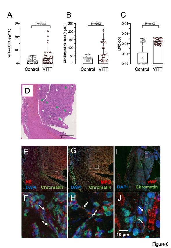
Supporting a role of NETs in VITT, three NET biomarkers cell-free DNA, citrullinated histones
and myeloperoxidase (MPO, marker for neutrophil activation), were elevated in VITT patient
sera compared to healthy controls (Figures 6A-C). CVST is a hallmark complication reported
in many VITT patients. We analyzed the composition of cerebral sinus vein thrombi obtained
by thrombectomy from one VITT patient or by autopsy from a second VITT patient,
11Mechanism of VITT Greinacher et al.
respectively. Histology of the thrombectomy-derived tissue showed regions with amorphous
material, likely representing fibrin deposition surrounded by nucleated cell-rich areas
throughout the thrombi (Figure 6D). Immunohistochemistry revealed that these cell-rich
areas contained abundant activated neutrophils and NETs. Antibodies against the NET
biomarker neutrophil elastase (NE, Figures 6E and 6F) and MPO (Figures 6G and 6H)
visualized degranulated neutrophils releasing elongated DAPI- and chromatin-positive NETs
into the platelet-rich (Figures 6I and J) thrombus. A highly similar NET staining pattern was
observed in a cerebral sinus vein thrombus of a second VITT patient obtained at the time of
Downloaded from http://ashpublications.org/blood/article-pdf/doi/10.1182/blood.2021013231/1824689/blood.2021013231.pdf by guest on 03 November 2021
autopsy, whereas cerebral sinus vein thrombus regions from a non-vaccinated control patient
contained less neutrophils (Supplemental Figure S5). Together, for the first time the data
visualize NETs in VITT-induced cerebral sinus vein thrombosis, indicating a causal
relationship of NET prothrombotic mechanisms and VITT.
12Mechanism of VITT Greinacher et al.
DISCUSSION
Based on a combination of biophysical imaging, mouse models and analysis of VITT patient
material, our study suggests a two-step mechanism underlying VITT-driven thrombosis that
is schematically shown in Figure 7. VITT pathogenesis is reminiscent of autoimmune HIT
pathology. Initially, neoantigens are generated by interaction of PF4 with vaccine
components. As visualized by TEM, 3D super-resolution microscopy and DLS, in
reconstituted systems PF4 has the capacity to bind to vaccine constituents leading to
Downloaded from http://ashpublications.org/blood/article-pdf/doi/10.1182/blood.2021013231/1824689/blood.2021013231.pdf by guest on 03 November 2021
formation of complexes that contain adenovirus proteins (Figure 1, Supplemental Figures
S1, S2). While PF4/vaccine aggregates are exposed on platelet surfaces and recognized ex
vivo by VITT patient anti-PF4 antibodies, the precise sequence of events occurring in vivo at
injection sites or in blood, requires additional studies. In humans, PF4 is enriched at the
vessel wall and is locally released in high concentrations following platelet activation. 32,33
Consistent with our imaging data, previous studies have shown that coronaviruses have the
34,35
capacity to activate platelets and that adenoviruses binding to platelets can lead to
platelet-activation and release of PF4.36
ChAdOx1 nCov-19 vaccine-derived adenovirus aggregates bind to platelet surfaces and are
transported via the bloodstream to the spleen where they are phagocytosed by
macrophages, ultimately inducing a pronounced B-cell activation in the marginal zone and
follicles in mice.29 In line with the animal model findings, we visualized an interaction of
adenovirus-derived hexon proteins, PF4, and VITT patient-derived anti-PF4 antibodies on
platelet surfaces by three independent techniques (Figure 1). Our observations suggest that
adenovirus binding to PF4 likely induces conformational changes in PF4 and creates
potential neoantigen(s). Consistent with the hypothesis that VITT antibodies target
neoantigen(s) in PF4, VITT patient anti-PF4 antibodies bind to PF4 following immobilization
on plastic surfaces. PF4 binding to surfaces is known to induce conformational changes.37
Additionally, mutagenesis studies have shown that VITT anti-PF4 antibodies, similarly to
polyanions (Figure 4A), induce PF4 clustering leading to platelet activation.38 Anti-PF4
antibody stimulated platelet aggregation is not a new concept as high-affinity anti-PF4
antibodies also cluster PF4, even in the absence of polyanions and induce platelet activation
in atypical HIT.13 In addition to adenovirus proteins, we found substantial amounts (~43-60%
by content) of non-viral proteins originating from the T-REx HEK293 (RRID:CVCL_D585)
human embryonic kidney-derived production cell line.39 HEK293 cells lack tissue-specific
gene expression signature and express an array of markers of renal progenitor, neuronal and
adrenal gland cells with undefined immunogenicity.40 Potential other immunologic
consequences triggered by intramuscular injection of approximately 15-24 µg T-REx HEK293
proteins per vaccination dose remain to be established. In addition, vaccine protein
13Mechanism of VITT Greinacher et al.
contaminants have the potential to induce an acute inflammatory co-signal that enhances B
cell responses (immunologic “‘danger signal”).15,41,42 Synergistically, disseminated viral
proteins potently activate innate immune reactions supporting early inflammatory reactions
following vaccination.15,41,42 The inflammatory response provides a co-stimulus for anti-PF4
antibody production by preformed B-cells in HIT.8,14 Consistently, western blotting revealed
that vaccination increased titers of preexisting antibodies that bind to an array of vaccine
components separated by SDS-PAGE. Additionally, the ChAdOx1 nCov-19 vaccine contains
EDTA, with the capacity for increasing capillary leakage at the inoculation site by a VE-
Downloaded from http://ashpublications.org/blood/article-pdf/doi/10.1182/blood.2021013231/1824689/blood.2021013231.pdf by guest on 03 November 2021
cadherin-dependent pathway.27 Disruption of the endothelial barrier facilitates dissemination
of vaccine constituents into the circulation (Figure 3). Alternatively, accidental intravenous
injection may contribute to vaccine dissemination.39 Furthermore, viruses can reach tissues
distant from their infection site by invading endothelial cells and gaining access into the
extravascular space. Viruses can also be transported across the vascular wall or be co-
transported in infected immune cells such as neutrophils, that have the capacity to
transmigrate through the endothelial barrier.43 Systemic dissemination of vaccine
components is not unique to ChAdOx1 nCov-19; consistent with our murine model data
(Figure 3B), another ChAdOx1 vector variant (with a hepatitis B vector insert) was
detectable in multiple organs, including liver, heart, and lymph nodes, at days 2 and 29 after
intramuscular injection in mice.44
Similar to HIT pathology,18 marginal zone B-cells stimulated by PF4/vaccine neoantigens in
the presence of inflammatory co-stimuli produce the pathogenic anti-PF4 antibodies
underlying VITT. The detailed role of marginal zone B-cells in anti-PF4 VITT antibody
production, and the mode of exposure of PF4/vaccine neoantigens (Figure 1A), remains to
be established in patients;39 however, in murine models an intravenous vaccine injection
induces a pronounced B-cell immune response.29 At day 5 to 20 post-vaccination, pathologic
anti-PF4 antibodies reach high titers and are capable of clustering PF4 on platelet surfaces
and activating platelets by binding FcγRIIA (Figure 4B). Anti-PF4 mediated platelet
activation likely involves extracellular polyanions such as DNA and polyphosphate (Figures
1C and 4A). Clustering of PF4 by pathologic anti-PF4 auto-antibodies is also a central
mechanism for platelet activation in autoimmune HIT.13 Cross-talk of PF4, activated platelets,
and VITT anti-PF4 antibodies activates neutrophils leading to NETs formation in VITT patient
45
serum (Figure 5). NETs are degraded by DNases and extracellular circulating DNA was
increased in VITT patients (Figure 6A), which amplifies platelet activation in VITT (Figure 4).
Furthermore, DNA within NETs binds PF4.46 The resulting PF4/DNA complexes create an
additional target for anti-PF4 antibodies and increase the resistance of NETs to DNase-
mediated degradation, further amplifying their procoagulant activity.47 This sequence of
31,47
events culminates in massive Fcγ receptor-dependent activation of neutrophils, platelets
14Mechanism of VITT Greinacher et al.
and—by analogy with autoimmune HIT—likely also monocytes and endothelial cells.48
Consistent with our data in VITT patients, activated neutrophils and NETs contribute to
venous thrombosis in HIT mouse models, and HIT antibodies selectively bind PF4-NET
complexes.47 The bimodal distribution of the neutrophil activation marker serum MPO
observed in healthy controls (Figure 6C) may reflect the impact of smoking and hormonal
contraceptive use, both of which increase MPO enzyme levels.49 Broadened reactivity of
antibodies in a boosted immune response is the hallmark of other immune disorders besides
VITT. Patients with severe COVID-19 disease often have anti-NET auto-antibodies which
Downloaded from http://ashpublications.org/blood/article-pdf/doi/10.1182/blood.2021013231/1824689/blood.2021013231.pdf by guest on 03 November 2021
likely impair NET clearance and may potentiate SARS-CoV-2-mediated
50,51
thromboinflammation. Autoimmune HIT often features initial heparin-dependent reactivity
that extends to heparin-independent hyperreactivity.9 Similarly, post-transfusion purpura
(PTP) reflects a strong alloimmune response that progresses to include platelet-autoreactive
properties.52 In this regard the numerous cell culture-derived human proteins in ChAdOx1
nCov-19 vaccine (Figure 2) we have identified raise concern. If such proteins express
immunogenic structures e.g., a genetic polymorphism absent in the corresponding
endogenous protein of the vaccinated individual, an alloimmune response with potential for
autoreactivity in a susceptible vaccine recipient requires attention. Protein impurities and
adverse immune reactions are not unique to ChAdOx1 nCov-19. Xenogeneic antigens
derived from the growth matrix used in the virus manufacturing process have been identified
by mass spectrometry in swine influenza vaccines.53,54 Bioprocess impurities, originating from
the production cell line of a bovine-virus diarrhea vaccine-induced alloreactive antibodies
causing feto-maternal incompatibility with severe thrombocytopenia in calves.55 In humans,
narcolepsy has been associated with Pandemrix influenza vaccine, however, the underlying
immune mechanisms remain to be completely understood.56,57
Our study has limitations; detailed specifications of the ChAdOx1 nCov-19 vaccine are not
publicly available and we focused on identification of protein and some small molecule
content without claiming completeness. Additionally, we have not investigated the specific
roles of B-cells or T-cells in the VITT immune response, nor a potential contribution of the
complement system that is known to contribute to immunogenicity and downstream
thrombosis in HIT.58-60 VITT is a rare adverse event with thrombosis occurring in 1 out of
30,000 – 50,000 vaccinated people. We currently can only speculate on the low incidence of
VITT by drawing parallels to HIT, an adverse reaction that occurs only in a small subset of
heparin-exposed individuals (around 1 to 100 to 1 in 1,000 patients receiving UFH). VITT
appears to share similarities with atypical HIT, as the latter is even more rare (Mechanism of VITT Greinacher et al.
e.g., severe drug-dependent immune thrombocytopenia following treatment with antibiotics
such as vancomycin, only a small fraction of treated individuals develops adverse effects and
underlying mechanisms have remained enigmatic. Furthermore, predominant localization of
VITT associated thrombi in cerebral sinus veins has remained puzzling. Preliminary findings
61
based on systems biology and transcriptomics indicate low DNASE1 expression in central
nervous system endothelial cells that can potentially lead to increased half-life and persistent
procoagulant activity of NETs. However, the hypothesis requires additional human subjects
and experimental confirmation.
Downloaded from http://ashpublications.org/blood/article-pdf/doi/10.1182/blood.2021013231/1824689/blood.2021013231.pdf by guest on 03 November 2021
Together, our data provide insight into the mechanisms by which the SARS-CoV-2 vaccine
ChAdOx1 nCov-19 initiates immune responses, leading to pathogenic anti-PF4 antibodies
that trigger downstream prothrombotic reactions. Our findings have implications for the
development of adenoviral vector vaccines with improved patient safety.
16Mechanism of VITT Greinacher et al.
ACKNOWLEDGMENTS
We acknowledge all colleagues for helpful discussions, especially Drs. Sabine Eichinger-
Hasenauer, Brigitte Keller-Stanislawski, Dirk Mentzer, Klaus Cichutek and Axel Karger. We
thank Drs. Elke Hammer, Manuela Gesell-Salazar and Sascha Blankenburg and Katrin
Schoknecht for supporting proteomics and immunoproteomics analyses, Dr. Martina Wurster
and Simone Seefeldt for help with metabolomics analyses, Dr. Konrad for provided VITT
patient material, and Dr. Frédéric Ebstein for the 25-mer DNA. We are grateful to Ulrike
Strobel, Carmen Freyer, Katrin Stein, Ines Warnig, Ricarda Raschke, Julia Klauke, Jessica
Downloaded from http://ashpublications.org/blood/article-pdf/doi/10.1182/blood.2021013231/1824689/blood.2021013231.pdf by guest on 03 November 2021
Fuhrmann, Petra Meyer, Mandy Jörn, and Anita Badbaran for excellent technical support.
The study has been funded by the Deutsche Forschungsgemeinschaft (DFG, German
Research Foundation) grants: 374031971 - TRR240, 398967434 -SFB/TR261, 25440785 -
SFB877, P6 - KFO306, 80750187 - SFB841, DFG-FR4239/1-1 and INST 2026/13-1 FUGG,
European Union (Marie Sklodowska-Curie grant agreement No 840189), the German Heart
Foundation (F/34/18), the Ministerium für Wirtschaft, Arbeit und Gesundheit Mecklenburg
Vorpommern (project COVIDPROTECT), Leibniz WissenschaftsCampus – ComBioCat –
W10/2018, the Südmeyer fund for kidney and vascular research (“Südmeyer-Stiftung für
Nieren- und Gefäßforschung”), and the Dr. Gerhard Büchtemann fund. The contents are
solely the responsibility of the authors and do not necessarily represent the official views of
the NIH, the U.S. Department of Veterans Affairs or the United States Government.
AUTHOR CONTRIBUTIONS
A.G. developed the concept, designed experiments, and wrote the first draft of the
manuscript; J.W., S.H., R.P., K.A., M.W. performed the platelet, granulocyte, DLS, ELISA,
and some confocal microscopy experiments; M.L., K.M. performed the NMR studies; U.V.,
C.H., S.M., L.St., A.R. performed the proteome studies; LS, KS, TT characterized patients,
contributed to the concept and helped write the manuscript; K.F. performed the electron
microscopy studies; M.B. analyzed data, discussed the concept, and helped write the
manuscript; S.K., L.K. analyzed the vaccine, and contributed to the study concept; F.S., N.E.
performed the 3D-super-resolution microscopy; T.E.W. contributed to the concept, discussed
results, helped write the manuscript; R.K.M., C.R., T.R. performed the DNase experiments,
NETs analyses, mouse studies, discussed data and wrote the revised manuscript. B.F.
designed the dPCR. M.F., H.E., E.X.S. performed and analyzed the immunohistochemistry
analyses. A.B. provided autopsy material. All authors have critically revised and approved
the final version of the manuscript. A.G., K.S., T.E.W., T.R have accessed and verified the
underlying data.
17Mechanism of VITT Greinacher et al.
DISCLOSURE OF CONFLICTS OF INTEREST
Dr. Greinacher reports grants and non-financial support from Aspen, Boehringer Ingelheim,
MSD, Bristol Myers Squibb (BMS), Paringenix, Bayer Healthcare, Gore Inc., Rovi, Sagent,
Biomarin/Prosensa, personal fees from Aspen, Boehringer Ingelheim, MSD, Macopharma,
BMS, Chromatec, Instrumentation Laboratory, non-financial support from Boehringer
Ingelheim, Portola, Ergomed, GTH e.V. outside the submitted work. Dr. Thiele reports
personal fees and other from Bristol Myers Squibb, personal fees and other from Pfizer,
Downloaded from http://ashpublications.org/blood/article-pdf/doi/10.1182/blood.2021013231/1824689/blood.2021013231.pdf by guest on 03 November 2021
personal fees from Bayer, personal fees and other from Chugai Pharma, other from Novo
Nordisk, personal fees from Novartis, other from Daichii Sankyo, outside the submitted work.
Dr. Beer reports grants from CureVac Company, Tübingen, Germany.
18Mechanism of VITT Greinacher et al.
References
1. Vaxzevria (priviously COVID-19 Vaccine Astra Zeneca): EPAR - Product information.
Vol. 2021: European Medicines Agency; 2021.
2. Greinacher A, Thiele T, Warkentin TE, Weisser K, Kyrle PA, Eichinger S. Thrombotic
Thrombocytopenia after ChAdOx1 nCov-19 Vaccination. New Engl J Med. 2021;
384(22):2092-2101.
3. Schultz NH, Sorvoll IH, Michelsen AE, et al. Thrombosis and Thrombocytopenia after
ChAdOx1 nCoV-19 Vaccination. N Engl J Med. 2021; 384(22):2124-2130.
Downloaded from http://ashpublications.org/blood/article-pdf/doi/10.1182/blood.2021013231/1824689/blood.2021013231.pdf by guest on 03 November 2021
4. Sánchez van Kammen M, Heldner MR, Brodard J, et al. Frequency of
Thrombocytopenia and Platelet Factor 4/Heparin Antibodies in Patients With Cerebral
Venous Sinus Thrombosis Prior to the COVID-19 Pandemic. JAMA. 2021; 326(4):332-
338.
5. Krauel K, Potschke C, Weber C, et al. Platelet factor 4 binds to bacteria, [corrected]
inducing antibodies cross-reacting with the major antigen in heparin-induced
thrombocytopenia. Blood. 2011;117(4):1370-1378.
6. Krauel K, Schulze A, Jouni R, et al. Further insights into the anti-PF4/heparin IgM
immune response. Thromb Haemost. 2016;115(4):752-761.
7. Zheng Y, Zhu W, Haribhai D, et al. Regulatory T Cells Control PF4/Heparin Antibody
Production in Mice. J Immunol. 2019;203(7):1786-1792.
8. Zheng Y, Wang AW, Yu M, et al. B-cell tolerance regulates production of antibodies
causing heparin-induced thrombocytopenia. Blood. 2014;123(6):931-934.
9. Greinacher A, Selleng K, Warkentin TE. Autoimmune heparin-induced
thrombocytopenia. J Thromb Haemost. 2017;15(11):2099-2114.
10. Greinacher A. Heparin-Induced Thrombocytopenia. N Engl J Med. 2015;373(19):1883-
1884.
11. Warkentin TE, Greinacher A. Spontaneous HIT syndrome: Knee replacement,
infection, and parallels with vaccine-induced immune thrombotic thrombocytopenia.
Thromb Res. 2021;204:40-51.
12. Cai Z, Yarovoi SV, Zhu Z, et al. Atomic description of the immune complex involved in
heparin-induced thrombocytopenia. Nat Commun. 2015;6:8277.
13. Nguyen TH, Medvedev N, Delcea M, Greinacher A. Anti-platelet factor 4/polyanion
antibodies mediate a new mechanism of autoimmunity. Nat Commun. 2017;8:14945.
14. Lubenow N, Hinz P, Thomaschewski S, et al. The severity of trauma determines the
immune response to PF4/heparin and the frequency of heparin-induced
thrombocytopenia. Blood. 2010;115(9):1797-1803.
15. Gong T, Liu L, Jiang W, Zhou R. DAMP-sensing receptors in sterile inflammation and
inflammatory diseases. Nat Rev Immunol. 2020;20(2):95-112.
16. Saito T, Chiba S, Ichikawa M, et al. Notch2 is preferentially expressed in mature B cells
and indispensable for marginal zone B lineage development. Immunity.
2003;18(5):675-685.
17. Garis M, Garrett-Sinha LA. Notch Signaling in B Cell Immune Responses. Front
Immunol. 2020;11:609324.
18. Zheng Y, Yu M, Podd A, et al. Critical role for mouse marginal zone B cells in
PF4/heparin antibody production. Blood. 2013;121(17):3484-3492.
19Mechanism of VITT Greinacher et al.
19. Arepally GM. Heparin-induced thrombocytopenia. Blood. 2017;129(21):2864-2872.
20. Scully M, Singh D, Lown R, et al. Pathologic Antibodies to Platelet Factor 4 after
ChAdOx1 nCoV-19 Vaccination. N Engl J Med. 2021;384(23):2202-2211.
21. Siegerist F, Ribback S, Dombrowski F, et al. Structured illumination microscopy and
automatized image processing as a rapid diagnostic tool for podocyte effacement. Sci
Rep. 2017;7(1):11473.
22. Schindelin J, Arganda-Carreras I, Frise E, et al. Fiji: an open-source platform for
biological-image analysis. Nat Methods. 2012;9(7):676-682.
23. Ramasamy MN, Minassian AM, Ewer KJ, et al. Safety and immunogenicity of
Downloaded from http://ashpublications.org/blood/article-pdf/doi/10.1182/blood.2021013231/1824689/blood.2021013231.pdf by guest on 03 November 2021
ChAdOx1 nCoV-19 vaccine administered in a prime-boost regimen in young and old
adults (COV002): a single-blind, randomised, controlled, phase 2/3 trial. Lancet.
2021;396(10267):1979-1993.
24. Greinacher A, Gopinadhan M, Günther JU, et al. Close approximation of two platelet
factor 4 tetramers by charge neutralization forms the antigens recognized by HIT
antibodies. Arterioscler Thromb Vasc Biol. 2006;26(10):2386-2393.
25. Tobaiqy M, Elkout H, MacLure K. Analysis of Thrombotic Adverse Reactions of COVID-
19 AstraZeneca Vaccine Reported to EudraVigilance Database. Vaccines (Basel).
2021;9(4):393.
26. Shi C, Yang L, Braun A, Anders HJ. Extracellular DNA-A Danger Signal Triggering
Immunothrombosis. Front Immunol. 2020;11:568513.
27. Gao X, Kouklis P, Xu N, et al. Reversibility of increased microvessel permeability in
response to VE-cadherin disassembly. Am J Physiol Lung Cell Mol Physiol.
2000;279(6):L1218-1225.
28. Martin F, Kearney JF. Marginal-zone B cells. Nat Rev Immunol. 2002;2(5):323-335.
29. Nicolai L, Leunig A, Pekayvaz K, et al. Thrombocytopenia and splenic platelet directed
immune responses after intravenous ChAdOx1 nCov-19 administration. bioRxiv.
2021:https://doi.org/10.1101/2021.1106.1129.450356.
30. Mailer RK, Allende M, Heestermans M, et al. Xenotropic and polytropic retrovirus
receptor 1 regulates procoagulant platelet polyphosphate. Blood. 2021;137(10):1392-
1405.
31. Perdomo J, Leung HHL, Ahmadi Z, et al. Neutrophil activation and NETosis are the
major drivers of thrombosis in heparin-induced thrombocytopenia. Nat Commun.
2019;10(1):1322.
32. Lopez Yomayuza CC, Preissner KT, Lorenz B, Stieger K. Optimizing Measurement of
Vascular Endothelial Growth Factor in Small Blood Samples of Premature Infants. Sci
Rep. 2019;9(1):6744.
33. Rauova L, Zhai L, Kowalska MA, Arepally GM, Cines DB, Poncz M. Role of platelet
surface PF4 antigenic complexes in heparin-induced thrombocytopenia pathogenesis:
diagnostic and therapeutic implications. Blood. 2006;107(6):2346-2353.
34. Comer SP, Cullivan S, Szklanna PB, et al. COVID-19 induces a hyperactive phenotype
in circulating platelets. PLoS Biol. 2021;19(2):e3001109.
35. Nazy I, Jevtic SD, Moore JC, et al. Platelet-activating immune complexes identified in
critically ill COVID-19 patients suspected of heparin-induced thrombocytopenia. J
Thromb Haemost. 2021;19(5):1342-1347.
20You can also read