Phase separation of Nur77 mediates celastrol-induced mitophagy by promoting the liquidity of p62/SQSTM1 condensates - Nature
←
→
Page content transcription
If your browser does not render page correctly, please read the page content below
ARTICLE
                  https://doi.org/10.1038/s41467-021-26295-8                 OPEN

                  Phase separation of Nur77 mediates celastrol-
                  induced mitophagy by promoting the liquidity of
                  p62/SQSTM1 condensates
                  Shuang-zhou Peng1,3, Xiao-hui Chen1,3, Si-jie Chen1, Jie Zhang1, Chuan-ying Wang1, Wei-rong Liu1, Duo Zhang1,
                  Ying Su1,2 & Xiao-kun Zhang 1 ✉
1234567890():,;

                  Liquid-liquid phase separation promotes the formation of membraneless condensates that
                  mediate diverse cellular functions, including autophagy of misfolded proteins. However, how
                  phase separation participates in autophagy of dysfunctional mitochondria (mitophagy)
                  remains obscure. We previously discovered that nuclear receptor Nur77 (also called TR3,
                  NGFI-B, or NR4A1) translocates from the nucleus to mitochondria to mediate celastrol-
                  induced mitophagy through interaction with p62/SQSTM1. Here, we show that the ubiqui-
                  tinated mitochondrial Nur77 forms membraneless condensates capable of sequestrating
                  damaged mitochondria by interacting with the UBA domain of p62/SQSTM1. However,
                  tethering clustered mitochondria to the autophagy machinery requires an additional inter-
                  action mediated by the N-terminal intrinsically disordered region (IDR) of Nur77 and the
                  N-terminal PB1 domain of p62/SQSTM1, which confers Nur77-p62/SQSTM1 condensates
                  with the magnitude and liquidity. Our results demonstrate how composite multivalent
                  interaction between Nur77 and p62/SQSTM1 coordinates to sequester damaged mito-
                  chondria and to connect targeted cargo mitochondria for autophagy, providing mechanistic
                  insight into mitophagy.

                  1 Schoolof Pharmaceutical Sciences, Fujian Provincial Key Laboratory of Innovative Drug Target Research, Xiamen University, Xiamen 361102, China.
                  2 NucMito  Pharmaceuticals Co. Ltd., Xiamen 361101, China. 3These authors contributed equally: Shuang-zhou Peng, Xiao-hui Chen.
                  ✉email: xkzhang@xmu.edu.cn

                  NATURE COMMUNICATIONS | (2021)12:5989 | https://doi.org/10.1038/s41467-021-26295-8 | www.nature.com/naturecommunications                            1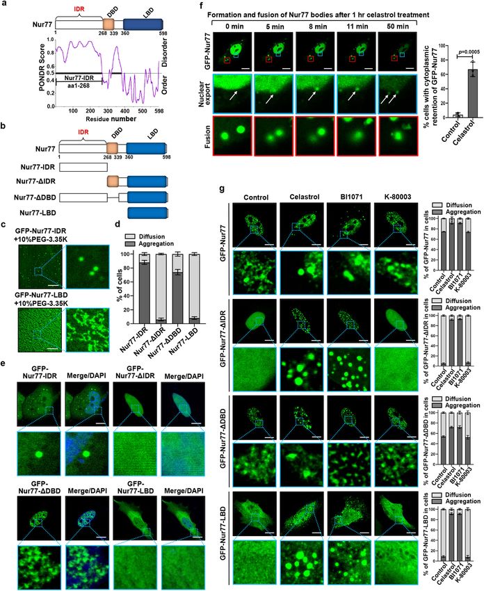
ARTICLE                                                                 NATURE COMMUNICATIONS | https://doi.org/10.1038/s41467-021-26295-8

M
           itophagy, special selective autophagy with an evolutio-           interaction with p62 promotes the autophagy of dysfunctional
           narily conserved process that eliminates impaired                 mitochondria remains unknown.
           mitochondria through autophagy, is important for                      Liquid-liquid phase separation (LLPS) is an important
cellular homeostasis and prevention of diseases and cancer1–4.               mechanism of how cells rapidly and reversibly compartmentalize
The mechanism by which dysfunctional mitochondria are                        their components into membraneless organelles to mediate
recognized and engulfed by autophagosomes involves ubiquiti-                 diverse biological processes31–36. Recent studies have revealed an
nation of mitochondrial proteins followed by their interaction               important role that phase separation plays in the condensation of
with cargo receptors connecting targeted cargo to the autopha-               misfolded and ubiquitin-positive proteins and their degradation
gosomal membrane via LC3/ATG8. Several cargo receptors                       by autophagy37,38. p62 undergoes phase separation upon binding
including OPTN, NDP52, p62/SQSTM1 (hereafter referred to as                  to polyubiquitin chain, driving misfolded and ubiquitinated
p62), NBR1, and TAXBP1, are known to interact with both                      protein aggregates into larger condensates in a PB1-dependent
ubiquitin chains attached to damaged mitochondria and with                   manner and their subsequent tethering to the autophagosomal
LC3/ATG8 proteins anchored to the inner surface of the autop-                membrane via its interaction with LC3 proteins16,39,40. However,
hagosome membrane5,6. Different types of autophagy receptors                 little is known about the role of phase separation in the seques-
are likely required for mitophagy depending on different phy-                tering and tethering of larger intracellular organelles such as
siological and pathological situations3.                                     dysfunctional mitochondria.
   p62, one of the best characterized selective autophagy receptors,             In this work, we show that Nur77 and p62 assemble con-
participates in different types of selective autophagy7–11. Like             densates to mediate celastrol-induced mitophagy. Celastrol-
other cargo receptors, p62 contains a ubiquitin-associating (UBA)            induced ubiquitination in the C-terminal LBD of Nur77 and
domain responsible for binding ubiquitinated proteins, and an                its interaction with p62 results in the formation of Nur77-LBD/
LC3-interacting region (LIR), thus acting as a cargo receptor by             p62 condensates capable of sequestering damaged mitochondria.
simultaneously binding to the cargo and LC312. In addition, p62              However, such condensates are insufficient to tether the clustered
harbors a Phor and Bem1p (PB1) domain at its NH2-terminal                    mitochondria to the autophagosomal membrane. The Nur77/p62
region, which mediates p62 oligomerization essential for its                 condensates require an additional interaction mediated by the
function as a selective autophagy receptor13–16. PB1-directed                N-terminal IDR of Nur77 with the PB1 of p62, which confers the
organization of p62 filamentous assemblies is a critical determi-             condensates with liquidity. These results reveal a critical role of
nant of the diverse functions of p62. While p62 was initially shown          coordinated multivalent interaction between Nur77 and p62 in
to be required for PINK1/Parkin-mediated mitophagy17, later                  celastrol-induced mitophagy and provide mechanistic insight into
studies revealed that the recruitment of p62 by ubiquitination of            nuclear receptor action.
mitochondrial outer membrane proteins only serves to aggregate/
cluster dysfunctional mitochondria through polymerization via its
PB1 domain18,19, in a manner analogous to its aggregation of                 Results
polyubiquitinated proteins8,9. This suggested that p62 fails to              Nur77 and p62 form condensates in celastrol-induced mito-
function as a full autophagy receptor in Parkin-mediated mito-               phagy. To study the role of phase separation in celastrol-induced
phagy. Interestingly, p62 is essential for inflammasome NLRP3                 mitophagy, we first used a sensitive fluorescence-activated cell
agonist-induced mitophagy20 and the elimination of damaged                   sorting (FACS)-based dual-color fluorescence-quenching assay
mitochondria through autophagy in leukemia cells21, demon-                   with an EGFP-mCherry-COX8 reporter localized to the mito-
strating that p62 is fully functional in mediating mitophagy under           chondrial matrix to monitor celastrol-induced mitophagy41,42.
different cellular contexts. However, how p62-mediated seques-               Normal mitochondria are yellow, having both green and red
tration of dysfunctional mitochondria and the recruitment of the             fluorescence in the matrix, whereas mitochondria engulfed into
autophagy machinery are coupled and regulated remains unclear.               acidic lysosomes environment show red-only fluorescence due to
   Nur77 (also called TR3, NGFIB, or NR4A1), a nuclear receptor              the selective sensitivity of EGFP fluorescence to low pH. One
superfamily member and an immediate-early response gene,                     advantage of such analysis allows the detection of mitochondrial
plays an integral role in a plethora of cellular processes including         proteins in lysosomes during mitophagy. Mitochondria are sus-
survival, apoptosis, autophagy, immunity, and inflammation in                 ceptible to inflammatory signaling, which can result in the accu-
response to diverse stimuli22–24. Like other nuclear receptors,              mulation of inflammatory mediators including tumor necrosis
Nur77 comprises an NH2-terminal intrinsically disordered region              factor receptor-associated factor 2 (TRAF2) at dysfunctional
(IDR) with a poorly defined function, a central DNA-binding                   mitochondria28. We showed previously that celastrol can induce
domain (DBD), and a COOH-terminal ligand-binding domain                      Nur77 translocation from the nucleus to damaged mitochondria
(LBD). Although nuclear receptors are conventionally considered              where it interacts with TRAF2 to initiate mitophagy for elim-
as transcription factors, we reported that Nur77 could translocate           inating dysfunctional mitochonria28. Thus, cells were treated with
from the nucleus to mitochondria to modulate mitochondrial                   TNFα and celastrol to evaluate celastrol-mediated mitophagy
activities25–27. Recently, we discovered that celastrol could bind           throughout the study. Upon treatment with celastrol and TNFα,
Nur77 to induce Nur77 translocation from the nucleus to                      significant amounts of mitochondria were engulfed by lysosomes
damaged mitochondria, where it mediates their elimination                    as indicated by their red fluorescence (Fig. 1a). Quantification of
through autophagy by interacting with p6228. Celastrol is a potent           red-only autophagic mitochondria by flow cytometry (Supple-
anti-inflammatory triterpenoid quinine-methide isolated from                  mentary Fig. 1a, b) revealed a temporal effect of celastrol on the
the root of a traditional Chinese medicinal herb Tripterygium                autophagy of dysfunctional mitochondria, which peaked between
wilfordii commonly known as “Thunder God Vine” that has been                 3 to 6 h of treatment (Supplementary Fig. 1c). The celastrol-
widely and successfully used for treating a number of auto-                  induced appearance of red-only mitochondria and degradation of
immune and inflammatory diseases including rheumatoid                         cytochrome c oxidase subunit II (COXII), a mitochondrial inner
arthritis and lupus28–30. We showed that the elimination of                  membrane protein, was attenuated in cells transfected with
dysfunctional mitochondria by celastrol-induced mitophagy                    Atg7 siRNA, which inhibited the expression of Atg7 protein
alleviates chronic inflammation in several disease models,                    (Supplementary Fig. 1d), thereby confirming the mitophagic effect
revealing a critical role of Nur77/p62-mediated mitophagy in                 of celastrol and the feasibility of using FACS-based EGFP-
controlling mitochondrial homeostasis. However, how Nur77                    mCherry-COX8 mitophagy assay.

2                NATURE COMMUNICATIONS | (2021)12:5989 | https://doi.org/10.1038/s41467-021-26295-8 | www.nature.com/naturecommunications
NATURE COMMUNICATIONS | https://doi.org/10.1038/s41467-021-26295-8                                                                ARTICLE

   The role of Nur77 in celastrol-induced mitophagy was                         expressed in Nur77−/−HeLa cells formed droplet-like condensates
illustrated by reduction of autophagic mitochondria (from 12.20                 colocalizing with mitochondria undergoing autophagy in auto-
to 0.02%) and diminished COXII degradation in HeLa cells                        lysosome (Fig. 1a), suggesting that Nur77 is directly involved in
depleted with Nur77 (Nur77−/−HeLa) (Fig. 1a and Supplementary                   both sequestering dysfunctional mitochondria and directing cargo
Fig. 1e). In addition, transfecting Myc-tagged Nur77 (Myc-Nur77)                mitochondria for autophagy.
into Nur77−/−HeLa rescued the effect of celastrol on inducing                      When the role of p62 was examined, we found that the
mitophagy (from 0.02 to 11.91%). Interestingly, Myc-Nur77                       mitophagic effect of celastrol was almost completely abolished in

NATURE COMMUNICATIONS | (2021)12:5989 | https://doi.org/10.1038/s41467-021-26295-8 | www.nature.com/naturecommunications                       3
ARTICLE                                                                    NATURE COMMUNICATIONS | https://doi.org/10.1038/s41467-021-26295-8

Fig. 1 Nur77 and p62 are required for celastrol-induced mitophagy. a Representative image of celastrol-induced mitophagy in HeLa, Nur77−/−HeLa, and
Nur77−/−HeLa cells transfected with Myc-Nur77 by EGFP-mCherry-COX8 assay as described in Methods. Scale bar, 10 μm. b Representative images of
celastrol-induced mitophagy in MEFs and p62−/−MEFs by EGFP-mCherry-COX8 assay as described in Methods. Scale bar, 10 μm. c Colocalization of
Nur77, LC3, and p62 with mitochondria within mitophagosome/autolysosome. Upper panel: Electron micrographs of HeLa cells stained with 15 nm
immunogold-conjugated Nur77 antibody to detect Nur77 (red), and 10 nm immunogold-conjugated p62 antibody to detect p62 (green). Bottom panel:
Electron micrographs of HeLa cells stained with 15 nm immunogold-conjugated LC3 antibody to detect LC3, and 10 nm immunogold-conjugated p62
antibody to detect p62. Cells were treated for 1 h with celastrol. The blue dotted line indicates mitophagosome/autolysosome. Mito mitochondrion, Scale
bar, 200 nm. d Representative images showing Hsp60, a mitochondrial marker, in the liver tissue from wild-type and Nur77−/−mice in aging model. Young
mice, 8 weeks old. Aged mice, 2 years old. Scale bar, 10 μm. e Statistical analysis of mitochondrial size was represented from liver tissue. Left graph,
n = 316, 253, 267, and 287, respectively; Right graph, n = 3 biologically independent samples. A two-tailed unpaired Student’s t-test was used for statistical
analysis, and data were presented as mean values ± SEM. f The expression of Nur77 protein in the liver tissue from wild-type and Nur77−/−mice in the
aging model. g Representative images of EGFP-mCherry-COX8 in the liver from wild-type or Nur77−/−mice in the aging model. Purple arrows indicate
mitophagy. Scale bar, 2 μm. h Quantification of cells showing mCherry-COX8 accumulation on liver tissue. Two-tailed unpaired Student’s t-test was used
for statistical analysis, and data were presented as mean values ± SEM (n = 5 mice per group). Data represent at least three independent experiments.
Source data are provided as a Source Data file.

p62−/−MEF cells, which could be restored by reexpressing Flag-                  with mitochondria and LC3 in the liver of young mice, which
tagged p62 (Flag-p62) (Fig. 1b). Knocking down p62 from HeLa                    decreased in the liver of aged mice (Supplementary Fig. 1l–p).
cells also impaired the mitophagic effect of celastrol (Supple-                 Thus, Nur77 and p62 are involved in mitophagy of the liver,
mentary Fig. 1f). Thus, p62 is also essential for celastrol-induced             which declines during aging. This was confirmed by our data
mitophagy. Interestingly, transfected Flag-p62, like Nur77                      showing that knocking out Nur77 caused defective mitophagy in
(Fig. 1a), was found in autolysosomes as droplet-like condensates               the liver of both young and aged mice (Supplementary Fig. 1o, p).
colocalizing with autophagic mitochondria (Fig. 1b). Moreover,                  To provide more direct evidence supporting the role of Nur77 in
the condensates of Nur77 and p62 colocalized not only with each                 mitophagy during aging, we injected adeno-associated virus ser-
other but also with mitochondria in cells treated with celastrol                otype 9 (AAV-9) plasmid that expresses the mitophagy biosensor
(Supplementary Fig. 1g). The role of p62 in celastrol-induced                   EGFP-mCherry-COX842 into the livers of young and aged mice.
mitophagy differs from Parkin-mediated mitophagy, in which                      Two weeks after injection, mCherry-COX8 exposure was detected
p62 failed to translocate to autolysosome in oligomycin/                        in the liver of young mice but not young Nur77−/−mice (Fig. 1g,
antimycin A (OA)-induced mitophagy (Supplementary Fig. 1h),                     h, and Supplementary Fig. 1q), demonstrating that Nur77 is
in agreement with previous observations18,19. Thus, p62 acts as a               required for hepatic mitophagy of young mice. Hepatic mito-
full selective autophagy cargo receptor for Nur77- but not Parkin-              phagy declined during aging as the mCherry-COX8 exposure was
mediated mitophagy.                                                             not detected in the liver of aged wild-type and Nur77−/−mice.
   We also conducted immunogold electron microscopy (EM)43                      Thus, Nur77/p62-mediated mitophagy plays a role in maintain-
to examine the localization of Nur77 and p62 condensates. Dual                  ing mitochondria homeostasis in the liver.
immunogold staining revealed no detectable Nur77 or p62 on
healthy mitochondria in control cells. When cells were treated                  Celastrol promotes phase separation of Nur77/p62. p62 is
with celastrol, damaged mitochondria were densely decorated at                  known to phase separate ubiquitinated proteins into condensates
their outer membrane with anti-p62 (10 nm) and anti-Nur77                       that become substrates for autophagy16,40. The fact that not only
(15 nm) immunogold labels, which could be found within                          Nur77 but also p62 displayed punctate structures colocalizing
mitophagosome or autolysosome (Fig. 1c). LC3 (15 nm) and                        with damaged mitochondria undergoing autophagy in autolyso-
p62 (10 nm) immunogold particles were also colocalized near the                 some (Fig. 1) suggested that the phase separation of Nur77 and
mitochondrial outer membrane, which again were engulfed by                      p62 are involved in celastrol-induced mitophagy. To test our
the mitophagosome or autolysosome. These results support the                    hypothesis, HeLa cells ectopically expressing Nur77 fused with
notion that Nur77/p62 condensates are capable of directing cargo                green fluorescent protein (GFP-Nur77) and mCherry-fused p62
mitochondria to the autophagy machinery for degradation.                        (mCherry-p62) were examined for their phase separation in the
                                                                                absence or presence of celastrol. Transfected GFP-Nur77 resided
Nur77-mediated mitophagy in aging. The physiological sig-                       exclusively in the nucleus, while mCherry-p62 was diffusely dis-
nificance of Nur77/p62-mediated mitophagy was examined dur-                      tributed in the cytoplasm. However, when cells were treated with
ing the aging process of the liver, in which the autophagy of                   celastrol, GFP-Nur77 formed cytoplasmic droplets colocalizing
damaged mitochondria plays a critical role in counterbalancing                  extensively with mCherry-p62 in a time dependent manner
age-related pathological conditions44. Confocal microscopy ana-                 (Fig. 2a). To understand how GFP-Nur77/mCherry-p62 con-
lysis revealed that the size of mitochondria in the liver of aged               densates are formed in the cytoplasm upon celastrol treatment,
mice significantly increased (Fig. 1d, e), in agreement with the                 cells were treated with celastrol for 1 h and subjected to real-time
enlargement of mitochondria termed megamitochondria in aged                     microscopy visualization of the formation of the GFP-Nur77/
liver45. The increase in the size of mitochondria in aged mice was              mCherry-p62 condensates. We found that transfected GFP-
accompanied with liver enlargement (Supplementary Fig. 1i, j)                   Nur77 and mCherry-p62 rapidly formed large cytoplasmic bodies
and increased hepatic inflammation (Supplementary Fig. 1k). The                  through the fusion of undetectable micro-sized droplets or
role of Nur77 was revealed by enlarged hepatic mitochondria in                  detectable droplets in response to celastrol treatment (Fig. 2b,
both young and aged Nur77−/−mice compared to wild-type mice                     Supplementary Fig. 2a, b, and Supplementary Movies 1–3). Thus,
(Fig. 1d, e) and decreased expression of Nur77 during aging                     celastrol induces the formation of Nur77/p62 bodies by triggering
(Fig. 1f). Alteration of p62-mediated Parkin-independent mito-                  their phase separation. The autophagosome marker LC3 is
phagy is responsible for the formation of megamitochondria in                   recruited to p62 bodies after p62 undergoes phase separation
the liver46. We, therefore, studied whether Nur77 and p62 could                 through ubiquitin-induced oligomerization16,40, which serves as
mediate mitophagy during aging, and found their colocalization                  a mechanism for linking p62-containing protein aggregates to

4                  NATURE COMMUNICATIONS | (2021)12:5989 | https://doi.org/10.1038/s41467-021-26295-8 | www.nature.com/naturecommunications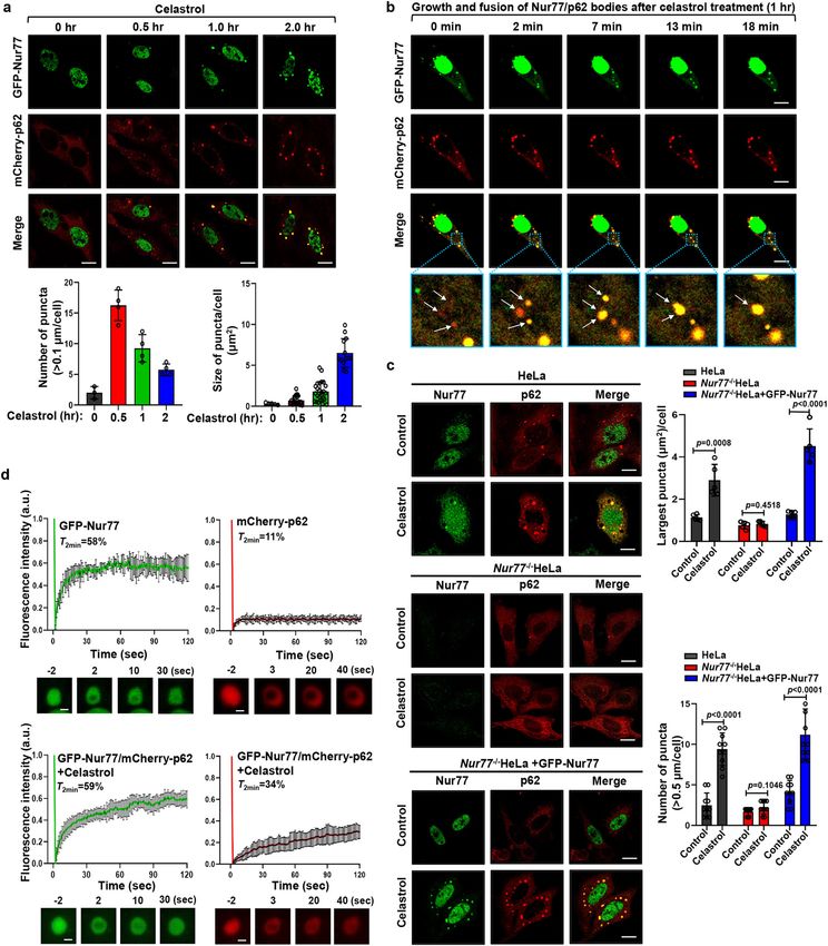
NATURE COMMUNICATIONS | https://doi.org/10.1038/s41467-021-26295-8                                                                        ARTICLE

Fig. 2 Celastrol promotes phase separation and liquidity of p62. a Representative images showing the time-dependent effect on celastrol induction of
cytoplasmic Nur77 body formation. Bottom panels: quantitative analysis of the number and size of Nur77/p62 body formation. Bottom left graph, n = 4
biologically independent samples; Bottom right graph, n = 20, 23, 25, and 19, respectively. Data were presented as mean values ± SEM. Scale bar, 10 μm. b
Real-time images showing the formation and fusion of GFP-Nur77 and mCherry-p62 droplets in HeLa cells after treatment with celastrol (2 μM) for 1 h.
White arrows indicate droplets formation and fusion (see also Supplementary Movie 1). Scale bar, 10 μm. c Representative images illustrating the role of
celastrol in promoting p62 body formation in a Nur77-dependent manner immunostaining. Nur77−/−HeLa cells were also transfected with GFP-Nur77 to
determine its effect on p62 body formation. The diameter of the biggest p62 puncta in each cell was measured. The number of p62 puncta >0.5 μm in each
cell was assessed. A two-tailed unpaired Student’s t-test was used for statistical analysis, and data are presented as mean values ± SEM (n = 5 biologically
independent samples). d FRAP analysis of the effect of Nur77 in regulating p62 mobility in HeLa cells. Data were presented as means ± SEM (n = 3
independent experiments). Scale bar, 1.5 μm. Data represent at least three independent experiments. Source data are provided as a Source Data file.

NATURE COMMUNICATIONS | (2021)12:5989 | https://doi.org/10.1038/s41467-021-26295-8 | www.nature.com/naturecommunications                                  5
ARTICLE                                                                 NATURE COMMUNICATIONS | https://doi.org/10.1038/s41467-021-26295-8

the autophagy machinery. However, whether LC3 can be                         reversible phase-separated condensates. To this end, GFP-Nur77
recruited to phase-separated p62 bodies during mitophagy is                  was allowed to form droplets in an initial solution, and then
unknown. We found that both LC3 and p62 localized at the outer               diluted sequentially by five times in equimolar protein con-
membrane of mitochondria undergoing autophagy in the mito-                   centration and crowding agent. The preformed GFP-Nur77
phagosome (Fig. 1c). Transfected Nur77 and p62 also formed                   droplets were reduced in size and turbidity with dilution,
condensates with LC3 and mitochondria in cells treated with                  demonstrating that the droplet formation was reversible (Sup-
celastrol (Supplementary Fig. 2c, d). These results confirmed the             plementary Fig. 3e). Taken together, these results demonstrated
capability of Nur77/p62 condensates in mediating celastrol-                  that Nur77 exhibits liquid-like properties in vitro.
induced mitophagy.                                                              To test whether Nur77 undergoes LLPS in vivo, we ectopically
                                                                             expressed GFP-Nur77 in HeLa cells. Transfected GFP-Nur77
Celastrol-induced phase separation of p62 depends on Nur77.                  formed punctum structures in the nucleus of cells (Fig. 3d), in
To determine the role of Nur77 in p62 phase separation, we                   which GFP-Nur77 diffused rapidly within the puncta (Fig. 3e).
studied the formation of p62 condensates in HeLa and Nur77−/                 3D-reconstruction of the GFP-Nur77 images revealed that it
−HeLa cells. In response to celastrol, p62 rapidly formed con-               exhibited as a single particle (Fig. 3f). Myc-Nur77 (Fig. 3g) and
                                                                             endogenous Nur77 (Fig. 3h) also formed puncta in the nucleus.
densates colocalizing with Nur77 in HeLa cells (Fig. 2c). How-               Thus, the Nur77 phase separates into condensates via LLPS.
ever, the ability of p62 to assemble condensates was impaired in
Nur77−/−HeLa cells, which was restored by reexpressing GFP-
Nur77. Thus, celastrol-induced formation of p62 condensates is               Nur77 phase separation is dependent on its N-terminal IDR
dependent on Nur77 expression. As the liquidity of condensate is             domain. Proteins containing large IDRs often phase separate
a critical determinant for its selective autophagy47,48, we studied          under physiologic conditions50. Using the prediction program
the effect of celastrol on the liquidity of Nur77/p62 condensates.           PONDR (VSL2) (http://www.pondr.com/)51, we found that the
Fluorescence recovery after photobleaching (FRAP) analysis of                N-terminal region of Nur77 displays the highest structural dis-
co-transfected GFP-Nur77 and mCherry-p62 revealed that the                   order propensity (Fig. 4a). To determine if the IDR of Nur77 is
fluorescence of GFP-Nur77 after photobleaching was recovered                  required for its phase separation, GFP-tagged IDR (GFP-Nur77-
rapidly and significantly (T2 min = 58%), while the fluorescence of            IDR) or LBD of Nur77 (GFP-Nur77-LBD) (Fig. 4b) was purified
p62 was only slightly recovered (T2 min = 11%) and remained                  and subjected to droplet formation in vitro. GFP-Nur77-IDR
static (Fig. 2d), indicating a more liquid-like property of GFP-             formed numerous spherical droplets in a buffer containing 10%
Nur77 than mCherry-p62. This is consistent with previous stu-                PEG-3.35 K (Fig. 4c). For comparison, GFP-Nur77-LBD exhib-
dies showing low mobility of p62 body16,40. However, when cells              ited amorphous condensates rather than spherical droplets. GFP-
were treated with celastrol, which induces the formation of                  Nur77-IDR also formed phase-separated droplets in cells (Fig. 4d,
Nur77/p62 bodies, the recovery rate of the p62 signal                        e). Removing IDR from Nur77 (GFP-Nur77-ΔIDR) significantly
(T2 min = 34%) was much improved, while the recovery of Nur77                attenuated its ability to form nuclear droplets, while deleting DBD
in Nur77/p62 bodies remained almost the same. These results                  (GFP-Nur77-ΔDBD) altered the morphology Nur77 bodies. GFP-
demonstrated that Nur77 is a critical determinant of the liquidity           Nur77-LBD also failed to assemble droplets in cells. Thus, the
and mobility of the Nur77/p62 condensates.                                   N-terminal IDR of Nur77 is responsible for Nur77 phase
                                                                             separation.
Nur77 forms liquid-like condensate through phase separation.                    The N-terminal IDR of Nur77 is involved in an intramolecular
The above FRAP data suggested that Nur77 might itself undergo                interaction with its carboxyl-terminal LBD52. We then asked
phase separation. Thus, GFP-Nur77 was expressed and purified                  whether GFP-Nur77-IDR could modulate the phase separation of
to study its phase separation. When added to droplet formation               mCherry-Nur77-LBD through the intramolecular interaction.
buffer containing a crowding agent (10% PEG-3.35 K) to simulate              Nur77-LBD alone exhibited amorphous condensates rather than
the densely crowded environment of the nucleus, GFP-Nur77 but                spherical droplets (Fig. 4c). However, when mCherry-Nur77-LBD
not GFP formed micro-sized droplets in solution with markedly                was mixed with GFP-Nur77-IDR, mCherry-Nur77-LBD formed
increased size (Fig. 3a). FRAP analysis demonstrated that GFP-               spherical droplets together with GFP-Nur77-IDR (Supplementary
Nur77 redistributed rapidly from the unbleached area to the                  Fig. 4a). For comparison, GFP-Nur77-IDR had no effect on droplet
bleached area (Fig. 3b). GFP-Nur77 droplets also fused to form               formation of mCherry and mCherry-DBD (Supplementary Fig. 4b).
larger droplets, revealing droplet coalescence (Fig. 3c). The for-           These studies revealed the potency of Nur77-IDR in LLPS.
mation of Nur77 droplets was a general result of macromolecular                 Nuclear receptor superfamily members are characterized by the
crowding rather than a specific effect of PEG, as Nur77 assembled             presence of IDRs in their N-terminal regions, whose function
into spherical droplets in the presence of Dextran, Ficoll-400, or           remains poorly defined. To address whether IDRs in other
in a highly concentrated solution of lysozyme (2 mM) (Supple-                nuclear receptor family members could also mediate phase
mentary Fig. 3a). It was dependent on the molecular weight of the            separation, we studied Nur77 subfamily members NOR1 and
crowding agent, illustrated by its assembling into spherical dro-            Nurr1. Similar to Nur77, NOR1, and Nurr1 contain a large IDR
plets only in solutions containing >1 kDa PEG. The size of                   in their N-terminal region (Supplementary Fig. 4c, d). We then
mCherry-Nur77 droplets in solution became bigger by increasing               tested whether Nurr1 and NOR1 underwent LLPS in vivo and
PEG molecular mass with the efficiency of condensate formation                found that ectopically expressed GFP-Nurr1 and GFP-NOR1 also
peaked in 8 kDa PEG (Supplementary Fig. 3b). Analysis of the                 formed punctate structures in the nucleus (Supplementary Fig. 4e,
biophysical properties of Nur77 droplets revealed a shift of the             f). Removal of IDR from Nurr1 (GFP-Nurr1-ΔIDR) or NOR1
distributions and opacity of the droplets toward greater droplets            (GFP-NOR1-ΔIDR) greatly reduced their ability to form nuclear
with increasing PEG concentration (Supplementary Fig. 3c).                   droplets. GFP-Nurr1-LBD and GFP-NOR1-LBD also failed to
Phase-separated droplets typically scale in size according to the            form droplets. Thus, the N-terminal IDR of Nurr1 and NOR1
concentration of components in the system49. Indeed, the size                also mediates their formation of phase-separated droplets,
distribution and turbidity of Nur77 droplets increased in a Nur77            suggesting that the IDR in the nuclear receptor superfamily
concentration-dependent manner (Supplementary Fig. 3d). We                   members plays an important role in regulating nuclear receptor
also tested whether the droplets were irreversible aggregates or             action through phase separation.

6                NATURE COMMUNICATIONS | (2021)12:5989 | https://doi.org/10.1038/s41467-021-26295-8 | www.nature.com/naturecommunications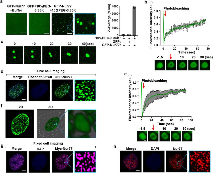
NATURE COMMUNICATIONS | https://doi.org/10.1038/s41467-021-26295-8 ARTICLE
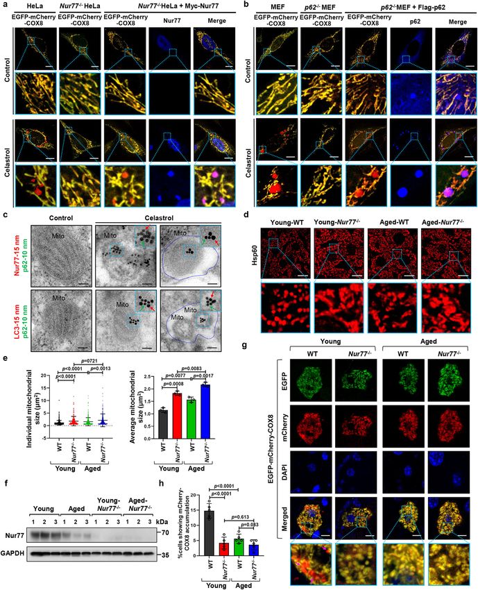
Fig. 3 Nur77 phase separates into a liquid-like condensate. a GFP-Nur77 (2 μM) undergoes phase separation. The size of Nur77 droplets was analyzed.
Data were presented as mean values ± SEM (n = 3 independent experiments). Scale bar, 10 μm. b Top, changes in fluorescence intensity of GFP-Nur77
droplets after photobleaching were plotted over time. Bottom, representative images of fluorescence recovery. Data were presented as mean values ± SEM
(n = 3 independent experiments). Scale bar, 1.5 μm. c Fusion of GFP-Nur77 droplets in 10% PEG-3.35 K. Scale bar, 20 μm. d Live imaging of GFP-Nur77 in
HeLa cells. Scale bar, 5 μm. e Time course analysis of GFP-Nur77 nuclear body recovery after photobleaching in HeLa cells. Representative images of
fluorescence recovery are shown. Data were presented as mean values ± SEM (n = 3 independent experiments). Scale bar, 1.5 μm. f Three-dimensional
(3D) images of Nur77 nuclear assemblies. An enlarged view of inset is also shown. Scale bar, 5 μm. g Fixed imaging of Myc-Nur77 in HeLa cells. Scale bar,
5 μm. h Endogenous Nur77 displays nuclear puncta in HeLa cells revealed by immunostaining with anti-Nur77. Scale bar, 5 μm. Data represent at least
three independent experiments. Source data are provided as a Source Data file.
Celastrol regulation of Nur77 phase separation. We next stu- effect. We also used FRAP analysis to study the mobility and
died how celastrol binding to Nur77 regulates its phase separa- liquidity of GFP-Nur77 and mutants and found that the fluor-
tion. Transfected GFP-Nur77 exclusively resided in the nucleus of escence recovery of GFP-Nur77 and GFP-Nur77-ΔDBD was
control cells (Figs. 1a and 2a). Figure 4f showed stills of movie much quicker than that of GFP-Nur77-ΔIDR and GFP-Nur77-
taken after GFP-Nur77-transfected cells were treated with celas- LBD (Supplementary Fig. 4g), revealing a critical role of Nur77-
trol for 1 h (Supplementary Movie 4). There was a significant IDR in promoting the mobility and liquidity of celastrol-induced
amount of cytoplasmic GFP-Nur77 bodies in cells with new Nur77 condensates.
bodies continuously emerging from the nuclear membrane. Thus,
celastrol promotes GFP-Nur77 nuclear export as we reported
Nur77 ubiquitination-induced p62 phase separation sequesters
before26, although we could not exclude the possibility that
mitochondria. Ubiquitinated substrates can trigger p62-mediated
celastrol may also promote the retention of cytoplasmic Nur77.
phase separation and segregation of the substrates into autop-
Our data also showed the growth of Nur77 droplets in the
hagic degradation16,39,40. We previously reported28 that celastrol
cytoplasm via fusion, revealing a role of phase separation in
binding to Nur77 induces Nur77 translocation to mitochondria
celastrol-induced growth of cytoplasmic Nur77 bodies. When the
where it is ubiquitinated at Lys536 located in its C-terminal LBD
effect of celastrol on phase separation of Nur77 mutants was
by TRAF2, an E3 ubiquitin ligase56, through interaction. The
analyzed (Fig. 4g), we found that celastrol could also facilitate the
ubiquitinated mitochondrial Nur77 then interacts with the UBA
conversion of Nur77 mutants, Nur77-ΔIDR, and Nur77-LBD,
of p62, leading to autophagy of Nur77-primed dysfunctional
from diffusely status to spherical particle structures in the
mitochondria28. To study the molecular detail of phase separation
nucleus, suggesting a ligand-dependent phase separation in the
in mediating the mitophagic effect of Nur77 and p62, we exam-
nucleus. The effect of celastrol is likely due to its binding to
ined the role of Nur77 ubiquitination in p62 phase separation.
Nur77 as BI1071, another Nur77 ligand53, also promoted Nur77
Consistent with our previous results28, TRAF2 was recruited to
phase separation, while K-80003, a RXRα ligand54,55, had no
the surface of Nur77 condensates in cells treated with celastrol,
NATURE COMMUNICATIONS | (2021)12:5989 | https://doi.org/10.1038/s41467-021-26295-8 | www.nature.com/naturecommunications 7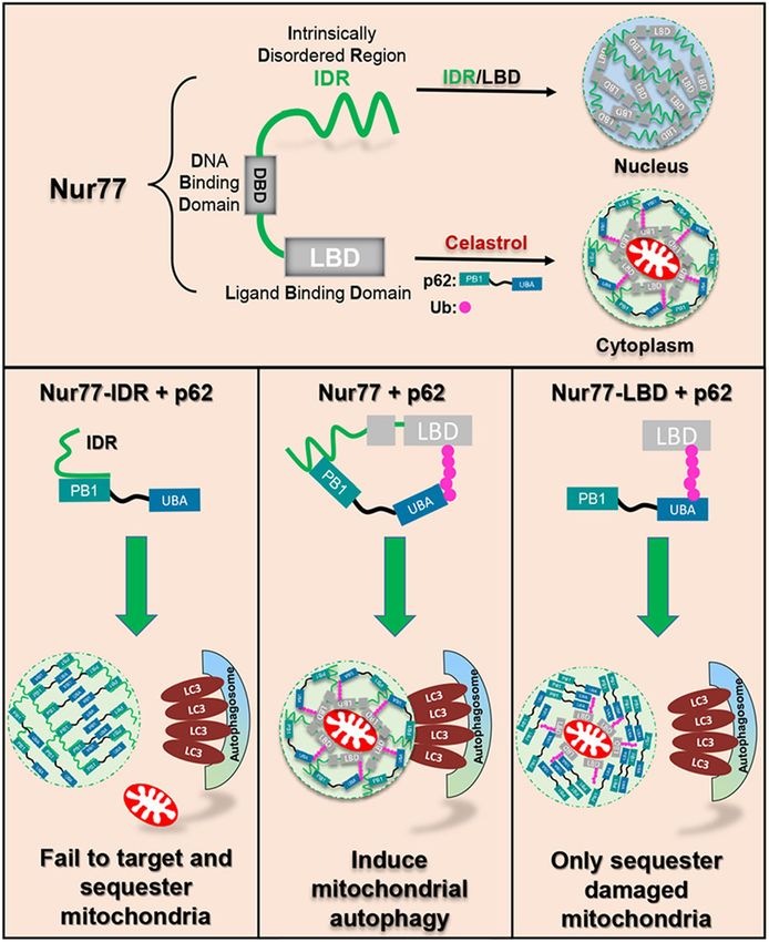
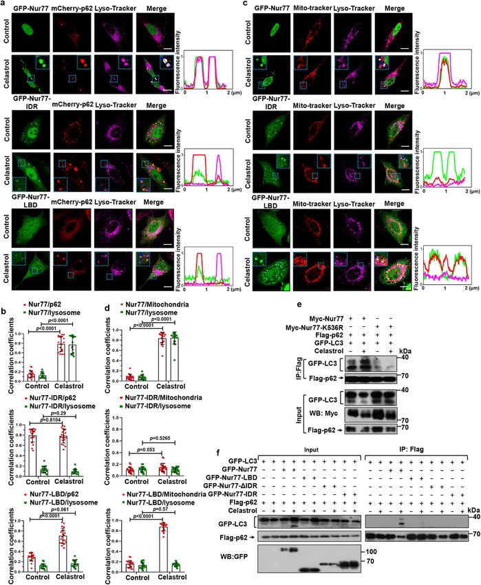
ARTICLE                                                               NATURE COMMUNICATIONS | https://doi.org/10.1038/s41467-021-26295-8

displaying spherical shell structure (Supplementary Fig. 5a).              celastrol on inducing their interaction with p62. To our surprise,
TRAF2-mediated Nur77 ubiquitination likely plays a role in p62             mutating Lys536 in Nur77 did not show an apparent inhibitory
body formation as celastrol-induced Nur77 condensates coloca-              effect on the celastrol-induced assembly of Nur77/p62 con-
lized not only with p62 but also with ubiquitin (Fig. 5a). To              densates (Fig. 5d). We suspected that this might be due to
confirm the role of Nur77 ubiquitination, we mutated its ubi-               celastrol-independent interaction between Nur77 and p62, which
quitination site and tested its effect on p62 body formation.              was not affected by Lys536 mutation (Fig. 5b). Notably, mutating
Mutating Lys536 in Nur77 (Nur77-K536R) (Fig. 5b) or Nur77-                 Lys536 in Nur77-LBD completely impaired its ability to interact
LBD (Nur77-LBD-K536R) (Fig. 5c) impaired the effect of                     with p62 even in the presence of celastrol (Fig. 5c), indicating that

8              NATURE COMMUNICATIONS | (2021)12:5989 | https://doi.org/10.1038/s41467-021-26295-8 | www.nature.com/naturecommunications
NATURE COMMUNICATIONS | https://doi.org/10.1038/s41467-021-26295-8                                                                        ARTICLE

Fig. 4 Nur77 phase separation is dependent on N-terminal IDR domain. a Intrinsic disorder tendency of Nur77. IDR intrinsically disordered region, DBD
DNA-binding domain, LBD ligand-binding domain. b Schematic representation of Nur77 and its mutants. c In vitro phase separation of GFP-Nur77-IDR and
GFP-Nur77-LBD (2 μM). Scale bar, 10 μm. d Quantification of Nur77 mutant droplets formation in absence of celastrol in HeLa cells. Data were presented
as mean values ± SEM (n = 3 independent experiments). e Representative images of Nur77 mutant droplets formation in absence of celastrol in HeLa cells.
Scale bar, 10 μm. f Representative real-time images showing the formation and fusion of cytoplasmic GFP-Nur77 droplets after treatment with celastrol
(2 μM) for 1 h in HeLa cells (see also Supplementary Movie 4). Right: quantification of the cytoplasmic retention of GFP-Nur77 protein. A two-tailed
unpaired Student’s t-test was used for statistical analysis, and data were presented as mean values ± SEM (n = 3 independent experiments). Scale bar,
10 μm. g Droplet formation of GFP-Nur77 and mutants in HeLa cells treated with the indicated compounds (2 μM). Left: Representative droplet images of
transfected GFP-Nur77 and mutants. An enlarged view of the inset is also shown. Scale bar, 10 μm. Right: Quantification of droplet formation of GFP-Nur77
and mutants. Data represent at least three independent experiments. Source data are provided as Source Data file.

Fig. 5 Ubiquitinated Nur77 interacts with p62 to sequester damaged mitochondria. a Immunofluorescence images of GFP-Nur77, mCherry-p62, and
HA-Ub transfected in HeLa cells treated with or without celastrol. Data illustrate the colocalization of Nur77 with p62 and Ub in the presence of celastrol.
Scale bar, 10 μm. b, c Interaction of indicated Nur77 or deubiquitinated mutant (K536R) and p62 was analyzed in HeLa cells treated with or without
celastrol by co-immunoprecipitation (co-IP) assay. d, e Immunofluorescence images showing the effect of celastrol-induced Nur77 ubiquitination on
mCherry-p62 droplet formation. Scale bar, 10 μm. f Representative images showing ubiquitination-dependent colocalization of Nur77 with p62 and
mitochondria in HeLa cells treated with celastrol. Scale bar, 10 μm. Data represent at least three independent experiments. Source data are provided as
Source Data file.

NATURE COMMUNICATIONS | (2021)12:5989 | https://doi.org/10.1038/s41467-021-26295-8 | www.nature.com/naturecommunications                                  9
ARTICLE                                                                 NATURE COMMUNICATIONS | https://doi.org/10.1038/s41467-021-26295-8

Nur77-LBD is not involved in celastrol-independent interaction               mitochondria but not their connection to the autophagy
between Nur77 and p62. Thus, we used Nur77-LBD to study the                  machinery in Parkin-dependent mitophagy18. To confirm the
effect of Nur77 ubiquitination on p62 body formation. Nur77-                 mitophagic role of p62 in Nur77-dependent mitophagy, we
LBD could assemble condensates with p62 in the presence of                   examined the colocalization of Nur77- and p62-positive con-
celastrol. However, such an effect of Nur77-LBD was completely               densates with core ATG proteins essential for autophagosome
abolished when Lys536 was mutated (Fig. 5e). These results,                  formation (Supplementary Fig. 6c–f). Our results showed that
therefore, revealed not only a critical role of celastrol-induced            ULK1 and FIP200 failed to colocalize with celastrol-induced p62
Nur77 ubiquitination in promoting p62 phase separation but also              condensates formed with Nur77 or Nur77-mutants (Nur77-ΔIDR
the existence of the celastrol-independent interaction between               and Nur77-K536R). In contrast, WIPI2 and ATG16L1 exhibited
Nur77 and p62.                                                               extensive colocalization with p62 condensates formed with Nur77
   We next studied the role of Nur77 ubiquitination-mediated                 but not Nur77 mutants (Nur77-ΔIDR and Nur77-K536R). These
p62 phase separation in celastrol-induced mitophagy. Mito-                   results demonstrated that Nur77/p62-mediated mitophagy is
phagy involves sequestering/clustering dysfunctional mito-                   independent on the ULK1/FIP200 complex but requires early
chondria and subsequently directing cargo mitochondria for                   autophagy proteins WIPI2 and ATG16L1 for autophagosome
degradation by the autolysosome system57. We first studied its                formation.
effect on sequestering dysfunctional mitochondria. Upon                         When p62 functions as a full autophagy receptor for protein
celastrol treatment, condensates formed by GFP-Nur77 and                     aggregates, ubiquitin-induced p62 phase separation generates high
mCherry-p62 colocalized with mitochondria (Fig. 5f and                       avidity interaction with LC3 on the inner surface of the
Supplementary Fig. 5b). By contrast, the condensates assembled               autophagosomal membrane, which is responsible for tethering of
by Nur77-K536R and p62 did not. Thus, p62 recruitment by                     targeted cargo to autolysosome for autophagy40. Thus, we examined
ubiquitinated Nur77 could mediate celastrol-induced seques-                  whether the lack of connecting Nur77-LBD/p62 condensates to the
tering of dysfunctional mitochondria, in a manner analogous to               autophagy machinery was due to their inability to interact with
its aggregation of misfolded proteins16,40 or dysfunctional                  LC3. Co-IP experiments demonstrated that p62 interacted with LC3
mitochondria during Parkin-mediated mitophagy18,19. This is                  in the presence of Nur77 but not Nur77-K536R when cells were
further confirmed by data showing that celastrol-induced                      treated with celastrol (Fig. 6e), revealing a high-affinity interaction
Nur77-LBD/p62 but not Nur77-LBD-K536R/p62 condensates                        of Nur77/p62 complex with LC3, which is dependent on Nur77
colocalized with mitochondria. As deleting IDR from Nur77                    ubiquitination. By contrast, cotransfection of Nur77-LBD, which
(Nur77-ΔIDR) did not affect its ability to aggregate mitochon-               binds p62 in a ubiquitination-dependent manner, could not induce
dria, the C-terminal LBD of Nur77 is sufficient to sequester                  p62 interaction with LC3 (Fig. 6f). Thus, p62 condensates induced
damaged mitochondria in a ubiquitination dependent manner                    by Nur77 ubiquitination alone is insufficient to direct cargo
(Fig. 5f and Supplementary Fig. 5b). Notably, the IDR of Nur77               mitochondria to the autophagosome.
could also assemble condensates with mCherry-p62 even
though it is not required for clustering mitochondria.
                                                                             The IDR of Nur77 interacts with PB1 of p62. Our observation
                                                                             that ubiquitin-induced p62 phase separation is insufficient to
Nur77 ubiquitination-mediated p62 phase separation is                        complete mitophagy prompted us to study the role of Nur77-IDR
insufficient to complete mitophagy. We next asked whether                     in modulating the cargo activity of p62. To this end, we found
Nur77 ubiquitination-induced p62 phase separation is sufficient               that Nur77-IDR could form condensates with p62 in the absence
to complete the mitophagic process, in analogous to its effect on            of celastrol, even though the resulting Nur77-IDR/p62 con-
misfolded proteins9. We reasoned that if it was sufficient, Nur77-            densates were not found either at mitochondria or in the lyso-
LBD, which promotes p62 phase separation through ubiquitina-                 some (Fig. 6a–d). We first asked whether Nur77-IDR is involved
tion, should also act to tether clustered mitochondria to auto-              in celastrol-independent interaction between Nur77 and p62
lysosome for degradation. Bodies assembled by cotransfected                  observed (Fig. 5b). Mutational analysis of p62 (Fig. 7a) demon-
GFP-Nur77 and mCherry-p62 were found in lysosome as indi-                    strated that deleting TB or ZZ domain from p62 had no effect on
cated by their colocalization with Lyso-Tracker (Fig. 6a, b). GFP-           celastrol-dependent and -independent interaction between Nur77
Nur77 also colocalized with both mitochondria and lysosome                   and p62 (Fig. 7b), excluding their involvement. Removing the
(Fig. 6c, d). Thus, condensates containing Nur77, p62, and                   UBA domain impaired celastrol-dependent but not -independent
mitochondria were engulfed by autolysosome. The role of Myc-                 interaction, in agreement with its role in binding ubiquitinated
Nur77 to support the mitophagic effect of celastrol was confirmed             Nur77 (Fig. 5b). When PB1 was deleted, however, both celastrol-
by EGFP-mCherry-COX8 mitophagy assay and its reduction of                    dependent and -independent interactions were lost, revealing a
COXII expression (Supplementary Fig. 6a, b). However, when                   critical role of PB1 in p62 high-affinity interaction with Nur77.
GFP-Nur77-LBD was evaluated, we found that the condensates                   The importance of UBA and PB1 was also illustrated by confocal
formed by GFP-Nur77-LBD and p62 were not detected in lyso-                   microscopy analysis showing that celastrol-induced p62 coloca-
some (Fig. 6a, b), even though GFP-Nur77-LBD colocalized                     lization with Nur77 was impaired by their deletion (Supple-
extensively with mitochondria (Fig. 6c, d). Unlike Myc-Nur77,                mentary Fig. 7a). Mutational analysis of Nur77 demonstrated that
transfected Myc-Nur77-LBD also failed to mediate the mito-                   DBD of Nur77 (Nur77-ΔDBD) is dispensable for celastrol-
phagic effect of celastrol (Supplementary Fig. 6a, b). This is               dependent and -independent interactions (Supplementary
reminiscent of the role of p62 in Parkin-mediated mitophagy, in              Fig. 7b). When GFP-Nur77-IDR was analyzed, we found that it
which it serves only to cluster damaged mitochondria but not                 strongly interacted with Flag-p62 but not Flag-p62 lacking PB1
their autophagy18,19. Collectively, these results showed that                (Flag-p62-ΔPB1) in a celastrol-independent manner (Fig. 7c). It
Nur77 ubiquitination-mediated p62 phase separation is insuffi-                also interacted with p62-PB1 but not p62-UBA (Fig. 7d). For
cient to direct targeted cargo to autophagosome and that the                 comparison, Nur77-LBD interacted with p62-UBA but not p62-
structural integrity of Nur77 is required to confer p62 with full            PB1 in a celastrol-dependent manner (Supplementary Fig. 7c).
autophagy receptor activity.                                                 Taken together, these data demonstrated that celastrol-induced
   Our finding that p62 is required for Nur77-dependent                       formation of Nur77-p62 condensates is mediated by their unique
mitophagy is interesting as p62 serves to sequester dysfunctional            celastrol-dependent and -independent interactions involving their

10               NATURE COMMUNICATIONS | (2021)12:5989 | https://doi.org/10.1038/s41467-021-26295-8 | www.nature.com/naturecommunications
NATURE COMMUNICATIONS | https://doi.org/10.1038/s41467-021-26295-8 ARTICLE Fig. 6 Nur77-LBD is insufficient to mediate celastrol-induced mitophagy. a–d Representative images showing colocalization of Nur77 or mutants with p62, mitochondria, and lysosome in HeLa cells after celastrol treatment. The blue arrow indicates line profiles of fluorescence intensities including Pearson’s correlation coefficients shown in b and d. A two-tailed unpaired Student’s t-test was used for statistical analysis, and data were presented as mean values ± SEM (n = 20 biologically independent samples). Dotted box: higher magnification of indicated region. Scale bar, 10 μm. e Mutating K536 in Nur77 inhibits celastrol-induced interaction between p62 and LC3. HeLa cells transfected with the indicated expression plasmids were treated with or without 2 μM celastrol and 20 ng/mL TNFα. Interaction of Flag-p62 with GFP-LC3 was examined by co-IP assay. f Characterization of domain requirement of Nur77 for promoting celastrol-induced p62 interaction with LC3. HeLa cells transfected with the indicated Flag-p62, GFP-LC3, and GFP-Nur77 or mutant were treated with celastrol and TNFα for 1 h and analyzed for Flag-p62 interaction with GFP-LC3 by co-IP assay. Data represent at least three independent experiments. Source data are provided as Source Data file. NATURE COMMUNICATIONS | (2021)12:5989 | https://doi.org/10.1038/s41467-021-26295-8 | www.nature.com/naturecommunications 11
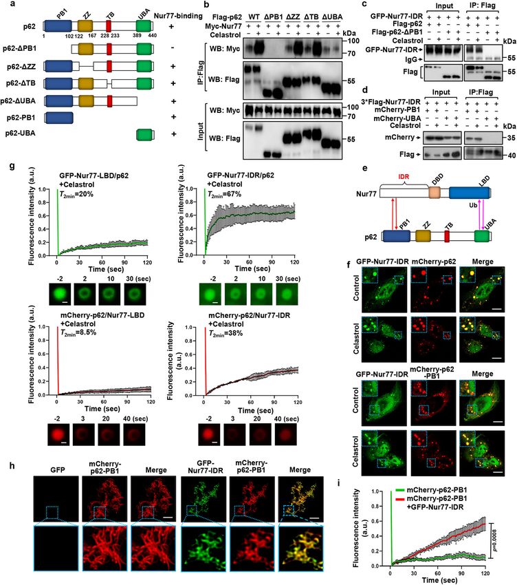
ARTICLE                                                                   NATURE COMMUNICATIONS | https://doi.org/10.1038/s41467-021-26295-8

Fig. 7 IDR interaction with PB1 promotes liquidity of p62 condensates. a Schematic representation of p62 and mutants and their interaction with Nur77.
PB1 Phox/Bem1p protein-protein binding domain. ZZ zinc-finger domain, TB TRAF6 binding domain, UBA ubiquitin-associated domain. b–d Interaction of
Nur77 and p62, as well as their mutants, was analyzed in HeLa cells treated with or without celastrol by co-IP assay. e Multivalent interaction between
Nur77 and p62. The interaction between the IDR of Nur77 and PB1 of P62 is ligand-independent (red), whereas the interaction between LBD of Nur77 and
UBA of p62 depends on celastrol that triggers Nur77-LBD ubiquitination (pink). f Immunofluorescence images showing colocalization of GFP-Nur77-IDR
with mCherry-p62 or mCherry-p62-PB1 after treatment with or without celastrol. Scale bar, 10 μm. g FRAP analysis of the effect of Nur77-IDR in regulating
p62 mobility in HeLa cells. Data were presented as mean values ± SEM (n = 3 independent experiments). Scale bar, 1.5 μm. h Representative images
showing the effect of GFP-Nur77-IDR on the filamentous structures of mCherry-p62-PB1 when mCherry-p62-PB1 was incubated with GFP or GFP-Nur77-
IDR at intermediate molar ratio (1:2). Scale bar, 10 μm. i FRAP analysis of the effect of GFP-Nur77-IDR on mCherry-p62-PB1 mobility in vitro. Two-tailed
unpaired Student’s t-test was used for statistical analysis, and data were presented as mean values ± SEM (n = 3 independent experiments). Data
represent at least three independent experiments. Source data are provided as Source Data file.

12                 NATURE COMMUNICATIONS | (2021)12:5989 | https://doi.org/10.1038/s41467-021-26295-8 | www.nature.com/naturecommunications
NATURE COMMUNICATIONS | https://doi.org/10.1038/s41467-021-26295-8                                                                        ARTICLE

C-terminal and N-terminal regions in a “head-to-head” and “tail-
to-tail” manner (Fig. 7e).

Role of IDR of Nur77 in celastrol-induced mitophagy. PB1 is
known to mediate p62 oligomerization15,58, which is critical for
not only sequestering ubiquitinated cargo but also their connec-
tion to the autophagosome14,15. Given the above observation that
Nur77-IDR could directly interact with p62-PB1, we suspected
that the interaction might play a crucial role in celastrol-induced
mitophagy. Nur77-IDR interacted with p62, resulting in the
formation of Nur77-IDR/p62 condensates (Fig. 7f), which how-
ever were not found either on mitochondria (Fig. 5f) or in the
lysosome (Fig. 6a). This suggested that the interaction is not
directly involved in sequestering and tethering dysfunctional
mitochondria, raising an intriguing question that the interaction
might play a modulatory role in directing engulfed mitochondria
to the autophagy machinery. Thus, we studied how IDR-
dependent phase separations could confer Nur77-p62 con-
densates with the ability to connect targeted mitochondria to the
autophagy machinery. We found that Nur77-IDR contributed
significantly to the increase in the sizes and number of p62 body
(Supplementary Fig. 7d), features that are important for effective
autophagy59. The flexibility in the size of condensates allows the
sequestration of different sizes and amounts of cargo for autop-
hagy. In case of mitophagy, removing large organelle such as
mitochondrion may depend on the size of the sequestering
compartment, which is essential to maintain high-avidity inter-
action of p62 with ubiquitin and LC3. The liquidity of protein
condensates is another critical determinant for their selective                 Fig. 8 Graphic summary of Nur77 phase separation and its role in
membrane sequestration, and a receptor with floatability is                      celastrol-induced mitophagy by promoting the liquidity of p62
essential for selective autophagy47. Our FRAP analysis also                     condensates. The phase separation of Nur77 and p62/SQSTM1 triggered
revealed that the liquidity of p62 was largely dependent on                     by their multivalent interaction sequesters damaged mitochondria and
Nur77-IDR but not Nur77-LBD, as Nur77-IDR increased sig-                        directs cargo mitochondria to the autophagic machinery. DBD DNA-binding
nificantly the mobility of p62 condensates with ~38% fluores-                     domain, LBD ligand-binding domain, IDR intrinsically disordered region, PB1
cence recovery within 120 s (Fig. 7g). For comparison, Nur77-                   Phor and Bem1p, UBA ubiquitin-associating, Ub ubiquitin.
LBD and p62 showed ~8.5% fluorescence recovery, reflecting
their lower fluidity. Incubation of purified mCherry-p62-PB1
                                                                                ubiquitin condensates. Although ubiquitin-induced p62 phase
with GFP-Nur77-IDR but not control GFP protein resulted in a
                                                                                separation is sufficient to condensate ubiquitinated proteins and
change of mCherry-p62-PB1 from elongated filamentous to
                                                                                connect them to autophagosome16,40, it is insufficient to direct
puncta structure (Fig. 7h), accompanied with increased liquidity
                                                                                clustered mitochondria to the autophagic machinery. This is
(Fig. 7i). Taken together, our data demonstrated that Nur77
                                                                                illustrated by our data that the phase separation of p62 by ubi-
through its IDR drives mitophagy by increasing the size and
                                                                                quitinated Nur77-LBD could sequester damaged mitochondria
fluidity of p62 condensates.
                                                                                but failed to connect cargo mitochondria to autolysosome
                                                                                (Fig. 6a, c). The mechanistic basis for such a difference appears to
Discussion                                                                      rely on the ability of phase separated p62 condensates to interact
Phase separation plays a critical role in the condensation of                   with LC3, which is essential for connecting cargo to the autop-
misfolded and ubiquitin-positive proteins and their degradation                 hagosome membrane12. p62–ubiquitin condensates sequestering
by autophagy37,38,60. We present evidence here that coordinated                 misfolded proteins are able to interact with LC3 when engaged in
phase separations of Nur77 and p62 through their unique “head-                  phase separation16,40, while p62–ubiquitin condensates clustering
to-head” and “tail-to-tail” interactions assemble membraneless                  damaged mitochondria fail to connect cargo mitochondria to the
Nur77-p62 condensates capable of driving celastrol-induced                      autophagosome membrane (Fig. 6e, f). Thus, ubiquitin-induced
mitophagy. During the celastrol-induced formation of Nur77-                     p62 bodies is insufficient to direct cargo mitochondria to the
p62 condensates, the ubiquitination-dependent “tail-to-tail”                    autophagic machinery, reminiscent of the role of p62 in PINK1/
interaction of Nur77 with p62 promotes p62 phase separation,                    Parkin-mediated mitophagy18,19. Selective autophagy in general
which serves to prime and sequester dysfunctional mitochondria,                 requires a high density of receptors on the cargo surface in order
whereas the “head-to-head” interaction involving IDR of Nur77                   to form tight and extensive contacts between the cargo and the
and PB1 of p62 confers the Nur77-p62 condensates with liquidity,                autophagic membrane13. p62 employs oligomerization via its PB1
which is necessary to connect targeted cargo mitochondria to the                domain to generate high avidity interaction with LC3 and ubi-
autophagic machinery (Fig. 8). These results reveal a spatio-                   quitin tags14. As LIR activity is subject to regulation by p62
temporal control of Nur77/p62 phase separation for both clus-                   oligomerization13, it is possible that p62 oligomerization required
tering dysfunctional mitochondria and tethering them to                         to sequester ubiquitinated mitochondria may allosterically impair
autolysosome for degradation.                                                   its LIR activity. Indeed, we showed that an additional interaction
   Our data demonstrated that the sequestration of dysfunctional                mediated by p62-PB1 and Nur77-IDR is required for efficient
mitochondria by p62 is analogous to its clustering of ubiquiti-                 delivery of cargo mitochondria to autolysosome. Zaffagnini et al.
nated proteins, both of which require the formation of p62-                     also found that NBR1 interaction with the PB1 domain has a

NATURE COMMUNICATIONS | (2021)12:5989 | https://doi.org/10.1038/s41467-021-26295-8 | www.nature.com/naturecommunications                                13
ARTICLE NATURE COMMUNICATIONS | https://doi.org/10.1038/s41467-021-26295-8
direct stimulatory effect on substrate condensation and autop- important for mitochondrial quality control, thereby contributing
hagy through its promotion of p62-mediated phase separation40. to cellular homeostasis and potentially preventing aggregation-
Recently, IDRs, especially those containing a low-complexity induced diseases such as metabolic69 or neurodegenerative
domain, have been shown to drive the formation of intracellular diseases70 and cancer71. The Nur77/p62-mediated mitophagy
membraneless organelles for compartmentalized biochemical likely plays a crucial role in physiological and pathological con-
reactions by LLPS61,62. The N-terminal IDR of Nur77 exhibits ditions such as aging and inflammatory disease as well as
liquid-like property, which is necessary not only for phase cancer2,72. Our discovery that the IDR of Nur77 could promote
separation of Nur77 but also for promoting the expansion and the autophagy of aggregated mitochondria in a ligand-dependent
migration of Nur77/p62 condensates. In the case of PGL granules manner offers an opportunity for developing new therapeutics for
in C. elegans embryos, cargo mobility is associated with increased treating aggregation-associated diseases and cancer.
PGL granule degradation by selective autophagy48. The liquidity
of Ape1 droplets further strengthens the concept that the fluidity
of biomolecular condensates determines their susceptibility to Methods
selective autophagy47. It is likely that there is optimal liquidity for Plasmids and reagents. Plasmids GFP-Nur77, GFP-Nur77-IDR, GFP-Nur77-
ΔIDR, GFP-Nur77-LBD, GFP-Nur77-ΔDBD, GFP-Nur77-K536R, GFP-Nur77-
biomolecular condensates to be an ideal cargo for selective LBD-K536R, mCherry-LC3, GFP-LC3, mCherry-p62, mCherry-p62-ΔPB1,
autophagy of mitochondria. The p62 body, as a specific receptor mCherry-p62-PB1, mCherry-p62-UBA, mCherry-TRAF2, Myc-Nur77-
protein, has been reported to be liquid-like in its droplet state, but ΔDBD,Myc-Nur77, Myc-Nur77-K536R, 3*Flag-Nur77-LBD, Flag-Nur77-LBD-
with low mobility16,40. The behavior of condensates may depend K536R, Myc-p62, Flag-p62, Flag-p62-ΔPB1, Flag-p62-ΔTB, Flag-p62-ΔZZ, Flag-
p62-ΔUBA, and HA-Ub were described previously28. Plasmid EGFP-mCherry-
on the physicochemical properties of the ubiquitinated substrates, COX8 was from Addgene (Plasmid #78520)42, and AAV-9-EGFP-mCherry-COX8
and damaged mitochondria are likely to show less mobility than plasmids was subcloned by Genomeditech. Plasmids His-GFP-Nur77, His-GFP-
misfolded proteins. Thus, the liquid-like properties of Nur77-IDR Nur77-IDR, His-GFP-Nur77-LBD, His-mCherry-p62-PB1, His-mCherry-Nur77,
could confer cargo mitochondria with fluidity for the high-avidity His-mCherry-Nur77-IDR, and His-mCherry-Nur77-LBD, were generated with the
pET-21a-mCherry or pET-28a-GFP vector using Hind III and Xho I restriction
interaction of p62 with ATG proteins anchored on the autop- sites, respectively (Thermo Fisher Scientific). Nur77 (NM_002135, siRNA ID:
hagosomal membrane. How substrate sequestration and autop- SASI_Hs02_00333289, SASI_Hs02_00333290, and SASI_Hs02_00333291),
hagosome formation are coordinated during mitophagy remains SQSTM1/p62 (NM_003900, siRNA ID: SASI_Hs01_00118616,
poorly understood. p62 has been implicated in the formation of SASI_Hs01_00118618, and SASI_Hs01_00118620), Atg7 (NM_006395, siRNA ID:
SASI_Hs01_00077648, SASI_Hs01_00077649, and SASI_Hs02_00341471) and
autophagosome by driving the bending of the membrane around control siRNAs from Sigma-Aldrich. siRNA was transfected into cells using
cargo13,63. The liquidity of p62 may also afford cargo with flex- Lipofectamine 2000 transfection reagent (Thermo Fisher Scientific, 11668019)
ibility for tight and extensive contact with the nascent autopha- according to the manufacturer’s protocols. Control siRNA served as a negative
gosomal membrane through LC3 binding, a mechanism that was control.
recently proposed to utilize cargo as a template for membrane
formation47. Interestingly, Nur77/p62 bodies could colocalize
Antibodies. For Western blot, the following antibodies were used: Myc (9E10)
with early autophagy proteins WIPI2 and ATG16L1 in an IDR- (Santa Cruz, sc-40, dilution: 1:1000), FLAG (Sigma-Aldrich, F1804, dilution:
dependent manner (Supplementary Fig. 6e, f), raising the possi- 1:1000), GFP (Santa Cruz, sc-9996, dilution: 1:1000), COXII (Abclonal, A3843,
bility that the fluidity of Nur77/p62 bodies may have a role in the dilution: 1:1000), LC3 (Abcam, AB51520, dilution: 1:5000), Nur77 for HeLa cell
initiation of autophagosome formation. Although inconclusive, study (CST, 3960S, dilution: 1:500), Nur77 for mouse study (Thermo Fisher Sci-
entific, 14-5965-82, dilution: 1:1000), SQSTM1/p62 (Abcam, AB56416, dilution:
we found that the interaction of Nur77-IDR with p62-PB1 could 1:5000), Atg7 (HUABIO, SC06-30, dilution: 1:1000), β-actin (Sigma-Aldrich,
shorten p62-PB1 filaments (Fig. 7h), reminiscent of the binding A5441, dilution: 1:10,000), GAPDH (Proteintech, 60004, dilution: 1:10,000). The
of the autophagy receptor NBR1 to the PB1 of p62, which leads to goat anti-mouse IgG F(ab′)2 secondary antibody (#31436, dilution: 1:10,000) and
shortening and solubilization of filamentous p62 structure14. The the goat anti-rabbit IgG F(ab′)2 secondary antibody (#31461, dilution: 1:10,000)
fact that overexpression of NBR1 can block the autophagic were from Pierce Chemical. For immunoprecipitation, FLAG (Sigma-Aldrich,
F1804, dilution: 1:100) was used. For immunostaining, the following antibodies
turnover of p62 bodies64 suggests a potential regulatory role of were used: Nur77 for HeLa cell study (CST, 3960S, dilution: 1:100), Nur77 (M-210)
NBR1 in Nur77-mediated mitophagy, which remains to be (Santa Cruz, sc-5569, dilution: 1:50), Nur77 for mouse study (Affinity, DF7850,
investigated. dilution:1:200), CD68 (Abcam, AB955, dilution: 1:200), SQSTM1/p62 (Abcam,
Nur77 shares similar structural domains with other members AB56416, dilution: 1:400), Myc (9E10) (Santa Cruz, sc-40, dilution: 1:100), FLAG
(Sigma-Aldrich, F1804, dilution: 1:400), HA (Santa Cruz, sc-7392, dilution: 1:100),
of the nuclear receptor superfamily. Compared to LBD and DBD, LC3 (MBL, PM036, dilution: 1:200), Hsp60 (Santa Cruz, sc-13115, dilution: 1:100),
the N-terminal IDR region is most variable among nuclear FIP200 (CST, 12436, dilution: 1:100), ATG16L1 (Proteintech, 19812, dilution:
receptor family members but accounts for a large portion of 1:100), WIPI2 (Proteintech, 15432, dilution: 1:100), ULK1 (Santa Cruz, Sc-390904,
nuclear receptor activities in a cell- and context-dependent dilution: 1:50), MitoTracker Red FM (Thermo Fisher Scientific, M22425, dilution:
1:20000), MitoTracker Deep Red FM (Thermo Fisher Scientific, M22426, dilution:
manner65. Moreover, numerous nuclear receptor subtypes dif- 1:20000), LysoTracker Deep Red FM (Thermo Fisher Scientific, L12492, dilution:
fering in their N-terminal A/B region are generated through 1:20,000), Goat anti-Rabbit (A10523) and anti-Mouse (A10524) IgG (H+L) Cross-
alternative splicing and translation initiation66 as well as proteo- Adsorbed Secondary Antibody, Cy5 (Thermo Fisher Scientific, dilution: 1:200),
lytic cleavage54, providing the complexity of nuclear receptor Cy3-AffiniPure Goat Anti-Rabbit (111-165-003) and Anti-Mouse (115-165-003)
IgG (H+L) (Jackson, dilution: 1:200), FITC-AffiniPure Rabbit Anti-Goat IgG (H
regulatory networks and cell/tissue-specific signaling. As LLPS- +L) FITC (Yeasen, 33707ES60, dilution: 1:200). For Immunogold EM, the fol-
mediated formation of membraneless organelles have emerged as lowing antibodies were used: Nur77 (CST, 3960S, dilution: 1:100), SQSTM1/p62
important platforms to mediate diverse biological processes, our (Abcam, AB56416, dilution: 1:100), LC3 (MBL, PM036, dilution: 1:200), Goat
finding that the IDRs of Nur77 and its subfamily members could Anti-rabbit IgG/Gold (AB-0295G-Gold, 15 nm), Goat Anti-mouse IgG/Gold (AB-
act by phase separation suggests that IDR-mediated phase 0296R-Gold, 10 nm), (Leading Biology Inc., California, USA), dilution: 1:100).
separation may represent an important mechanism utilized by
nuclear receptor family members to regulate diverse biological Cell culture. Human cervical cancer cell line HeLa and Nur77−/−HeLa53 were
activities. cultured in RPMI-1640 (Sigma-Aldrich) with 10% fetal bovine serum (FBS), 100 U/
Membraneless condensates resulting from the interaction ml penicillin-streptomycin (Sigma-Aldrich). HEK293T cells were maintained in
between Nur77 and p62 may also serve as an important signaling DMEM (Sigma-Aldrich) containing 10% FBS and grown under standard tissue-
culture conditions (37 °C, 5.0% CO2). Primary mouse embryonic fibroblasts
hub for diverse cellular events that two proteins are known to (MEFs) and p62−/−MEFs were obtained from M.T.D.-M and J.M.’s lab73. Trans-
play, such as cell survival, apoptosis, metabolism, and amino acid fection was performed using lipofectamine 2000 or PEI reagent according to the
sensing and the oxidative stress response67,68. Mitophagy is manufacturer’s instructions (Thermo Fisher Scientific).
14 NATURE COMMUNICATIONS | (2021)12:5989 | https://doi.org/10.1038/s41467-021-26295-8 | www.nature.com/naturecommunicationsYou can also read