The functional impact of nuclear reorganization in cellular senescence
←
→
Page content transcription
If your browser does not render page correctly, please read the page content below
Briefings in Functional Genomics, 00(00), 2021, 1–11

                                                                              doi: 10.1093/bfgp/elab012
                                                                              Advance Access Publication Date: 00 Month 0000
                                                                              Review Paper

The functional impact of nuclear reorganization

                                                                                                                                                         Downloaded from https://academic.oup.com/bfg/advance-article/doi/10.1093/bfgp/elab012/6182491 by guest on 15 December 2021
in cellular senescence
Azucena Rocha† , Audrey Dalgarno† and Nicola Neretti
Corresponding author: Nicola Neretti, Department of Molecular Biology, Cell Biology and Biochemistry, Brown University, Providence, RI.
Tel.: +1 (401) 863 6702; E-mail: nicola_neretti@brown.edu

† Authors   contributed equally

Abstract
Cellular senescence is the irreversible cell cycle arrest in response to DNA damage. Because senescent cells accumulate with
age and contribute to chronic inf lammation, they are promising therapeutic targets for healthspan extension. The
senescent phenotype can vary depending on cell type and on the specific insults that induce senescence. This variability is
also ref lected in the extensive remodeling of the genome organization within the nucleus of senescent cells. Here, we give
an overview of the nuclear changes that occur in different forms of senescence, including changes to chromatin state and
composition and to the three-dimensional organization of the genome, as well as alterations to the nuclear envelope and to
the accessibility of repetitive genomic regions. Many of these changes are shared across all forms of senescence, implicating
nuclear organization as a fundamental driver of the senescent state and of how senescent cells interact with the
surrounding tissue.

Key words: cellular senescence; nuclear organization; chromatin; epigenome; aging

Introduction                                                                  The causal link between senescent cell accumulation and tissue
Cellular senescence is the irreversible arrest of cell proliferation          dysfunction has been demonstrated in rodents, as clearance of
in response to stressors that cause irreparable DNA damage.                   senescent cells led to a significant increase in both healthspan
Stressors such as telomere shortening, irradiation, oncogenic                 and lifespan [8].
or oxidative stress, and exposure to genotoxic agents, trigger                    In this review article, we focus on the changes in nuclear
cell cycle arrest via the DNA damage response (DDR) signaling                 organization in senescent cells and on their functional conse-
pathway [1].                                                                  quences, particularly on their roles in triggering an irreversible
    Cellular senescence occurs at different life stages and has               cell cycle arrest and the subsequent inflammatory response.
both beneficial and detrimental effects. During embryonic devel-              Most studies have been conducted in commonly used models of
opment, programmed senescence orchestrates tissue growth                      senescence, including replicative senescence (RS) in response
and patterning [2]. Senescence also plays a role in cellular plas-            to telomere erosion through consecutive cell divisions [9],
ticity and stemness [3] and facilitates tissue remodeling and                 oncogene-induced senescence (OIS) caused by replication fork
healing [4]. In later life, the accumulation of senescent cells               collapse due to the activation of oncogenes [10] and DNA-
contributes to a wide range of age-associated diseases by pro-                damage induced senescence caused by different DNA damaging
moting chronic inflammation [5, 6] and tumor progression [7].                 chemical agents or increased levels of reactive oxygen species

Azucena Rocha is a PhD candidate in the Molecular Biology, Cell Biology and Biochemistry program at Brown University. Her research focuses on the
mechanisms of DNA damage sensing in senescence induction and aging.
Audrey Dalgarno is a PhD student in the Molecular Biology, Cell Biology and Biochemistry program at Brown University studying the connection between
genomic architecture and function in the context of aging.
Nicola Neretti, PhD, is an Associate Professor in the Department of Molecular Biology, Cell Biology and Biochemistry at Brown University. His research
focuses on transcriptional and epigenetic changes in cellular senescence and aging.
© The Author(s) 2021. Published by Oxford University Press.
This is an Open Access article distributed under the terms of the Creative Commons Attribution License (http://creativecommons.org/licenses/by/4.0/),
which permits unrestricted reuse, distribution, and reproduction in any medium, provided the original work is properly cited.

                                                                                                                                                1
2     Rocha et al.

(ROS) [11, 12]. These forms of primary senescence can induce
secondary senescence on neighboring cells through the release
of inflammatory factors or via the NOTCH signaling pathway
(NOTCH-induced senescence, or NIS) [13].

The changing chromatin landscape
Chromatin changes significantly with senescence and these
changes have important functional consequences. Post-translational
modifications of histones such as methylation and acety-

                                                                                                                                                         Downloaded from https://academic.oup.com/bfg/advance-article/doi/10.1093/bfgp/elab012/6182491 by guest on 15 December 2021
lation create two distinct chromatin states: compact, inac-
cessible heterochromatin and open, accessible euchromatin
[14]. Senescent cells display both a global loss and focal
gains of heterochromatin [15–17]. Most prominently in OIS,
senescence features the formation of senescence-associated
heterochromatic foci (SAHF) [18]. All forms of senescence
display broad changes in the landscape of histone variants and
post-translational modifications [19–27]. These changes yield
altered transcriptional programs, contributing to the senescent
phenotype [24–27].

Global loss and focal gains of heterochromatin
Early work on senescence supported the heterochromatin
loss model of aging, which posits that senescence-associated
changes in gene expression are due to the progressive loss
of heterochromatin and the consequent transcription of
otherwise-silenced genes [28]. In support of this model, drug-
induced demethylation impairing heterochromatin formation
shortens proliferative lifespan [29], and histone deacetylase
inhibitors also induce premature senescence [30].
    Heterochromatin is associated with H3K9, H3K27 and H4K20
methylation, low acetylation levels, and the presence of hete-
rochromatin protein 1 (HP1) [14]. The loss of heterochromatin
contributes to senescence as knocking down HP1α or SUV39H1,
which methylates H3K9, induces premature senescence [31].
Recently, DGCR8 and ZKSCAN3 were found to stabilize het-
erochromatin. Deficiency in either protein causes premature
senescence, and overexpressing either of the two diminishes the
senescent phenotype. Both DGCR8 and ZKSCAN3 interact with
heterochromatin components, including HP1 proteins [32, 33].         Figure 1. Senescence-associated chromatin changes. Globally, heterochromatin
    Global loss of heterochromatin is contrasted with local gains    (shown in blue) is lost, but there are additional focal gains of heterochromatin
                                                                     [15–17]. In OIS, SAHFs (shown in green) are formed [18]. Canonical histones
in heterochromatin (Figure 1). In RS, although the loss of methy-
                                                                     become less prevalent, whereas increased proportions of histone variants are
lation occurs in gene-poor late-replicating regions associated
                                                                     observed [19–23, 47, 49]. Modifications to histones drive a senescence-associated
with the nuclear envelope (NE), the gains of hypermethylation        transcriptional program [24–27, 65]. Acetylation = Ac, Methylation = Me.
occur in promoter regions, including those of genes which, when
repressed, inhibit cell cycle progression. The hypomethylation is
attributed to aberrant localization of DNA methyltransferase 1       genes responsive to E2F, a transcription factor associated with
(DNMT1) [15]. Studies on chromatin accessibility support these       cell cycle progression [18].
findings, as RS cells show increased accessibility in gene-poor          High-mobility group (HMG) proteins such as HMGA1 and
heterochromatin and decreased accessibility at promoters and         HMGA2 have been implicated in SAHF formation [36]. HMG pro-
enhancers [16]. Overall, chromatin accessibility increases in both   teins are abundant non-histone regulatory proteins that asso-
OIS and NIS, including at gene-distal sites such as enhancers        ciate with chromatin and alter its structure. HMGA, HMGB and
and repeat regions, although largely at separate sites in the        HMGN are three distinct families of HMG proteins and differ
two different types of senescence. NIS also shows a decrease in      based on their DNA binding motifs [37]. Ectopic overexpression
accessibility at gene-distal sites [17].                             of HMGA2 can induce senescence and SAHF formation. GSK3β,
                                                                     a Wnt pathway regulator, is essential for HMGA2-induced SAHF
                                                                     formation [38]. The Wnt pathway is associated with stem cell
Senescence-associated heterochromatic foci                           regulation, implicated in cancer and repressed in senescence
SAHF are regions of compacted heterochromatin observed               [39, 40]. Additionally, NOTCH-signaling represses HMGA1, dis-
primarily in OIS (Figure 1). SAHF exhibit enrichment in H3K9me3,     rupting SAHF formation [17]. Another HMG protein, HMGB2,
HP1 proteins, H4K20me3 and H3K27me3 around the SAHF                  does not affect SAHF formation but promotes the senescence-
periphery [18, 34, 35], all markers of heterochromatin. SAHF         associated secretory phenotype (SASP) [41], i.e. the secretion of
contribute to cell cycle arrest by forming on loci containing        soluble signaling factors, insoluble proteins, extracellular matrix
The functional impact of nuclear reorganization     3

components and proteases. The SASP creates a proinflamma-             inhibits cyclin d-dependent kinases CDK4 and CDK6, activating
tory environment, which can lead to chronic inflammation, drive       the G1-S cell cycle checkpoint and preventing proliferation [52].
neighboring cells to senescence and aid tumor progression [42].       The downregulation of EZH2, the aforementioned INK4A-ARF
In OIS, HMGB2 localizes to SASP genes and prevents their incor-       inhibitor, leads to DDR activation and thus senescence. Later,
poration into SAHF by fending off the spread of heterochromatin       EZH2 downregulation is followed by H3K27me3 loss, notably at
marks [41]. This contrasts with findings that indicate HMGB2          p16 and activation of the SASP [26]. Additionally, in RS, histone
does not play a similar role in RS. Although there are de novo        acetyltransferase p300 drives a senescence-associated tran-
HMGB2 peaks observed with RS, these do not include loci associ-       scriptional program because of its induction of super-enhancers
ated with the SASP [43]. One plausible reason for this discrepancy    enriched in several acetylation marks and H3K4me1 [27].
is the heterogeneity of the SASP across different types of cellular

                                                                                                                                           Downloaded from https://academic.oup.com/bfg/advance-article/doi/10.1093/bfgp/elab012/6182491 by guest on 15 December 2021
senescence [13, 42]. Notably, OIS has a SASP profile distinct from
                                                                      Alterations to the NE
other types of senescence such as RS. The OIS SASP features
higher secretion levels of more general SASP factors such as IL-      A key function of the NE is to protect the genetic material
6 and IL-8 as well as the secretion of OIS-specific SASP factors      enclosed in the nucleus. Insults to the NE result in pathological
including ENA-78 and G-CSF [42]. Additionally, NIS has a distinct     states because of genomic instability and altered gene regula-
SASP profile with prominent TGF-β [44]. Secondary senescence          tion. RS and OIS cells display NE blebbing and the formation
has a composite SASP profile with contributions from both             of cytoplasmic chromatin fragments (CCFs). Nuclear blebbing
paracrine and NOTCH signaling [45].                                   can also be observed in Hutchinson–Gilford progeria syndrome
    Finally, recent work has implicated nuclear pores in the          (HGPS) cells and in aged cells from healthy individuals because
formation of SAHF, as the increase in nuclear pores density           of progerin accumulation. HGPS is a rare disease characterized
observed in OIS is required for SAHF formation. The association       by premature aging because of mutations in LMNA (lamin A/C)
of the nucleoporin translocated promoter region to the nuclear        causing progerin, a protein product with an internal deletion
pore complex was shown to be necessary for SASP activation in         [53]. Nuclear lamins are intermediate filament proteins that play
OIS [46].                                                             a role in maintaining the structural properties of the nucleus, as
                                                                      well as in the regulation of DNA replication, transcription and
                                                                      chromatin organization [54].
Histone modifications and variants                                        Alterations to NE integrity have profound functional conse-
The prevalence of canonical histone proteins decreases with           quences in senescent cells. The release of DNA fragments from
senescence (Figure 1). In RS, stress from telomere shortening         the cell nucleus into the cytosol triggers an innate immune
causes reduced expression of histones H3 and H4. The decreased        response via the recognition of cytosolic DNA by cGAS (cGAMP
quantity of histones compromises the chromatin landscape and          synthase). cGAS activates STING, inducing the phosphorylation
thus amplifies local damage to a larger scale [21]. Levels of H1,     and nuclear translocation of IFN (interferon) regulatory factors
a linker histone, also decrease in senescent cells containing         (IRFs) and promoting the SASP [55–60].
SAHF [20].                                                                CCFs contain genomic DNA, the DNA damage marker γ H2AX,
    Histone variants become more abundant as cells progress           but not the DSB repair regulator 53BP1, and heterochromatin
into the senescent state (Figure 1). For example, increased lev-      markers H3K9me3 and H3K27me3 [61–63]. This suggests that
els of macroH2A, a family of transcription-silencing histone          CCFs are derived from damaged heterochromatic regions and
variants, have been found in SAHF [19]. In OIS, macroH2A1, a          involve the DDR. Whether the content of CCFs preferentially
member of the macroH2A family, redistributes with help from           contains specific chromatin elements remains unclear. CCFs are
ATM, notably moving away from SASP genes and allowing their           later degraded by an autophagic/lysosomal pathway [61]. The
transcription [47]. ATM also mediates the DDR associated with         autophagy protein LC3 binds directly to lamin B1. This inter-
senescence and phosphorylates the histone variant H2AX [48,           action mediates lamin B1 degradation upon oncogenic insults,
49]. In its phosphorylated state H2AX (γ H2AX) may ‘anchor’           playing a key role in reinforcing cellular senescence [62]. The
the ends of double-stranded breaks (DSBs) in close proximity,         lamin B1 receptor (LBR) is also lost when DNA is damaged by
enabling repair [50]. Another histone variant, H2A.J, accumulates     γ -radiation in cancer cells, which causes changes in chromatin
with DNA-damage-associated senescence. This rare variant is           structure including blebbing, micronuclei, and CCFs and pro-
important to upregulating inflammatory and immune response            motes senescence in cancer cells [64]. The mechanisms that ini-
genes, including those associated with the SASP [23]. Further-        tiate CCF formation in the nucleus are not well understood, and
more, the histone variant H3.3 becomes more prevalent in senes-       whether CCFs preferentially contain specific genomic elements
cence, and its cleavage leads to cell cycle arrest through the        is still not clear. Evidence exists that a large fraction of these
silencing of cell cycle regulators [22].                              elements originates from LINE-1 retrotransposons [65].
    Histone modifications also play significant roles in senes-           Mitochondria have been shown to play a role in CCF forma-
cence by altering the transcriptional landscape (Figure 1). For       tion via mitochondria-to-nucleus retrograde signaling via ROS
example, H3K9ac and H4K16ac at the promoters of the SASP              and the stress-activated c-Jun N-terminal kinase (JNK). Senes-
genes IL-8 and IL-6 increase with senescence, promoting their         cent cells lacking mitochondria show a strong suppression of
transcription. Sirtuin 1 (SIRT1), an NAD+-dependent protein           CCFs [66]. Additionally, 53BP1 has been shown to interact with
deacetylase, normally prevents acetylation of these SASP              JNK and negatively regulate CCF formation.
genes, but its expression is decreased with senescence [24].              Lukášová et al. also reported that euchromatin and hete-
Additionally, The MYST-family histone acetyltransferase MOZ           rochromatin are extruded from the nucleus independently. In
maintains H3K9ac and H3K27ac at several INK4A-ARF pathway             irradiated MCF7 cells, most DNA-carrying vesicles contained
inhibitors, including CDC6, EZH2 and E2F2, thus inhibiting            low-density chromatin, lamin B1 and lamin A/C, but lacked LBR
senescence [25]. Accordingly, inhibiting MOZ promotes senes-          and heterochromatin markers, which is indicative of euchro-
cence [51]. p16 (p16INK4A ) is an essential tumor suppressor          matin release. Senescent cells, on the other hand, extruded
and senescence-marker encoded at the INK4A-ARF locus. p16             CCFs through ruptures in the lamin A/C meshwork. The CCFs
4 Rocha et al.
were made of heterochromatin not coated by lamins but some- The first Hi-C study of RS found a decrease in long-range
times attached to LBR. Similarly, several other NE proteins are and an increase in short-range contacts. TAD boundaries were
downregulated in OIS [67]. overall conserved between proliferating, quiescent and senes-
NE rupture frequency correlates inversely with lamin A/C cent cells, but a subset of TADs switched compartment in senes-
levels and can be reduced in genome-edited LMNA knockout cent cells with respect to both quiescent and proliferating cells
cells by the inhibition of actomyosin contractility or the acetyl- (Figure 2) [79]. The study also reported a significant reduction in
transferase protein NAT10 [68]. Also, the downregulation of chromosome volumes and interpreted it as a consequence of
lamin B1 increases CCF levels, whereas the overexpression of their detachment from the lamina caused by the depletion of
lamin B1 impairs CCF generation [61, 62]. Given both lamin A and lamin B1 [80], which had been proposed previously as a potential
lamin B are altered in aging, it is possible that NE ruptures also mechanism for retrotransposon activation in senescent cells
Downloaded from https://academic.oup.com/bfg/advance-article/doi/10.1093/bfgp/elab012/6182491 by guest on 15 December 2021
increase with aging. lamin A depletion leads to the weakening [16]. In contrast, Zirkel et al. investigated chromosome organiza-
of the NE, where cytoskeletal pressure might originate NE blebs, tion in RS using three cell lines at higher resolution and observed
culminating in rupture [69]. an increase in long-range interactions and only limited com-
Extracellular vesicles (EVs) contain exosome components. partment switching. This discrepancy could be explained by this
DNA damage activates the ceramide synthetic pathway, leading latter study using cells entering senescence (‘early’ senescence),
to increased senescence-associated EV biogenesis [70]. EVs con- whereas the former one used cells kept in a senescent stage for
tain proteins, lipids and chromosomal DNA fragments, which a long period of time (‘deep’ senescence). There was shifting,
indicates that exosome secretion may play a role in removing fusing and separating of TADs with RS [43]. However, these TADs
harmful cytoplasmic DNA from cells. The inhibition of exosome were identified using a less commonly used method, which was
secretion results in the accumulation of nuclear DNA in the not included in a study that evaluated and compared many
cytoplasm, leading to ROS-dependent DDR and a senescence- TAD callers [81]. HMGB2 was identified as being depleted in the
like cell cycle arrest in human cells [71]. Jeppesen et al. inves- nucleus during senescence and was found to affect genomic
tigated micronuclei and found their morphology and mark- architecture. HMGB2 is located at a subset of TAD boundaries,
ers were consistent with their identity as multivesicular endo- helps insulate CTCF loops, and its depletion is sufficient to
somes, proposing a new model for active secretion of extracellu- form senescence-induced CTCF clusters, a reorganization of
lar DNA through an autophagy- and multivesicular-endosome- CTCF that occurs with senescence. In accordance with HMGB2’s
dependent but exosome-independent mechanism [72]. insulating role, de novo long-range CTCF loops were observed
with RS across locations where HMGB2 was formerly present in
proliferating cells (Figure 2) [43].
Chromosome reorganization More recent studies have examined both RS and OIS at higher
Recent technological advancements have revolutionized our Hi-C resolution [82, 83]. Sati et al. found a loss of local interactions
knowledge of chromosome organization. Hi-C, a chromosome and gain of distal interactions for both types of senescence
conformation capture technique, has revealed the genome is (Figure 2). For OIS, compartmentalization increases because of
organized hierarchically in three-dimensional (3D) space into the loss of A-B contacts and gain of B-B contacts. For RS, com-
open and active A compartments and more closed and repressed partmentalization decreases because of the gain of A-B contacts
B compartments [73]. Locally, chromosomes are organized and loss of A-A contacts [83]. Some compartment switching
into topologically associating domains (TADs), ∼1 Mb regions occurs, with a higher proportion of B to A switches than A
with elevated levels of intra-domain contacts [74]. At higher to B switches (Figure 2). The regions undergoing compartment
resolution, DNA is folded into loops, formed through a process switching are significantly conserved between OIS and RS and
mediated by cohesin called loop-extrusion. DNA loops often exhibit transcriptional changes: upregulation in B to A switches
connect promoters and enhancers and are associated with gene and downregulation in A to B switches. Condensin is important
activation; convergent CCCTC-binding factor (CTCF) motifs are to maintaining senescence, as it enforces the A compartment
normally found at loop anchors [75]. and is implicated in B to A transitions, allowing the expres-
One of the first studies on chromosome organization in sion of senescence-related genes [82]. Both RS and OIS fea-
an age-related context examined HGPS [76]. McCord et al. [76] ture senescence-associated heterochromatin domains (SAHDs),
performed Hi-C on HGPS fibroblasts and identified a loss of areas enriched for H3K9me3, which develop into SAHF in OIS.
compartments in late passage HGPS cells, albeit the compart- DNMT1 is important for SAHF formation because it increases
ment loss, the study still identified an increase in compartment the expression of HMGA2 [83]. The formation of SAHF alters
switching (i.e. switching from A to B or B to A) as compared gene expression. Although Iwasaki et al. found genes 500–700 kb
with controls. Compartment switching can affect gene expres- from SAHF exhibit statistically significant downregulation, Sati
sion. Regions that switch compartments tend to reflect the et al. found that SAHF bring together specific loci to enable their
transcriptional level of the compartment they join [77]. gene expression; these include genes relating to cancer and
Relatively low-resolution Hi-C analysis of OIS cells informed inflammatory response, but do not include SASP genes. Polymer
how chromosome organization changes in senescence. The modeling revealed lamina detachment and SAHD decompaction
analysis indicated a sequence- and lamin-specific heterochro- may cause SAHF formation [83].
matic loss of local interactions; these regions were inaccessible, Cancer and senescence exhibit similar epigenetic changes.
GC poor, enriched for H3K9me3 and associated with lamins. With colorectal cancer, the A and B compartments lose
Although these regions lose local interactions, they also come spatial segregation, becoming more homogeneous in 3D space.
together in space, which is indicative of SAHF formation [78]. Additionally, there is a novel self-interacting compartment, I,
This study contrasted their findings with those in HGPS to intermediate to A and B, with distinct intermediate patterns of
support a two-step formation of SAHF, as OIS and HGPS share contacts between the A and B compartments within nuclear
similar changes in local interactions, but HGPS does not exhibit space. In tumors, the I compartment becomes hypomethylated,
a gain of distal interactions. TAD boundaries were mainly resembling the B compartment and enriched for H3K27me3. Very
conserved between proliferating and OIS cells. similar changes were observed with late passage fibroblasts,The functional impact of nuclear reorganization 5
Downloaded from https://academic.oup.com/bfg/advance-article/doi/10.1093/bfgp/elab012/6182491 by guest on 15 December 2021
Figure 2. Overview of chromosome organization changes in RS and OIS. Long-range interactions increase while short-range interactions decrease [83]. There is evidence
of compartment switching with more B to A than A to B compartment switches [82]. TADs are largely unchanged in both types of senescence, although a less commonly
used method identified changes such as fusing and shifting TADs in RS [43, 79, 86]. In RS, there is evidence of de novo long-range CTCF loops formed across sites occupied
by HMGB2 in proliferating but not senescent cells [43]. In OIS, de novo loops connecting enhancers and promoters were observed [86].
indicating that the compartmental changes are a feature of repressed. They are tandemly arrayed in the centromeric region
excess cell division rather than cancer and thus aid in impeding of chromosomes, at telomeres at the end of chromosomes, or
malignant progression [84]. interspersed across the genome, such as transposable elements
Polymer modeling was able to recapitulate senescence- (TEs) [89]. These regions experience changes in organization
associated changes in chromosome organization. A polymer during senescence.
model with varying heterochromatin-heterochromatin and
heterochromatin-nuclear lamina interactions identified four
Telomeres
chromatin states, including those resembling growing cells,
senescent cells and progeroid cells. According to the model, the Telomeres are heterochromatic DNA repeat regions found at
transition between proliferating and senescent cells is abrupt the ends of chromosomes that form protective structures for
and is stabilized by hysteresis [85]. chromosome integrity. Telomeres gradually shorten with cellu-
Although previous studies mainly addressed senescence- lar replication, leading to a permanent cell cycle arrest known as
associated changes in macro-domains such as A/B compart- RS [90].
ments and TADs, a recent study from the Narita lab focused Telomeres are redistributed to the nuclear center in late
on how chromatin loops change in OIS. OIS features altered passage human fibroblasts [91], whereas in OIS cells they are
enhancer-promoter interactions, indicative of loop formation, preferentially associated with the nuclear lamina (NL) [92]. In
notably at the IL1 cluster, which contains important SASP and mice, the loss of lamin A results in telomere accumulation at the
cell cycle-associated genes (Figure 2). OIS-associated changes nuclear periphery, in addition to telomere shortening, defects in
in enhancer-promoter contacts support that inflammation- telomeric chromatin and increased genomic instability [93]. This
related genes are upregulated, and cell cycle-related genes are indicates that telomere relocalization might be associated with
downregulated [86]. Some of these changes can be attributed to telomere dysfunction.
transcription-dependent cohesin repositioning following the Telomeres are protected by t-loops, whose formation requires
observation of ‘cohesin islands’ by Busslinger et al. Cohesin the shelterin component TRF2. lamin A/C reduction or progerin
islands form at the 3 ends of activated genes after cohesin is expression associated with premature aging disorders results
loaded on to the transcription start site and progresses because in reduced t-loop formation and telomere loss, demonstrating
of transcription, but there is inefficient offloading and no the impact of the interaction between TRF2 and lamin A/C
impeding CTCF; this creates de novo cohesin peaks and plausibly on chromosome structure [94]. In addition to telomere com-
de novo loops [86, 87]. Regarding macro-domains, Olan et al. found partmentalization, lamins also affect telomere mobility in the
TAD borders and A/B compartments to be mostly conserved with nucleus [95].
OIS, but the TAD containing HMGA2 was among those most The depletion of AKTIP, a shelterin-interacting protein, corre-
changed (Figure 2) [86]. lates with senescence-associated markers and recapitulates the
progeroid phenotype in cells. AKTIP is required for replication
of telomeric DNA, localizes at the nuclear periphery, interacts
Repetitive regions with A- and B-type lamins and affects lamin A expression in
Repetitive DNA sequences make up a major proportion of interphase cells. These results confirm AKTIP’s role in lamin-
nuclear DNA in the eukaryotic genome and are composed related processes and its effect on nuclear architecture, telomere
of hundreds of thousands of repeated sequence motifs [88]. homeostasis and cellular senescence [96].
Repetitive DNA sequences act as nucleation centers for Higher-order chromatin organization at telomeres has been
heterochromatin formation and are usually transcriptionally reported to alter gene expression in cells, a phenomenon known6 Rocha et al.
as telomere position effect over long distances, suggesting a silencing of pericentric satellites by removing H3K18 acetylation
potential mechanism for the contribution of telomere shorten- in proliferative cells. The depletion of SIRT6 leads to senescence
ing to aging. Also, Hi-C experiments on human chromosome 6p and accumulation of pericentric satellite transcripts, which in
revealed that as telomeres shorten during rounds of cell division, turn increases mitotic errors, chromosome missegregation and
the loss of gene-telomere interactions leads to alterations in the aneuploidy, cytoplasmic micronuclei, and cellular senescence
expression of genes near telomeres [97]. [106]. Because SIRT6 is one of the first factors recruited at the
Telomeres contribute to the integrity of eukaryotic genomes sites of DSBs, it is possible that in the presence of extensive
by acting as sensors of both intrinsic and extrinsic stress. irreparable DNA damage, SIRT6 sequestration away from
Additionally, telomeres aid in DSB repair. Uncapped telomeres pericentric satellite DNA initiates centromere unraveling and
lead to a double-strand DNA repair response, inducing the cells distension.
Downloaded from https://academic.oup.com/bfg/advance-article/doi/10.1093/bfgp/elab012/6182491 by guest on 15 December 2021
to become senescent [98]. Mammalian telomeres are protected Using chromosome-orientation fluorescent in situ hybridiza-
from the DDR by the shelterin protein complex; removal of tion and super-resolution microscopy to monitor centromeric
TRF2 from shelterin leads to the derepression of the DNA repair repeat stability in human cells, Giunta et al. showed that
pathways, including the phosphorylation and accumulation of the depletion of CENP-A and members of the constitutive
53BP1 [99]. Dysfunctional telomeres and DSBs are more mobile centromere-associated network (CCAN) proteins leads to an
than undamaged chromatin, and new studies have shown increase in centromere aberrations. This suggests a role
that 53BP1-dependent mobility of dysfunctional telomeres is for CENP-A and CCAN in protecting centromere integrity.
a LINC/microtubule-dependent process that promotes non- Additionally, CENP-A protects α-satellite repeat integrity [107].
homologous end-joining. However, this mechanism promotes The analysis of nuclei labeled with a CENP-B antibody and
the mobility of ionizing radiation-induced DSBs and contributes DamID to investigate the changes in genome–NL interactions
to their misrepair in poly ADP ribose polymerase inhibitor- in an OIS model showed that centromeres in OIS cells tend to
treated BRCA1-deficient cells [100]. This feature of the DDR move toward the nuclear lamina [92].
might lead to aberrant DNA repair in the presence of extensive Anchoring of heterochromatin to the NE contributes to the
damage. spatial organization of chromatin structure in the nucleus. In
DNA damage foci co-localize with telomere regions and cancer cells, heterochromatin is tethered to the inner nuclear
increase in cardiomyocytes with age independently of telomere membrane (INM) by LBR. Both LBR and lamin B1 are downregu-
length, telomerase activity or DNA replication [101]. Recent lated at the onset of cell senescence [108]. This downregulation
studies induced regulated DSBs within telomeric DNA (T-DSB), leads to the detachment of centromeric repetitive sequences
indicating the DDR is more muted in response to telomeric DSBs. from the INM, relocation to the nucleoplasm and satellite dis-
The muted response allows mitosis to proceed in cells with tension, which in turn results in changes in chromatin archi-
residual damage or fused chromosomes, leading to an increased tecture and gene expression [64]. Genotoxic stress conditions
number of micronuclei compared with controls. Additionally, trigger transcriptional activation of centromeric repeats, fol-
after the induction of T-DSBs, there is increased phosphorylation lowed by disorganization of centromeres with delocalization
of cytosolic DNA-mediated immune signaling pathway markers of nucleosomal CENP-A, which in turn leads to the accumu-
such as STING, TBK1 and IRF3. DNA sensor cGAS recruitment lation of micronuclei [109]. The increase of satellite RNA has
to CCFs correlates positively with the activation of immune been shown to have effects also on human cancers. Genomic
signaling in response to telomeric DSBs, and this process instability induced by satellite RNAs occurs through interactions
triggered senescence in cells, independent of telomere length. with BRCA1-associated protein networks, which are required
This study confirms that imperfect DDR signaling because of to stabilize DNA replication forks. Consequently, de-stabilized
dysfunctional telomeres, such as in cells with fused chromo- replication forks might promote the formation of RNA–DNA
somes, can cause the accumulation of chromatin fragments hybrids [110].
in the cytosol, leading to a premature senescence phenotype
independently of telomere shortening [102].
Transposons
TEs comprise almost half of our genomes, and they are enriched
Centromeres in constitutive heterochromatin [111]. Retrotransposons are a
Centromeres are heterochromatic chromatin domains that dis- type of TE that can mobilize themselves, integrating into the
play dramatic structural alterations in senescence. Histone vari- genome using a copy-and-paste mechanism. Interspersed ele-
ant CENP-A protein levels are diminished with age in human ments, a type of retrotransposon, can be classified based on
islet cells and are also reduced in senescent human primary their length as short interspersed elements (SINEs) and long
fibroblasts [103, 104]. Additionally, the reduction of CENP-A by interspersed elements (LINEs). Alu elements are a type of SINEs
shRNA causes premature senescence in fibroblasts. that show upregulated transcription in senescence, and they
Human centromeres harbor a class of DNA repeats named are associated with persistent DNA damage foci and loss of
satellites, which are normally constitutively repressed, but in RS efficient DNA repair in pericentric chromatin [112]. Within the
cells, pericentric satellites HSATII distend and become acces- LINE class, LINE-1 comprises about 17% of the human genome
sible [16]. Additional studies confirmed the distension of cen- and is normally repressed [88]. LINE-1 expression can result
tromeres, or senescence-associated distension of satellites, on in insertional mutagenesis, genomic rearrangements, deletions
pericentric satellite II and centromeric alpha satellite as an early and DSBs [113–115]. Transposition can contribute to genetic dis-
event in both RS and OIS [79, 105]. The satellite sequences are ease, aging and cancer [116]. TEs also regulate gene expression
also hypomethylated, consistent with distension and derepres- by reshaping chromatin structure or by providing transcription
sion [15]. factor binding sites [117, 118].
Pericentric heterochromatin silencing at centromeres is cru- Cellular senescence is characterized by extensive epige-
cial for genomic stability and protection against mitotic defects netic remodeling involving changes to the chromatin of Alu,
and senescence. SIRT6, a histone deacetylase, maintains the SINE-VNTR-Alus and LINE-1 elements, which become moreThe functional impact of nuclear reorganization 7
Downloaded from https://academic.oup.com/bfg/advance-article/doi/10.1093/bfgp/elab012/6182491 by guest on 15 December 2021
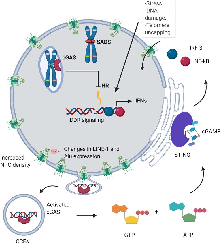
Figure 3. The DDR response is triggered as a response to stress (irradiation, ROS), DNA damage or telomere uncapping, leading to the senescent state. Changes of
nuclear localization of telomeres disrupt their maintenance and homeostasis. Nuclear cGAS accumulates at LINE-1 s and centromeres [123], and it interferes with DNA
repair [124, 125]. SIRT-6 sequestration to DNA damage sites impacts LINE-1 expression [120]. These changes lead to nuclear blebbing and the expulsion of CFFs [53],
which elicit an interferon response via the cGAS-STING pathway, leading to chronic inflammation [58].
open. This derepression increases RNA expression and mobi- LINE-1 replication extends the healthspan and the lifespan of
lization of these elements, stimulating the cGAS-STING pathway SIRT6 KO mice [120]. SIRT7 has also been shown to play a role in
and eliciting a type-1 IFN response and the SASP [16, 65, the epigenetic transcriptional repression of LINE-1 in mouse and
79]. Retrotransposons are repressed in heterochromatin by human cells. The depletion of SIRT7 leads to increased LINE-1
epigenetic factors, including DNMT1, SUV39h, HP1 and SIRT6. In expression and retrotransposition by promoting interaction with
senescent cells, LINE-1s are derepressed by the loss of epigenetic lamin A/C and via H3K18 deacetylation, which is associated with
inactive marks and are activated by the transcription factor gene repression [122].
FOXA1 [65]. Nuclear cGAS has been reported to localize preferentially to
Retrotransposons are also activated in cancer cells as well centromeres and LINE-1 elements, which points at an additional
as in old and progeroid mice [119–121]. SIRT6-deficient cells contribution of cGAS as a ‘transposition sensor’ [123]. Nuclear
and tissues accumulate cytoplasmic LINE-1 cDNA. This triggers cGAS also interferes with homologous recombination repair in
a strong type-I IFN response via activation of cGAS. Inhibiting the nucleus, but the mechanisms by which this happens are yet8 Rocha et al.
to be elucidated [124, 125]. One hypothesis is that cGAS prevents 6. Franceschi C, Campisi J. Chronic inflammation (inflam-
strand invasion by Rad 51 by compacting chromatin via phase maging) and its potential contribution to age-associated
separation [126, 127] (Figure 3). diseases. J Gerontol A Biol Sci Med Sci 2014;69(Suppl 1):S4–S9.
In mouse embryo fibroblasts, transfection with LINE-1 7. Lecot P, Alimirah F, Desprez P-Y, et al. Context-dependent
expression constructs induces an IFN-β response, dependent effects of cellular senescence in cancer development. Br J
on LINE-1’s ORF2 endonuclease activity, suggesting that IFN- Cancer 2016;114:1180–4.
β induction requires active LINE-1 transposition. Additionally, 8. Baker DJ, Wijshake T, Tchkonia T, et al. Clearance of
either induced IFN-β or exogenous IFN-β inhibited LINE-1 trans- p16Ink4a-positive senescent cells delays ageing-associated
position showed that IFN-β induced by LINE-1 transposition disorders. Nature 2011;479:232–6.
suppresses LINE-1 transposition in a negative feedback loop 9. Campisi J, d’Adda di Fagagna F. Cellular senescence: when
Downloaded from https://academic.oup.com/bfg/advance-article/doi/10.1093/bfgp/elab012/6182491 by guest on 15 December 2021
[128]. bad things happen to good cells. Nat Rev Mol Cell Biol
Recent studies have shown differential methylation of retro- 2007;8:729–40.
transposons in chronic lymphocytic leukemia that modulates 10. Serrano M, Lin AW, McCurrach ME, et al. Oncogenic ras
the expression of proximal genes [129]. LINE-1 hypomethyla- provokes premature cell senescence associated with accu-
tion is observed with increasing age and as a result of expo- mulation of p53 and p16INK4a. Cell 1997;88:593–602.
sure to ionizing radiation in vivo [130, 131] and global genome 11. Passos JF, Simillion C, Hallinan J, et al. Cellular senescence:
hypomethylation can be observed during premature cell senes- unravelling complexity. Age 2009;31:353–63.
cence induced by oxidative stress. Additionally, hydrogen perox- 12. Debacq-Chainiaux F, Boilan E, Le Moutier JD, et al. p38MAPK
ide treatment causes translocation from non-CpG-rich to CpG- in the senescence of human and murine fibroblasts. Adv
rich areas of proteins such as DNMT1, DNMT3B and SIRT1 [132]. Exp Med Biol 2010;694:126–37.
More studies are needed to better understand whether the gen- 13. Kirschner K, Rattanavirotkul N, Quince MF, et al. Functional
eral decrease of methylation observed in senescence preferen- heterogeneity in senescence. Biochem Soc Trans 2020;48:765–
tially occurs for a particular subset of TEs. 73.
14. Kouzarides T. Chromatin modifications and their function.
Cell 2007;128:693–705.
Key Points 15. Cruickshanks HA, McBryan T, Nelson DM, et al. Senescent
• Changes to the chromatin landscape promote a cells harbour features of the cancer epigenome. Nat Cell Biol
senescence-associated expression profile including 2013;15:1495–506.
activation (p16, the SASP, LINE-1) and repression (cell 16. De Cecco M, Criscione SW, Peckham EJ, et al. Genomes
cycle promoting genes). of replicatively senescent cells undergo global epigenetic
• RS and OIS cells display NE blebbing and the formation changes leading to gene silencing and activation of trans-
of CCFs, which triggers an immune response via the posable elements. Aging Cell 2013;12:247–56.
cGAS-STING pathway. 17. Parry AJ, Hoare M, Bihary D, et al. NOTCH-mediated non-
• Genomic architecture is altered in RS and OIS, enabling cell autonomous regulation of chromatin structure during
the senescent phenotype through changes such senescence. Nat Commun 2018;9:1840.
as compartment switching and altered enhancer- 18. Narita M, Nũnez S, Heard E, et al. Rb-mediated heterochro-
promoter contacts. matin formation and silencing of E2F target genes during
• Repetitive regions of the genome, including telom- cellular senescence. Cell 2003;113:703–16.
eres, centromeres and retrotransposons, experience 19. Zhang R, Poustovoitov MV, Ye X, et al. Formation of
changes in organization during senescence. MacroH2A-containing senescence-associated heterochro-
matin foci and senescence driven by ASF1a and HIRA. Dev
Cell 2005;8:19–30.
20. Funayama R, Saito M, Tanobe H, et al. Loss of linker histone
Funding H1 in cellular senescence. J Cell Biol 2006;175:869–80.
This work was supported by the following National Insti- 21. O’sullivan RJ, Kubicek S, Schreiber SL, et al. Reduced his-
tutes of Health grants: T32AG041688 to A.R.; T32GM128596 tone biosynthesis and chromatin changes arising from a
to A.D.; R01AG050582 to N.N. damage signal at telomeres. Nat Struct Mol Biol 2010;17:
1218.
22. Duarte LF, Young ARJ, Wang Z, et al. Histone H3.3 and its
proteolytically processed form drive a cellular senescence
References programme. Nat Commun 2014;5:5210.
1. Hernandez-Segura A, Nehme J, Demaria M. Hallmarks of 23. Contrepois K, Coudereau C, Benayoun BA, et al. Histone
cellular senescence. Trends Cell Biol 2018;28:436–53. variant H2A.J accumulates in senescent cells and promotes
2. Muñoz-Espín D, Cañamero M, Maraver A, et al. Programmed inflammatory gene expression. Nat Commun 2017;8:14995.
cell senescence during mammalian embryonic develop- 24. Hayakawa T, Iwai M, Aoki S, et al. SIRT1 suppresses the
ment. Cell 2013;155:1104–18. senescence-associated secretory phenotype through epi-
3. Ritschka B, Storer M, Mas A, et al. The senescence- genetic gene regulation. PLoS One 2015;10:e0116480.
associated secretory phenotype induces cellular plasticity 25. Sheikh BN, Phipson B, El-Saafin F, et al. MOZ (MYST3,
and tissue regeneration. Genes Dev 2017;31:172–83. KAT6A) inhibits senescence via the INK4A-ARF pathway.
4. Demaria M, Ohtani N, Youssef SA, et al. An essential role for Oncogene 2015;34:5807–20.
senescent cells in optimal wound healing through secre- 26. Ito T, Teo YV, Evans SA, et al. Regulation of cellular senes-
tion of PDGF-AA. Dev Cell 2014;31:722–33. cence by polycomb chromatin modifiers through distinct
5. López-Otín C, Blasco MA, Partridge L, et al. The hallmarks of DNA damage- and histone methylation-dependent path-
aging. Cell 2013;153:1194–217. ways. Cell Rep 2018;22:3480–92.The functional impact of nuclear reorganization 9
27. Sen P, Lan Y, Li CY, et al. Histone Acetyltransferase p300 46. Boumendil C, Hari P, Olsen KCF, et al. Nuclear pore density
induces de novo super-enhancers to drive cellular senes- controls heterochromatin reorganization during senes-
cence. Mol Cell 2019;73:684–698.e8. cence. Genes Dev 2019;33:144–9.
28. Villeponteau B. The heterochromatin loss model of aging. 47. Chen H, Ruiz PD, McKimpson WM, et al. MacroH2A1 and
Exp Gerontol 1997;32:383–94. ATM play opposing roles in paracrine senescence and
29. Gray MD, Jesch SA, Stein GH. 5-Azacytidine-induced the senescence-associated secretory phenotype. Mol Cell
demethylation of DNA to senescent level does not 2015;59:719–31.
block proliferation of human fibroblasts. J Cell Physiol 48. Burma S, Chen BP, Murphy M, et al. ATM phosphorylates
1991;149:477–84. histone H2AX in response to DNA double-strand breaks. J
30. Ogryzko VV, Hirai TH, Russanova VR, et al. Human fibroblast Biol Chem 2001;276:42462–7.
Downloaded from https://academic.oup.com/bfg/advance-article/doi/10.1093/bfgp/elab012/6182491 by guest on 15 December 2021
commitment to a senescence-like state in response to 49. Mallette FA, Gaumont-Leclerc M-F, Ferbeyre G. The
histone deacetylase inhibitors is cell cycle dependent. Mol DNA damage signaling pathway is a critical mediator
Cell Biol 1996;16:5210–8. of oncogene-induced senescence. Genes Dev 2007;
31. Zhang W, Li J, Suzuki K, et al. A Werner syndrome stem cell 21:43–8.
model unveils heterochromatin alterations as a driver of 50. Bassing CH, Alt FW. H2AX may function as an anchor to
human aging. Science 2015;348:1160–3. hold broken chromosomal DNA ends in close proximity.
32. Deng L, Ren R, Liu Z, et al. Stabilizing heterochromatin Cell Cycle 2004;3:149–53.
by DGCR8 alleviates senescence and osteoarthritis. Nat 51. Baell JB, Leaver DJ, Hermans SJ, et al. Inhibitors of histone
Commun 2019;10:3329. acetyltransferases KAT6A/B induce senescence and arrest
33. Hu H, Ji Q, Song M, et al. ZKSCAN3 counteracts cellular tumour growth. Nature 2018;560:253–7.
senescence by stabilizing heterochromatin. Nucleic Acids 52. Mirzayans R, Andrais B, Hansen G, et al. Role of p16(INK4A)
Res 2020;48:6001–18. in replicative senescence and DNA damage-induced pre-
34. Chandra T, Kirschner K, Thuret J-Y, et al. Independence mature senescence in p53-deficient human cells. Biochem
of repressive histone marks and chromatin compaction Res Int 2012;2012:951574.
during senescent heterochromatic layer formation. Mol Cell 53. Capell BC, Erdos MR, Madigan JP, et al. Inhibiting farnesyla-
2012;47:203–14. tion of progerin prevents the characteristic nuclear bleb-
35. Nelson DM, Jaber-Hijazi F, Cole JJ, et al. Mapping H4K20me3 bing of Hutchinson-Gilford progeria syndrome. Proc Natl
onto the chromatin landscape of senescent cells indicates Acad Sci 2005;102:12879–84.
a function in control of cell senescence and tumor sup- 54. Dechat T, Pfleghaar K, Sengupta K, et al. Nuclear lamins:
pression through preservation of genetic and epigenetic major factors in the structural organization and func-
stability. Genome Biol 2016;17:1–20. tion of the nucleus and chromatin. Genes Dev 2008;22:
36. Narita M, Narita M, Krizhanovsky V, et al. A novel role for 832–53.
high-mobility group a proteins in cellular senescence and 55. Li X-D, Wu J, Gao D, et al. Pivotal roles of cGAS-cGAMP Sig-
heterochromatin formation. Cell 2006;126:503–14. naling in antiviral defense and immune adjuvant effects.
37. Reeves R. Nuclear functions of the HMG proteins. Biochim- Science 2013;341:1390–4.
ica et Biophysica Acta (BBA) - Gene Regulatory Mechanisms 56. Sun L, Wu J, Du F, et al. Cyclic GMP-AMP synthase is a
2010;1799:3–14. cytosolic DNA sensor that activates the type I interferon
38. Shi X, Tian B, Ma C, et al. GSK3β activity is essential for pathway. Science 2013;339:786–91.
senescence-associated heterochromatin foci (SAHF) for- 57. Wu J, Sun L, Chen X, et al. Cyclic GMP-AMP is an endogenous
mation induced by HMGA2 in WI38 cells. Am J Transl Res second messenger in innate immune Signaling by cytosolic
2017;9:167–74. DNA. Science 2013;339:826–30.
39. Reya T, Clevers H. Wnt signalling in stem cells and cancer. 58. Glück S, Guey B, Gulen MF, et al. Innate immune sensing
Nature 2005;434:843–50. of cytosolic chromatin fragments through cGAS promotes
40. Ye X, Zerlanko B, Kennedy A, et al. Downregulation of senescence. Nat Cell Biol 2017;19:1061–70.
Wnt signaling is a trigger for formation of facultative 59. Yang H, Wang H, Ren J, et al. cGAS is essential for cellular
heterochromatin and onset of cell senescence in primary senescence. Proc Natl Acad Sci U S A 2017;114:E4612–20.
human cells. Mol Cell 2007;27:183–96. 60. Ishikawa H, Ma Z, Barber GN. STING regulates intracel-
41. Aird KM, Iwasaki O, Kossenkov AV, et al. HMGB2 lular DNA-mediated, type I interferon-dependent innate
orchestrates the chromatin landscape of senescence- immunity. Nature 2009;461:788–92.
associated secretory phenotype gene loci. J Cell Biol 2016; 61. Ivanov A, Pawlikowski J, Manoharan I, et al. Lysosome-
215:325–34. mediated processing of chromatin in senescence. J Cell Biol
42. Coppé J-P, Desprez P-Y, Krtolica A, et al. The senescence- 2013;202:129–43.
associated secretory phenotype: the dark side of tumor 62. Dou Z, Xu C, Donahue G, et al. Autophagy mediates degra-
suppression. Annu Rev Pathol 2010;5:99–118. dation of nuclear lamina. Nature 2015;527:105–9.
43. Zirkel A, Nikolic M, Sofiadis K, et al. HMGB2 loss upon senes- 63. Harding SM, Benci JL, Irianto J, et al. Mitotic progression
cence entry disrupts genomic organization and induces following DNA damage enables pattern recognition within
CTCF clustering across cell types. Mol Cell 2018;70:730– micronuclei. Nature 2017;548:466–70.
744.e6. 64. Lukášová E, Kovařík A, Kozubek S. Consequences of lamin
44. Hoare M, Ito Y, Kang T-W, et al. NOTCH1 mediates a switch B1 and lamin B receptor Downregulation in senescence. Cell
between two distinct secretomes during senescence. Nat 2018;7:11.
Cell Biol 2016;18:979–92. 65. De Cecco M, Ito T, Petrashen AP, et al. L1 drives IFN in
45. Voan Teo Y, Rattanavirotkul N, Olova N, et al. Notch medi- senescent cells and promotes age-associated inflamma-
ates secondary senescence. Cell Rep 2019;27:997–1007. tion. Nature 2019;566:73–8.10 Rocha et al.
66. Vizioli MG, Liu T, Miller KN, et al. Mitochondria-to- 86. Olan I, Parry AJ, Schoenfelder S, et al. Transcription-
nucleus retrograde signaling drives formation of cytoplas- dependent cohesin repositioning rewires chromatin loops
mic chromatin and inflammation in senescence. Genes Dev in cellular senescence. Nat Commun 2020;11:6049.
2020;34:428–45. 87. Busslinger GA, Stocsits RR, van der Lelij P, et al. Cohesin is
67. Lukášová E, Kovarˇík A, Bacˇíková A, et al. Loss of lamin B positioned in mammalian genomes by transcription, CTCF
receptor is necessary to induce cellular senescence. Biochem and Wapl. Nature 2017;544:503–7.
J 2017; 474:281–300 88. Lander ES, Linton LM, Birren B, et al. Initial sequencing and
68. Robijns J, Molenberghs F, Sieprath T, et al. In silico synchro- analysis of the human genome. Nature 2001;409:860–921.
nization reveals regulators of nuclear ruptures in lamin A/C 89. Biscotti MA, Olmo E, Heslop-Harrison JS. Repetitive DNA in
deficient model cells. Sci Rep 2016;6:30325. eukaryotic genomes. Chromosome Res 2015;23:415–20.
Downloaded from https://academic.oup.com/bfg/advance-article/doi/10.1093/bfgp/elab012/6182491 by guest on 15 December 2021
69. Hatch EM, Hetzer MW. Nuclear envelope rupture is 90. Harley CB, Bruce Futcher A, Greider CW. Telomeres shorten
induced by actin-based nucleus confinement. J Cell Biol during ageing of human fibroblasts. Nature 1990;345:458–
2016;215:27–36. 60.
70. Hitomi K, Okada R, Loo TM, et al. DNA damage regu- 91. Hänzelmann S, Beier F, Gusmao EG, et al. Replicative senes-
lates senescence-associated extracellular vesicle release cence is associated with nuclear reorganization and with
via the Ceramide pathway to prevent excessive inflamma- DNA methylation at specific transcription factor binding
tory responses. Int J Mol Sci 2020;21:3720. sites. Clin Epigenetics 2015;7:19.
71. Takahashi A, Okada R, Nagao K, et al. Exosomes maintain 92. Lenain C, de Graaf CA, Pagie L, et al. Massive reshaping
cellular homeostasis by excreting harmful DNA from cells. of genome-nuclear lamina interactions during oncogene-
Nat Commun 2017;8:15287. induced senescence. Genome Res 2017;27:1634–44.
72. Jeppesen DK, Fenix AM, Franklin JL, et al. Reassessment of 93. Gonzalez-Suarez I, Redwood AB, Perkins SM, et al. Novel
exosome composition. Cell 2019;177:428–445.e18. roles for A-type lamins in telomere biology and the DNA
73. Lieberman-Aiden E, van Berkum NL, Williams L, et al. damage response pathway. EMBO J 2009;28:2414–27.
Comprehensive mapping of long-range interactions 94. Wood AM, Rendtlew Danielsen JM, Lucas CA, et al. TRF2 and
reveals folding principles of the human genome. Science lamin A/C interact to facilitate the functional organization
2009;326:289–93. of chromosome ends. Nat Commun 2014;5:5467.
74. Dixon JR, Selvaraj S, Yue F, et al. Topological domains in 95. Bronshtein I, Kepten E, Kanter I, et al. Loss of lamin A func-
mammalian genomes identified by analysis of chromatin tion increases chromatin dynamics in the nuclear interior.
interactions. Nature 2012;485:376–80. Nat Commun 2015;6:8044.
75. Rao SSP, Huntley MH, Durand NC, et al. A 3D map of the 96. Burla R, Carcuro M, Torre ML, et al. The telomeric pro-
human genome at kilobase resolution reveals principles of tein AKTIP interacts with A- and B-type lamins and is
chromatin looping. Cell 2014;159:1665–80. involved in regulation of cellular senescence. Open Biol
76. McCord RP, Nazario-Toole A, Zhang H, et al. Correlated 2016;6:160103.
alterations in genome organization, histone methylation, 97. Robin JD, Ludlow AT, Batten K, et al. Telomere posi-
and DNA-lamin A/C interactions in Hutchinson-Gilford tion effect: regulation of gene expression with progres-
progeria syndrome. Genome Res 2013;23:260–9. sive telomere shortening over long distances. Genes Dev
77. Dixon JR, Jung I, Selvaraj S, et al. Chromatin architec- 2014;28:2464–76.
ture reorganization during stem cell differentiation. Nature 98. d’Adda di Fagagna F, Reaper PM, Clay-Farrace L, et al. A
2015;518:331–6. DNA damage checkpoint response in telomere-initiated
78. Chandra T, Ewels PA, Schoenfelder S, et al. Global reorgani- senescence. Nature 2003;426:194–8.
zation of the nuclear landscape in senescent cells. Cell Rep 99. Dimitrova N, Chen Y-CM, Spector DL, et al. 53BP1 promotes
2015;10:471–83. non-homologous end joining of telomeres by increasing
79. Criscione SW, De Cecco M, Siranosian B, et al. Reorgani- chromatin mobility. Nature 2008;456:524–8.
zation of chromosome architecture in replicative cellular 100. Lottersberger F, Karssemeijer RA, Dimitrova N, et al. 53BP1
senescence. Sci Adv 2016;2:e1500882. and the LINC complex promote microtubule-dependent
80. Shah PP, Donahue G, Otte GL, et al. Lamin B1 depletion in DSB mobility and DNA repair. Cell 2015;163:880–93.
senescent cells triggers large-scale changes in gene expres- 101. Anderson R, Lagnado A, Maggiorani D, et al. Length-
sion and the chromatin landscape. Genes Dev 2013;27:1787– independent telomere damage drives post-mitotic car-
99. diomyocyte senescence. EMBO J 2019;38:e100492.
81. Zufferey M, Tavernari D, Oricchio E, et al. Comparison of 102. Abdisalaam S, Bhattacharya S, Mukherjee S, et al. Dysfunc-
computational methods for the identification of topolog- tional telomeres trigger cellular senescence mediated by
ically associating domains. Genome Biol 2018;19:217. cyclic GMP-AMP synthase. J Biol Chem 2020;295:11144–60.
82. Iwasaki O, Tanizawa H, Kim K-D, et al. Involvement of con- 103. Lee S-H, Itkin-Ansari P, Levine F. CENP-A, a protein required
densin in cellular senescence through gene regulation and for chromosome segregation in mitosis, declines with age
compartmental reorganization. Nat Commun 2019;10:5688. in islet but not exocrine cells. Aging 2010;2:785–90.
83. Sati S, Bonev B, Szabo Q, et al. 4D genome rewiring dur- 104. Maehara K, Takahashi K, Saitoh S. CENP-A reduction
ing oncogene-induced and replicative senescence. Mol Cell induces a p53-dependent cellular senescence response to
2020;78:522–538.e9. protect cells from executing defective mitoses. Mol Cell Biol
84. Johnstone SE, Reyes A, Qi Y, et al. Large-scale topologi- 2010;30:2090–104.
cal changes restrain malignant progression in colorectal 105. Swanson EC, Manning B, Zhang H, et al. Higher-order
cancer. Cell 2020;182:1474–1489.e23. unfolding of satellite heterochromatin is a consistent and
85. Chiang M, Michieletto D, Brackley CA, et al. Polymer mod- early event in cell senescence. J Cell Biol 2013;203:929–42.
eling predicts chromosome reorganization in senescence. 106. Tasselli L, Xi Y, Zheng W, et al. SIRT6 deacetylates
Cell Rep 2019;28:3212–3223.e6. H3K18ac at pericentric chromatin to prevent mitoticYou can also read