Perlecan, A Multi-Functional, Cell-Instructive, Matrix-Stabilizing Proteoglycan With Roles in Tissue Development Has Relevance to Connective ...
←
→
Page content transcription
If your browser does not render page correctly, please read the page content below
REVIEW
                                                                                                                                                 published: 01 April 2022
                                                                                                                                          doi: 10.3389/fcell.2022.856261

                                            Perlecan, A Multi-Functional,
                                            Cell-Instructive, Matrix-Stabilizing
                                            Proteoglycan With Roles in Tissue
                                            Development Has Relevance to
                                            Connective Tissue Repair and
                                            Regeneration
                                            Anthony J. Hayes 1, Brooke L. Farrugia 2, Ifechukwude J. Biose 3, Gregory J. Bix 3 and
                                            James Melrose 4,5*
                                            1
                                              Bioimaging Research Hub, Cardiff School of Biosciences, Cardiff University, Wales, United Kingdom, 2Department of Biomedical
                                            Engineering, Melbourne School of Engineering, The University of Melbourne, Melbourne, VIC, Australia, 3Departments of
                                            Neurosurgery and Neurology, Clinical Neuroscience Research Center, Tulane University School of Medicine, New Orleans, LA,
                                            United States, 4Graduate School of Biomedical Engineering, University of New South Wales, Sydney, NSW, Australia, 5Raymond
                                            Purves Bone and Joint Research Laboratories, Kolling Institute of Medical Research, Royal North Shore Hospital, The Faculty of
                          Edited by:        Medicine and Health, The University of Sydney, St. Leonard’s, NSW, Australia
               Sissel Beate Rønning,
Norwegian Institute of Food, Fisheries
 and Aquaculture Research (Nofima),          This review highlights the multifunctional properties of perlecan (HSPG2) and its potential
                              Norway
                                            roles in repair biology. Perlecan is ubiquitous, occurring in vascular, cartilaginous, adipose,
                        Reviewed by:
                                            lymphoreticular, bone and bone marrow stroma and in neural tissues. Perlecan has roles in
                         Linda Hiebert,
 University of Saskatchewan, Canada         angiogenesis, tissue development and extracellular matrix stabilization in mature weight
                        Chiara Falciani,    bearing and tensional tissues. Perlecan contributes to mechanosensory properties in
               University of Siena, Italy
                                            cartilage through pericellular interactions with fibrillin-1, type IV, V, VI and XI collagen and
                 *Correspondence:
                     James Melrose          elastin. Perlecan domain I - FGF, PDGF, VEGF and BMP interactions promote embryonic
       james.melrose@sydney.edu.au          cellular proliferation, differentiation, and tissue development. Perlecan domain II, an LDLR-
                                            like domain interacts with lipids, Wnt and Hedgehog morphogens. Perlecan domain III
                    Specialty section:
         This article was submitted to
                                            binds FGF-7 and 18 and has roles in the secretion of perlecan. Perlecan domain IV, an
             Cell Growth and Division,      immunoglobulin repeat domain, has cell attachment and matrix stabilizing properties.
               a section of the journal
                                            Perlecan domain V promotes tissue repair through interactions with VEGF, VEGF-R2 and
  Frontiers in Cell and Developmental
                               Biology      α2β1 integrin. Perlecan domain-V LG1-LG2 and LG3 fragments antagonize these
         Received: 17 January 2022          interactions. Perlecan domain V promotes reconstitution of the blood brain barrier
        Accepted: 28 February 2022          damaged by ischemic stroke and is neurogenic and neuroprotective. Perlecan-VEGF-
           Published: 01 April 2022
                                            VEGFR2, perlecan-FGF-2 and perlecan-PDGF interactions promote angiogenesis and
                              Citation:
Hayes AJ, Farrugia BL, Biose IJ, Bix GJ
                                            wound healing. Perlecan domain I, III and V interactions with platelet factor-4 and
    and Melrose J (2022) Perlecan, A        megakaryocyte and platelet inhibitory receptor promote adhesion of cells to implants
     Multi-Functional, Cell-Instructive,
                                            and scaffolds in vascular repair. Perlecan localizes acetylcholinesterase in the
  Matrix-Stabilizing Proteoglycan With
    Roles in Tissue Development Has         neuromuscular junction and is of functional significance in neuromuscular control.
Relevance to Connective Tissue Repair       Perlecan mutation leads to Schwartz-Jampel Syndrome, functional impairment of the
                     and Regeneration.
     Front. Cell Dev. Biol. 10:856261.
                                            biomechanical properties of the intervertebral disc, variable levels of chondroplasia and
      doi: 10.3389/fcell.2022.856261        myotonia. A greater understanding of the functional working of the neuromuscular junction

Frontiers in Cell and Developmental Biology | www.frontiersin.org                   1                                            April 2022 | Volume 10 | Article 856261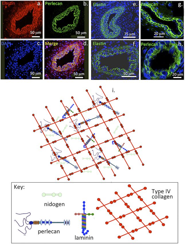
Hayes et al.                                                                                                                           Perlecan and Repair Biology

                                           may be insightful in therapeutic approaches in the treatment of neuromuscular disorders.
                                           Tissue engineering of salivary glands has been undertaken using bioactive peptides
                                           (TWSKV) derived from perlecan domain IV. Perlecan TWSKV peptide induces
                                           differentiation of salivary gland cells into self-assembling acini-like structures that
                                           express salivary gland biomarkers and secrete α-amylase. Perlecan also promotes
                                           chondroprogenitor stem cell maturation and development of pluripotent migratory stem
                                           cell lineages, which participate in diarthrodial joint formation, and early cartilage
                                           development. Recent studies have also shown that perlecan is prominently expressed
                                           during repair of adult human articular cartilage. Perlecan also has roles in endochondral
                                           ossification and bone development. Perlecan domain I hydrogels been used in tissue
                                           engineering to establish heparin binding growth factor gradients that promote cell
                                           migration and cartilage repair. Perlecan domain I collagen I fibril scaffolds have also
                                           been used as an FGF-2 delivery system for tissue repair. With the availability of
                                           recombinant perlecan domains, the development of other tissue repair strategies
                                           should emerge in the near future. Perlecan co-localization with vascular elastin in the
                                           intima, acts as a blood shear-flow endothelial sensor that regulates blood volume and
                                           pressure and has a similar role to perlecan in canalicular fluid, regulating bone development
                                           and remodeling. This complements perlecan’s roles in growth plate cartilage and in
                                           endochondral ossification to form the appendicular and axial skeleton. Perlecan is thus
                                           a ubiquitous, multifunctional, and pleomorphic molecule of considerable biological
                                           importance. A greater understanding of its diverse biological roles and functional
                                           repertoires during tissue development, growth and disease will yield valuable insights
                                           into how this impressive proteoglycan could be utilized successfully in repair biology.
                                           Keywords: perlecan, repair biology, vascular repair, cartilage repair, repair of blood brain barrier, perlecan domain-I,
                                           perlecan domain-V, growth factor delivery

INTRODUCTION                                                                         (Bengtsson et al., 2002). The basement membrane is a widely
                                                                                     distributed, specialized, thin, dense, sheet-like structure tailored
This review highlights the interactive properties of perlecan                        to the needs of specific tissues and organs with roles as a cellular
(HSPG2) and its potential applications in repair biology.                            scaffold and cell signaling platform (Timpl, 1996; Pozzi et al.,
Perlecan is often referred to as a large heparan sulfate                             2017). While cartilaginous tissues are devoid of sheet-like
proteoglycan (HS-PG) but exists as a HS/chondroitin sulfate                          basement membrane structures it has been suggested that the
(CS) hybrid form in most tissues, endothelial cells however                          pericellular matrix (PCM) surrounding each chondrocyte serves a
synthesize a monosubstituted HS glycoform (Melrose et al.,                           similar role (Kvist et al., 2008). Von Willebrand factor A-domain-
2008). Keratinocytes produce a form of perlecan substituted                          related protein (WARP) also interacts with perlecan, as a bridging
with keratan sulfate (KS), HS and CS side chains and it is one                       structure to type VI collagen in the chondrocyte PCM (Allen
of only a few PGs, which are found with such a                                       et al., 2006; Fitzgerald, 2020), and stabilizes the basement
glycosaminoglycan (GAG) substitution pattern (Knox et al.,                           membrane of basal structures in peripheral nerves (Allen
2005). Perlecan, domain I contains three 70–100 kDa HS or                            et al., 2008; Allen et al., 2009). WARP is found in a distinct
CS chains attached to serine 65, 71, and 76, mouse perlecan                          sub-set of nerve basement membrane and neural vasculature
also contain a HS or CS substitution site on Ser 3593 or Serine                      (Allen et al., 2008; Fitzgerald, 2020).
3250 respectively on domain V (Tapanadechopone et al., 1999).                           Perlecan colocalizes with elastin in blood vessels and this
Perlecan has a ubiquitous distribution and occurs in vascular,                       contributes to their visco-elastic properties in vasculogenesis
poorly vascularized cartilaginous, fibro-cartilaginous, adipose,                      during establishment of early vascular networks and tissue
lymphoreticular systems, neural, bone and bone marrow                                development (Hayes et al., 2011a) and is also a key
stromal tissues (Melrose et al., 2008). Perlecan is a major                          component of basement membranes (Figure 1). Perlecan
basement membrane component (Figure 1) as are type IV                                occurs in specialized neuroprogenitor stem cell niches termed
collagen, laminin and nidogen. PRELP (proline/arginine-rich                          fractones in the sub-ventricular and sub-granular dentate gyrus of
end leucine-rich repeat protein), a HS binding SLRP (small                           the hippocampus where it promotes neural cell survival and
leucine rich proteoglycan) and type IV collagen both interact                        proliferation through sequestered FGF-2 (Kerever et al., 2014;
and anchor perlecan in the vascular basement membrane                                Kerever et al., 2007). Perlecan is also associated with

Frontiers in Cell and Developmental Biology | www.frontiersin.org                2                                          April 2022 | Volume 10 | Article 856261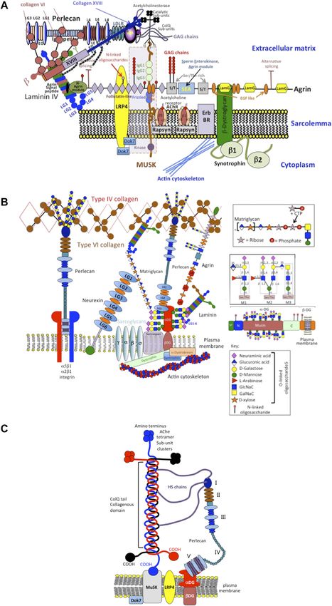
Hayes et al.                                                                                                                                 Perlecan and Repair Biology

  FIGURE 1 | Immunofluorescent colocalisation of perlecan and elastin in transverse sections of blood vessels (A–D) and immunolocalisation of elastin (E,F) and
  perlecan (G,H) in venules (E,G) and capillaries (F,H). The underlying schematic depicts the major extracellular matrix components of basement membranes highlighting
  perlecan’s interactive role in maintaining integrity of this structure (I). (A–H) reproduced from (Hayes et al., 2011a) with permission.

chondroprogenitor cell niches in cartilage rudiments where it                           establish the primary and secondary ossification centers in the
promotes the attainment of stem cell pluripotency and migratory                         rudiments, these will give rise to the long bone cartilage growth
stem cell lineages that participate in joint cavitation, cartilage                      plates (Melrose et al., 2004; Smith et al., 2010). Perlecan also
development and the expansion of cartilaginous rudiments                                promotes the establishment of primitive vascular networks in the
(Hayes et al., 2016a; Melrose and Melrose, 2016) (Figure 2).                            stromal tissues surrounding fetal rudiments that provide
Perlecan promotes chondrocyte proliferation and differentiation                         nutrition to the rapidly expanding cell numbers within the
and ECM production and the assembly of a transient                                      mesenchymal condensations and cartilage rudiments (Melrose
developmental cartilaginous scaffold (Melrose et al., 2016).                            et al., 2003; Shu et al., 2013). Perlecan provides mechanical
Perlecan is up-regulated in hypertrophic chondrocytes that                              stability to the developing ECM through interactions with a

Frontiers in Cell and Developmental Biology | www.frontiersin.org                   3                                            April 2022 | Volume 10 | Article 856261
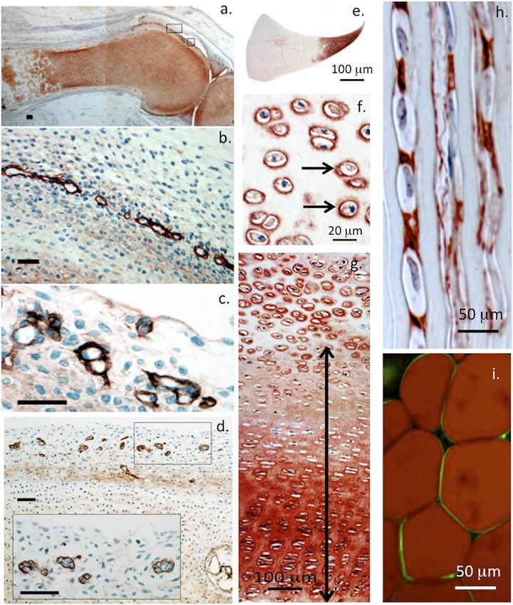
Hayes et al.                                                                                                                                        Perlecan and Repair Biology

  FIGURE 2 | Immunolocalisation of perlecan in 14-week-old human foetal knee joint (A–D), newborn ovine cartilaginous tissues (E–H) and mature human
  adipocytes (I). Perlecan has a diffuse distribution in cartilage rudiments in the femur and tibia of a foetal knee joint (A). The boxed areas in (A) are depicted at higher
  magnification in (B,C). These show strong localization of perlecan in putative stem cell niches in the surface region of the cartilage rudiments (B,C) and diffuse
  extracellular staining in the rudiment. Perlecan localization in putative stem cell niches within the surface regions of a hip rudiment (D). The boxed area in (D) is
  depicted at higher magnification in the inset. Scale bars in (A–D), 50 μm. Macroscopic immunolocalisation of perlecan in a newborn ovine medial meniscus. Perlecan is
  concentrated predominantly within the inner cartilage-like meniscal zone (E). Pericellular immunolocalisation (arrows) of perlecan within neonatal ovine femoral head
  articular cartilage (F) and resting zone tibial growth plate chondrocytes. Double headed arrow indicates extracellular gradient of perlecan immunolabel extending from the
  resting zone through to the columnar proliferating and hypertrophic growth plate chondrocytes (G). Polarised pericellular immunolocalisation of perlecan in strings of cells
  in the newborn ovine ACL (H). Pericellular immunolocalization of perlecan around human adipocytes (I). (A–D) modified from (Melrose and Melrose, 2016) with
  permission, images © Melrose 2016, (E–H) modified from (Smith et al., 2010), (I) reproduced from (Yamashita et al., 2018) with permission.

range of HS-binding structural matrix components. Pericellular                              type VI and XI collagen, fibrillin-1 and elastin (Hayes et al., 2013;
type VI and XI collagen interactions with perlecan aids in the                              Hayes et al., 2016b; Smith and Melrose, 2019; Melrose, 2020;
stabilization and functional properties of mature cartilaginous                             Guilak et al., 2021; Hayes et al., 2021).
ECMs (Hayes et al., 2016b; Smith and Melrose, 2019; Guilak et al.,                             Perlecan is a modular multifunctional PG with five distinct
2021). Interactions of perlecan with a wide range of HS interactive                         domains. Domain I is unique to perlecan and its GAG chains bind
structural matrix proteins and cell adhesive glycoproteins, ensure                          fibroblast growth factors (FGFs), platelet-derived growth factor
efficient cell-matrix communication with weight bearing and                                  (PDGF), vascular endothelial growth factor (VEGF) and bone
tensional stresses providing mechanotransductive signals to                                 morphogenetic proteins (BMPs) promoting cellular proliferation,
chondrocytes to maintain tissue homeostasis (Knox and                                       differentiation and tissue development (Melrose et al., 2008; Knox
Whitelock, 2006; Melrose et al., 2008; Melrose, 2020). Perlecan                             and Whitelock, 2006; Melrose, 2020; Whitelock et al., 2008)
contributes to mechano-sensory regulatory properties in                                     (Figure 3). Perlecan-HS mediated interactions with ECM
cartilaginous tissues mediated by pericellular interactions with                            components stabilize tissues (Arikawa-Hirasawa et al., 1999;

Frontiers in Cell and Developmental Biology | www.frontiersin.org                       4                                               April 2022 | Volume 10 | Article 856261
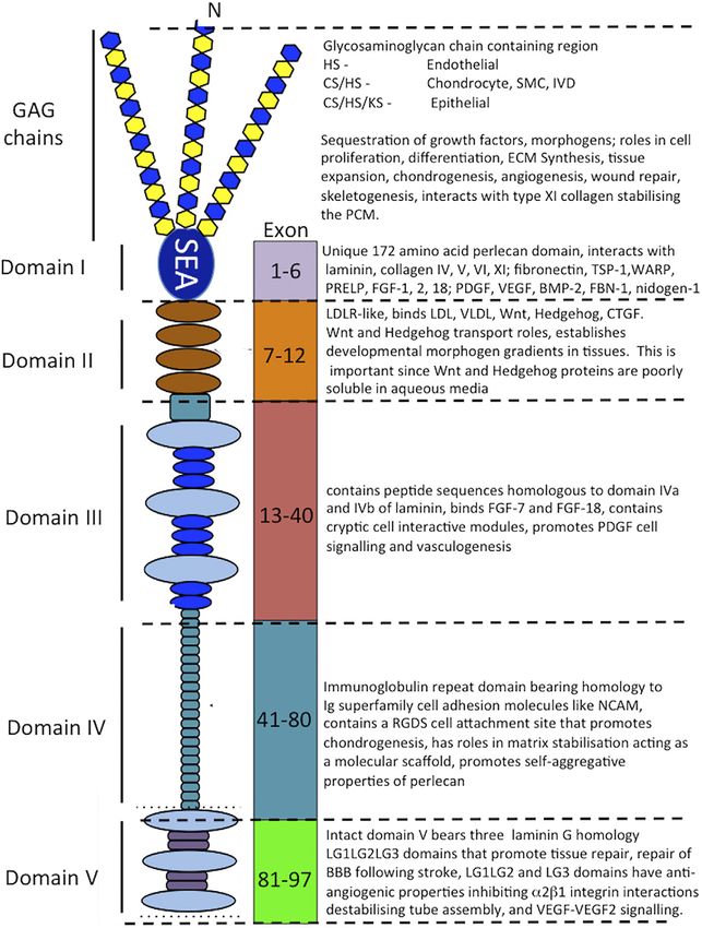
Hayes et al.                                                                                                                               Perlecan and Repair Biology

  FIGURE 3 | Schematic of the modular structure of perlecan and the interactive and cell instructive properties of each of its domains.

Costell et al., 1999; Nicole et al., 2000; Gomes et al., 2002; Mongiat                 IV, an immunoglobulin (Ig) repeat domain has cell attachment
et al., 2003; Knox and Whitelock, 2006; Melrose et al., 2006;                          properties, bears homology with the cell membrane Ig receptor
Farach-Carson and Carson, 2007; Rodgers et al., 2007; Kvist et al.,                    family and neural cell adhesion molecule (NCAM) and also has
2008; Melrose et al., 2008; Smith et al., 2010; Hayes et al., 2011a;                   roles as a scaffolding material (Noonan et al., 1991; Murdoch
Hayes et al., 2011b; Singhal and Martin, 2011; Thompson et al.,                        et al., 1992; Hopf et al., 1999; Farach-Carson et al., 2008; Martinez
2011; Hayes et al., 2013; Shu et al., 2013; Hayes et al., 2014; Wilusz                 et al., 2018).
et al., 2014; Shu et al., 2016a; Hayes et al., 2016b; Sadatsuki et al.,                    The carboxyl terminal domain V of perlecan contains three
2017; Smith and Melrose, 2019; Melrose, 2020; Ocken et al.,                            laminin-type G domains (LG) and four EGF-like repeats
2020). Domain II bears homology with low-density lipoprotein                           (Noonan et al., 1991; Murdoch et al., 1992). These LG
(LDL) receptor and has roles in the clearance of LDL and very                          domains are homologous with the α chain globular domains
low-density lipoprotein (VLDL) from the bloodstream. Domain                            of laminin and facilitate cell-ECM interactions (Timpl, 1996) and
II binds the poorly soluble Wnt and Hedgehog morphogens and                            unique divergent roles in angiogenesis, vascular cell interactions,
acts as a transport PG, establishing gradients of these                                wound healing and autophagy (Gubbiotti et al., 2017). Missense
morphogens in tissues. Domain III of perlecan binds FGF-7                              mutations, alternatively spliced, truncated or ablated sequences in
and 18 (Mongiat et al., 2000; Smith et al., 2007) and domain                           perlecan domain V are evident in an autosomal recessive disorder

Frontiers in Cell and Developmental Biology | www.frontiersin.org                  5                                            April 2022 | Volume 10 | Article 856261
Hayes et al.                                                                                                            Perlecan and Repair Biology

called Schwartz Jampel Syndrome (SJS) characterized by                     V supports angiogenesis, vascular cell interactions and wound
neuromuscular deficits and myotonia (Gubbiotti et al., 2017).               healing (Lee et al., 2011; Clarke et al., 2012; Kahle et al., 2012;
Domain V interacts with α2β1 integrin on endothelial cells in the          Marcelo and Bix, 2014; Marcelo and Bix, 2015; Poluzzi et al.,
assembly of angiogenic capillary tubes (Lord and Whitelock,                2016; Rnjak-Kovacina et al., 2017). Interactions between
2013). Recombinant perlecan domain V has been expressed                    perlecan PDGF BB, FGF-2, and TGF-β1 promote fibroblast
using human, mouse and Drosophila (domain V homologue                      migration and collagenous repair of the corneal stroma and
unc-52) DNA sequences in bacterial and mammalian                           other connective tissues to regulate tissue fibrosis. Perlecan-
expression systems (Lord and Whitelock, 2013). Human                       VEGF-VEGF-R2 interactions promote angiogenesis and wound
domain V sequence from Glu3687 to Ser4391 when expressed                   healing (Lord et al., 2014). Perlecan domain III and V
in HEK-293 cells resulted in the synthesis of a GAG-free perlecan          interactions with platelet factor 4 and domain I of perlecan
domain V, this was termed endorepellin. However when mouse                 with megakaryocyte and platelet inhibitory receptor G6b-R
perlecan domain V (Brown et al., 1997; Friedrich et al., 1999;             promote adhesion of cells to implants and scaffolds in
Tapanadechopone et al., 1999), or human domain V                           vascular repair applications (Rnjak-Kovacina et al., 2017).
encompassing 37 amino acids of the C-terminal region of                    With the availability of recombinant perlecan, applications
perlecan domain IV from Leu3626 to Ser439 was expressed in                 are now emerging for perlecan in repair biology. Perlecan
HEK-293 cells a perlecan domain V containing HS and CS chains              expression by adipocytes and smooth muscle cells mechano-
was produced. rhPerlecan domain V is a fully functional vascular           regulate lipid and glucose catabolism and oxidative muscle fiber
PG in its own right supporting endothelial cell interactions as            composition and the systemic metabolism of adipose tissue and
effectively as full-length perlecan thus it has considerable promise       skeletal muscle (Yamashita et al., 2018). Perlecan thus has
in repair biology (Lin et al., 2020).                                      physiological roles in obesity, the onset of metabolic
    Endogenously produced perlecan domain V released by                    syndrome and in lipoprotein retention in diabetic
MMPs from full-length perlecan also promotes tissue repair                 atherosclerosis (Tannock and King, 2008; Ng et al., 2021).
through angiogenic interactions with VEGF, VEGF-R2, α2β1                   Polarised M2 macrophages retain LDL in atherosclerotic
integrin, ECM-1 and progranulin (Brown et al., 1997; Bix, 2013;            plaques through interactions with perlecan domain I HS
Bix et al., 2013). LG1-LG2 and LG3 modules of perlecan domain              chains and the LDL receptor of perlecan domain II. Perlecan
V antagonize these interactions and inhibit tube formation and             as a basement membrane component of blood vessels, blood
in-growth of new blood vessels. Perlecan domain V promotes                 brain barrier and nerve basal structures in moto-neuron
repair of the disrupted blood brain barrier that occurs after              synapses of the NMJ is thus a ubiquitous pleomorphic
ischemic stroke (Bix, 2013; Bix et al., 2013). Interactions                molecule involved in the regulation of many physiological
between perlecan           domain V and pro-angiogenic                     processes in tissues of varied form and function. With a full
glycoproteins such as extracellular matrix protein 1 (ECM1)                appreciation of perlecan’s biology in tissues this insightful
(Mongiat et al., 2003) and progranulin (Gonzalez et al., 2003)             information would greatly aid in future tissue repair
promote angiogenesis and tissue repair. Perlecan complexes                 strategies involving this remarkable PG.
with dystroglycan and acetylcholinesterase occur in the
neuromuscular junction (NMJ) and are of functional
significance with essential roles in the assembly and function              THE DIVERSE ROLES OF PERLECAN
of this structure. Perlecan domain V α5β1 integrin interactions
promote pericyte migration, enhance PDGF-BB-induced                        Perlecan’s Roles in Tissue Development
phosphorylation of platelet-derived growth factor receptor β               Perlecan is a large multi-domain ECM HSPG detectable in
(PDGFRβ), SHP-2 (Src homology region 2 domain-containing                   four cell blastocysts during embryogenesis, its expression
phosphatase-2), and focal adhesion kinase (FAK) (Nakamura                  changes spatiotemporally during implantation of the
et al., 2019). This supports the maintenance of the normal blood           embryo and in the placentation stage of embryonic
brain barrier and repair processes of this structure following             development (Smith et al., 1997). Perlecan is expressed by
ischemic stroke (Nakamura et al., 2015a; Nakamura et al., 2019).           many embryonic cell types and tissues (Roediger et al., 2009)
Perlecan domain V and its LG1LG2 and LG3 modules                           including the trophoblast and tropho-ectoderm, the basal
differentially modulate interactions with VEGF-1, 2; FGF-7;                lamina that underlies the uterine epithelia and endothelia,
PDGF promoting or inhibiting angiogenesis. Perlecan domain                 and developing decidua (Smith et al., 1997). Perlecan
V is neuroprotective, and has anti-inflammatory properties                  promotes many biological processes in the embryo during
leading to the proposal of domain-V for the treatment of                   cell adhesion, growth factor binding, and modulation of
Alzheimer’s disease (AD), stroke patients (Lee et al., 2011;               apoptotic events (Girós et al., 2007; Soulintzi and Zagris,
Clarke et al., 2012; Kahle et al., 2012; Bix, 2013; Bix et al.,            2007). Perlecan expression and activity is controlled at the
2013; Guell and Bix, 2014; Marcelo and Bix, 2014; Edwards and              transcriptional level, through alternative splicing, and
Bix, 2019a; Edwards and Bix, 2019b), vascular dementia                     proteolytic processing of perlecan in the extracellular
(Marcelo and Bix, 2015) and brain trauma (Badaut and Bix,                  environment (Gomes et al., 2002). Full length perlecan acts
2014). Perlecan and VEGF incorporated into bio-scaffolds                   as an extracellular scaffold supporting cell attachment, growth
promote angiogenesis and tissue repair (Rnjak-Kovacina                     factor and morphogen sequestration that promoting cell
et al., 2016; Lord et al., 2017). Recombinant perlecan domain              proliferation and differentiation, matrix production and

Frontiers in Cell and Developmental Biology | www.frontiersin.org      6                                     April 2022 | Volume 10 | Article 856261
Hayes et al. Perlecan and Repair Biology
Perlecan’s Roles in Vascularized Tissues
In a carotid artery injury model using MΔ3/Δ3 mice lacking
perlecan HS chains the medial thickness, medial area/lumen ratio,
and macrophage infiltration were significantly increased (Gotha
et al., 2014). Perlecan lacking HS side chains has a reduced ability
to inhibit SMC proliferation in vitro. The HS side chains of
perlecan have critical roles to play in the vessel wall, as an
interleukin (IL)-2 receptor for vascular SMCs (Arumugam
et al., 2019). IL-2, a 15 kDa immunoregulatory cytokine
secreted by T cells, promotes peripheral immune cell growth
and development initiating a defensive immune response as an
initial response in the wound repair process. While SMC
proliferation is inhibited by HS-PGs, digestion of HS chains
from perlecan reverses this effect. SMC perlecan is a hybrid
HS/CS PG while endothelial perlecan is mono-substituted with
HS (Whitelock et al., 2008; Lord et al., 2014; Melrose, 2020).
SMCs bind to perlecan core protein only when perlecan’s GAG
side chains have been removed. This involves a novel binding site
in perlecan domain III, perlecan domain V and α2β1 integrin.
Endothelial cells, however, adhere to perlecan core protein
containing intact GAG substitution (Whitelock et al., 1999;
Lord et al., 2014).
SMC perlecan binds FGF-1 and FGF-2 via HS side chain
interactions promoting FGF-2, but not FGF-1 signaling (Lord
et al., 2014). Endothelial cell perlecan also binds both FGF
isoforms through HS but, in this case, promotes signaling
through both FGF-1 and FGF-2. Perlecan differentially
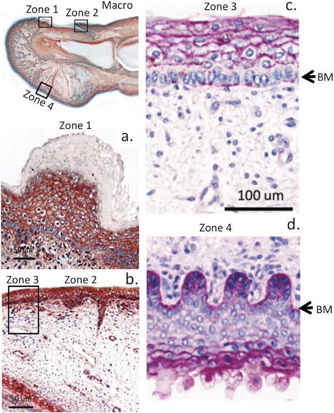
FIGURE 4 | Immunolocalisation of perlecan in a 14-week gestational age
regulates cellular proliferation and cell signaling promoted by
human hallux (big toe). Top left panel shows a macroscopic view with boxed
regions depicted at higher magnification in subjacent panels (A,B). Right-hand
growth factors to regulate tissue repair processes (Segev et al.,
panel shows periodic acid-schiff (PAS) staining of the foetal hallux 2004; McGrath et al., 2005; Hultgårdh-Nilsson and Durbeej,
showing selected regions of its anterior and posterior surfaces. The strong 2007). The form of perlecan present in tissues can vary with
reaction (purple staining) indicates the presence of perlecan HS and other different cell populations assembling variable GAG side chain
matrix polysaccharides (e.g., hyaluronic acid, HA) within the basement
components in domain I. Perlecan is also subject to post-
membrane (BM) and epidermis (C,D) of the digit. Images modified from (Smith
and Melrose, 2015) with permission. Images © the authors 2015. translational modifications including truncations, mis-sense
and point mutations in its core protein and proteolytic
modifications leading to the generation of perlecan fragments
tissue expansion during development (Farach-Carson and (Melrose, 2020). Domain IV of full-length perlecan is particularly
Carson, 2007). Perlecan also stabilises the ECM through susceptible to cleavage by matrix metalloproteinases (MMPs)
multi-component interactions mediated by its core protein leading to the generation of bioactive domain I and V
and its GAG chain substitution in domain I. Perlecan is a matricryptic fragments.
ubiquitous modular instructive multifunctional extracellular Attachment of perlecan to the endothelial cell surface as a
and pericellular PG that regulates cellular migration, dynamic flow sensor at the endothelium-blood interface (Siegel
differentiation and proliferation (McGrath et al., 2005; et al., 2014) can also modulate charge density at the endothelial
Whitelock et al., 2008; Lord et al., 2014; Nakamura et al., cell surface affecting membrane polarization (Siegel et al., 2014).
2015b). Perlecan in cartilaginous tissues promotes Shear stress signaling to endothelial cells regulates vascular ECM
proliferation and differentiation of chondrogenic cell types, remodeling and induction of angiogenesis (Russo et al., 2020).
stimulates matrix synthesis and contributes to tissue Membrane polarization regulates cell proliferation, cell signaling,
expansion and skeletogenesis. It also helps stabilize the cytoskeletal organization and gene expression (Gradilla et al.,
extracellular matrix (ECM) and promotes various tissue 2018). Cell polarization facilitates cell-cell signalling and is
repair processes (Costell et al., 1999; French et al., 2002; interfaced with stimulatory biophysical forces at the cell
Gomes et al., 2004; Sadatsuki et al., 2017; Gao et al., 2021; surface that regulate cell differentiation and tissue
Garcia et al., 2021). In vascular tissues, perlecan has different development (Saha et al., 2018). Calcium signalling initiated
effects; for example, it promotes angiogenic repair of skin through transient receptor potential (TRP) endothelial cell
wounds through FGF-2 sequestered by its domain I HS chains channels (Thakore and Earley, 2019) drives vasculogenesis and
(Zhou et al., 2004) but inhibits smooth muscle cell (SMC) controls the contractile properties of SMCs, vasodilation and
proliferation and migration (Koyama et al., 1998; Gotha et al., blood pressure (Thompson et al., 2011). Perlecan domain II is a
2014) (Figures 3, 4). LDL receptor that facilitates lipid clearance from the circulation
Frontiers in Cell and Developmental Biology | www.frontiersin.org 7 April 2022 | Volume 10 | Article 856261Hayes et al. Perlecan and Repair Biology
FIGURE 5 | Schematic depiction of perlecan’s role as an integral structural component involved in the assembly and function of the neuromuscular junction (NMJ)
showing its interactions with cell surface integrins, type IV, VI, XVIII, ColQ collagens, MuSK (Muscle-Specific Kinase) and dystroglycan (DG) and localization of catalytically
active acetylcholinesterase sub-units (A). Other structural components of the NMJ include extracellular components such as matriglycan, neurexin, laminin, type XVIII
collagen and agrin and the cell membrane components MuSK, sarcoglycan, dystrophin and sarcospan (B). This schematic is a simplified interpretation of data from
the many publications that have outlined the very complex structure and function of the NMJ (Geppert et al., 1992; Crosbie et al., 1999; Arikawa-Hirasawa et al., 2002b;
(Continued )
Frontiers in Cell and Developmental Biology | www.frontiersin.org 8 April 2022 | Volume 10 | Article 856261Hayes et al. Perlecan and Repair Biology
FIGURE 5 | Steen and Froehner, 2003; Rotundo et al., 2008; Sigoillot et al., 2010; Knight et al., 2011; Singhal and Martin, 2011; Ohno et al., 2013; Arredondo et al.,
2014; Yoshida-Moriguchi and Campbell, 2015; Banerjee et al., 2017; Cescon et al., 2018; Belhasan and Akaaboune, 2020; Legay and Dobbertin, 2020) and illustrates
how mutations in perlecan evident in SJS that result in severely diminished tissue levels of perlecan compromise the functional properties of the NMJ manifesting in the
neurological and muscular deficits evident in SJS. A better understanding of the functional basis of the NMJ is also of relevance to synaptic functions in musculoskeletal
disorders in general. Perlecan has key roles to play in the assembly, function and regulation of the NMJ (Aldunate et al., 2004; Cartaud et al., 2004; Kimbell et al., 2004;
Rotundo et al., 2005; Rudenko, 2017; Südhof, 2018; Noborn and Sterky, 2021). Perlecan has central roles in the clustering of acetylcholinesterase (Ache) at the synaptic
basal membrane through formation of a ternary complex with MuSK, DG and ColQ (C). The collagen-tailed form of AChE is localized at the NMJ through interaction with
the transmembrane DG complex by binding to perlecan (Kimbell et al., 2004). HS binding domains in ColQ anchor it to the synaptic basal lamina. ColQ-AChE/perlecan
complex co-localizes in the NMJ with dystroglycan, rapsyn, laminin and MuSK (Rotundo et al., 2005). MuSK is a receptor tyrosine kinase with important roles to play in
the clustering of active AChE sub units at the NMJ in a ternary complex with ColQ and perlecan (Cartaud et al., 2004) of functional importance (Aldunate et al., 2004).
Neurexin HS chains also recruit HS-binding proteins required for synaptic assembly and the maintenance of synaptic plasticity (Südhof, 2018; Noborn and Sterky, 2021).
A large collection of synaptic adhesion/organizing molecules (SAMs) exist in the mammalian brain with roles in synapse development and maintenance. SAMs, include
neurexins, neuroligins, cadherins, and contactins implicated in neuropsychiatric and neurodevelopmental diseases, including autism, schizophrenia, and bipolar disorder
(Rudenko, 2017). A greater understanding of the process of synaptic assembly, function and regulation at the molecular level may further the development of novel
synaptic therapeutics. A recent publication proposes that HS-PGs are key players in Alzheimer’s disease (AD). In a unifying hypothesis HS-PGs are considered central to
all aspects of AD neuropathology, i.e., plaque/tangle development amyloid deposition, neuroinflammation and apolipeprotein E (ApoE) accumulation (Snow et al., 2021).
(Ebara et al., 2000). Membrane de-polymerization resulting from proliferation and differentiation of neuroprogenitor stem
binding of LDL to perlecan leads to vasoconstriction, lowering of cell populations in the sub-ventricular and dentate gyrus of
cyclic guanosine monophosphate (cGMP) SMC levels and the hippocampus (Kerever et al., 2007; Douet et al., 2013;
deleteriously contributes to atherosclerosis. Lipid binding to Kerever et al., 2014; Mercier, 2016; Kerever and Arikawa-
perlecan in bone may also modulate its flow sensory Hirasawa, 2021; Kerever et al., 2021; Kim et al., 2021).
properties and the regulatory properties it conveys to
osteocytes (Thompson et al., 2011; Wang, 2018).
PERLECAN MUTATIONS AND ANIMAL
Perlecan’s Roles in Cartilaginous Tissues MODELS: FUNCTIONAL CLUES THEY
Perlecan is localized in the periphery of stem cell niches in foetal PROVIDE ON THE BIOLOGICAL ROLES OF
cartilage rudiments (Melrose and Melrose, 2016) (Figures 2B–D).
Perlecan regulates the attainment of stem cell pluripotency and the
PERLECAN
development of migratory chondroprogenitor stem cell lineages with Schwartz-Jampel Syndrome
roles in diarthrodial joint development, expansion of the cartilage
rudiments and development of primary and secondary ossification
(Chondrodystrophic Myotonia) and
center precursors to the cartilage growth plate cartilages. These are Dyssegmental Dysplasia
important features of relevance in potential repair applications that Silverman-Handmaker Type
might be developed using perlecan in repair biology. The importance of perlecan to the functional weight bearing and
While perlecan is a component of basement membranes in tensile properties of cartilaginous tissues is well illustrated in SJS
vascularised tissues it also has a wide distribution in tensional and (Schwartz-Jampel syndrome; chondrodystrophic myotonia)
weight bearing cartilages such as the meniscus, tendon, ligament (Arikawa-Hirasawa et al., 2002a) and Dyssegmental dysplasia
and intervertebral disc (IVD). These are predominantly avascular Silverman-Handmaker type (DDSH) (Arikawa-Hirasawa et al.,
tissues devoid of a basement membrane, however the PCM of 2001a; Arikawa-Hirasawa et al., 2001b). The former is a relatively
chondrocytes has been suggested to represent an intrinsic mild skeletal condition characterized by reduced perlecan levels in
basement membrane around each cell (Kvist et al., 2008). tissues; however, DDSH is a very severe condition where perlecan
Atomic force microscopy (AFM) studies demonstrate that levels in tissues are severely depleted or totally absent. SJS is an
perlecan imparts compliancy to the PCM and is cytoprotective autosomal recessive disease caused by mutation in the HSPG2 gene
(Guilak et al., 2021). Cell-ECM interconnections in cartilages and resulting in skeletal dysplasia and neuromuscular hyperactivity
perlecan as a biosensor, facilitates cell-matrix communication (Nicole et al., 2000; Bauché et al., 2013). In this condition mutant
allowing cells to perceive and respond to perturbations in their fibroblasts secrete reduced levels of perlecan and display impaired
biomechanical microenvironments and to orchestrate tissue migratory properties but normal proliferative rates (Arikawa-
homeostasis. Perlecan also monitors the flow of cannalicular Hirasawa et al., 2002a). DDSH (MIM 224410) in contrast is a
fluid in the osteocyte PCM and acts as a mechanosensor that very severe but extremely rare condition caused by functional null
regulates bone development (Thompson et al., 2011; Wang et al., mutations in the perlecan HSPG2 gene (Arikawa-Hirasawa et al.,
2014; Wijeratne et al., 2016). 2001a). Less than forty DDSH cases have been reported in the
literature, and only four of these were detected prenatally.
Perlecan’s Roles in Neural Tissues
Perlecan has critical roles in basement membrane in the blood Perlecan Knockout Model
brain barrier and important roles in NMJ assembly and Perlecan knockout is a lethal condition. Conventional perlecan
function (Figures 5, 6). Perlecan-FGF-2 interactions in the knockout (i.e., Hspg2−/−, KO) mice die just after birth mainly due
neural stem cell niche (fractone) regulate the survival, to respiratory failure (Arikawa-Hirasawa et al., 1999; Costell et al.,
Frontiers in Cell and Developmental Biology | www.frontiersin.org 9 April 2022 | Volume 10 | Article 856261Hayes et al. Perlecan and Repair Biology
1999). The few mice that survive to birth display macroscopic
abnormalities in cephalic development and have short squat
frames with severely distorted axial and appendicular skeletal
development. Furthermore, internal examination shows major
abnormalities in the development of the major outflow tracts
from the heart in these mice. Perlecan-null mice, form normal
basement membranes but these soon deteriorate at areas of
increased mechanical stress e.g., areas of myocardial
contraction and brain vesicle expansion. Perlecan-null mice
die around E10–12, due to heart, lung, and brain defects.
Weakened embryonic heart basements membranes and “leaky”
cardiomyocyte-endothelial cell interfaces result in cardiac arrest
due to blood leakage into the pericardial space. Major defects in
lung development also contribute to the lethality of perlecan
deficiency. Abnormalities in cephalic development, distortion in
normal brain laminar architecture and development of holes in
the fore- and midbrain also occur. Perlecan-null mice experience
severe bleeding in the lung, skin, and brain, due to weakened
blood vessels. Perlecan null mice also display distorted growth
plate architecture and a disturbed chondrocyte organization with
a loss of normal columnar chondrocyte spatial organization and
expansion and distortion of the resting, proliferative and
hypertrophic zones consistent with the massive disruptions
seen macroscopically in skeletal development in this mouse
model. Perlecan knock-out mice are not suitable for the
examination of perlecan’s roles in postnatal tissue
development, but starkly demonstrate the importance of
perlecan in the development of pre-natal vascular and non-
vascular tissues.
Perlecan Conditional Transgenic Model
In order to avoid the lethality of perlecan KO mice, conditional
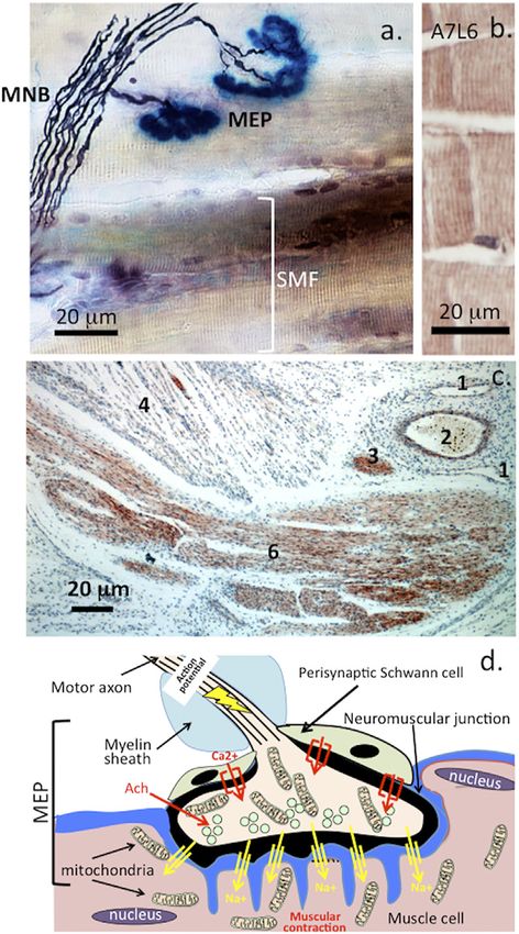
FIGURE 6 | Histochemical localization of a motor neuron attaching to
perlecan-deficient (Hspg2−/−, TG) mice were developed that
muscle fibre showing the myelinated nerve bundle (MNB), motor endplates
(MEP) and striated muscle fibres (SMF) (A). and perlecan-positive striated express the perlecan transgene only in cartilage using the
muscle fibres stained with MAb A7L6 to perlecan domain IV in murine Col2a1promoter and enhancer (Xu et al., 2010; Xu et al.,
extensor digitorum longus muscle (B). A lower power image depicting 2016). These perlecan TG mice develop a phenotype similar to
perlecan-positive features in a foetal human elbow joint (C). 1) flattened DDSH but also share features of SJS (Arikawa-Hirasawa et al.,
venule, 2) capillary with entrapped red blood cells, 3) small nerve -bundle in
cross-section, 4) muscle fibres, 5) longitudinal nerve fibre bundles in ulnar
2001a; Arikawa-Hirasawa et al., 2002a). Perlecan knockdown
nerve of the elbow. Schematic of the major features of the motor end plate (C). impacts on cartilage development and skeletogenesis and
Figure segment a, stock image 2AD3P00 from Alamy Science Photolibrary impairs the normal weight bearing properties of cartilaginous
reproduced under license. (B,C) modified from (Shu et al., 2019) Open tissues and functional properties of the pericellular matrix of
Access under CC BY NC-ND License to Publish. (D). A wave of membrane chondrocytes (Xu et al., 2016; Ocken et al., 2020).
depolarization emanating from the nerve soma produces an action potential
that travels down the nerve axon resulting in activation of voltage gated Ca2+
channels in the nerve synapse and a resultant influx of Ca2+. This results in
mobilization of synaptic vesicles in the nerve to the post synaptic membrane.
Perlecan Exon 3 Null (HS Deletion) Model
These merge with the post-synaptic membrane releasing their In Hspg2−/−-Tg (Hspg2−/−; Col2a1-Hspg2Tg/−) mice, perlecan
neurotransmitter contents which include acetylcholine into the synaptic gap. is only expressed in cartilage but not the synovium. This results
Acetylcholine is captured by acetylcholine receptors on the muscle synaptic in less development of osteophytic spurs in the tibial and
membrane which results in an influx of Na+ ions through Na+ channels into the
femoral joint margins (Arikawa-Hirasawa et al., 1999; Xu et al.,
muscle causing muscular contraction. Acetylcholinesterase released by the
nerve mops up any excess of acetylcholine. Acetylcholinesterase is one of
2010; Ishijima et al., 2012; Kaneko et al., 2013). A perlecan
nature’s most efficient enzymes and hydrolyses acetylcholine regulating exon 3 null mouse model has also been developed. Perlecan
muscular relaxation (Vigny et al., 1978). The myotonia found in SJS is due to a domain-I encoded by exon 3 contains its GAG attachment
breakdown in this mechanism due to a deficiency of perlecan at the NMJ and points thus perlecan exon 3 null mice produce perlecan
a deficiency of AChE that is normally clustered in synapses by perlecan
deficient in HS. A 20 kDa drop in the size of the perlecan
(Silman and Sussman, 2008).
core protein is also evident. This model has yielded important
Frontiers in Cell and Developmental Biology | www.frontiersin.org 10 April 2022 | Volume 10 | Article 856261Hayes et al. Perlecan and Repair Biology information on the role of perlecan HS in the regulation of Schwartz-Jampel Syndrome chondrocyte behavior and in tissue homeostasis and perlecan (Chondrodystrophic Myotonia) Model HS mediated interactions with ECM components (Whitelock A model of SJS in which a 4595G to A point mutation occurs in et al., 2008; Smith et al., 2010; Hayes et al., 2011a; Hayes et al., the perlecan gene displays reduced perlecan secretion and 2011b; Hayes et al., 2013; Shu et al., 2013; Hayes et al., 2014; incorporation into the PCM similar to the clinical features of Shu et al., 2016a; Hayes et al., 2016b; Shu et al., 2018; Shu et al., human SJS (Rodgers et al., 2007; Stum et al., 2008). Mice 2019; Smith and Melrose, 2019). This exon 3 null GAG free homozygous for Hspg2C1532Y-Neo (Neo/Neo) have short form of perlecan does not participate in growth factor and stature, impaired mineralization, misshapen bones, OA-like morphogen sequestration and cell signaling like the full length joint dysplasias and myotonia (Rodgers et al., 2007; Stum GAG substituted form of perlecan resulting in a loss in its et al., 2008). ability to act as a co-receptor for the presentation of these A model of SJS has also been developed that displays growth factors to their cognate receptors (Zhou et al., 2004; congenital peripheral nerve hyper-excitability, neuromyotonia, Whitelock et al., 2008). However, there are GAG-free regions demyelination and peripheral neuropathies (Bangratz et al., of the perlecan core protein that can also bind certain growth 2012). This model revealed roles for perlecan in the regulation factors. For example, perlecan domain III can bind FGF-7 and of longitudinal elongation of myelin by Schwann cells FGF-18, however it is not known to what extent this (Echaniz-Laguna et al., 2009; Bangratz et al., 2012). interaction can provide the same cell proliferative and Perlecan-deficient mice displaying shorter internodes, had differentiative properties provided by growth factors that increased levels of impaired functional voltage-gated K (+) bind to the GAG chains of perlecan domain I. Perlecan channels (Echaniz-Laguna et al., 2009). Electrophysiological exon 3 null HS-deficient mice do not store TGF-β1 in skin studies have demonstrated muscle fiber hyper-excitability tissues like the full-length perlecan does (Shu et al., 2016a). arising from such alterations in peripheral nerve Participation in the wound healing response, previously organization, muscle hypertrophy and compositional provided by FGF-2 and VEGF perlecan interactions and the changes (Xu et al., 2010). angiogenic responses they provide, is also lost in perlecan exon 3 null mice (Zhou et al., 2004). As previously discussed, full length HS substituted perlecan has major roles in tissue and organ development and wound healing orchestrated by the Perlecan Cerebral Artery Occlusion Stroke binding and signaling of mitogens and morphogens with cells Model in a temporal and dynamic fashion. FGF-7, −18 and PDGF can Cerebral artery occlusion stroke models in Yucatan miniature also bind to perlecan domains III, IV and V, such interactions pigs, dogs and mice (Platt et al., 2014; Vasquez et al., 2019; may also mediate wound healing and cell signaling responses. Llovera et al., 2021) have facilitated examination of perlecan’s Binding of PDGF-BB has been mapped to domain III-2 (Kd = roles in the repair of the blood brain barrier following stroke. 8 nM), lower binding affinities are evident for domains I, IV-1 Perlecan domain V has neurogenic and neuroprotective and V (Kd = 34–64 nM). PDGF-AA binds to domain III-2 properties and promotes angiogenic repair of the blood (Göhring et al., 1998). Perlecan HS deficiency impairs brain barrier (Lee et al., 2011; Bix, 2013; Marcelo and Bix, pulmonary vascular development (Chang et al., 2015). 2014). Perlecan transgenic mice have demonstrated Perlecan HS chains also recruit pericytes to pulmonary important roles for perlecan domain V in pericyte vessels. HS deficiency in perlecan attenuates hypoxia- recruitment in the promotion of BBB repair processes induced pulmonary hypertension involving impaired FGF- (Nakamura et al., 2019). IL-1 is also neuroprotective and 2/FGFR1 interactions (Chang et al., 2015). Perlecan exon 3 has neuron restoring properties in experimental ischemic null mice display reduced healing responses due to impaired stroke studies (Salmeron et al., 2019). Recombinant FGF-2 and VEGF signaling (Zhou et al., 2004). The perlecan domain V is now available for blood brain barrier chondroprotection evident in perlecan exon 3 null mice in a repair strategies (Rnjak-Kovacina et al., 2017). Recombinant post traumatic OA model may be attributable to the perlecan domain V decorated with HS and CS chains is a preservation of FGFR-3-FGF-18 signaling and perlecan vascular PG in its own right and supports endothelial cell domain III-mediated interactions (Shu et al., 2016b), interactions as well as full-length perlecan (Rnjak-Kovacina contributing to significantly reduced joint margin et al., 2017). Repair of the blood brain barrier involves osteophytosis, synovial perlecan is required for osteophyte pericyte recruitment, triggered by an up-regulation in formation in knee OA (Kaneko et al., 2013). Hspg2 exon 3 PDGFRβ (Arimura et al., 2012; Makihara et al., 2015; null murine chondrocytes display increased hypertrophic Shibahara et al., 2020), this drives pericyte migration maturational changes and chondrocyte proliferative rates required for pericyte endothelial tube repair interactions in in vitro and in vivo, accelerated growth plate maturation, the neurovascular unit (Hellstrom et al., 1999). Perlecan elevated GAG deposition, and exostosis formation in the binds PDGF and promotes pericyte migration and integrin IVD (Shu et al., 2019). Perlecan HS may thus exert α5β1 and α2β1 mediated interactions in endothelial tube repressive control over chondrocytes in mature cartilage formation (Hellstrom et al., 1999; Arimura et al., 2012; explaining why cartilage has such a poor healing response Shen et al., 2012; Makihara et al., 2015; Nakamura et al., (Garcia et al., 2021). 2019). Frontiers in Cell and Developmental Biology | www.frontiersin.org 11 April 2022 | Volume 10 | Article 856261
Hayes et al.                                                                                                               Perlecan and Repair Biology

APPLICATION OF PERLECAN IN REPAIR                                           promotes bone formation and tissue remodeling at the
BIOLOGY                                                                     osteochondral interface during endochondral ossification
                                                                            (Kram et al., 2006).
Cell-Mediated Effects of Perlecan:
Modulation of Cell Proliferation
While perlecan promotes the proliferation and differentiation of            Application of Perlecan in Vascular Tissue
endothelial cells, as well as many other cell types, it inhibits SMC        Repair
proliferation through the tumor repressor PTEN (tumor                       Given the crucial role that perlecan plays in multiple biological
suppressor phosphatase and tensin homolog), including                       processes, it is unsurprising that perlecan, or its components,
upregulation of FRNK (focal adhesion kinase–related non-                    have been utilized in multiple therapeutic applications. Perlecan’s
kinase) and down regulation of FAK signalling (Walker et al.,               known ability to bind, sequester, and deliver a myriad of growth
2003; Garl et al., 2004).                                                   factors and other bioactive molecules is a key feature that has been
                                                                            harnessed to translate this molecule into potential therapeutics
                                                                            for multiple applications, from angiogenesis to the regeneration
Perlecan’s HS Mediated Interactions and                                     of cartilage (Gao et al., 2021; Garcia et al., 2021). Perlecan’s ability
Their Relevance to Tissue Repair                                            to modulate processes in cardiovascular applications has been
HS’s interactions with FGF-2, PDGF, VEGF, HGF, BMP2, GM-                    explored through an immune-purified form of perlecan from
CSF, angiopoietin-3, and activin A illustrate the potential of              human coronary arterial endothelial cells. This immunopurified
perlecan domain I as a co-receptor for growth factor delivery               perlecan was used to coat expanded polytetrafluoroethylene
and receptor activation. The ITIM megakaryocyte-platelet                    (ePTFE) vascular grafts. Implantation of the vascular grafts
receptor (G6b-B-R) is an additional ligand for the HS chains                into the carotid arteries of an ovine model demonstrated that
of perlecan domain I (Vögtle et al., 2019). G6b-B-R is highly               the perlecan-coated grafts reduced platelet adhesion and
expressed in mature megakaryocytes (MKs) that regulates platelet            enhanced endothelial cell growth along the implanted graft
activation (Lord et al., 2018; Vögtle et al., 2019). Binding of G6b-        when compared with the uncoated vascular graft (Lord et al.,
B-R to perlecan HS chains mediates functional responses in MKs              2009). While this study demonstrated the ability for perlecan to
and platelets, negatively regulating platelet adhesion to fibrinogen         improve vascular graft patency, isolation of perlecan from tissues
and collagen. It also modulates platelet adhesion to vascular graft         or purified from conditioned medium, can be cost-prohibitive
materials and explains the varied roles of perlecan in fibrosis              due to the small amounts available and the quantities required.
(Lord et al., 2009; Lord et al., 2018).                                         An alternative option that has been explored is through the use
                                                                            of recombinant fragments of perlecan. The protein core of
                                                                            perlecan contains five domains, many with bioactive
Understanding Perlecan’s Cell Regulatory                                    properties, though use of recombinant perlecan fragments has
Roles in Blood Vessels                                                      focused on the use of domain-I and -V due to these domains
Perlecan attached to endothelial cells in the lumen of blood                containing GAG attachment sites. The protein component of
vessels acts as a shear flow sensor that interacts with Ca2+ or              perlecan domain I contains three GAG attachment sites. The
Na+ regulating charge density at the endothelial cell surface. This         GAGs that decorate the perlecan domain I core protein are
also regulates membrane polarization in endothelial cells and               predominantly HS, though it may also be decorated with CS.
SMCs, ion transport regulates vasoconstriction and relaxation in            Recombinant perlecan domain I decorated with HS has been
blood vessels (Siegel et al., 2014) and is crucial for cell-cell            incorporated into 3D structures or scaffolds for multiple
signalling coupled with stimulatory biophysical forces that                 therapeutic applications. The incorporation of perlecan
promote cell differentiation and tissue development (Saha                   domain-I into 3D structures has resulted in increased
et al., 2018). Calcium signalling through endothelial cell TRP              retention of FGF-2 (Yang et al., 2005), as well as BMP2 (Jha
channels (Thakore and Earley, 2019) drives vasculogenesis                   et al., 2009; Srinivasan et al., 2012; Chiu et al., 2016) for cartilage
(Moccia et al., 2019) and SMC contractility which in turn                   repair and regeneration. More recently, advances in fabrication
regulates vasodilation and blood pressure (Ishai-Michaeli et al.,           and microfluidics has enabled the ability to produce growth factor
1990).                                                                      gradients (Hubka et al., 2019), an approach that has been utilized
                                                                            to generate chemotactic gradients with FGF-2R The ability to
                                                                            create gradients using perlecan, perlecan fragments and other
                                                                            bioactive components of the ECM, has significant potential in
Heparanase has Roles in Wound Repair and                                    tissue repair and regeneration, including the development of
Tissue Remodelling                                                          smart biomaterials and constructs. It will also greatly improve
While the HS chains of perlecan mediate growth factor                       the understanding of many developmental and disease processes,
interactions in skeletogenesis, degradation of HS in situ has               e.g., in cancer biology.
been shown to improve wound healing through the re-                             As mentioned above, perlecan domain V, like domain I,
mobilization of sequestered growth factors locally at sites of              contains a GAG attachment site. Recombinant perlecan has
tissue repair (Ishai-Michaeli et al., 1990; Zcharia et al., 2005;           been explored in several applications due to its growth factor
Nasser, 2008). Heparanase expression in osteoblastic cells also             interactions. Recombinant perlecan domain V is substituted with

Frontiers in Cell and Developmental Biology | www.frontiersin.org      12                                       April 2022 | Volume 10 | Article 856261
Hayes et al. Perlecan and Repair Biology
HS and CS chains and promotes angiogenesis by enhancing formation of epithelial spheroids, and promotes the expansion of
growth factor signaling (Lin et al., 2020). When perlecan or 3D progenitor cell populations, representing the first step toward
perlecan DNA is immobilized on silk or chitosan scaffolds the development of an engineered salivary gland (Fowler et al.,
(Lord et al., 2017) increased vascular ingrowth and integration 2021). Primary salivary human progenitor stem cells undergo
in vivo underlies the importance of perlecan in angiogenesis/ acinar-like differentiation in HA hydrogel cultures and
vasculogenesis. incorporation of basement membrane peptides derived from
The complexity and nuances of perlecan’s ability to modulate perlecan and laminin-111 further directs the development of
biological processes has been explored by immobilization 3D salivary gland-like spheroids (Srinivasan et al., 2017). Full-
technique, and the presence of GAG chains plays a key role in length perlecan also has directive roles over the 3D development
the orientation of this PG (Rnjak-Kovacina et al., 2016). of submandibular salivary glands. Heparanase colocalizes with
Immobilization of perlecan domain V by physisorption or perlecan in submandibular gland basement membrane. Cleavage
covalently, in addition with immobilization of the protein core of perlecan HS side chains by heparanase regulates salivary gland
of perlecan domain V only, or protein core decorated with GAG branching morphogenesis by modulating FGF-10 mediated cell
chains, modulated the interaction and attachment of both signaling. Heparanase releases FGF-10 from perlecan HS in the
endothelial cells and platelets. The ability to control the basement membrane. This increases MAPK signaling, epithelial
interaction of different cell types holds tremendous clefting, and lateral branching thus increasing submandibular
importance to tissue repair, for example in cardiac and gland branching morphogenesis (Patel et al., 2007). The size and
vascular repair procedures where tissue grafts and implants, sulfation patterns of the perlecan HS side chains regulate FGF-10-
require cues to facilitate migration and re-endothelization of mediated interactions during proliferation, salivary gland duct
the repair tissue, whist minimizing platelet adhesion. The elongation, bud expansion, and differentiation. The spatio-
potential of perlecan domain V to modulate platelet adhesion temporal localization of specific HS structures in salivary
has been explored by incorporating this domain onto different tissues provides a mechanistic insight as to how salivary gland
polymeric surfaces including poly (vinyl chloride) (Chandrasekar developmental processes mediated by FGF10 occur in vivo (Patel
et al., 2021), silk (Lau et al., 2021) and chitosan (Lord et al., 2017). et al., 2008). FGF-10 is a multi-functional paracrine growth factor
Incorporation of perlecan domain I plasmid DNA in conjunction that mediates mesenchymal-epithelial signaling during tissue
with VEGF189 in a rodent wound model demonstrated increased development, growth and disease and has significant relevance
re-epithelialization of the wound, formation of sub-endothelial to regenerative medicine (Itoh and Ohta, 2014; Itoh, 2016).
tissue and neo-angiogenesis within the wound bed (Lord et al.,
2017).
Potential Roles for Perlecan in Cartilage
Application of Stimulatory Peptides Derived Repair
From ECM Components in Repair Biology: Once damaged, articular cartilage has a notoriously poor
ability to repair itself (Armiento et al., 2019). Perlecan’s
In Vitro Engineering of Salivary Glands important well-established roles in chondrogenesis and
Using Perlecan Domain IV, Laminin-111 and cartilage development (Smith et al., 2010; Melrose et al.,
Fibronectin Peptide Scaffolds 2012) imply potential roles in cartilage repair by
Peptides derived from perlecan domain IV (TWSKV), laminin- recapitulating developmental roles in damaged/diseased
111 (YIGSR, IKVAV), and fibronectin (RGDSP) have been tissues (Gao et al., 2021; Garcia et al., 2021). Perlecan
incorporated into HA scaffolds with RGDSP and TWSKV promotes chondroprogenitor stem cell maturation and
peptide HA scaffolds significantly accelerating cell proliferation development of pluripotent migratory stem cell lineages
(Fowler et al., 2021). Perlecan peptide TWSKV triggers with roles in diarthrodial joint formation and early cartilage
differentiation of salivary gland cells into self-assembling acini- development (Hayes et al., 2016a; Melrose and Melrose, 2016).
like structures expressing salivary gland biomarkers that secrete Perlecan’s cartilage stabilizing properties through interactions
α-amylase (Pradhan et al., 2009; Pradhan et al., 2010). Purified with ECM components also establish its potential in cartilage
ECM-derived peptides have been suggested as stimulatory repair (SundarRaj et al., 1995; Gomes et al., 2002; Melrose
molecules that direct the proliferation and differentiation of et al., 2006; Melrose et al., 2008). Perlecan domain I hydrogels
progenitor cell populations. These are of potential application have been used to establish heparin binding growth factor
in tissue repair processes using human embryonic stem cells gradients that promote cell migration of potential use to
(hESCs) and induced pluripotent stem cells (iPSCs) (Rowland promote cartilage repair (Hubka et al., 2019). Perlecan
et al., 2013). Laminin-111 peptide fibrin hydrogels restore salivary domain I BMP-2 hydrogels have also been shown to
gland function (Nam et al., 2017). An extensive range of laminin- promote chondrogenesis and cartilage repair in a murine
111-derived peptides conjugated to chitosan scaffolds also show early OA model (Yang et al., 2006; Jha et al., 2009;
promise in tissue engineering applications designed to promote Srinivasan et al., 2012). BMP-2 and BMP-9 can also
tissue regeneration (Hozumi et al., 2012). Laminin-111 peptide- promote chondrogenic differentiation of human
HA hydrogels have also been shown to act as a synthetic multipotential mesenchymal stem cells (Majumdar et al.,
basement membrane (Yamada et al., 2013). Conjugation of 2001; Shestovskaya et al., 2021). Perlecan domain I collagen
RGDSP peptide to HA gels improves cell viability, accelerates I fibril scaffolds have been used as an FGF-2 delivery system
Frontiers in Cell and Developmental Biology | www.frontiersin.org 13 April 2022 | Volume 10 | Article 856261You can also read