Pediatric HIIT Workout: Abdominal Pain & Limping Child - Daniel Wood PA-C UT Health San Antonio/Pediatric Center of North Austin - Skin ...
←
→
Page content transcription
If your browser does not render page correctly, please read the page content below

Pediatric HIIT Workout:
Abdominal Pain
& Limping Child
Daniel Wood PA-C
UT Health San Antonio/Pediatric Center of North Austin

Understand the causes of acute abdominal pain in childhood.
Develop a differential diagnosis based on age and symptoms.
Formulate a plan for evaluation and management of acute
abdominal pain.
W.O.D Discuss imaging study for various diagnoses.
Green vomit is surgical

Vomiting and distention is
                                                                          Mantra
surgical.
Vomiting and shock is surgical.

  Accessed 4.23.2020:http://pemplaybook.org/podcast/the-pediatric-surgical-abdomen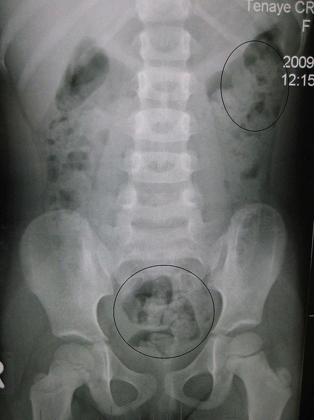

Causes range from “belly aches" to an emergency requiring
immediate attention.
Differential to abdominal pain is a very long list
Age based approach
Introduction Divided into the following categories:
SERIOUS/Common
LESS serious/ Common
SERIOUS/UNcommon
LESS serious/UNcommon
< 5 yo are not able to localize pain well.
1000 children: ages 4-18yo
Background 10% with one episode that sought medical
attention
Constipation accounted for 48%
Good history, physical and exam.
https://socratic.org/questions/from-the-lumen-outward-what-are-the-layers-of-the-gastrointestinal-tract
Neonate
Child is a former 36 week premature infant.
Diagnosis of ToF a birth, awaiting surgery at 3 month of age.
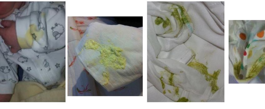
7 day old infant Child is feed Similac Neosure.
“Change in Parents complain of feeding intolerance and more spit up(non-
feeding.” bilous) over the last 2 days and hematochezia x 1. most frequent
sign of NEC is a sudden change in feeding toleranceo Vitals: T: 99 F, HR: 70, RR: 30, BP: 70/40
o Physical exam:
o Gen: neonate irritable and crying
o CV: RRR with + murmur
Physical exam o Lungs: b/l CTA
o Abd: +mild distended, BS hypoactive, No HSM, NTTP
o Skin is without lesions.KUB
Accessed3.25.2020 : www. Uptodate.coma. Bacteremia Which of the b. Dietary protein allergy following is the c. Necrotizing Enterocolitis likely diagnosis d. Volvulus in this patient?
Necrotizing NEC occurrence inverse to gastrointestinal age Enterocolitis Presentation: (NEC) Diagnosis: Bell staging Niño DF et al. Necrotizing enterocolitis: new insights into pathogenesis and mechanisms. Nature. 2016; 13:590-600.
Modified Bell
staging
Management depends upon the severity of illness
Supportive care
Bowel rest with discontinuation of enteral intake
Gastric decompression with intermittent nasogastric suction,
Correction of metabolic, fluid/electrolyte,
NEC Broad spectrum antibiotics
management Surgical intervention
Intestinal perforation occurs
Unremitting clinical deterioration
Prognosis: Survival rates at 70 to 80 %.
Lin PW, Stoll BJ. Necrotising enterocolitis. Lancet 2006; 368:1271. Child is a term infant.
2 week old
Diagnosis of ASD and hetrotaxy.
”Has spit up Child is direct breast feed.
after History of poor feeding that was thought to be reflux.
feedings.”Bilious?
Vitals: T: 99.0 F, HR: 170, RR: 30, BP: 70/40
Exam:
Gen: calm
Physical exam CV: RRR without murmur
Lungs: b/l CTA
Abd: + distended, BS active, No HSM
Skin is without lesions.Which of the
a. Abdominal radiograph
following is the b. Abdominal CT
best exam to c. Abdominal Ultrasound
order in this d. Upper GI series
patient? Pathophysiology
Clinical Presentation:
Vomiting
Abdominal distension
Volvulus &
Abdominal tenderness
Hematochezia
Malrotation Imaging modalities:
Plain radiograph: midgut
Ultrasound
Upper GI series should be performed under fluoroscopyVolvulus & Malrotation
Management:
Immediate surgical intervention if decompensation.
UGI is the best study to order
Barium enema and ultrasonography can be useful
adjuncts. DOES NOT exclude malrotation.
Ladd procedure : Surgical intervention
Aboagye J, Goldstein SD, Salazar JH, et al. Age at presentation of common pediatric surgical conditions: Reexamining dogma.
J Pediatr Surg 2014; 49:995. Cow's milk allergy (CMA) : most common food allergy in young
children.
Dietary protein Non-immunoglobulin E (IgE)-mediated CMA tends to resolve by
early childhood.
allergy Diagnosis
Management
Luyt D, Ball H, Makwana N, et al. BSACI guideline for the diagnosis and management of cow's milk allergy. Clin
Exp Allergy 2014; 44:642.Serious Less Serious
Causes of
Common NEC Colic
abdominal
Uncommon Volvulus Dietary protein pain in the
Testicular allergy
torsion
neonate. Recent diagnosis of adenovirus a month ago.
Non-bilious, non-bloody emesis
18 mo.
Not responsive to laxatives and Zofran
”He has 3 days Episodes lasting of screaming and then seems fine.
of vomiting Denies diarrhea.
and his belly
hurts.”Vitals: T: 99 F, HR: 120, RR: 30, BP: 90/50
Exam:
Gen: asleep in mothers arms
Physical exam CV: RRR without murmur
Lungs: b/l CTA
Abd: No hepatosplenomegaly appreciated. Bowel
sounds diminished. Soft abdomen
Skin is without lesions.Imaging
https://litfl.com/ultrasound-case-064/Which of the a. Constipation
b. Gastroenteritis
following is c. Intussusception
this patient's d. Viral illness
likely
diagnosis? Ileocecal junction accounts for 90 percent < 2 yo.
Often occurs at the ileocecal junction
Presentation: typical vs. atypical presentation
Intussusception Classic triad found in approximately 15 percent of cases.
Males > females 3:1
Peaks in spring and autumn: correlated with adenovirus infections
“Lead point” - Meckel diverticulum, intestinal polyps, lymphomas,
cystic fibrosisAbdominal plain film
Exclude perforation
Sensitivity Hydrostatic enema:
Contrast (barium or water-soluble contrast with fluoroscopy
Saline (with ultrasound)
Air-contrast enema: air or carbon dioxide
Management
(with either fluoroscopy or ultrasound);
Higher risk for perforation than hydrostatic (1% risk)
Generally safer than perforation from contrast
Consider involving surgical service early
KitagawS, Miqdady M. Intussusception in children. Uptodate.com. Accessed April 17, 2020.14 mo.
“Sudden onset
of chocking
and gagging.”
https://litfl.com/top-10-foreign-bodies/#jp-carousel-174423a. When the patient shows signs of airway compromise
b. When there is evidence of near-complete esophageal
Which of the obstruction
following is an c. When the ingested object is sharp, long.
d. When the ingested object is a high-powered magnet or
indication for magnets.
referral in this e. When a disk battery is in the esophagus and/or stomach.
patient? f. When there are signs or symptoms suggesting inflammation or
intestinal obstruction A battery lodged in the esophagus should be removed urgently.
Suspected ingestion of a high-powered magnet(s) requires urgent
Foreign body evaluation.
ingestion A sharp object in the esophagus or proximal gastrointestinal tract
should be removed promptly.
management Patients with a food bolus impaction who are in acute distress or
unable to swallow oral secretions require immediate attention.Serious Less Serious
Common FB ingestion Constipation
Gastroenteritis
Viral Illness Causes of
UTI acute
UNCommon Adhesions Hepatitis
HUS
abdominal
Hirshsprung disease pain in infants
Intussusception
Sickle cell crisis
& toddlers.
Incarcerated hernia
Tumor5 yo. “Intermittent Intermittent abdominal pain lasting various amounts of time. abdominal No vomiting, + occasional watery diarrhea pain for 2 Denies fever weeks.”
Vitals: T: 99.5 F, HR: 100, RR:27, BP: 90/50
                Exam:
                     Gen: Awake, active
                     HEENT: no erythema or exudate
Physical exam        CV: RRR without murmur
                     Lungs: b/l CTA
                     Abd: BS active,soft, No HSM, no masses palpated
                     Skin is without lesions.
Imaging By James Heilman, MD - https://iasotea10.com/perdida-de-peso/iaso-tea/, CC BY 3.0, https://commons.wikimedia.org/w/index.php?curid=6876565
a.   Adhesions
Which of the       b. Constipation
following is the   c.   Bowel obstruction

likely diagnosis   d. Pneumonia

in this patient?
Most frequently identified cause of acute abdominal pain.
History
Bristol stool chart
Overflow
Constipation Physical Exam:
Abdominal exam
Medical Management: Child with a 2 day history of anorexia, periumbilical pain, nausea
8 yo. and fever of 101.
“Abdominal Physical Exam reveals an ill appearing child with an equivocal
obturator/psoas sign who is febrile.
pain x 2 days.” You suspect appendicitis.Which of the a. Abdominal x ray following b. Abdominal Ultrasound would be the c. Abdominal CT scan best first test d. Abdominal MRI in this patient?
Uncommon < 5 years old
Symptoms: School age
Often have abdominal pain first; vomiting comes later
Typical migration of periumbilical pain to the right lower quadrant
may not occur
Appendicitis Symptoms: Adolescents: Adult like
Fever, anorexia, periumbilical abdominal pain that migrates to the
right lower quadrant, and vomiting.
Involuntary guarding and rebound tenderness are present more
often with perforation.
Scoring systems: Scores vary in their performanceItem Score
Pediatric Appendicitis Anorexia 1
Score Nausea or vomiting 1
Low Risk PAS ( 100.5 1
Equivocal PAS (4-6)
Surgical consults are warranted for patients with Pain with cough or 2
equivocal scores and imaging where the appendix percussion
cannot be visualized.
RLQ tenderness 2
High Risk PAS (>6)
Surgical consult is warranted for these patients. WBC > 10,000 cells/mcl 1
Imaging may still be pursued, but patients should only
undergo ultrasound prior to a surgical consult. Neutrophils > 75% 1
TOTAL 10
Accessed April 27, 2020:https://litfl.com/abdominal-pain-ddx/Ultrasound:
Advantages: readily available, non invasive, highly
specific
US confirmed dx of appendicitis in 51% of patients
Limited: obese and uncooperative children
Imaging CT scan
Advantages: Most accurate study for diagnosis
High radiation
MRI:
May become the imaging modality of choice Term birth
Past medical history
A 6 yo male with unremarkable.
2 day Denies fever, ill contacts, or
recent exposure to children
history of bloody with diarrhea.
diarrhea Recently attended a birthday
party consuming hamburgers
and hotdogs. Vitals: T: 100 F, P 150, R 28, BP 100/45, O2:100% on RA.
Gen: Alert but fussy, pale, and non-toxic appearing.
Eyes: conjunctiva are pale.
HEENY: No nasal flaring or palatal petechiae. Pale tongue.
Physical Exam CV: RRR + murmur
Lungs: b/l CTA
Abdomen: soft, NT liver edge palpable 3cm below the RCM.
Skin: edema, rash, or petechiae CBC: WBC 16,000 with 56% segs, 12% bands, 27% lymphs, 3% eos,
2% basos,
H/H:8 mg/dl/24.6, platelet count 75,000
peripheral smear shows schistocytes, helmet cells, and
Labs polychromasia.
BMP:Na 133, K 5.9, Cl 96, bicarbonate 16, BUN 45, creatinine 1.3,
glucose 145 mg/dL, Ca 7.8, PO4 7.1,
Uric acid 7.3, and LDH 300.
Coagulation studies are normal.Which of the
a. Inflammatory bowel diseases
following is the b. Appendicitis
most likely c. Acute gastroenteritis
diagnosis in d. Hemolytic uremic syndrome
this patient? Triad of HUS: microangiopathic hemolytic anemia,
thrombocytopenia, and acute kidney injury.
Pathogenesis
Presentation
Management - supportive
Red blood cell transfusions for anemia when the hemoglobin level
reaches 6 to 7 g/dL or hematocrit https://www.uptodate.com/contents/overview-of-hemolytic-uremic-syndrome-in-childrenSerious Less Serious
Common Adhesions Constipation
Appendicitis Viral illness
DKA Gastroenteritis
IBD
Trauma
Pharyngitis
UTI Causes of
Uncommon HUS
Pneumonia
Abdominal migraine Abdominal
Perforated Ulcer
Peritonitis
Cholecystitis
HSP Pain in 6-11 yo
Intrabdominal absces
Myocarditis
Hepatitis
children
Pancreatitis
Testicular torsion
Sickle cell crisis
Tumor Playing soccer this afternoon when was elbowed in the stomach at
a soccer game. Had a syncope episode on sidelines while
stretching without LOC.
17 yo. M with ROS: Sore throat, cough and fatigue x 1 week.
abdominal Physical Exam:
pain and T 98.3 °F | HR 90 | BP 129/60 | RR 16 | SpO2 100%
syncope Pale but comfortable and alert. CV: RRR without m/g/r Lungs: CTA
Abdomen diffusely tender, guarding in the upper quadrants.Which of the a. Acute appendicitis following is the b. Intestinal perforation most likely c. Liver laceration diagnosis in d. Splenic laceration this patient?
Splenic conservation through non-operative management is
preferred in hemodynamically stable children and adolescents.
>90%: no surgical intervention necessary
Serious                  Less Serious
              Common     Adhesions                Constipation
                         Appendicitis             Viral illness
                         DKA                      Gastroenteritis
                         IBD                      Pharyngitis
                         HUS                      UTI
Causes of                PID                      Dysmenorrhea

Abdominal     Uncommon
                         Trauma
                         Perforated Ulcer
                                                  Pneumonia
                                                  Abdominal migraine
Pain in                  Pertonitis               Cholecystitis

Adolescents              Intraabdominal abscess   HSP

                         Myocarditis              Hepatitis
                         Pancreatitis             Urolitiasis
                         Sickle cell disease
                         Ectopic pregnancy
                         Tumor
Acute abdominal pain is a common
occurrence. Most episodes are not
emergencies.
Acute appendicitis remains the most
Summary common surgical emergency in
pediatrics.
Constipation is the most frequent
identified cause of acute abdominal pain
in children 29% Moderate improvement after Motrin.
Mom denies any trauma or fall.
Afebrile, but has had a mild coryzal illness early in the week.
An 2 year old Physical Examination:
with a 1 day T 36.9 C, pulse 120, BP 100/60, RR 22, SpO2 100%.
history of No specific tenderness, swelling or redness noted
Alert, active, lying on the bed, with his hip flexed, abducted, and in
refusing to external rotation.
bear weight No overlying erythema of the hip, knee, or ankle.
to the left leg. The left hip has mild restriction in abduction in comparison the
right hip
Patient is able to bear weight but has an antalgic gait.a. CBC
b. ESR/CRP
c. X ray: A/P, lateral and frogleg
Which labs and d. Ultraound
imaging would e. MRI
you order? f. All the above
g. None of the abovea. Toddler’s fracture
Which is the b. Septic arthritis
most likely c. Transient synovitis
cause of his d. Osteomyelitis
e. Limb length discrepancy
symptoms?S: Septic arthritis (hip>knee)
T: Toddler’s fracture
O: Osteomyelitis (2% of those children presenting with limp)
P: Perthes disease
L: Limb length discrepancy
Cause of a limp I: Inflammatory
in a child M: Malignancy
P: Pyomyositis
I: Iliopsoas abscess
N: Neurologic
G: Gastrointestinal /Genitourinary
Accessed on April 21, 2020: https://pemplaybook.org/ American College of Radiology Appropriateness Criteria
Traumatic – XR
Labs and Atraumatic, no signs of infection – XR, if negative then US hip
Imaging Atraumatic, signs of infection – US hip, if negative consider XR, if
negative and still concerned for septic arthritis consider MRI
Labs : CBC, ESR, CRP
Accessed April 27, 2020.American College of Radiology. ACR appropriateness criteria. Acutely limping child up
to age 5. Revised 2018. https://acsearch.acr.org/docs/69361/Narrative). Erythrocyte sedimentation 0/4 = 0%
rate >40
1/4 = 3%
WBC >12
Kocher’s Non-weight bearing on the
2/4 = 40%
criteria affected joint 3/4 = 93%
Fever >38.5 C 4/4 = >99%
Kocher, MS Differentiating between septic arthtisi ans transient synovitisis of the hip J Bone Joint Surg AM, 1999 Dec (12)
1662-1670.Child with a fever who presents with
refusal to walk has septic arthritis or
osteomyelitis until proven otherwise.
Transient synovitis is a diagnosis of
Take Home
exclusion!
Points
2 or more Kocher criteria should prompt
orthopedic consultation for consideration
of joint aspiration.NEC Volvulus Intussusception
Review Splenic rupture Constipation Appendicitis
Transient
HUS
SynovitisYou can also read