Osseous Ankle Morphology and Recurrent Lateral Sprains: a Case-Control Study
←
→
Page content transcription
If your browser does not render page correctly, please read the page content below
ORIGINAL ARTICLE                                                                                              Nr 2021;11 (3):427-432

    Osseous Ankle Morphology and Recurrent Lateral
    Sprains: a Case-Control Study
    V. Nabi1, M. N. Doral2, O. Bilge3, G. Huri4, F. Familiari5, J. Nyland6
    1
        Ministry Of Health, Department of Orthopaedics and Traumatology, University of Health Science, Antalya
        Research and Education Hospital, Antalya, Turkey
    2
        School of Medicine, Department of Orthopaedics and Traumatology, Ufuk University, Dr. Ridvan Ege Hospital,
        Ankara, Turkey
    3
        Department of Orthopaedics and Traumatology, Department of Sports Medicine, Meram School of Medicine,
        N.E. University Konya, Konya, Turkey
    4
        Department of Orthopaedics and Traumatology, School of Medicine, Hacettepe University, Ankara, Turkey
    5
        Department of Orthopaedics and Traumatology, Magna Græcia University of Catanzaro, Catanzaro, Italy
    6
        Department of Orthopaedic Surgery, University of Louisville and Spalding University, Louisville (KY), U.S.A.

CORRESPONDING AUTHOR:                      SUMMARY
Mahmut Nedim Doral                         Background. This study evaluated the relationship between intrinsic osseous ankle
Department of Orthopaedics and             morphological characteristics and recurrent lateral ankle sprain history.
Traumatology                               Methods. Fifty-two patients with recurrent lateral ankle sprain history were age- and
School of Medicine, Ufuk University        sex-matched with 46 healthy control group subjects. Standardized anterior-posterior
Dr. Rıdvan Ege Hospital                    and lateral ankle-foot radiographs were taken and morphometric tibial arc length (TiAL),
Mevlana Bulvarı (Konya Yolu) No:86-88      ankle stability angle (γ), sagittal tibial mortise radius (SRTi), trochlea tali length (TaAL),
06520 Balgat, Ankara, Turkey
                                           talar tenon height (h), sagittal radius of the trochlea tali arc (SRTa), tibiotalar sector (a),
                                           and talar height (H) measurements were performed by an independent radiologist. Step-
E-mail: mndoral@gmail.com
                                           wise multiple regression, receiver operator characteristics (ROC) and area under the
DOI:                                       curve (AOC) analyses helped to determine clinical decision-making efficacy.
10.32098/mltj.03.2021.06                   Results. SRTi:SRTa and TaAL:TiAL ratios displayed strong efficacy for clini-
                                           cal decision-making. The AOC for the SRTi:SRTa ratio was 0.95 (95% confi-
LEVEL OF EVIDENCE: 3                       dence interval of 0.93-1.0). For the SRTi:SRTa ratio, a value of 0.68 produced a
                                           sensitivity of 0.96 and a specificity of 0.85. The AOC for the TaAL:TiAL ratio
                                           was 0.97 (95% confidence interval of 0.89-1.0). For the TaAL: TiAL ratio,
                                           a value of 1.15 produced a sensitivity of 0.98 and a specificity of 0.91.
                                           Conclusions. SRTi:SRTa and TaAL:TiAL ratio intrinsic osseous ankle morphological
                                           characteristics were related to recurrent lateral ankle sprain history providing excellent
                                           sensitivity and good specificity. Intrinsic osseous ankle morphological characteristics
                                           can help guide doctors and surgeons as they make treatment decisions for patients
                                           with recurrent lateral ankle sprain injuries.

                                           KEY WORDS
                                           Ankle; chronic instability; morphometry; sprain; Talo-crural joint; X-rays.

                                                                                                                                        427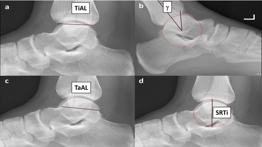
Osseous Ankle Morphology and Recurrent Lateral Sprains

INTRODUCTION                                                                patients with a history of recurrent lateral ankle sprain inju-
Ankle injuries are one of the most common injuries in sports                ries were recruited from the Hacettepe University, Depart-
and daily activities, and 77% of ankle sprains involve the                  ment of Orthopaedics and Traumatology (table I).
lateral compartment (1). The ankle joint complex compris-                   After ankle injury, initial treatment was conservative, such
es contractile and non-contractile anatomical structures that               as functional rehabilitation (7-9). Chronic ankle instability,
contribute to its function and stability (2). Although ankle                which is characterized by persistent pain, instability, re-injury
anatomy is well known, intrinsic osseous ankle morpholog-                   and permanent functional disability, may develop in 50% of
ical characteristics that contribute to sprain injury risk have             patients (10). Surgical treatment may be an option in patients
not been as well described (3, 4). Several intrinsic (e.g., rear-           with chronic ankle instability who fail appropriate conser-
foot alignment, capsuloligamentous laxity, neuromuscu-                      vative therapy after more than 6 months (11). Inclusion and
lar control) and extrinsic risk factors (e.g., shoe type, sports            exclusion criteria was conducted based on the International
participation, surface conditions) are known to contribute to               Ankle Consortium recommendations (10). Only patients who
chronic ankle instability (5). Although often managed success-              had undergone lateral ankle ligament reconstruction after
fully with stabilization surgery or conservative physiotherapy,             experiencing ≥ 3 recurrent lateral ankle sprains were includ-
up to 20-40% of patients who sustain a lateral ankle sprain                 ed in the case group. To be included for study participation
experience injury recurrence (6). This concern has prompt-                  subjects had to have experienced a “buckling” or “giving way”
ed many researchers to attempt to improve their understand-                 episode within three months prior to surgery (10, 12). Study
ing for how additional intrinsic osseous ankle morphological                exclusion criteria included the presence of any new acute
characteristics might contribute to ankle instability.                      lower extremity injury in the last 3 months, rearfoot deformity,
The purpose of this study was to evaluate the relationship                  ankle osteoarthritis, and posterior tibial tendon dysfunction,
between osseous ankle morphological characteristics and                     ankle dorsiflexion of < 10°, neurological disorders, or rheu-
recurrent lateral ankle sprain history. The study hypoth-                   matoid arthritis. It is known that Brostrom-Gould modified
esis was that specific intrinsic osseous ankle morpholog-                   surgery is an effective approach that strengthens the original
ical characteristics predict recurrent lateral ankle sprain                 ligaments with the extensor retinaculum without sacrificing
injury history.
                                                                            Table I. Descriptive analyses of patients in the study.

MATERIALS AND METHODS                                                       Variables            CAI            Normal group p value
The Ethics Committee of the Medical Sciences (IRB)                          Age (mean ± SD)       29.5 ± 2.7     30.9 ± 3.2        0.269
approved the present study and participants provided writ-                  Height (cm)          175.2 ± 6,0    177.2 ± 5.25       0.075
ten informed consent prior to study participation. Between                  Weight, (kg)          82.6 ± 9.8         78 ± 11.2     0.012
July 2009 and January 2015, 52 (18 men and 34 women)                        Data are presented with Mean ± SD (range).

Table II. Study group intrinsic osseous ankle morphological characteristic comparisons.
                                                        Abbreviation         Recurrent Inversion       Healthy                   p-value
                                                                             Ankle Sprain Group        Control Group
                                                                             (n = 52)                  (n = 46)
Tibial Arc Length                                       TiAL                 29.0 ± 3.6 mm             31.4 ± 3.6 mm              0.001
Sagittal Radius of the Trochlea                         SRTi                 23.3 ± 2.3 mm             16.1 ± 5.4 mm             < 0.0001
Trochlea Tali Length                                    TaAL                 41.3 ± 4.4 mm             29.0 ± 5.2 mm             < 0.0001
Sagittal Radius of the Trochlea Tali Arc                SRTa                 25.9 ± 2.8 mm             25.4 ± 5.9 mm             < 0.0001
Height of Talar Tenon                                   h                     7.7 ± 1.1 mm              9.9 ± 14.9 mm              0.31
Height of Talus                                         H                    24.5 ± 6.2 mm             21.2 ± 4.1 mm               0.002
Tibiotalar Sector Angle                                 a                    76.8 ± 6.0°               81.0 ± 6.6°                 0.001
Ankle Stability Angle                                   γ                    14.0 ± 2.5°               22.1 ± 7.3°               < 0.0001
Sagittal Radius of the Trochlea:                        SRTi : SRTa          0.90 ± 0.07               0.47 ± 0.19               < 0.0001
Sagittal Radius of the Trochlea Tali Arc Ratio
Trochlear Tali Length : Tibial Arc Length Ratio         TaAL : TiAL           1.4 ± 0.25               0.93 ± 0.16               < 0.0001
Independent sample t-tests were used to determine mean group differences.

428                                                                                          Muscles, Ligaments and Tendons Journal 2021;11 (3)
V. Nabi, M. N. Doral, O. Bilge, et al.
other normal structures, limiting the inversion to stabilize the
subtalar joint, and provides a stable ankle (7, 11, 13-15). Based
on our database, it was observed that the Brostrom-Gould
procedure was planned for patients with unstable ankle.
An age- and sex- matched control group consisting of
46 healthy adults (26 men and 20 women) with no previ-
ous ankle sprain injury history was recruited to create a
case-control research study design. Subject demographic
information is reported in (table II).
To better enable sagittal plane osseous structure identifica-
tion, standardized lateral and antero-posterior radiographs
were obtained with the ankle in neutral weightbearing align-
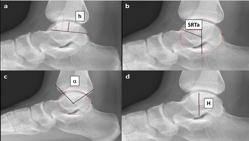
ment. Radiographs were restricted to the distal half of the leg. Figure 2. Talar tenon height (h): distance between the talar
Measurement calibration was obtained using a pure hydroxy- trochlear dome and the trochlear arch (a); Sagittal plane radi-
apatite phantom. To control for potential magnification us of the trochlea tali arc (SRTa): a circle digitally fit to the talar
effects all radiographs were performed using consistent fixed joint surface approximating the center of the talus. The distance
between the talar center and the circle is the radius (b); Tibio-
tube-detector distance. Digital radiographic images were then
talar sector angle (α): line from the talar center to the anterior
reviewed by the senior author and an independent radiologist. and posterior margins of the distal tibia. These lines enclose
Eight measurements were performed (16-19) (Sectra Worksta- the angle a (tibiotalar sector) which defines the size of the tibial
tion IDS7, version 21.2.8.6195 software, Linköping, Sweden) coverage of the talus (c); talus height (H): a line perpendicular to
as shown in figure 1 a-d and figure 2 a-d. Measurement reli- the ground through the talar center representing the distance
ability revealed intraclass correlation coefficients of ≥ 0.75 between the talar surface and the inferior border of the talus (d).
(good to excellent reliability). Additionally, the ratio between
SRTa:SRTi and TaAL:TiAL was determined. The case group
radiographs used for study measurements were taken prior to Statistical methods
ankle surgery. Similar to the human glenohumeral joint circle Data normality was confirmed using Kolmogorov-Smirn-
concept these ratios were evaluated to determine the relation- ov and Shapiro Wilk tests. Stepwise multiple regression
ship of talus (ball) and tibial surface (socket) dimensions to analysis was performed to identify variables that displayed
patient recurrent lateral ankle sprain history (20). predictive value based on patient recurrent lateral ankle
sprain injury history. Following this, independent sample
t-tests were used to determine mean group differences. Last-
ly, receiver operator characteristic (ROC) and area under
the curve (AOC) analysis was performed to identify thresh-
old cut-off values for diagnostic test sensitivity and speci-
ficity. Post-hoc statistical power analysis was performed
using Decision Support Systems Software, SPH Analyt-
ics, Alpharetta, GA, USA). All other statistical analysis was
performed using SPSS version 26.0 software (IBM-SPSS,
Armonk, NY, USA). An alpha value of p < 0.05 indicated
statistical significance.
RESULTS
Figure 1. Tibial arc length (TiAL): the sagittal plane length Radiographic intrinsic osseous ankle morphological char-
between the most anterior and posterior tibial mortise locations
acteristic values are reported in table II (mean ± standard
(a); Ankle stability angle (γ): the angle formed by the tibial axis
deviation). The patient group displayed a shorter TiAL
and a vertical line from the talar dome center with the ankle
in maximal plantar flexion (b); Trochlea tali length (TaAL): the and h than the healthy control group. The patient group
length of the line connecting the most anterior and posterior also displayed a greater SRTi, TaAL, SRTa, and H than the
points of the trochlea tali sagittal arc (c); and the Sagittal plane healthy control group. The patient group also displayed
radius of the tibial mortise (SRTi): the radius of the tibial mortise lesser α, γ, SRTi:SRTa ratio, and TiAL:TaAL ratio than the
circumference using a least-square fitting technique (d). healthy control group.
Muscles, Ligaments and Tendons Journal 2021;11 (3) 429Osseous Ankle Morphology and Recurrent Lateral Sprains
Forward stepwise regression with recurrent lateral ankle
sprain patient group assignment serving as the dependent
variable identified the following independent variables as
contributing to a highly predictive statistical model: Patient
Group (recurrent lateral ankle sprain history) = 1.45 + 0.37
TaAL + - 0.43 SRTa + - 0.23 γ + 0.15 H + -0.14 h + 0.17
SRTi (P < 0.0001). This equation displayed high predictive
value with R = 0.96, R2 = 0.91. This verified that as TaAL, H
and SRTi beta values increased there was a lower likelihood
for patient group assignment. As SRTa, γ, and h beta values
decreased there was a greater likelihood for patient group
assignment. The variables TiAL and α did not provide statis-
tically significant contributions to the prediction equation.
Following this, ROC and AUC analysis of each independent
variable was performed to identify which measurements
had the greatest sensitivity and specificity for identifying Figure 4. Receiver operating curve characteristics for SRTi:
patients with a history of recurrent lateral ankle sprain inju- SRTa and TaAL:TiAL osseous characteristic ratios.
ry. This revealed that the strongest independent radiograph-
ic predictors for the patient group were TaAL (Area = 0.97)
and SRTi (Area = 0.84) (figure 3). As individual measure- DISCUSSION
ments these values displayed generally excellent and good The most important result of this study was that recurrent
diagnostic value, respectively. lateral ankle joint stability was directly influenced by intrin-
To better delineate their clinical decision-making efficacy, sic osseous ankle morphological characteristics. Of greatest
the following ratio values were assessed: SRTi:SRTa and clinical significance were the ratio between the sagittal plane
TaAL:TiAL (figure 4). The AOC for the SRTi:SRTa ratio radius of the tibial mortis (SRTi) and the sagittal radius of
was 0.95 (95% confidence interval of 0.93-1.0). For the the trochlea tali arc (SRTa) and the ratio between trochlea
SRTi:SRTa ratio, a value of 0.68 produced a sensitivity of tali length (TaAL) and tibial arc length (TiAL).
0.96 and a specificity of 0.85. The AOC for the TaAL:TiAL The SRTi:SRTa ratio displayed excellent sensitivity and
ratio was 0.97 (95% confidence interval of 0.89-1.0). For good specificity. Using this measurement method 95% of
the TaAL:TiAL ratio, a value of 1.15 produced a sensitivity subjects would be classified correctly and only 15% who
of 0.98 and a specificity of 0.91. Post-hoc statistical power were deemed negative would be incorrectly classified. For
analysis for both ratio values revealed excellent statistical the TaAL:TiAL ratio, at a value of 1.15, sensitivity was 0.98
power (100%) at an alpha level of 0.05. and specificity was 0.91. This suggests that in using this
method, 98% of subjects would be classified correctly and
only 9% who were deemed negative would be incorrectly
classified. In managing patients with recurrent lateral ankle
sprain history, these ratio measurements can assist the physi-
cian and surgeon who treats a patient with recurrent lateral
ankle sprain history as they consider whether or not surgical
ankle ligament stabilization may be indicated to achieve a
positive outcome.
The ratio measurements we recommend provided excel-
lent sensitivity and very good specificity for distinguishing
individuals with the intrinsic osseous ankle morphologi-
cal characteristics that are consistent with recurrent lateral
ankle sprain injury. Individuals with a larger radius, corre-
sponding to a flatter talus, and a smaller tibiotalar sector,
corresponding to less talus restraint in the tibia were more
likely to have recurrent lateral ankle sprain injury history.
Figure 3. Receiver operating characteristic plots for indepen- In conjunction with a comprehensive clinical examination
dent TaAL and SRTi osseous morphology characteristics. including patient history, neuromuscular control and capsu-
430 Muscles, Ligaments and Tendons Journal 2021;11 (3)V. Nabi, M. N. Doral, O. Bilge, et al. loligamentous laxity assessments, radiographic evaluation of ous landmark ratios we describe provided insight as to the these intrinsic risk factors might help doctors and surgeons complex three-dimensional nature of the functional ankle more effectively make decisions about the potential effica- joint (17). Ankle capsuloligamentous and neuromuscular cy of surgical or conservative rehabilitation treatments for structures provide essential non-contractile and contractile improving patient outcomes. constraints to dynamic ankle joint stability. However, stan- Persistent pain, recurrent sprains, and repeated “giving dardized single plane radiographs as commonly ordered way” or “buckling” are hallmarks of debilitating chron- during an initial clinical examination potentially possess ic ankle instability (21). Although deformity (e.g., rearfoot greater diagnostic value than currently believed. When varus, first ray plantar flexion, mid-foot cavus, and gener- the ankle is evaluated in terms of chronic instability, the alized laxity) contributes to ankle sprain predisposition, most important intrinsic mechanical factor such as sagit- the combination of mechanical (pathologic laxity, synovial tal alignment (SRTi:SRTa and TaAL:TiAL ratio), is often changes, abnormal morphology and degenerative changes) overlooked because functional factors are more prominent. and functional (impaired proprioception, postural control, All factors should be considered in a holistic, rehabilitative and neuromuscular control) insufficiencies in association treatment to better target this condition and reduce health with recurrent lateral ankle sprain injuries are key contribu- and economic burden. Recently, there is an increasing trend tors to chronic ankle instability (22, 23). towards earlier surgical treatment in patients with mechan- Osteophyte excision is commonly recommended for ical instability, which reduces or prevents the progress of patients with anterior ankle impingement (24). In the more serious problems in the future (33). studies, it has been reported at a rate of 12-26% and may contribute to decreased tibial mortise coverage of the talus, further disposing it to anterior dislocation and greater CONCLUSIONS ankle (25, 26). In these patients, osteophyte excision with Intrinsic osseous ankle morphological characteristics were ligament repair produces satisfactory results by increasing related to recurrent lateral ankle sprain injuries. The ratio functional results and range of motion (27). When ankle between the sagittal plane radius of the tibial mortis (SRTi) instability is not diagnosed and treated in a timely manner, and the sagittal radius of the trochlea tali arc (SRTa) and 78% of individuals develop posttraumatic ankle osteoar- the ratio between trochlea tali length (TaAL) and tibial arc thritis, being a negative prognostic factor for poor results in length (TiAL) provide excellent sensitivity and good spec- terms of functional scores and negatively affecting patients’ ificity clinical decision-making measurement characteris- motivation, and this entails significant healthcare costs (28, tics. Intrinsic osseous ankle morphological characteristics 29). The accumulative effects of chronic ankle instability on such as these can help doctors and surgeons make treat- long-term joint health and disability are not as well under- ment decisions for patients with recurrent lateral ankle stood (30). Whether they originate from an occupational or sprain injuries. athletic initial injury mechanism chronic ankle instability or incongruency increases contact stresses and shear forc- es contribute to articular cartilage degeneration or defect ACKNOWLEDGMENTS development (31, 32). We express our gratitude to Prof. E. Turhan, for his This study has several limitations. Most importantly, it sole- assistance. ly evaluated intrinsic osseous ankle morphological charac- teristics, not capsuloligamentous or neuromuscular contri- butions to joint stability. Although the imaging used relied CONFLICT OF INTERESTS solely on standard clinical uniplanar radiographs, the osse- The authors declare that they have no conflict of interests. REFERENCES 1. Acevedo JI, Mangone P. Ankle instability and arthroscopic 3. Bozkurt M, Doral MN. Anatomic factors and biomechanics in lateral ligament repair. Foot Ankle Clin 2015;20(1):59-69. ankle instability. Foot Ankle Clin 2006;11(3):451-63. 2. Buchhorn T, Sabeti-Aschraf M, Dlaska CE, Wenzel F, Graf A, 4. Erkman MJ, Walker PS. A study of knee geometry applied to Ziai P. Combined medial and lateral anatomic ligament recon- the design of condylar prostheses. Biomed Eng 1974;9(1):14-7. struction for chronic rotational instability of the ankle. Foot 5. Halabchi F, Angoorani H, Mirshahi M, Pourgharib Shahi MH, Ankle Int 2011;32(12):1122-6. Mansournia MA. The Prevalence of Selected Intrinsic Risk Muscles, Ligaments and Tendons Journal 2021;11 (3) 431
Osseous Ankle Morphology and Recurrent Lateral Sprains

      Factors for Ankle Sprain Among Elite Football and Basketball              musculo-skeletal modelling and optimization in gait analysis. J
      Players. Asian J Sports Med 2016;7(3):e35287.                             Biomech 2017;62:77-86.
6.    Kuo CC, Lu HL, Lu TW, et al. Effects of positioning on radio-       19.   Stagni R, Leardini A, Catani F, Cappello A. A new semi-auto-
      graphic measurements of ankle morphology: a computerized                  mated measurement technique based on X-ray pictures for
      tomography-based simulation study. Biomed Eng Online                      ankle morphometry. J Biomech 2004;37(7):1113-8.
      2013;12:131.                                                        20.   Warren RF, Kornblatt IB, Marchand R. Static factors affecting
7.    Doherty C, Bleakley C, Hertel J, Caulfield B, Ryan J, Delahunt            posterior shoulder stability. Orthop Trans 1984;8:89.
      E. Recovery From a First-Time Lateral Ankle Sprain and the          21.   Evans DL. Recurrent instability of the ankle; a method of surgi-
      Predictors of Chronic Ankle Instability: A Prospective Cohort             cal treatment. Proc R Soc Med 1953;46(5):343-4.
      Analysis. Am J Sports Med 2016;44(4):995-1003.                      22.   Frigg A, Magerkurth O, Valderrabano V, Ledermann HP,
8.    McKeon PO, Mattacola CG. Interventions for the preven-                    Hintermann B. The effect of osseous ankle configuration on
      tion of first time and recurrent ankle sprains. Clin Sports Med           chronic ankle instability. Br J Sports Med 2007;41(7):420-4.
      2008;27(3):371-82, viii.                                            23.   Ferran NA, Oliva F, Maffulli N. Ankle instability. Sports Med
9.    van Rijn RM, van Ochten J, Luijsterburg PA, van Middelkoop                Arthrosc Rev 2009;17(2):139-45.
      M, Koes BW, Bierma-Zeinstra SM. Effectiveness of additional         24.   Ross KA, Murawski CD, Smyth NA, et al. Current concepts
      supervised exercises compared with conventional treatment                 review: Arthroscopic treatment of anterior ankle impingement.
      alone in patients with acute lateral ankle sprains: systematic            Foot Ankle Surg 2017;23(1):1-8.
      review. BMJ 2010;341:c5688.                                         25.   Odak S, Ahluwalia R, Shivarathre DG, et al. Arthroscopic Eval-
10.   Gribble PA, Delahunt E, Bleakley C, et al. Selection criteria for         uation of Impingement and Osteochondral Lesions in Chronic
      patients with chronic ankle instability in controlled research: a         Lateral Ankle Instability. Foot Ankle Int 2015;36(9):1045-9.
      position statement of the International Ankle Consortium. Br J      26.   Hua Y, Chen S, Li Y, Chen J, Li H. Combination of modified
      Sports Med 2014;48(13):1014-8.                                            Broström procedure with ankle arthroscopy for chronic ankle
11.   Gould N, Seligson D, Gassman J. Early and late repair of later-           instability accompanied by intra-articular symptoms. Arthrosco-
      al ligament of the ankle. Foot Ankle 1980;1(2):84-9.                      py 2010;26(4):524-8.
12.   Pourkazemi F, Hiller CE, Raymond J, Black D, Nightingale            27.   Yang Q, Zhou Y, Xu Y. Arthroscopic debridement of anterior
      EJ, Refshauge KM. Predictors of recurrent sprains after an                ankle impingement in patients with chronic lateral ankle insta-
      index lateral ankle sprain: a longitudinal study. Physiotherapy           bility. BMC Musculoskelet Disord 2018;19(1):239.
      2018;104(4):430-7.                                                  28.   Harrington KD. Degenerative arthritis of the ankle secondary to
13.   Cao Y, Hong Y, Xu Y, Zhu Y, Xu X. Surgical management of                  long-standing lateral ligament instability. J Bone Joint Surg Am
      chronic lateral ankle instability: a meta-analysis. J Orthop Surg         1979;61(3):354-61.
      Res 2018;13(1):159.                                                 29.   Shah S, Thomas AC, Noone JM, Blanchette CM, Wikstrom EA.
14.   Gerstner Garces JB. Chronic ankle instability. Foot Ankle Clin            Incidence and Cost of Ankle Sprains in United States Emergen-
      2012;17(3):389-98.                                                        cy Departments. Sports Health 2016;8(6):547-52.
15.   Maffulli N, Del Buono A, Maffulli GD, et al. Isolated ante-         30.   Bosien WR, Staples OS, Russell SW. Residual disability following
      rior talofibular ligament Broström repair for chronic later-              acute ankle sprains. J Bone Joint Surg Am 1955;37-a(6):1237-43.
      al ankle instability: 9-year follow-up. Am J Sports Med             31.   Hintermann B. Medial ankle instability. Foot Ankle Clin
      2013;41(4):858-64.                                                        2003;8(4):723-38.
16.   Kwon DG, Sung KH, Chung CY, et al. Preliminary findings of          32.   van Ochten JM, de Vries AD, van Putte N, et al. Associ-
      morphometric analysis of ankle joint in Korean population. J              ation between Patient History and Physical Examination
      Foot Ankle Surg 2014;53(1):3-7.                                           and Osteoarthritis after Ankle Sprain. Int J Sports Med
17.   Leardini A. Geometry and mechanics of the human ankle                     2017;38(9):717-24.
      complex and ankle prosthesis design. Clin Biomech (Bristol,         33.   Michels F, Pereira H, Calder J, et al. Searching for consensus
      Avon) 2001;16(8):706-9.                                                   in the approach to patients with chronic lateral ankle insta-
18.   Leardini A, Belvedere C, Nardini F, Sancisi N, Conconi M,                 bility: ask the expert. Knee Surg Sports Traumatol Arthrosc
      Parenti-Castelli V. Kinematic models of lower limb joints for             2018;26(7):2095-102.

432                                                                                         Muscles, Ligaments and Tendons Journal 2021;11 (3)
You can also read


