Oligodendroglia Generated From Adult Rat Adipose Tissue by Direct Cell Conversion
←
→
Page content transcription
If your browser does not render page correctly, please read the page content below
ORIGINAL RESEARCH
                                                                                                                                            published: 11 February 2022
                                                                                                                                          doi: 10.3389/fcell.2022.741499

                                            Oligodendroglia Generated From
                                            Adult Rat Adipose Tissue by Direct Cell
                                            Conversion
                                            Lara Vellosillo 1,2†, Jorge Pascual-Guerra 1†, Maria Paz Muñoz 1,
                                            José Antonio Rodríguez-Navarro 1,3, Daniel González-Nieto 2, Luis Carlos Barrio 4,
                                            Maria del Val Toledo Lobo 5 and Carlos Luis Paíno 1,2*
                                            1
                                              Servicio de Neurobiología-Investigación, IRYCIS, Hospital Universitario Ramón y Cajal, Madrid, Spain, 2Center for Biomedical
                                            Technology (CTB), Universidad Politécnica, Madrid, Spain, 3Departamento de Biología Celular, Universidad Complutense,
                                            Madrid, Spain, 4Unidad de Neurología Experimental, IRYCIS, Hospital Universitario Ramón y Cajal, Madrid, Spain,
                                            5
                                              Departamento de Biomedicina y Biotecnología, IRYCIS, Universidad de Alcalá, Alcalá de Henares, Spain

                              Edited by:
                          Stefanie Robel,   Obtaining oligodendroglial cells from dispensable tissues would be of great interest for
             Virginia Tech, United States   autologous or immunocompatible cell replacement therapy in demyelinating diseases, as
                          Reviewed by:      well as for studying myelin-related pathologies or testing therapeutic approaches in
                        Chih-Yen Wang,
             Baylor College of Medicine,
                                            culture. We evaluated the feasibility of generating oligodendrocyte precursor cells
                           United States    (OPCs) from adult rat adipose tissue by expressing genes encoding transcription
                            Tim Czopka,
                                            factors involved in oligodendroglial development. Adipose-derived mesenchymal cells
                University of Edinburgh,
                        United Kingdom      were lentivirally transduced with tetracycline-inducible Sox10, Olig2, Zfp536, and/or
                    *Correspondence:        Nkx6.1 transgenes. Immunostaining with the OPC-specific O4 monoclonal antibody
                      Carlos Luis Paíno     was used to mark oligodendroglial induction. O4- and myelin-associated glycoprotein
                    carlos.paino@hrc.es
                                            (MAG)-positive cells emerged after 3 weeks when using the Sox10 + Olig2 + Zfp536
     †
         These authors have contributed
                    equally to this work
                                            combination, followed in the ensuing weeks by GFAP-, O1 antigen-, p75NTR (low-affinity
                                            NGF receptor)-, and myelin proteins-positive cells. The O4+ cell population progressively
                    Specialty section:      expanded, eventually constituting more than 70% of cells in culture by 5 months. Sox10
         This article was submitted to
                                            transgene expression was essential for generating O4+ cells but was insufficient for
    Molecular and Cellular Pathology,
               a section of the journal     inducing a full oligodendroglial phenotype. Converted cells required continuous
  Frontiers in Cell and Developmental       transgene expression to maintain their glial phenotype. Some vestigial characteristics
                               Biology
                                            of mesenchymal cells were maintained after conversion. Growth factor withdrawal and
               Received: 14 July 2021
           Accepted: 19 January 2022        triiodothyronine (T3) supplementation generated mature oligodendroglial phenotypes,
          Published: 11 February 2022       while FBS supplementation produced GFAP+- and p75NTR+-rich cultures. Converted
                              Citation:     cells also showed functional characteristics of neural-derived OPCs, such as the
        Vellosillo L, Pascual-Guerra J,
                                            expression of AMPA, NMDA, kainate, and dopaminergic receptors, as well as similar
   Muñoz MP, Rodríguez-Navarro JA,
         González-Nieto D, Barrio LC,       metabolic responses to differentiation-inducing drugs. When co-cultured with rat dorsal
    Lobo MdVT and Paíno CL (2022)           root ganglion neurons, the converted cells differentiated and ensheathed multiple axons.
Oligodendroglia Generated From Adult
        Rat Adipose Tissue by Direct        We propose that functional oligodendroglia can be efficiently generated from adult rat
                       Cell Conversion.     mesenchymal cells by direct phenotypic conversion.
     Front. Cell Dev. Biol. 10:741499.
      doi: 10.3389/fcell.2022.741499        Keywords: ADSC, oligodendrocyte precursor cells, myelination, benztropine, adult rat, direct lineage conversion

Frontiers in Cell and Developmental Biology | www.frontiersin.org                   1                                        February 2022 | Volume 10 | Article 741499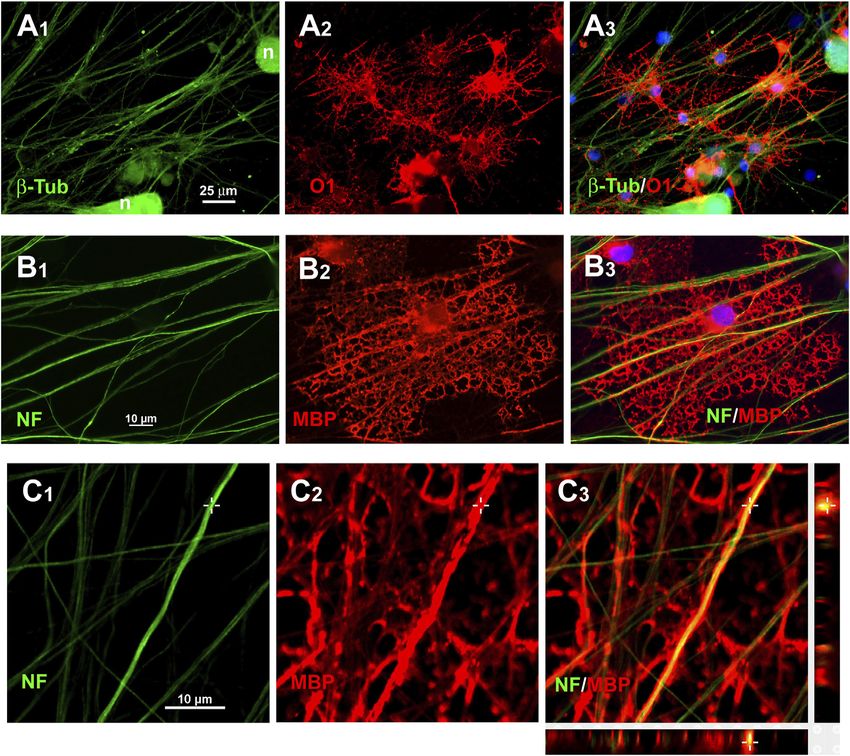
Vellosillo et al.                                                                                           Oligodendroglia Generated From Rat Fat

1 INTRODUCTION                                                             2015), and trophic properties (Bai et al., 2012; Drago et al.,
                                                                           2013; Pires et al., 2016), rendering them potentially suitable
Oligodendrocyte transplantation has long been proposed to be a             for use in the treatment of demyelinating diseases.
feasible strategy for repairing demyelinated lesions (Blakemore               Here, we show that cultured mesenchymal cells from adult rats
and Franklin, 1991; Archer et al., 1997; Wang et al., 2013), an            can be directly converted into induced oligodendroglia-like cells
approach that would be facilitated by the use of autologous or             as well as other macroglial cell types through the expression of
immunocompatible cells. Moreover, cultures of oligodendroglia              three transgenes coding for transcription factors involved in
from patients suffering genetic leukodystrophies would allow               oligodendroglial development. We further demonstrate that
studying the basis of their impaired myelination as well as                these induced cells show a repertoire of molecular traits
testing therapeutic approaches in a dish, aiming to identify               similar to those of, and respond to the same pharmacological
personalized treatments. However, there are obvious                        cues as, neural-derived oligodendroglia.
limitations to obtaining oligodendrocytes from the central
nervous system (CNS) of patients or donors. Consequently, it
would be of great interest to identify alternative means of                2 MATERIALS AND METHODS
generating oligodendroglia from the peripheral tissues of
patients.                                                                  A list of commercial product references is provided in
    Several procedures for obtaining expandable and                        Supplementary Table S1.
myelinogenic oligodendrocytes from pluripotent stem cells
(PSCs) in culture have been reported (Keirstead et al., 2005;              2.1 Animals and Procedures
Izrael et al., 2007; Hu et al., 2009; Najm et al., 2011; Douvaras          All procedures involving animals were performed by qualified
et al., 2014; Douvaras and Fossati, 2015; Piao et al., 2015),              personnel and in accordance with Directive 2010/63/UE of the
including protocols that can promote the differentiation of                European Union on the protection of animals used for scientific
oligodendrocytes from induced PSCs (iPSCs), themselves                     purposes and its transposition to Spanish law (RD53/2013). Rats
generated through the reprogramming of adult somatic cells                 were bred at the animal facilities of the Hospital Universitario
(Wang et al., 2013; Douvaras et al., 2014; Douvaras and                    Ramón y Cajal (ES280790002001). For tissue collection, the rats
Fossati, 2015). More recently, some groups have proposed the               were first deeply anesthetized with isoflurane and subsequently
rapid generation of oligodendroglia from human iPSCs via the               euthanized by decapitation. Ethics committee approval was not
exogenous expression of a combination of Sox10, Olig2, and                 required for these procedures.
Nkx6.2 (Ehrlich et al., 2017) or only Sox10 (Garcia-Leon et al.,
2018). This process involves three steps, namely, the generation,
selection, characterization, and the growing of iPSCs from                 2.2 Adipose Tissue-Derived Mesenchymal
cultures of adult somatic cells; deriving neural progenitor cells          Stem Cells Culture
from the iPSCs; and using transduction to express genes encoding           Adipose tissue was dissected under aseptic conditions from the
oligodendroglial transcription factors in these cells. However, in         inguinal pads of 16 Sprague–Dawley rats weighing 220–250 g.
addition to the protracted time needed for cell reprogramming,             The tissue was cleaned of fasciae and major blood vessels, cut into
the risk remains that, unless all cells are differentiated, residual       1-mm2 pieces, and digested for 40 min at 37°C with 1 mg/ml
PSCs might continue to proliferate and produce teratomas.                  collagenase A in αMEM supplemented with 20% FBS, non-
    Two laboratories have simultaneously reported that                     essential amino acids, glutamine, and antibiotics/antimitotics
embryonic rat or mouse fibroblasts can be directly converted                (Gibco; henceforth referred to as α20 medium). DNAse I
into oligodendroglial cells through the exogenous expression of            (20 μg/ml) was added to prevent cell clumping due to DNA
genes coding for transcription factors involved in                         released from dead cells and the tissue was then dispersed by
oligodendrocyte development (Najm et al., 2013; Yang et al.,               repeated passage through a P1000 automatic pipette using sterile
2013). Furthermore, these cells were shown to be capable of                filter tips. Large, undispersed clumps of tissue were removed by
myelinating axons both in vivo and in culture. However, cell               passing the suspension through 100-μm mesh cell strainers. After
replacement therapies, as well as the search for personalized              centrifugation at 400 ×g for 3 min, the supernatant, including the
treatment strategies, would require the use of adult somatic               layer of floating adipose cells, was discarded. The pellet was
cells, but evidence that these procedures can be used to                   resuspended in 1 ml of α20 medium and seeded in a T75 flask
directly convert cells from adult somatic tissues into                     (Falcon) containing the same medium. The next day, the medium
oligodendrocytes is lacking.                                               with unattached cells was removed, the flask was rinsed once with
    The vasculo–stromal fraction of adipose tissue contains                Hank’s balanced salt solution (HBSS), and fresh α20 medium was
mesenchymal stem cells (MSCs) that can be easily grown in                  added. These adipose-derived stromal cells (ADSCs) proliferated
culture, and thus represents an accessible source of mesenchymal           and reached near-confluence after 4–5 days, at which point they
cells. These cells display characteristics such as                         were detached with 0.05% trypsin + 0.02% EDTA, centrifuged,
immunomodulatory (Bai et al., 2009; Fransson et al., 2014; Ma              resuspended in α20 medium, and quantified using a
et al., 2014; Mattar and Bieback, 2015), anti-inflammatory                  hemocytometer. The cells were then seeded in new T flasks at
(Marfia et al., 2016; van Velthoven et al., 2017),                          660 cells/cm2 in α20 medium. The medium was changed on day 4
neuroprotective (Otero-Ortega et al., 2015; Ribeiro et al.,                and the cells were passaged on day 7, at which time they were

Frontiers in Cell and Developmental Biology | www.frontiersin.org      2                                 February 2022 | Volume 10 | Article 741499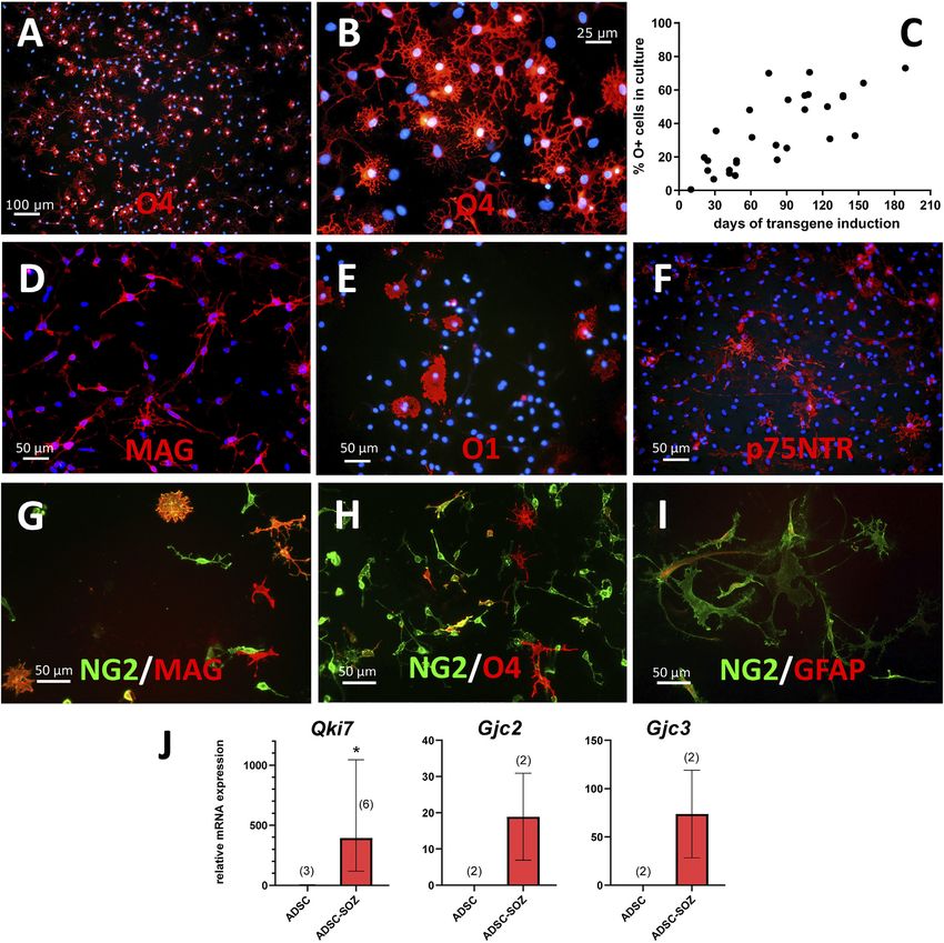
Vellosillo et al.                                                                                            Oligodendroglia Generated From Rat Fat

again confluent, their number having increased 30–40-fold. After             doxycycline-mediated activation of transgene expression. Four
4 passages, the cultures showed signs of cellular senescence and            days after ADSC transduction with the Sox10 + Olig2 + Zfp536
slow proliferation. Consequently, the experiments were                      (+M2rtTA) transgenes (subsequently referred to as S + O +
performed using cells at passages 1–3.                                      Z-transduced cells), an average of 8.9% of cells showed nuclear
   The mesenchymal characteristics of these cells were tested by            labeling for Sox10. After 12 days, 38.9% of the transduced cells
assessing their adipogenic, chondrogenic, and osteogenic                    were Sox10+, and the cell numbers had increased by 496% relative
differentiation potential (Pittenger et al., 1999) using standard           to control cultures transduced with M2rtTA only. After 45 days,
procedures.                                                                 53.7% of the cells were Sox10+, and after 100 days, nearly all the
                                                                            cells showed intense nuclear Sox10 staining. These observations
2.3 Lentiviral Particle Production                                          indicated that cells expressing the Sox10 transgene were
Self-inactivating, replication-incompetent lentiviruses were                proliferating and that the proportion of Sox10+ cells increased
produced in HEK293T cells by co-transfecting the lentiviral                 when cultures were maintained in neurobasal medium with B27
plasmid (see below), the packaging plasmid psPAX2, and the                  supplement (NBB27 medium) (Invitrogen) containing EGF +
envelope vector pCMV-VSV-G using Lipofectamine 2000. The                    basic FGF (bFGF) + PDGF-AA + doxycycline (see below).
cells were maintained in Dulbecco’s minimal essential medium                Additionally, the low numbers of Sox10+ cells observed at day
(DMEM) supplemented with 10% FBS. Culture supernatants                      4 (cells that had been simultaneously transduced with the Sox10
were collected every 24 h for 3 days, pooled, centrifuged at                and the M2rtTA transgenes) were nevertheless sufficient to
1,000 ×g for 10 min, passed through sterile 0.45-µm filters,                 generate pure cell cultures expressing Sox10 in 3 months or
layered over 2 ml of 20% sterile sucrose, pelleted by                       less because of their selective proliferation in the present
ultracentrifugation at 50,000 ×g for 3 h at 4°C, resuspended in             medium conditions.
500 µL of DMEM, and aliquoted.
    Viral titers were measured with the Lentivirus qPCR kit                 2.6 Culture Medium for Transduced Cells
(Applied Biological Materials; contained reverse transcriptase,             After transduction, the cells were cultured in NBB27 medium. To
standards, and primers specific for the lentiviral 5′-LTR) using             induce transgene expression, doxycycline (1 μg/ml) was added to
LightCycler 480 with SybrGreen I Master kit (Roche Applied                  the growth medium. For OPC growth, NBB27 was supplemented
Science). Titers ranged between 120,000 and 500,000 lentiviral              with EGF (20 ng/ml), bFGF (20 ng/ml), PDGF-AA (10 ng/ml),
particles per µL. Viral particle-to-cell ratios of 1:1, 5:1, and 10:1       d-biotin (10 ng/ml), and doxycycline hydrochloride (OPC
were tested for ADSC transduction.                                          medium). To increase the efficiency of oligodendroglial
                                                                            conversion, several pre-treatments were tested (Supplementary
2.4 Plasmids                                                                Figure S6; Supplementary Table S2). This included attempts to
The FUW-M2rtTA plasmid was a gift from Rudolf Jaenisch                      neuralize the cells by exposure to 1 mM all-trans retinoic acid
(Addgene plasmid # 20342) (Hockemeyer et al., 2008); Tet-O-                 (RA) for 5 days, to prevent ADSCs from adopting a chondrogenic
FUW-Sox10 (Addgene plasmid # 45843), Tet-O-FUW-Zfp536                       phenotype when Sox10 was expressed (as suggested in
(Addgene plasmid # 45845), Tet-O-FUW-Nkx6.1 (Addgene                        Supplementary Figure S2) (Chimal-Monroy et al., 2003) by
plasmid # 45846), Tet-O-FUW-Olig2 (Addgene plasmid #                        incubating in adipogenic differentiation cocktail (see below)
30131), and Tet-O-FUW-EGFP (Addgene plasmid # 30130)                        for 5 days, or to treat with 25 nM Repsox before the initial
were gifts from Marius Wernig (Vierbuchen et al., 2010; Yang                doxycycline exposure. The adipocyte pre-differentiation
et al., 2013); psPAX2 was a gift from Didier Trono (Addgene                 cocktail    initially   included     10 μg/ml    insulin, 1 µM
plasmid # 12260); and pCMV-VSV-G was a gift from Bob                        dexamethasone, 200 µM indomethacin, and 250 µM 3-isobutyl-
Weinberg (Addgene plasmid # 8454) (Steward et al., 2006).                   1-methylxanthine (IBMX), but this was later reduced to
                                                                            indomethacin + IBMX to improve cell adhesion to the culture
2.5 Lentiviral Transduction                                                 surface.
MSCs were seeded at 15,000 cells/cm2 in 24-well plates and
allowed to grow for 24 h in α20 medium. Viral particles and                 2.7 Immunocytochemistry
8 μg/ml polybrene were added to the culture medium and the                  Cells were cultured on glass coverslips in the wells of 24-well
plates were incubated at 37°C for 1 h in an Eppendorf centrifuge            plates for at least 48 h, and then fixed in buffered 4%
at 1,000 ×g and then for 1 h in an incubator with 5% CO2,                   paraformaldehyde for 10 min and washed in PBS, pH 7.4. To
following which the medium was replaced with fresh α20                      detect surface antigens, after blocking with 5% normal goat serum
medium. After 24 h, the cells were detached with trypsin (as                in PBS, cells were incubated with antibodies targeting O4
above) and seeded in new flasks or plates for expansion or                   sulfatide, O1/galactocerebroside (GalC), NG2, p75NTR or
experimental analysis. Where appropriate, after transfection,               A2B5 without permeabilization for 90 min at room
cells were cryopreserved at −80°C in FBS with 10% DMSO.                     temperature. For the immunostaining of intracellular antigens,
    To prevent cells from receiving multiple transgene doses,               cells were post-fixed and permeabilized with ethanol: acetic acid
lentiviral titers were limited to that necessary for transducing            (19:1) for 10 min at −20°C and then incubated with the respective
approximately 30% of cells in each culture. Transduction                    primary antibodies overnight at 4°C. The next day, the cells were
efficiency      was    assessed    by    analysis    of    SOX10             incubated with anti-mouse, anti-rat, or anti-rabbit antibodies
immunofluorescence at different time points following the                    conjugated to Alexa fluorochromes (1:500 dilution) for

Frontiers in Cell and Developmental Biology | www.frontiersin.org       3                                 February 2022 | Volume 10 | Article 741499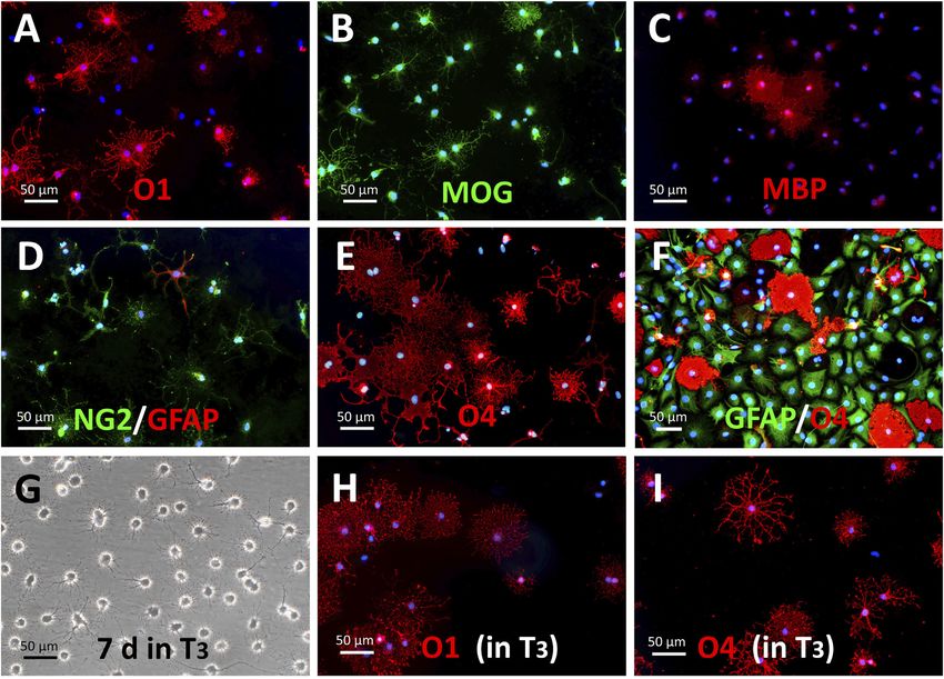
Vellosillo et al.                                                                                           Oligodendroglia Generated From Rat Fat

30–45 min at room temperature in the dark. Both primary and                (20 ng/ml) and bFGF (20 ng/ml). The cell suspension was
secondary antibodies were diluted in blocking solution. Nuclei             collected and pelleted by centrifugation at 400 ×g for 3 min
were counterstained with bisBenzimide hydrochloride (Hoechst               and seeded in a new flask every day for the first 3 days to
33342, 3 × 10⁻⁵ M) in PBS for 5 min. The coverslips were rinsed in         discard adherent cells. To produce single-cell suspensions and
distilled water and mounted on slides with Prolong Gold. The               to eliminate cell debris without the need for gradient
samples were analyzed and imaged using an Olympus BX51                     centrifugation, samples were digested with accutase first for
fluorescence microscope or a Nikon Eclipse Ti confocal                      10 min at room temperature and then for 10 min at 37°C,
microscope. The list of antibodies and suppliers is shown in               followed by trituration with fire-narrowed P1000 sterile filter
Supplementary Table S5.                                                    tips. These cultures were transferred to new flasks and provided
                                                                           with fresh NBB27 medium supplemented with EGF + bFGF every
2.8 RT-PCR                                                                 2–3 days. Numerous medium-sized neurospheres (i.e., cell
Total RNA was isolated from transduced cells, control ADSCs, or            aggregates rich in neural progenitors) had formed by day 10.
neural tissue-derived oligospheres using the RNeasy Mini Kit or            Thereafter, the medium was additionally supplemented with
the GeneJET RNA Purification Kit. RNA was reverse transcribed               10 ng/ml PDGF-AA to promote the oligodendroglial
using the First-Strand RNA Synthesis Kit and PCR was                       commitment of neural progenitors, eventually leading to the
performed in a thermal cycler (Applied Biosystems) using a                 generation of “oligospheres,” floating aggregates highly
reaction mixture containing specific primers, dNTPs, and                    enriched in oligodendroglial cells (58%–95% O4+ cells and
AmpliTaq DNA polymerase. Amplified cDNAs were                               ~90% NG2+ cells; see Supplementary Figure S14). For the
electrophoretically separated in 1.5%–2% agarose gels loaded               seeding of these cells on adherent surfaces (poly-L-ornithine-
with the fluorochrome GreenSafe Premium. Bands were                         coated flasks, wells, or coverslips), the oligospheres were digested
documented and analyzed using Molecular Imager Gel Doc                     with accutase and disaggregated mechanically as described above.
(Bio-Rad).                                                                 The seeded cells were maintained in NBB27 medium
   For RT-qPCR, 1,000 ng of total RNA was reverse transcribed              supplemented with either EFG + bFGF + PDGF-AA, for
using the NZY First-Strand cDNA Synthesis Kit. Real-time PCR               OPCs, or triiodothyronine (T3), for differentiated
was performed using TB Green Premix Ex Taq (Takara Bio) or                 oligodendroglia showing high O1 positivity (as shown in
LightCycler 480 II reagents (Roche Applied Science). The initial           Supplementary Figure S14).
denaturation step was 95°C for 5 min, followed by 45 cycles at
95°C for 5 s and 60°C for 30 s (for Takara reagents) or 95°C for           2.10 Metabolic Flux Analysis
10 s, 60°C for 15 s, and 72°C for 15 s (when using LightCycler             For the assessment of mitochondrial function, a Cell Mito Stress
reagents). The melting curves for the PCR products were                    Test Kit (Agilent) was used to assay the oxygen consumption rate
evaluated at the end of the amplification reactions. The final               (OCR) on a Seahorse XFp Extracellular Flux Analyzer (Agilent). S
PCR reaction products were separated on 2% agarose gels                    + O + Z-transduced ADSCs and neural stem cell (NSC)-OPCs
containing GreenSafe Premium to confirm the presence of a                   were seeded at 20,000 cells/well in poly-L-ornithine + laminin-
single band. The efficiency of the reaction for each primer pair            coated Seahorse XFp Cell Culture Miniplates (Agilent) and
was calibrated by amplifying serial dilutions (1:10, 1:100, 1:1,000,       maintained for 24 h in proliferative growth medium (NBB27
and 1:10,000) of cDNA from positive controls. The association              medium supplemented with EGF + bFGF + PDGF-AA; for
between the threshold cycle (Ct) and the log[RNA] was linear               converted cells, doxycycline was also added).
(−3.45 < slope < −3.32). The relative expression levels of target             On the day of the experiment, the medium was changed to
genes were normalized to that of the housekeeping gene                     Seahorse XF DMEM, pH 7.4 supplemented with 1 mM pyruvate,
glyceraldehyde 3-phosphate dehydrogenase (Gapdh) using the                 25 mM glucose, and 4 mM L-glutamine, and the plates were
ΔCt method (Pfaffl, 2001). For quantification, cDNA samples                  incubated for 1 h at 37°C without CO2. The assay is based on
were diluted 1/10 in the reaction mix. The primers used in real-           optical sensors that measure the oxygen concentration after the
time and end-point PCR are shown in Supplementary Tables S3                sequential addition of drugs that modulate cellular respiration
and S4.                                                                    (1 µM oligomycin, which blocks ATP synthase; 2 µM FCCP, a
                                                                           protonophore that enables maximum electron flux through the
                                                                           electron transport chain; and rotenone and antimycin A [RAA],
2.9 Cultures of Neural Tissue-Derived                                      which inhibit complexes I and III, respectively, thus shutting
Oligospheres                                                               down mitochondrial activity), allowing to estimate the basal,
Control oligodendroglia-rich cultures were generated from the              ATP-linked, or maximal capacity of mitochondrial respiration.
cervical spinal cords of E16 rats (the same animals from which the
dorsal root ganglia were obtained; see below) or the cerebral              2.11 Myelination in Culture
cortices of 3–6-day-old postnatal rat pups. In both cases, the             Dorsal root ganglion neurons (DRGns) were obtained from the
neural tissue, free of meninges, was mechanically dispersed by             cervical enlargement of E16 rat embryos (E0 being the day after
passing through a P1000 automatic pipette fitted with a fire-                overnight insemination) and dispersed using 0.05% trypsin +
smoothened sterile filter tip, following which the cell suspension          0.02% EDTA digestion for 15 min at 37°C on a shaker, followed
was passed through a 100-µm mesh cell strainer and seeded                  by mechanical trituration with a P1000 automatic pipette fitted
directly in flasks in NBB27 medium supplemented with EGF                    with a fire-smoothened sterile filter tip. The cell suspension was

Frontiers in Cell and Developmental Biology | www.frontiersin.org      4                                 February 2022 | Volume 10 | Article 741499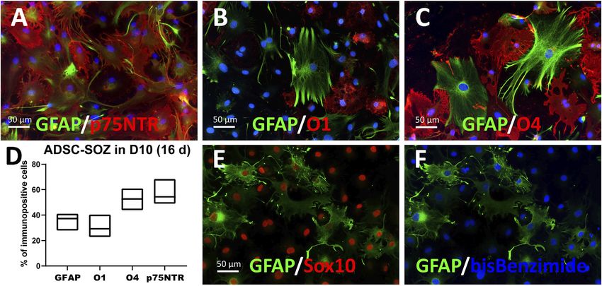
Vellosillo et al.                                                                                         Oligodendroglia Generated From Rat Fat

passed through a 70-µm mesh cell strainer to eliminate                   paraformaldehyde + 2.5% glutaraldehyde in PBS at 4°C until
undigested tissue (containing blood vessels, meninges, and               processing for TEM. Post-fixing/staining in 1% OsO4,
nerve fragments) and the cells were then seeded on poly-L-               dehydration in alcohol, and embedding in epoxy resin were
ornithine + laminin-coated coverslips (diameter: 12 mm,                  performed in the Petri dish, leaving a thin layer of epoxy resin
placed inside 24-well plates) at approximately 1,000 cells/              covering the cells that was allowed to harden overnight at 56°C (as
coverslip in DMEM supplemented with 10% FBS and 50 ng/                   shown in Supplementary Figure S15E). Fragments of the central
ml β-NGF overnight. The next day, the cultures were changed              portion of the axonal cable were cropped and positioned in resin
to NBB27 medium containing β-NGF + 10 µM                                 molds for transverse cutting in an ultramicrotome (Leica Ultracut
fluorodeoxyuridine + 10 µM uridine for 2–3 days to                        S). Ultrathin (60 nm) sections were contrasted on grids with
eliminate dividing cells (comprising mostly Schwann cells                uranyl acetate and lead citrate and examined in a Jeol JEM1010
and perineurial fibroblasts). The medium was subsequently                 (100 kV) microscope at the TEM facilities of the School of
replaced with NBB27 medium supplemented only with β-NGF                  Medicine of the Autonomous University of Madrid or a Jeol
for 2–3 days. The cycle (with/without antimitotics) was                  JEM1400 Flash at the Electron Microscopy Service of the Centro
repeated twice more. After 2 weeks, the culture consisted of             de Biología Molecular “Severo Ochoa” (CSIC/UAM, Madrid).
small clusters of neurons interconnected by a mesh of axonal
fascicles free of Schwann cells. A 20 µL drop of S + O +
Z-transduced ADSCs was carefully deposited at the center
of the neuronal culture. After a few minutes, these cells had
                                                                         3 RESULTS
dispersed and adhered to the axons and the coverslip.                    3.1 Immature Oligodendroglia Can Be
Myelination was allowed to progress for 3–7 more weeks in
NBB27 medium containing 0.5 μg/ml doxycycline. For some
                                                                         Efficiently Generated by Expressing Sox10,
experiments, 1.5 µM benztropine mesylate was added to co-                Olig2, and Zfp536 Transgenes in Adult Rat
cultures for the last 1–2 weeks.                                         Adipose Tissue-Derived MSCs
                                                                         ADSCs were transduced with tetracycline-inducible Sox10,
2.12 Transmission Electron Microscopy                                    Olig2, Zfp536, and/or Nkx6.1 (N) transgenes to test if the
Transmission electron microscopy (TEM) was performed                     exogenous expression of the encoded transcription factors
using various procedures, the most consistent one of which               could induce the conversion of these adult rat cells into
is described here. A 20-mm2 area of a 35-mm Petri dish was               oligodendroglia. The expression of Sox10, Olig2, and Zfp536
coated with rat tail collagen type I (4.36 mg/ml in 0.02 N acetic        in a defined medium containing EGF (20 ng/ml) + bFGF
acid, spread with cell scrapers), polymerized under NH4OH                (20 ng/ml) + PDGF-AA (10 ng/ml) led to the generation of
vapor for 2 min, and allowed to dry. Straight tracks (12 ×               colonies containing a few small, branched, and refringent cells
1.5 mm) were drawn on the collagen with Geltrex (LDEV-Free               within 3–4 weeks (Figure 1). Immunofluorescence staining of
Reduced Growth Factor Basement Membrane Matrix, from                     these cultures showed the presence of O4+ cells (Figures
Gibco) or laminin (each at 4 µL per track), and allowed to dry.          2A,B), which we considered to be presumptive OPCs as this
The Petri dish was rinsed once with HBSS, filled with 1 ml of             marker has not been previously observed in ADSCs (Vellosillo
DMEM supplemented with 10% FBS (D10 medium) + 100 ng/                    et al., 2017). Although morphological changes could be
ml β-NGF, and kept in an incubator until DRG seeding. For                observed in these branched and refringent cells from the
DRG seeding, 400 µL of medium was removed from the plate,                first week (Figure 1B), it was not until they decreased their
leaving only a thin layer of liquid, and two DRG were                    soma size, divided, and became separated from the flat,
positioned at each end of the tracks. The ganglia were                   mesenchymal-like cells that they could be labeled by the O4
induced to attach to the substrate by incubating overnight               antibody. These oligodendroglia-like cells proliferated and
inside a humidified chamber, after which the medium was                   formed larger colonies during the following weeks, while
switched to NBB27 medium supplemented with β-NGF                         the rest of the cells in the culture either died or proliferated
(50 ng/ml) + fluorodeoxyuridine (10 µM) + uridine (10 µM),                slowly. The proliferation of these oligodendroglia-like cells
as described above. After three cycles of antimitotic treatment,         accelerated from month two of the induction of Sox10 + Olig2
the DRGns had extended numerous straight axons along the                 + Zfp536 transgene expression, when there was a doubling of
tracks, merging with those extending from the opposite                   the cell population every 3.9 days, to month three and
ganglions, such that a cable of nude axons had formed.                   subsequently, when there was a doubling every 1.8 days,
Then, the medium was changed to NBB27 medium +                           such that culture maintenance required weekly detachment
doxycycline, and 4 µL of a concentrated suspension                       of cells with accutase and the seeding in a new flask at a 1:15
(20,000 cells/µL) of S + O + Z-transduced ADSCs were                     dilution. By month 3, O4+ cells constituted approximately 40%
carefully deposited along each axonal cable under a                      of the total cell population (Figures 2A–C; Supplementary
microscope (after 10 min, numerous oligodendroglia-like                  Figure S1). Cells in these cultures could continue proliferating
cells had attached to axons; see Supplementary Figure S15).              for longer than 7 months, whereas the original ADSCs
   After at least 4 weeks of co-culturing, the cells were fixed for       typically became senescent by 4–5 weeks). At these times,
5 min by adding to the medium an equal volume of 4%                      the proportion of O4+ cells in the culture was >70%,
paraformaldehyde in PBS, and then maintaining them in 2%                 without any type of selection.

Frontiers in Cell and Developmental Biology | www.frontiersin.org    5                                 February 2022 | Volume 10 | Article 741499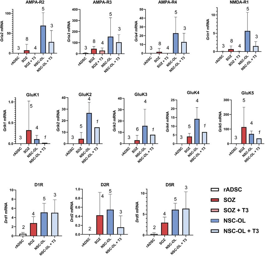
Vellosillo et al. Oligodendroglia Generated From Rat Fat
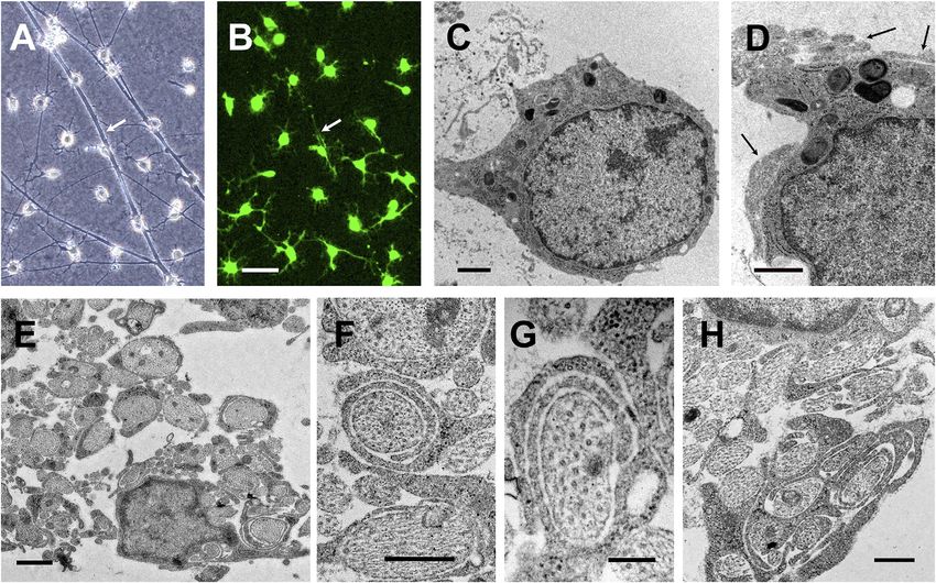
FIGURE 1 | Time course of phenotypic conversion from adult rat ADSCs into oligodendroglia-like cells. Phase contrast images at different stages after the activation
of the transgenes. (A) Confluent culture of ADSCs in α20. (B) During the first week of transgene activation, some refringent cells are visible between groups of flat cells.
(C) Refringent cells migrate to empty areas of the culture surface but keep contact with clusters of flat cells. (D) By 3–4 weeks, independent branched and refringent cells
are observed. (E) By 6 weeks after transgene induction, colonies of small and ramified cells are prominent. These cells proliferate in the culture conditions here
provided (arrow points to a cell undergoing mitosis). (F) At 3 months, a high proportion of cells in the culture show small and refringent cell bodies with a few branches.
Myelin-associated glycoprotein (MAG) was also expressed distinguishable from that of O4+ cells, and also different
in the small, refringent, and branched cells from very early from that of typical Schwann cells in culture.
after the induction of Sox10 + Olig2 + Zfp536 transgene No membrane labelling with A2B5 antibody was observed in
expression (Figure 2D). MAG could also be considered an any of our transduced cultures. In contrast, staining with this
oligodendroglial-specific marker as its expression was not antibody was strong in neural OPC cultures (see Supplementary
observed in mesenchymal cells (Vellosillo et al., 2017). Figure S14D). The repeated absence of A2B5 labelling in S + O +
Indeed, MAG positivity could be considered the most Z-transduced cells suggested that oligodendroglia and astroglia-
reliable marker of ADSC conversion into oligodendroglia- like cells were not generated through the so-called O2A stage.
like cells (see below). Additionally, MAG+ cells could Unlike A2B5, numerous NG2+ cells were present in S + O +
constitute >90% of the total number of cells after 3 months Z-transduced ADSC cultures (Figures 2G–I) from the earliest
or longer of transgene expression. stages. However, NG2 also labeled untransduced ADSCs, and thus
The S + O + Z-transduced cultures also contained minor could not serve as a marker of oligodendroglial conversion. Indeed,
populations of poorly-developed O1+ cells (Figure 2E). This at shorter induction times (i.e.,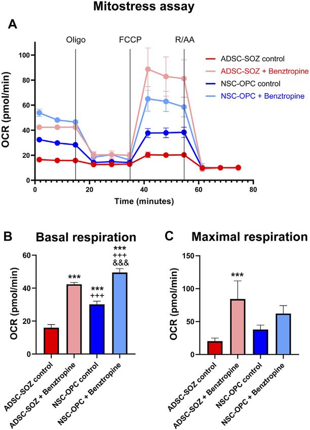
Vellosillo et al.                                                                                                                      Oligodendroglia Generated From Rat Fat

  FIGURE 2 | Phenotypical characterization of cells generated by transduction of Sox10 + Olig2 + Zfp536. (A, B) After 3 months of continuous expansion, around
  50% of cells in the culture are O4+ (shown in red at two different magnifications; in blue, bisBenzimide Hoechst 33342- counterstain of nuclei). (C) Percentage of O4+ cells
  in cultures of converted cells, examined at different times of S + O + Z transgene expression. (D) MAG is the earliest oligodendroglial-specific marker of cell conversion
  that we have observed, shown here in a 2-months culture. (E) Under continuous induction of transgene expression by doxycycline in proliferating cultures, a few
  cells show O1 staining with limited branching. (F) Multiple cells show p75NTR expression at their surface. (G–I) NG2+ cells (green) in double labelling (in red) with MAG
  (G), O4 (H) or GFAP (I) after 2-months of transgene activation. Double-labelled cells show orange color. Notice that, besides many double-labelled cells, there are singly-
  labelled cells for MAG or O4, as well as cells that are labelled for NG2 only. Light expression of GFAP can be observed in some NG2+ cells (I). Scale bars, indicated in each
  picture. (J) Demonstration of Qki7 and oligodendroglial-specific connexins Cx47 (gene Gjc2) and Cx29 (gene Gjc3) mRNA expression in S + O + Z-transduced ADSCs
  (ADSC-SOZ), while control rat ADSC in basal conditions show no or very low expression. Columns represent medians ± range, with the sample number (each with
  technical triplicates) indicated between parentheses. For Qki7, Mann-Whitney U test, shows statistically significant differences (p < 0.05) to control ADSC.

expression could be detected in S + O + Z-transduced cells                                     Additionally, RT-qPCR analysis also showed the presence of
maintained in the same medium and under the same                                            mRNAs for oligodendroglial-specific connexins in converted cells
conditions (Figure 2J; see also Supplementary Figure S7A).                                  (Figure 2J). Both Gjc2 (encoding Cx47) and Gjc3 (encoding

Frontiers in Cell and Developmental Biology | www.frontiersin.org                       7                                           February 2022 | Volume 10 | Article 741499
Vellosillo et al.                                                                                                                     Oligodendroglia Generated From Rat Fat

  FIGURE 3 | Maturation of S + O + Z-transduced cells in oligodendroglial-differentiating conditions. (A–E) Withdrawal of growth factors from the culture medium
  (i.e., switching to NBB227 + doxycycline) initiates the differentiation of OPC-like into mature oligodendrocyte-like cells. (A) Numerous O1+ cells with extended cytoplasms
  after 10 days of growth factor withdrawal are shown. (B) MOG+ cells (same field as in (A)). (C) MBP+ cells. (D) NG2+ cells increase morphological complexity but there is
  not further increase in size or number of GFAP+ cells. (E) O4 antigen keeps its expression in these conditions but most cells show profuse morphologies. (F) In the
  presence of DRG neurons and kept in NBB27 + doxycycline for 6 days, GFAP+ (in green) of the protoplasmic type and well-developed O4+ cells (in red) make up most of
  the culture cell population. (G–I) Supplementation of NBB27—doxycycline medium with T3 further enhances morphological differentiation. (G) Phase contrast image of
  living 5 month-old S + O + Z-transduced ADSC culture maintained for the last 7 days in NBB27 + T3 + doxycycline. The overall morphology is homogeneous and
  resembles pure oligodendrocyte cultures. Sibling cultures of the cells in G, immunostained for O1 (H) or for O4 (I), show enhanced differentiation, taking shorter times to
  reach larger and more profuse branching.

Cx29) were expressed in S + O + Z-transduced cells, but not in                             undisclosed concentration (Brewer et al., 1993). O4 antigen
untransduced ADSCs, when maintained in the same medium                                     expression was maintained when cells were differentiated through
(NBB27 medium supplemented with EGF + bFGF + PDGF-AA)                                      growth factor withdrawal or T3 stimulation (Figures 3E,I). The
and under the same conditions.                                                             withdrawal of EGF and bFGF, but not PDGF-AA from the medium
                                                                                           could also enhance the maturity of converted cells. Under this
                                                                                           condition, the expression of PDGF receptor alpha gene (Pdgfra)
3.2 Differentiation of S + O + Z-Transduced                                                was reduced (Supplementary Figure S7A), implying that the cells
Cells                                                                                      behaved as if they had been maintained in growth factor-free NBB27
The withdrawal of growth factors from the culture medium elicited                          medium.
the appearance of large numbers of O1+ (from approximately                                     Protoplasmic GFAP+ cells, presumably astroglia, could be
1%–28.35 ± 4.35% in quantification of four cultures) and MBP+                               preferentially generated by switching the defined medium to
cells with more mature morphologies (i.e., larger and more complex                         D10 medium + doxycycline (Figure 4). Intensely-labeled
arborizations). Addition of T3 (60 nM) to the medium further                               GFAP+ cells constituted a significant proportion of the total
stimulated the O1+ cell abundance and morphological complexity                             cell population (averaging 35.86% of cells in 3–4 months
(Figures 3G,H). It should be considered, however, that the                                 cultures; range from 27.9% to 41.05%). In these differentiating
qualitative formula of B27 supplement includes T3 at an                                    conditions, S + O + Z-transduced cells exhibited reduced

Frontiers in Cell and Developmental Biology | www.frontiersin.org                      8                                          February 2022 | Volume 10 | Article 741499
Vellosillo et al. Oligodendroglia Generated From Rat Fat
FIGURE 4 | Differentiation to various glial phenotypes by culturing in 10% FBS-containing medium. (A–D) ADSCs expressing S + O + Z for 3 months and cultured
for the last 16 days in 10%-supplemented medium (D10 + doxycyline) change their morphology and cell composition. In (A), many GFAP+ cells, of typical astrocyte-like
morphology, are double-labelled as p75NTR+ cells. (B) O1+ cells showed a flat, non-branching morphology in D10 and no O1+ cell co-stained for GFAP. (C) By contrast,
part of O4+ cells showed more elaborated morphologies and approximately 20% were co-stained for GFAP. In (D) graphical representation of % cells that were
immunofluorescently labelled for each marker (means and range, n = 4–6 cultures). (E, F) ADSCs expressing S + O + Z for 5 months but maintained for the last 18 days in
D10 + doxycycline medium. Sox10+ nuclei (in red immunofluorescence) made up 98% of all cell nuclei (stained with bis-benzimide, in blue) at this time of transgene
activation. In (E), it is shown that most (if not all) GFAP+ cells express the Sox10 transgene.
proliferation as well as enlarged nuclei and cytoplasm. Seven days containing or not the signal peptide), Mag, and Plp1/Dm20
after switching to D10 medium containing doxycycline, most of were expressed in S + O + Z-transduced cultures. As expected,
the cells displayed a flat morphology and expressed p75NTR untransduced ADSCs expressed Cnp, the Dm20 variant of the
(56.3%) and/or O4 (51.4%) in the same proportions as observed Plp1 gene, and low levels of Mbp and Mog (Vellosillo et al., 2017).
in proliferation medium (see also Supplementary Figure S11). Oligospheres, which showed little spontaneous glial
Although double immunostaining for p75NTR and O4 could not differentiation, also expressed Cnp and low levels of Mbp,
be performed, since both single-labeled cells exceeded the 50% it Mag, Mog, and Plp1/Dm20. Notably, S + O + Z-transduced
was evident that, at least, a subpopulation of the cells expressed ADSCs mainly expressed Mbp variant 5 (the shortest) as well
both markers. Similarly, many NG2+ cells were co-labeled with as the rest of non-Golli variants to varying degrees, showing more
GFAP. Interestingly, after 1 month or longer in D10 medium, an intense bands with continued transgene activation. Untransduced
O1+ sub-population of cells displayed a flat morphology with ADSCs displayed very weak expression of some Mbp variants,
multiple peripheral processes, and did not co-express GFAP. Also while oligospheres did not show differentiated bands of Mbp
interestingly, nearly all GFAP+ cells showed nuclear Sox10 variants.
expression (due to transgene expression) (Figures 4E,F); A well-defined Gfap band was shown in S + O + Z-transduced
additionally, large numbers of intensely stained GFAP+ cells ADSCs from 24 days of transgene activation as well as in
were observed in NBB27 medium when S + O + Z-transduced oligospheres, but not in untransduced, control ADSCs.
cells were co-cultured with DRGns (Figure 3F). The endogenous expression of Sox10, Olig2, and Zfp536 was
also evaluated using primers directed at the 5′ untranslated region
of their mRNAs (including an intron), as the transgenes
3.3 Molecular Characteristics of Converted contained only the coding sequences and would not be
Cells amplified using these primers. Consistent expression of
End-point RT-PCR analyses at 24 days, 6 weeks, 2, 4, and endogenous rat Olig2 and Zfp536 was observed in S + O +
7 months of continuous Sox10, Olig2, and Zfp536 transgene Z-transduced cells; however, the expression of endogenous
expression in ADSCs identified the presence of mRNAs for Sox10 was close to the limit of detection after 35 PCR cycles.
the expected isoforms of key proteins involved in myelination. Oligospheres expressed the genes coding for all three
For comparison, untransduced ADSCs and oligospheres derived transcription factors. In contrast, no Sox10, Olig2, or Zfp536
from neural tissues were simultaneously analyzed. As shown in expression was detected in untransduced ADSCs maintained in
Figure 5, Cnp (encoding CNPase), Mbp, Mog (isoforms OPC medium (Figures 5A,B).
Frontiers in Cell and Developmental Biology | www.frontiersin.org 9 February 2022 | Volume 10 | Article 741499Vellosillo et al. Oligodendroglia Generated From Rat Fat FIGURE 5 | End-point RT-PCR of S + O + Z-transduced adult rat ADSCs. Expression of endogenous mRNAs of Sox10, Olig2 and Zfp536 transcription factors, as well as those of myelin-related proteins (CNPase, MAG, MBP, PLP1/DM20, and MOG), and of astroglial protein GFAP, in different samples. SOZ1, SOZ7, SOZ8 and SOZ9 correspond to S + O + Z-transduced ADSCs from different animals at different times of transgene activation, ranging between 24 days and 7 months. Additionally, mRNA expression in proliferating neural oligospheres and in untransduced adipose-derived MSC (control ADSC) is shown for comparison. Amplicon sizes are detailed in Supplementary Table S3. (A) Comparative RT-PCR of the different glial molecules. Notice that endogenous Sox10 is barely expressed. Endogenous Olig2 and Zfp536 mRNAs are not detected in control ADSC, but they are expressed in S + O + Z-transduced cells, from very early, as well as in oligospheres. CNPase is expressed in all samples, including control ADSC. MAG is also expressed in converted cells from early times, is very low in oligospheres, and totally absent in ADSC. Expression of the various isoforms of myelin-related proteins is shown for PLP1/DM20 (PLP1 and DM20 isoforms), MBP (non-Golli variants 1–5) and MOG (with or without signal peptide). (B) Comparison of the set of RT-PCRs assayed in oligospheres, control ADSCs and 4 months-activated S + O + Z-transduced cells. mwm1, mwm2: DNA molecular weight markers. (C) Expression of the two isoforms of MAG (S-MAG and L-MAG) at two different times of transgene activation (24 days and 4 months) showing that both isoforms are neatly expressed at early times. All PCRs were performed for 35 cycles using the same starting amount of cDNA. Other transcription factor combinations were less efficient at and were positive for O4 immunostaining. However, these cells inducing the generation of oligodendroglia-like cells from adult displayed a flat, unbranched, and vacuolated morphology, with rat ADSCs. As shown by RT-qPCR, Sox10 transgene expression only sporadic cells expressing MAG. This demonstrated that alone could induce the endogenous expression of Olig2 and Sox10 transgene expression was sufficient to induce both O4 Zfp536, whereas endogenous Sox10 expression was barely positivity and a proliferative response to EGF, bFGF, and/or detectable. The mRNA expression of Mag, Mbp, and Mog was PDGF-AA, but not for efficiently generating myelination-capable also induced by exogenous Sox10, while GFAP was expressed at cells. ADSC cultures transduced with both Sox10 and Olig2 low levels. The co-expression of the Sox10 and Olig2 transgenes contained cells with some branching and O4 staining; MAG further increased the expression levels of myelin proteins. expression was also detected sporadically, as above, and only Expression of the full set of transgenes (Sox10, Olig2, and these MAG+ cells had the morphological characteristics of Zfp536) intensified Mbp, Mag, and Mog expression. No oligodendroglia (Supplementary Figure S3). GFAP transgene was observed to stimulate the expression of its immunostaining was not observed under any of the above- corresponding endogenous mRNA (Figures 6A,B). mentioned conditions. On the other hand, cells expressing the Sox10 transgene, but The exogenous expression of other transcription factor not those transduced with the M2rtTA (expressing the combinations either was inefficient at inducing the production transactivator) alone, could proliferate when cultured in of O4+ cells or led to the generation of cells devoid of NBB27 medium supplemented with EGF + bFGF + PDGF-AA oligodendroglial phenotypical characteristics. Combinations Frontiers in Cell and Developmental Biology | www.frontiersin.org 10 February 2022 | Volume 10 | Article 741499
Vellosillo et al.                                                                                                                  Oligodendroglia Generated From Rat Fat

  FIGURE 6 | Effect of transgene activation on mRNA expression of endogenous transcription factors and glial genes. (A) end-point RT-PCR of control rat ADSCs,
  transduced only with the transactivator (TA) and of ADSCs additionally transduced with Sox10 (S), Sox10 + Olig2 (SO) or Sox10 + Olig2 + Zfp536 (SOZ). (B) real time RT-
  qPCR in the same conditions. Endogenous Sox10 expression remains practically invariable in all these conditions, while Olig2 and Zfp536 mRNA expression is induced
  by S transduction, with additional changes in SO or SOZ transgene combinations. Expression of the myelin-related proteins (Mbp, Mag, Mog) is strongly induced
  by the SOZ transgene combination. In SO combination, only Mag shows statistically significant increase, although at low level. By contrast, Gfap mRNA is induced by S,
  SO or SOZ transgene expression. Except for Sox10, all gene statistics show ANOVA significant differences (p < 0.001). Post-hoc Tukey’s multiple comparison tests were
  performed; n = 3. Symbols: *, statistical significant differences with respect to TA; +, with respect to S; $, with respect to SO. One symbol, p < 0.05; two symbols, p <
  0.01; three symbols, p < 0.001. (C) end-point RT-PCR and (D) real-time RT-qPCR, after 6 months of S + O + Z expression followed by doxycycline maintenance (Dox
  ON) or withdrawal (Dox OFF) from the culture medium for 15 additional days. Turning-off transgene expression in Dox OFF cultures greatly diminished or even abolished
  the expression of endogenous Sox10, Olig2 and Zfp536 mRNAs, as well as those of Mbp, Mag, Mog, Plp1 and Gfap. n = 3. Data shown in graphics are normalized to
  each respective Dox ON sample average and analyzed by paired data t test. *p < 0.05; **p < 0.01; ***p < 0.001. mwm: DNA molecular weight marker.

that did not include the Sox10 transgene (Olig2 + Zfp536 or Olig2                         3.4 Continuous Sox10, Olig2, and Zfp536
only) did not generate cells with oligodendrocyte morphology unless                       Transgene Expression Was Required for the
the transduced cells were treated with 1 mM RA for 4 days before
transgene activation, in which case it took 6 weeks for small,
                                                                                          Maintenance of the Oligodendroglial
refringent, branched cells to appear (see Supplementary Table S2).                        Phenotype
    The inclusion of the Nkx6.1 transgene (encoding a                                     The continuous induction of transgene expression with
transcription factor) in the transgene combination was                                    doxycycline was required to maintain the oligodendroglial
unfavorable for the generation of O4+ cells from adult rat                                phenotype. In cultures expressing the Sox10, Olig2, and Zfp536
ADSCs. The S + O + N combination did not produce colonies                                 transgene combination for less than 3 months, the withdrawal of
                                                                                          doxycycline from the culture medium was accompanied by an
of cells with oligodendroglial morphology (Supplementary Table
                                                                                          increase in size in former oligodendroglia-like cells, as well as the
S2); additionally, while the S + O + Z + N combination produced
                                                                                          frequent build-up of lipid droplets, which confirmed their
small, refringent, and branched cells, relatively few were O4+.                           reversion into adipocytes (Supplementary Figure S4).
However, pre-treating S + O + Z + N-transduced cultures with RA                           Oligodendroglia-like cells reappeared if the culture medium
increased the number of O4+ cells, even though their morphology                           was again supplemented with doxycycline. Even after
was, to some extent, aberrant, and also led to the generation of                          6 months of continuous oligodendroglial-like growth, the
larger numbers of p75NTR+ cells (Supplementary Figure S5).                                withdrawal of doxycycline for 14 days arrested cell

Frontiers in Cell and Developmental Biology | www.frontiersin.org                    11                                         February 2022 | Volume 10 | Article 741499
Vellosillo et al. Oligodendroglia Generated From Rat Fat FIGURE 7 | Expression of oligodendroglia-related neurotransmitter receptors mRNA by of S + O + Z-transduced ADSCs. Expression of all AMPA, kainate and NMDA receptor subunits present in the different glutamate receptors as well as DR1, DR2 and DR5 dopamine receptors were analyzed by RT-qPCR. Data represent the ratio of each gene cDNA relative to GAPDH cDNA multiplied by 1000. Numbers of assayed culture samples -each one with triplicate replicas-are shown on top of histogram columns. Our analysis demonstrates that S + O + Z-transduced ADSCs (SOZ) express many of these neurotransmitter receptors, some at low level and others, like the high-affinity GluK5 receptor, at high-level. By comparison, control, unconverted ADSCs showed null expression of most receptors and minimal expression, compared to SOZ, of AMPA-R3, GluK5 and D1-type receptors. Neural-derived oligodendroglia (NSC-OL) showed neat expression of all glutamatergic receptor subunits, except for GluK1, and low, but consistent, expression of DR1, DR2 and DR5 dopamine receptors. T3-treated cultures are also shown to check neurotransmitter receptor expression under oligodendroglial differentiating conditions. No statistical comparisons between all culture groups are provided due to low sample number in relation to data variability. proliferation, induced a morphological change into flat and to proliferate and showed oligodendroglial morphology. Analyses elongated cells, and led to cell apoptosis. This indicated that of mRNA expression in these long-term cultures confirmed that the cells were not responsive to the EGF, bFGF, and/or PDGF-AA doxycycline withdrawal for 14 days was sufficient to lower the growth factors still present in the medium, resulting in fewer cell expression of endogenous Olig2, Zfp536, and Gfap to barely numbers. Sibling cultures maintained with doxycycline continued detectable levels, as well as abolish the expression of genes Frontiers in Cell and Developmental Biology | www.frontiersin.org 12 February 2022 | Volume 10 | Article 741499
Vellosillo et al.                                                                                                Oligodendroglia Generated From Rat Fat

(Plp1, Mog, Mag, and Mbp) coding for myelin-related proteins
(Figures 6C,D).

3.5 The Capacity of S + O + Z-Transduced
Cells to Function as Oligodendroglia
Three different studies were performed to analyze the capacity of
these converted cells to function like oligodendroglia.

3.5.1 Evaluation of the Expression of Neurotransmitter
Receptors With Roles in OPC Function
RT-qPCR was used to evaluate the expression of AMPA, NMDA,
kainate, and dopamine receptors in S + O + Z-transduced ADSCs.
These factors have been shown to participate in proliferative,
developmental, and/or metabolic support of neural OPCs. We
measured the levels of the AMPA R2, R3, and R4 subunits, which
account for all AMPA receptors, as well as that of the NMDA-R1
subunit, which associates with all NMDA receptors. We also
assessed the expression levels of the required subunits of the
kainate receptor (GluK1, GluK2, and GluK3), as well as those of
the non-essential but high-affinity-conferring subunits GluK4
and GluK5 (formerly KA1 and KA2, respectively). Finally, we
sought to detect the presence of D1-type (DR1 and DR5) and D2-
type (DR2, DR3, and DR4) dopamine receptors, which we had
observed were expressed in OPCs and participated in their
proliferative and differentiation processes (unpublished data).
    As expected, neural-derived OPCs in culture expressed all
three AMPA receptor subunits as well as the NMDA-R1 subunit
at various levels. Rat ADSCs maintained under basal conditions
did not express mRNAs for these receptors, except for AMPA-R3
(although its mRNA levels were at the limit of detection using            FIGURE 8 | Analysis of mitochondrial function. (A) Mitochondrial
real-time PCR). In contrast, S + O + Z-transduced cells                   function (Oxygen consumption rate, OCR) of S + O + Z-transduced ADSCs
consistently expressed all these glutamate receptor subunits              (ADSC-SOZ) and neural OPCs (NSC-OPC), in basal proliferative conditions or
(Figure 7). Neural OPCs also expressed kainate receptors, a               after treatment with benztropine (0.5 μM) for 6 days. OCR was
                                                                          measured at baseline, as well as after the successive addition of 1 μM
type of often disregarded glutamate receptor. In culture, neural
                                                                          oligomycin (Oligo), 2 μM FCCP and 2 μM rotenone and antimycin A (RAA), as
OPCs expressed all five kainate receptor subunits at different             indicated by vertical lines in the graphics. (B) Average measurement of basal
levels, with GluK1 displaying the lowest levels of expression and         respiration index. (C) Average of maximal respiration, from the above data.
GluK5 the highest. In untransduced ADSCs, the mRNA                        The differentiating effect of benztropine is accompanied by increases in cell
expression of the five kainate receptor subunits was                       respiration both in NSC-OPC and in ADSC-SOZ. Data show mean ± s.d. (n =
                                                                          3–7 different samples). Symbols: *: statistical differences with respect to
undetectable. In contrast, the mRNA expression of all five
                                                                          ADSC-SOZ; +: differences with respect to ADSC-SOZ with benztropine; &:
subunits was detected in S + O + Z-transduced ADSCs (Figure 7).           differences with respect to NSC-OPC. One symbol, p < 0.05, two symbols,
    The mRNA expression of the type 1 dopamine receptors (DR1             p < 0,01, three symbols, p < 0.001.
and DR5) was also detected in cultured neural OPCs and T3-
differentiated oligodendrocytes. DR2, a type 2 dopamine
receptor, was also expressed in neural OPCs, although its                S13). This test demonstrated a very significant statistical
expression was downregulated in T3-differentiated cells. The             difference (p < 0.0001) in the expression of the
expression of the DR3 and DR4 subtypes was not detected in               neurotransmitter receptor set of these two types of cells.
neural oligodendroglia (not shown). Meanwhile, S + O +
Z-transduced ADSCs consistently expressed DR1 and DR5                    3.5.2 Comparison of the Metabolic and Respiration
mRNAs; also, type-2 dopamine receptor mRNA was detected                  Rates Between Converted Cells and OPCs
at low levels. Under basal culture conditions, untransduced rat          Cellular bioenergetics characterizes the differentiation state of
ADSCs only showed very low mRNA expression levels of type 1              oligodendroglia (Schoenfeld et al., 2010; Ziabreva et al., 2010).
dopamine receptors (Figure 7).                                           When OPCs differentiate, their mitochondrial metabolism rate is
    Non-parametric Wilcoxon’s test was used to compare the               drastically increased. Benztropine is a repurposed drug that has
expression of glutamatergic and dopaminergic receptors as a              been shown to promote oligodendrocyte differentiation
set, pairing the means of each receptor, in untransduced vs. S           (Deshmukh et al., 2013) and to increase the OCR (an index of
+ O + Z-transduced adult rat ADSC (Supplementary Figure                  mitochondrial functionality) in OPCs (Beyer et al., 2018). Using

Frontiers in Cell and Developmental Biology | www.frontiersin.org   13                                        February 2022 | Volume 10 | Article 741499
Vellosillo et al. Oligodendroglia Generated From Rat Fat FIGURE 9 | Immunostaining of axonal ensheathing by S + O + Z-transduced ADSCs. A1–A3: immunolabelling for β-tubulin isotype III (β-Tub) in green, to show axons, and O1/galactocerebroside, in red, to show oligodendrocyte surfaces, in co-cultures of DRG neurons (n) with S + O + Z-transduced cells. B1–B3: Immunolabelling for 200 kDa neurofilament, clone RT97 (NF, in green) and MBP (in red) showing an induced oligodendrocyte that forms large tubules of ensheathment along several axons. Nuclei are counterstained with bisBenzimide, in blue. C1–C3: confocal images of MBP+ processes around NF-labelled axons to show axonal ensheathment by converted cells; below and to the right of C3, the projections in the X-Z and Y-Z axes at the level indicated by the cross show that MBP labelling encircles NF-labelled axons. this oligodendroglia-specific action of benztropine, we tested and 11.1 pmol/ml, respectively). Therefore, when treated with whether it stimulated similarly S + O + Z-transduced ADSCs. benztropine, S + O + Z-transduced ADSCs show enhanced NSC-OPC cultures showed a high rate of mitochondrial mitochondrial metabolic rates concomitantly with increased metabolism, in line with that previously reported (Sanchez- differentiation (see below), a property that is characteristic of Abarca et al., 2001; Amaral et al., 2016), and S + O + neural oligodendroglia. Z-transduced ADSCs exhibited similar characteristics (Figure 8). Treatment with benztropine increased the basal 3.5.3 The Myelinating Capacity of Converted Cells respiratory capacity of NSC-OPCs and, to a greater extent, Finally, we tested whether the converted cells could ensheathe that of S + O + Z-transduced ADSCs (14.5 pmol/ml and and myelinate axons. Cultures of DRGns were obtained from E16 21.9 pmol/ml increases of their OCRs, respectively). In rat fetuses and purified by killing dividing fibroblasts and untransfected ADSCs, meanwhile, benztropine treatment led Schwann cells using antimitotic treatment. Cells from to a substantially lower OCR increase (2.7 pmol/ml, not shown converted cultures, containing more than 50% O4+ cells, were in the graphic). The maximal respiratory capacity (in the presence seeded onto purified DRGn cultures and maintained in NBB27 of the ionophore FCCP) was also substantially increased in S + O medium supplemented with doxycycline for up to 7 weeks. + Z-transduced ADSCs and in NSC-OPCs (OCR: 47.9 pmol/ml Benztropine (1.5 µM) was added for the final 1–2 weeks to Frontiers in Cell and Developmental Biology | www.frontiersin.org 14 February 2022 | Volume 10 | Article 741499
You can also read