Observations on a Novel Bacterial Pathogen of Root-Knot Nematodes (Meloidogyne spp.)
←
→
Page content transcription
If your browser does not render page correctly, please read the page content below
pathogens
Article
Observations on a Novel Bacterial Pathogen of Root-Knot
Nematodes (Meloidogyne spp.)
Aurelio Ciancio

                                          Istituto per la Protezione Sostenibile delle Piante, Consiglio Nazionale delle Ricerche, 70126 Bari, Italy;
                                          aurelio.ciancio@ipsp.cnr.it

                                          Abstract: A novel Gram-negative pathogenic bacterium (BN) was discovered in second-stage ju-
                                          veniles (J2) of root-knot nematodes (RKN, Meloidogyne spp.). Mature bacteria showed a peculiar
                                          rod morphology characterized by four cells sequentially joined at septa. Mature rods measured
                                          4–5 × 0.5–0.6 µm and were characterized by the emptying and tapering of both apical cells. The data
                                          showed an electron-dense external matrix forming a coating capsule involved in host attachment.
                                          The rods were not motile and packed in parallel inside the J2 body. After J2 penetration by adhering,
                                          germinating cells, the bacterium proliferated until the host body content was completely digested,
                                          producing a lethal disease. Parasitized hosts were recognized using light microscopy by a pale
                                          creamy-brown color assumed at parasitism completion. At death, the whole nematode body was
                                          filled with cells and only a few sclerotized esophageal structures (i.e., stylet, median bulb) remained
                                          visible. The BN cells were quickly released at host body rupture, suggesting that J2 infection occurs
                                          through passive adhesion of cells dispersed in soil. The bacterium appeared fastidious, as attempts
                                          to obtain pure cultures on common nutritive media failed.


                                   Keywords: bacteriosis; disease; infection; juvenile; parasitism; septa

Citation: Ciancio, A. Observations
on a Novel Bacterial Pathogen of
Root-Knot Nematodes (Meloidogyne
spp.). Pathogens 2021, 10, 1226.
                                          1. Introduction
https://doi.org/10.3390/pathogens               Nematodes and bacteria have a long history of coevolutionary links, as shown by the
10101226                                  number of highly specialized species reported in strict association with nematodes from
                                          soil or marine environments [1–3]. Progress in genome and metagenome studies showed
Academic Editor: Anton Hartmann           an increased number of associations based on a range of trophic relationships varying
                                          from parasitism to different types of symbiosis [2,3]. Such a broad diversity of taxa is
Received: 1 September 2021                expected to increase further as far as studies on the nematode-bacteria associations proceed
Accepted: 20 September 2021               considering the diversification of the phylum Nematoda, the richness of environmental
Published: 22 September 2021              niches colonized, and the specialized metabolic interactions established [1–4].
                                                The most common bacterial group often reported in, but not limited to, plant parasitic
Publisher’s Note: MDPI stays neutral      nematodes includes the Gram-positive, spore-forming obligate parasites of the genus
with regard to jurisdictional claims in   Pasteuria (Firmicutes: Pasteuriaceae) [5,6]. Further interactions include Corynebacterium spp.
published maps and institutional affil-
                                          associated to foliar nematodes and Streptomyces costaricanus, a biocontrol agent of root-knot
iations.
                                          nematodes (Meloidogyne spp.), isolated from a suppressive soil [7–9]. Several Bacillus and
                                          Pseudomonas spp. and/or strains have also been described or reported as biocontrol agents
                                          of plant parasitic nematodes [10–14] or free-living species [15].
                                                Many interactions with bacteria have also been discovered in animal parasitic and
Copyright: © 2021 by the author.          marine nematodes, including, i.e., intestine-inhabiting bacteria causing cuticular lesions
Licensee MDPI, Basel, Switzerland.        in horse-parasitic ascarid and strongylid species [16,17]. Moraxella osloensis, a bacterium
This article is an open access article    associated with the slug-parasitic nematode Phasmarhabditis hermaphrodita, was found to be
distributed under the terms and
                                          lethal to the slug and necessary for the nematode’s biological activity [18]. Species from gen-
conditions of the Creative Commons
                                          era Xenorhabdus and Photorhabdus are well-known endosymbionts of the entomopathogenic
Attribution (CC BY) license (https://
                                          nematodes Steinernema and Heterorhabditis, respectively, playing a fundamental role in their
creativecommons.org/licenses/by/
                                          lifecycle [1,19].
4.0/).

Pathogens 2021, 10, 1226. https://doi.org/10.3390/pathogens10101226                                           https://www.mdpi.com/journal/pathogens
Pathogens 2021, 10, 1226 2 of 10
Intracellular endosymbionts have been repeatedly reported in cyst nematodes [20–22],
whereas endosymbionts related to Wolbachia spp. have been found in filarial and plant
parasitic nematodes as well [3,23–25]. Nematode endosymbiotic associations include “Can-
didatus Paenicardinium” from Heterodera glycines [26]. Finally, a further lineage of vertically
transmitted endosymbionts belonging to the phylum Verrucomicrobia was described in
Xiphinema spp., characterized by a nutritional mutualistic relationship with the host [27,28].
Root-knot nematodes (RKN, Meloidogyne spp.) are damaging and economically im-
portant plant pests, distributed all over the world and endemic in many regions on horti-
cultural or perennial crops. In some populations sampled in Southern Italy at high juvenile
(J2) densities (in the order of 103 J2/100 mL soil−1 ), a novel undescribed Gram-negative
bacterial parasite (herein termed BN = “Bacterium nematophagum”) was observed producing
a previously unknown disease lethal for nematodes. The parasite displayed a particular
morphology and appeared as a novel rhizosphere bacterium. Different BN strains charac-
terized by a unique, specific morphology, were consistently observed in J2 from distinct
populations of M. javanica or M. incognita proceeding from Apulia or Sicily.
The pathology induced and the bacterium itself did not match any disease or species
previously reported in or associated with RKN. The diseased hosts were easily recognized
with light microscopy because of a pale creamy-brown color they assumed after infection.
This peculiar condition is also new and was observed only in the dead specimens, from
which several BN cells were released after body rupture.
Due to the interest and potentialities related to new undescribed bacterial species and
because of the increasing attention dedicated today to biological control agents of plant
pests, this novel host–parasite association was investigated. The first observations and
the data showing BN morphology, ultrastructure, and host parasitism are presented and
discussed herein.
2. Results
Nematodes showing symptoms of BN-induced infection were found among the
second-stage juveniles (J2) of four root-knot nematode populations, with the highest
prevalence levels (up to 50%) observed by late summer. The dead specimens lost any
body turgor and released thousands of bacterial cells after body collapse or when ruptured
with gentle pressure on agar or under a cover glass (Figure 1A,B). Light microscopy
(LM) examinations showed that the pathology was always associated to a color switch of
parasitized hosts, which turned from pale brown to creamy as the disease progressed. This
feature allowed the recognition of infected specimens during routine J2 counts. Moreover,
diseased nematodes often showed aligned oil droplets within their bodies (Figure 1C,D).
No internal anatomical structure of the J2 remained intact at disease completion as
the whole nematode body content was digested apart from a few sclerotized parts such as
the median bulb, esophagus, cuticle, and stylet (Figures 1D,E and 2A). When smeared on
slides from squashed J2, BN cells stained Gram-negative, as did all the bacterial cells with
the same morphology remaining inside the squashed specimens.
TEM examinations of BN cells from the diseased specimens showed the bacterium
as a segmented rod, confirming previous LM observations (Figure 2B,C). Cell walls in
the mature cells remained joined after cell division. They were not observed during the
first vegetative stages, in which the BN cells appeared as elongated, electron dark rods
(Figure 2A). The mature segmented rods measured 4–5 µm in length and 0.5–0.6 µm in
width (Figures 3 and 4).Pathogens 2021, 10, 1226
x FOR PEER REVIEW 3 of 10
Pathogens 2021, 10, x FOR PEER REVIEW 3 of 10
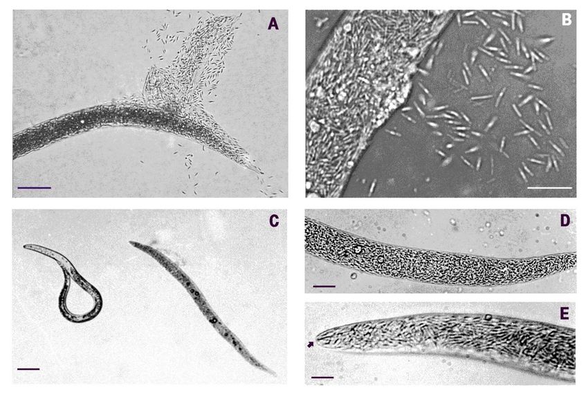
Figure
Figure1. LM images ofofthe nematode-parasitic bacterium (BN) in juveniles of (A) Meloidogyne javanica,
(A) Meloidogyne javanica, population:BN- BN-
Figure 1.1.LMLMimages
imagesof thenematode-parasitic
the nematode-parasiticbacterium
bacterium(BN)
(BN) inin juveniles
juveniles ofof (A) Meloidogyne javanica, population:
population: BN-
RAG;
RAG; and
and (B)(B)M.M.incognita,
incognita, population:
population:BN-MOL.
BN-MOL.Parasitism
Parasitism(C)(C)was
wasrevealed
revealedbyby
bya aacreamy-brown
creamy-brown color
colorof diseased hosts (a
RAG; and (B) M. incognita, population: BN-MOL. Parasitism (C) was revealed creamy-brown color of
of diseased
diseasedhosts
hosts
parasitized
(a M.
parasitized javanica
M. J2
javanica on
J2 the
on right,
the a
right,healthy
a nematode
healthy nematode ononthe
theleft;
left;population:
population: BN-LEV).
BN-LEV). (D,E)
(a parasitized M. javanica J2 on the right, a healthy nematode on the left; population: BN-LEV). (D, E) The nematodeswere
(D, E) The nematodes
nematodes were
were
completely
completely filled
filled with
with bacterial
bacterial cells
cells (population:
(population: BN-LEV).
BN-LEV). (E) At the end of the infection,
infection, only
only the
the
completely filled with bacterial cells (population: BN-LEV). (E) At the end of the infection, only the stylet and few other stylet
stylet and
and few
few other
other
cuticular
cuticular structures
structures
structures remained
remained
remained visible
visible
visible under
under
under LMLM
LM (arrow).
(arrow).
(arrow). Scale
Scale bars:
bars:
Scale (A):
bars: (A): 50
5050
(A): µm;μm; (B):
(B):(B):
μm; 12 μm;
12 µm;
12 μm;
(C):(C): 40
40 μm;
40 µm;
(C): (D):(D):
μm; 14
14 μm;
14 µm;
(D): (E): (E):
μm; 6(E):
µm. 66
μm.
μm.
Figure2.2.Transmission
Figure Transmissionelectron
electronmicroscopy
microscopy(TEM)(TEM)images
imagesof ofmature
matureBN BN cells
cells (population: BN-LEV) filling
(population: BN-LEV) filling the
thewhole
wholebody
body
Figure 2. Transmission
ofofM.M.javanica
javanica J2J2(A).
(A).Theelectron
The cells microscopy
cellsappeared
appeared as (TEM) rods
asseptate
septate images
rods rich ofin
rich mature
in BN
reserve cells (population:
granules C). The BN-LEV)
(B,C).
(B, filling the whole
peculiar morphology
morphology ofthe
of body
the BN
BN
of M. javanica
resulted
resulted from
from J2 (A).
the
the The cells appeared
persistence
persistence as septa
ofofdivision
division septate rods rich
septabetween
between in reserve
cells
cells granules
surrounded
surrounded by an(B,external
C). The peculiar
coat andmorphology
coat and the
the flattening ofof
flattening the
of BN
both
both
resulted
apicalcells
apical from
cells(C).the
(C). persistence
Scale
Scale (A)of
bars:(A)
bars: ==3division
3µm; (B)septa
μm;(B) between
== 11 µm;
μm; cells
(C) == 300
(C) 300 nm.
nm.surrounded by an external coat and the flattening of both
apical cells (C). Scale bars: (A) = 3 μm; (B) = 1 μm; (C) = 300 nm.
TEM examinations of BN cells from the diseased specimens showed the bacterium as
TEM examinations
a segmented of BN cells
rod, confirming fromLM
previous the observations
diseased specimens
(Figureshowed the bacterium
2B, C). Cell as
walls in the
a segmented rod, confirming previous LM observations (Figure 2B, C). Cell walls in themature cells remained joined after cell division. They were not observed during the first
vegetative stages, in which the BN cells appeared as elongated, electron dark rods (Figure
Pathogens 2021, 10, 1226 4 of 10
2A). The mature segmented rods measured 4–5 μm in length and 0.5–0.6 μm in width
(Figures 3 and 4).
Figure
Pathogens 3.TEM
2021,3.10,
Figure TEM
x images
FOR imagesofofthe
PEER REVIEWthenematode
nematodeinfection
infectionprocess
processshowing
showing germination
germination of
of BN
BN cells
cells adhering to the
adhering to the host
host cuticle
cuticle5 of 10
(population: BN-LEV).
(population: BN-LEV).After
Afteradhesion
adhesiontotothe
thehost,
host,aalateral
lateralgrowing
growingcell
cellpenetrated
penetrated the
the nematode body through
through its
its cuticle
cuticle
and
and hypodermis
hypodermis layers(A,B).
layers (A, B). Scale
Scale bars:
bars: (A)= =650
(A) 650nm;
nm;(B)
(B)==425
425nm.
nm.
TEM and SEM observations showed that BN cells adhered to the host nematode cu-
ticle (Figures 3 and 4B). Serial sections showed that, after adhesion, the parasite germi-
nated and eventually penetrated the host cuticle and hypodermis layers through a lateral
branch or budding, reaching the internal host tissues (Figure 3A, B). TEM images showed
that these stages were associated with the multiplication phase, spreading the infection
within the host and filling its whole body with septate cells at different maturing stages
(Figure 2A). TEM examinations confirmed total body digestion as observed with LM, with
the exclusion of the more external cuticular layers (Figures 1E and 2A–C). The induction
of germination was not related to any host metabolic activity or motility since it was also
observed in the parasitized or already killed nematodes (Figure 3).
TEM sections showed a complex cell wall, typical of Gram-negative bacteria. The in-
ner BN cytoplasmic membrane was separated by a periplasmic space from an 8–10 nm
wide electron-dense peptidoglycan layer (Figures 3B and 4D). After this layer, a 30–40 nm
thick and layered outer membrane was present, covered by a further electron dense coat,
12–20 nm thick. An additional external layer of O-side chains forming a matrix of adhesive
fibers 50–62 nm thick followed the coat. O-side chains show a thin electron dark peripheric
contour, 12–15 nm thick (Figure 4D).
The coat and fiber layers enveloped the whole rod, which at maturity was mostly
composed of four attached cells. These layers were also visible in immature, unsegmented
rods, but were not found in the wall of the germinating pegs, which appeared simpler and
thinner (Figure 3A, B). At the round, blunt apex of the mature terminal cells, either the
outer membrane or the coat gradually tapered to form a progressively thinner wall, 18 nm
thick (Figure 4D). The outer surface of the rods attached to the nematode cuticle frequently
showed adherent electron dark aggregates of clay-like microparticles or minerals, sug-
gesting the presence of superficial electric charges (Figure 3A, B).
Figure 4. Cells of BN from the collapsed M. incognita J2 (population: BN-LEV), showing the typical morphology of the bacterium
Figure 4. Cells of BN from the collapsed M. incognita J2 (population: BN-LEV), showing the typical morphology of the
(A). A SEM (A).
bacterium image Aof the cells
SEM image released
of the(A) or adhering
cells released (B)
(A)(arrowhead)
or adheringto(B)
the(arrowhead)
host nematodetocuticle (c). After
the host the release,
nematode the(c).
cuticle BNAfter
cells
appeared frequently packed in rows (C). A TEM image showing the bacterium’s apical tapering, giving a
the release, the BN cells appeared frequently packed in rows (C). A TEM image showing the bacterium’s apical tapering,hairpin-like appearance
(D). Cell
giving a wall section shows
hairpin-like appearancethe outer
(D).membrane, the coat,
Cell wall section and the
shows an external thin layer of
outer membrane, theadhesive
coat, and fibers. Scale bar
an external (A):
thin 2 µm;
layer of
(B): 4.6 µm;
adhesive (C): 2.7
fibers. (D):(A):
µm;bar
Scale 3752nm.
μm; (B): 4.6 μm; (C): 2.7 μm; (D): 375 nm.
BN rods were characterized by a unique shape and morphology resulting from the
symmetric emptying and flattening of both terminal cells or compartments (Figures 2B,C
and 3A,B). Empty apical cells were mainly found in mature rods at the end of the parasitic
stage or in the nematodes already dead and filled with rods (Figures 2C and 4D). The
terminal cells appeared collapsed and flattened due to the loss of their cytoplasmic con-
tent. Their opposite walls often approached in direct contact, originating a typical hairpin-Pathogens 2021, 10, 1226 5 of 10
TEM and SEM observations showed that BN cells adhered to the host nematode cuticle
(Figures 3 and 4B). Serial sections showed that, after adhesion, the parasite germinated and
eventually penetrated the host cuticle and hypodermis layers through a lateral branch or
budding, reaching the internal host tissues (Figure 3A,B). TEM images showed that these
stages were associated with the multiplication phase, spreading the infection within the
host and filling its whole body with septate cells at different maturing stages (Figure 2A).
TEM examinations confirmed total body digestion as observed with LM, with the exclusion
of the more external cuticular layers (Figures 1E and 2A–C). The induction of germination
was not related to any host metabolic activity or motility since it was also observed in the
parasitized or already killed nematodes (Figure 3).
TEM sections showed a complex cell wall, typical of Gram-negative bacteria. The
inner BN cytoplasmic membrane was separated by a periplasmic space from an 8–10 nm
wide electron-dense peptidoglycan layer (Figures 3B and 4D). After this layer, a 30–40 nm
thick and layered outer membrane was present, covered by a further electron dense coat,
12–20 nm thick. An additional external layer of O-side chains forming a matrix of adhesive
fibers 50–62 nm thick followed the coat. O-side chains show a thin electron dark peripheric
contour, 12–15 nm thick (Figure 4D).
The coat and fiber layers enveloped the whole rod, which at maturity was mostly
composed of four attached cells. These layers were also visible in immature, unsegmented
rods, but were not found in the wall of the germinating pegs, which appeared simpler
and thinner (Figure 3A,B). At the round, blunt apex of the mature terminal cells, either
the outer membrane or the coat gradually tapered to form a progressively thinner wall,
18 nm thick (Figure 4D). The outer surface of the rods attached to the nematode cuticle
frequently showed adherent electron dark aggregates of clay-like microparticles or minerals,
suggesting the presence of superficial electric charges (Figure 3A,B).
BN rods were characterized by a unique shape and morphology resulting from
the symmetric emptying and flattening of both terminal cells or compartments
(Figures2B,C and 3A,B). Empty apical cells were mainly found in mature rods at the end of
the parasitic stage or in the nematodes already dead and filled with rods
(Figures 2C and 4D). The terminal cells appeared collapsed and flattened due to the
loss of their cytoplasmic content. Their opposite walls often approached in direct con-
tact, originating a typical hairpin-like contour visible in longitudinal sections and also
observed with SEM (Figure 4A–D). This morphology was also visible with LM at highest
magnifications when BN smears from squashed nematodes from any population were
examined in temporary water mounts (Figure 1B). This peculiar shape of the bacterium
was consistently observed among the different nematode and BN populations examined
and appeared useful in the identification and visual diagnosis of the disease. Under LM,
the rods presented with a refractile central region corresponding to the inner cell pairs filled
with the cytoplasm, with an opaque peripheric portion corresponding to the terminal flat
ends (Figure 1B). The combination of cell shape, symmetry, and apical flattening gave rise
to a particular and distinctive morphology of the rods characterized by a typical bilateral
flute-like appearance (Figures 2B and 4A–C).
BN cells multiplied through a centripetal formation of a transverse septum (data not
shown). Lateral budding was observed in immature forms or at germination during host
infection (Figures 2A and 3B). At cell wall formation, the external coat layer of the mother
cell was kept, filling the inner space between the newly formed opposite walls, separating
the walls of two adjacent cells and acting as a cement between them (Figures 2C and 3B).
At septum completion, the cells remained embedded and covered by the coat and adhesive
matrix (Figure 3B).
Several electron transparent granules similar to poly-β-hydroxybutyrate inclusions
were frequently observed in the BN cytoplasm, as well as in immature rods
(Figures2B,C, 3A,B and 4D). Further cell organelles included electron dark vesicles of
variable sizes, mainly found scattered in the inner cell pairs, nucleoids, and ribosomes
(Figure 3B).Pathogens 2021, 10, 1226 6 of 10
TEM and SEM examinations did not show any flagella or flagella-like filaments at
any stage of BN multiplication. Furthermore, no direct or gliding motility was recorded
when examining clusters of BN rods dispersed on water agar or CMA with LM at weekly
intervals. The bacterium appeared fastidious in nature, and any attempt to obtain pure
cultures or induce its multiplication on the nutritive media tested failed.
Prevalence data from the M. javanica population sampled at Pedalino (Ragusa) showed
that the parasite was almost undetectable when J2 numbers were low, increasing its
frequency in a density-dependent manner as the nematode population increased.
3. Discussion
The large amount of metagenome data accumulated thus far has shown that a large
portion of the bacterial diversity present in the environment, including agricultural soils,
remains still unknown or not classified [29]. This consideration is supported by studies from
a wide range of microenvironments, suggesting that obligate and fastidious metabolism
characterizes a significant fraction of the total bacterial diversity occurring in nature [30].
The preliminary data shown herein, although limited to the BN morphology and biology,
suggest that the bacterium represents a novel Gram-negative species specialized in obligate
nematode parasitism. No record about similar microorganisms or pathologies was found
in the literature, whereas no correspondence could be directly assigned between the BN
and other bacteria known as associated with nematodes.
Filamentous bacteria have been reported in different nematodes, including marine
species [31] or from cyathostomins (Nematoda: Strongylidae) parasitic in African equids [32].
The species inhabiting the cuticle of the marine nematode Eubostrichus dianae showed a
complex community of distinct bacterial taxa, as shown by 16S rDNA sequencing [31].
The general morphology of these species, however, was distinct from that of BN. The
bacteria reported in association with cyathostomid nematodes included septate and very
long (>500 µm) Arthromitus-like species found attached to the nematodes’ natural openings.
They also appeared morphologically distinct from the BN, apart from some similarities in
the cell wall structure [32]. The cyathostomid microbial community also included other
smaller, blunt-ended organisms. The data available on their biology and ultrastructure
were, however, not sufficient to establish a relationship with the BN, if any [33]. Finally,
the BN differed from non-pathogenic, segmented filamentous bacteria with blunt ends
attached externally to the cuticle in the anal or vulvar regions or the reproductive tract of
cyathostomins from the hindgut of zebras. The diverse community of bacteria reported
presented with different and concomitant taxa, some of which were characterized by
a Gram-positive reaction, greater dimensions than that of the BN, production of septa,
and/or presence of Clostridium-like endospores [34].
The observations reported suggest that the BN is a highly specialized and obligate
rhizosphere bacterium dependent for growth on the host metabolism. The in vitro repro-
duction of the pathology and the validation of Koch’s postulates, however, were limited
by the absence of available BN-culturing media and by the low numbers of cells that it
was possible to handle from crushed hosts. The association of the bacterial parasite with
the disease was assessed anyway. Together with the consistent isolation of BN cells from
the J2 cadavers with the creamy color typical of the disease, the host-parasite association
provided support to consider the first and second of Koch’s postulates to be at least satisfied.
Adhesion of BN cells (proceeding from disrupted nematodes) to RKN J2 exposed in vitro
was also assessed (data not shown).
The disease may have practical implications as the BN appeared to be a specialized
antagonist, endemic in Meloidogyne-infested soils, as indicated by the density-dependent
host relationship. Because of J2 parasitism, the bacterium holds a potential in RKN biocon-
trol, as it kills the nematodes before root penetration and any tissue damage occur. It may
hence be added to the other microbial components of the soil microflora that contribute to
natural regulation of RKN, with a possible reference to soil suppressiveness [5,6,35].Pathogens 2021, 10, 1226 7 of 10
The systematic position of the BN remains unclear, pending culturing and 16S rRNA
ribosomal gene sequencing. Attempts to produce unambiguous PCR products for the 16S
gene consistent among different populations and experiments failed (data not shown).
Cell wall organization appeared typical of Gram-negative species [36] and more complex
than that of other nematode endosymbionts, i.e., Verrucomicrobia [27]. Some similarities
with Rhizobium suggested a first putative assignment to proteobacteria (R. Favre, personal
communication). The thick external coat covering the whole rod appeared, however, as a
specific characteristic involved in host adhesion and likely responsible for the host–parasite
specificity.
Some lifecycle similarities with Gram-positive parasites such as Pasteuria spp. arose
from a number of biological processes including propagule adhesion, host penetration,
outgrowth, and release from cadavers. This behavior, however, appears to be the outcome
of convergent evolution, mainly determined by the host nematode anatomy and structural
organization of the body. Differing from Pasteuria, no sporulating stage or endospore was
observed in the BN at any stage.
Although of limited value in bacterial taxonomy, the peculiar morphology exhibited
by the BN is a phenotypic trait likely reflecting functional adaptation to host parasitism.
Flat and empty apical cells did not appear as artefacts as they were consistently observed in
several replicates at high resolution with LM, TEM, or SEM. The blunt rod apex pairs likely
have a role in keeping firm cell adhesion to the J2 cuticle and, together with the matrix of
the coating capsule, appeared sufficiently strong to hold the central infective cells in place
during their subsequent activation and germination.
4. Materials and Methods
4.1. Sampling of the Host Populations
Cells of the BN were initially observed in Apulia in association with a population
of M. javanica parasitizing carnations in a greenhouse at Leverano (bacterial population
identified as BN-LEV). Further BN populations were observed in Apulia in association to J2
from an M. incognita population parasitizing tomatoes at Molfetta (population: BN-MOL),
an M. javanica population attacking tomatoes at Canosa and, in Sicily, from a further M.
incognita population parasitizing almonds at Pedalino (Ragusa, population: BN-RAG).
Nematode and BN populations were maintained in the greenhouse on cherry tomatoes at
25–28 ◦ C, in distinct pots with the original soils.
4.2. Light Microscopy (LM)
Nematodes were extracted from soil with Cobb’s sieving and decanting technique
using a set of 720 µm and 45 µm sieves [37]. The diseased specimens were identified
during J2 counts in a 1 mL Hawksley chamber under a Leitz Orthoplan light microscope
at 40×. Non-motile J2 showing the color changes associated to the BN disease were
gently recovered from the chamber using an eyelash glued to a needle tip. The specimens
collected this way were then stored in tap water in a watch glass and/or directly placed
on a glass slide in a temporary water mount for inspection at 400–1000×. Digital images
were captured using a Hamamatsu C 5810 CCD camera mounted on the microscope and
subsequently digitally processed. Images of clustered BN cells on CMA were captured
with the CCD and compared at one-week intervals to check mobility and division. Plates
with other media were also periodically examined for BN proliferation. Gram staining was
performed on the cells released from the nematodes ruptured on a glass cover slide, fixed
by fast flame exposure and stained with safranin and crystal violet (SIGMA HT90-A) [38].
4.3. Transmission Electron Microscopy (TEM)
The diseased M. javanica J2 filled with BN-LEV cells were extracted from soil as
previously described and selected for TEM preparation. The nematodes were handpicked
from the suspension obtained by sieving and examined in temporary water mounts with
LM at 250–400× to check occurrence of the disease. The diseased specimens were thenPathogens 2021, 10, 1226 8 of 10
embedded in 2% agarose, fixed in the glutaraldehyde–cacodylate buffer for 3 h or overnight,
post-fixed in 1% OsO4 in the cacodylate buffer, dehydrated in an ethanol series with 0.5%
uranyl acetate at 70% ethanol, and then washed three times in 100% ethanol and propylene
oxide before embedding the blocks in the Polybed resin. Serial sections 60–80 nm thick
were cut with a Reichert microtome, stained with uranyl acetate and lead citrate solutions,
and examined with a Philips EM 208 electron microscope at 100 kV.
4.4. Scanning Electron Microscopy (SEM)
The morphology of BN cells and their morphometrics were checked with SEM, exam-
ining cell clusters released from the ruptured nematodes. The diseased J2 of the M. javanica
population from Leverano were handpicked from the sieving suspension and identified as
described above. The nematodes were dehydrated with glycerol with the slow method [37]
and maintained in glycerol until used. For scanning, the specimens were handpicked and
mounted on a metal stub cleared of excess glycerol, cut or crushed directly on the stub
to release the BN cells, and sputter-coated with gold in vacuum. The samples were then
examined with a Stereoscan 360 Cambridge Instruments SEM at 3 kV [39].
4.5. Culturing Assays
The infected nematodes identified with LM were collected from different populations.
BN cells collected from the crushed nematodes with a sterile pipette or whole infected J2
handled with an ethanol-sterilized and air-dried eyelash were used in attempts to produce
BN cultures. The solid media tested were as follows: 1.5% water agar (WA), corn meal
agar (CMA); Grace’s insect medium (Difco, Detroit, MI, USA) with agar (15 g/L) with
or without hemin (5 mg/L); yeast extract (1 g/L) with agar (15 g/L), peptone (5 g/L),
and NaCl (5 g/L), with or without lactic acid (30 mL/L). Some BN-infected nematodes
were also ruptured and smeared on Petri dishes with the tested media after gentle rinsing
in sterile distilled water. Petri dishes were stored at 25 ◦ C in the dark and examined
periodically for growth during the following weeks [40].
Funding: This research was partially funded by Ministero Politiche Agricole e Forestali, projects
“Orticoltura” and “BIOMED”, and CNR Project “Agenzia 2000”.
Institutional Review Board Statement: Not applicable.
Informed Consent Statement: Not applicable.
Data Availability Statement: Not applicable.
Acknowledgments: This paper is dedicated to the memory of Renée Favre† (IIGB, CNR, Naples).
Michele Cermola (CREA, Caserta)and Gaetano Grasso (Tecnopolis, Valenzano, Bari) are gratefully
acknowledged for production of TEM and SEM images, respectively; Marino Binetti (Molfetta) is
gratefully acknowledged for assistance in field soil samplings.
Conflicts of Interest: The author declares no conflict of interest. The funder had no role in the design
of the study; in the collection, analyses, or interpretation of data; in the writing of the manuscript, or
in the decision to publish the results.
References
1. Poinar, G.O., Jr.; Hansen, E.L. Associations between nematodes and bacteria. Helminth. Abstr. Ser. B 1986, 55, 61–81.
2. Bandi, C.; Anderson, T.J.; Genchi, C.; Blaxter, M.L. Phylogeny of Wolbachia in filarial nematodes. Proc. R. Soc. Lond. B Biol. Sci.
1998, 265, 2407–2413. [CrossRef]
3. Brown, A.; Wasala, S.; Howe, D.; Peetz, A.B.; Zasada, I.A.; Denver, D.R. Genomic evidence for plant-parasitic nematodes as the
earliest Wolbachia hosts. Sci. Rep. 2016, 6, 34955. [CrossRef] [PubMed]
4. Palomares-Rius, J.E.; Archidona-Yuste, A.; Cantalapiedra-Navarrete, C.; Prieto, P.; Castillo, P. Molecular diversity of bacterial
endosymbionts associated with dagger nematodes of the genus Xiphinema (Nematoda: Longidoridae) reveals a high degree of
phylogenetic congruence with their host. Mol. Ecol. 2016, 25, 6225–6247. [CrossRef]
5. Siddiqi, Z.A.; Mahmood, I. Role of bacteria in the management of plant parasitic nematodes: A review. Biores. Technol. 1999, 69,
167–179. [CrossRef]Pathogens 2021, 10, 1226 9 of 10
6. Stirling, G. Biological Control of Plant-Parasitic Nematodes: Soil Ecosystem Management in Sustainable Agriculture; CABI: Wallingford,
UK, 2014.
7. Bird, A.F.; Riddle, D.L. Effect of attachment of Corynebacterium rathayi on movement of Anguina agrostis larvae. Int. J. Parasitol.
1984, 14, 503–511. [CrossRef]
8. Taylor, C.E. Nematode interactions with other pathogens. Ann. Appl. Biol. 1990, 116, 405–416. [CrossRef]
9. Esnard, J.; Potter, T.L.; Zuckerman, B.M. Streptomyces costaricanus sp. nov., isolated from nematode-suppressive soil.
Int. J. Syst. Bacteriol. 1995, 45, 775–779. [CrossRef]
10. Hackenberg, C.; Muehlchen, A.; Forge, T.; Vrain, T. Pseudomonas chlororaphis strain Sm3, bacterial antagonist of Pratylenchus
penetrans. J. Nematol. 2000, 32, 183–189.
11. Kluepfel, D.A.; Nyczepir, A.P.; Lawrence, J.E.; Wechter, W.P.; Leverentz, B. Biological control of the phytoparasitic nematode
Mesocriconema xenoplax on peach trees. J. Nematol. 2002, 34, 120–123.
12. Meyer, S.L.F.; Roberts, D.P. Combinations of biocontrol agents for management of plant-parasitic nematodes and soilborne
plant-pathogenic fungi. J. Nematol. 2002, 34, 1–8.
13. Meyer, S.L.F.; Roberts, D.P.; Chitwood, D.J.; Carta, L.K.; Lumsden, R.D.; Mao, W. Application of Burkholderia cepacia and
Trichoderma virens, alone and in combinations, against Meloidogyne incognita on bell pepper. Nematropica 2001, 31, 75–86.
14. Tian, H.; Riggs, R.D.; Crippen, D.L. Control of soybean cyst nematode by chitinolytic bacteria with chitin substrate. J. Nematol.
2000, 32, 370–376. [PubMed]
15. Huang, X.W.; Niu, Q.H.; Zhou, W.; Zhang, K.Q. Bacillus nematocida sp. nov., a novel bacterial strain with nematotoxic activity
isolated from soil in Yunnan, China. Syst. Appl. Microbiol. 2005, 28, 323–327. [CrossRef]
16. Anderson, W.R.; Madden, P.A.; Tromba, F.G. Histopathologic and bacteriologic examinations of cuticular lesions of Ascaris suum.
J. Parasitol. 1971, 57, 1010–1014. [CrossRef]
17. Anderson, W.R.; Madden, P.A.; Colglazier, M.L. Microbial flora of cuticular lesions on Strongylus edentatus. Proc. Helm. Soc. Wash.
1978, 45, 219–225.
18. Tan, L.; Grewal, P.S. Pathogenicity of Moraxella osloensis, a bacterium associated with the nematode Phasmarhabditis hermaphrodita,
to the slug Deroceras reticulatum. Appl. Environ. Microbiol. 2001, 67, 5010–5016. [CrossRef] [PubMed]
19. Forst, S.; Nealson, K. Molecular biology of the symbiotic-pathogenic bacteria Xenorhabdus spp. and Photorhabdus spp.
Microbiol. Rev. 1996, 60, 21–43. [CrossRef] [PubMed]
20. Sheperd, A.M.; Clark, S.A.; Kempton, A. An intracellular micro-organism associated with tissues of Heterodera spp. Nematologica
1973, 19, 31–34.
21. Endo, B.Y. The ultrastructure and distribution of an intracellular bacterium-like microorganism in tissue of larvae of the soybean
cyst nematode, Heterodera glycines. J. Ultrastr. Res. 1979, 67, 1–14. [CrossRef]
22. Walsh, J.A.; Lee, D.L.; Sheperd, A.M. The distribution and effect of intracellular rickettsia-like micro-organisms infecting adult
males of the potato cyst-nematode Globodera rostochiensis. Nematologica 1983, 29, 227–239.
23. Ferri, E.; Bain, O.; Barbuto, M.; Martin, C.; Lo, N.; Uni, S.; Landmann, F.; Baccei, S.G.; Guerrero, R.; Lima, S.D.S.; et al. New
insights into the evolution of Wolbachia infections in filarial nematodes inferred from a large range of screened species. PLoS ONE
2011, 6, e20843. [CrossRef] [PubMed]
24. Wasala, S.K.; Brown, A.M.V.; Kang, J.; Howe, D.K.; Peetz, A.B.; Zasada, I.A.; Denver, D.R. Variable abundance and distribution of
Wolbachia and Cardinium endosymbionts in plant-parasitic nematode field populations. Front. Microbiol. 2019, 10, 964. [CrossRef]
[PubMed]
25. Sironi, M.; Bandi, C.; Sacchi, L.; Di Sacco, B.; Damiani, G.; Genchi, C. Molecular evidence for a close relative of the arthropod
endosymbiont Wolbachia in a filarial worm. Mol. Biochem. Parasitol. 1995, 74, 223–227. [CrossRef]
26. Noel, G.R.; Atibalentja, N. “Candidatus Paenicardinium endonii” an endosymbiont of the plant-parasitic nematode Heterodera
glycines (Nemata: Tylenchida), affiliated to the phylum Bacteroidetes. Int. J. Syst. Evol. Microbiol. 2006, 56, 1697–1702. [CrossRef]
27. Vandekerckhove, T.T.M.; Willems, A.; Gillis, M.; Coomans, A. Occurrence of novel verrucomicrobial species, endosymbiotic and
associated with parthenogenesis in Xiphinema americanum-group species (Nematoda, Longidoridae). Int. J. Syst. Evol. Microbiol.
2000, 50, 2197–2205. [CrossRef]
28. Brown, A.M.V.; Howe, D.K.; Wasala, S.K.; Peetz, A.B.; Zasada, I.A.; Denver, D.R. Comparative genomics of a plant-parasitic
nematode endosymbiont suggest a role in nutritional symbiosis. Genome Biol. Evol. 2015, 7, 2727–2746. [CrossRef] [PubMed]
29. Kellenberger, E. Exploring the unknown: The silent revolution of microbiology. EMBO Rep. 2001, 2, 5–7. [CrossRef] [PubMed]
30. Overmann, J.; Abt, B.; Sikorski, J. Present and future of culturing bacteria. Ann. Rev. Microbiol. 2017, 71, 711–730. [CrossRef]
[PubMed]
31. Polz, M.F.; Harbison, C.; Cavanaugh, C.M. Diversity and heterogeneity of epibiotic bacterial communities on the marine nematode
Eubostrichus dianae. Appl. Environ. Microbiol. 1999, 65, 4271–4275. [CrossRef]
32. Els, H.J.; Krecek, R.C. Developmental stages of a smooth-walled filamentous bacterium associated with equine Cyathostomes. J.
Helm. Soc. Wash. 1993, 60, 174–182.
33. Krecek, R.C.; Sayre, R.M.; Els, H.J.; Van Niekerk, J.P.; Malan, F.S. Fine structure of a bacterial community associated with
chyatostomes (Nematoda: Strongylidae) of zebras. Proc. Helm. Soc. Wash. 1987, 54, 212–219.Pathogens 2021, 10, 1226 10 of 10
34. Mackie, R.I.; Krecek, R.C.; Els, H.J.; Van Niekerk, J.P.; Kirschner, L.M.; Baecker, A.A.W. Characterization of the microbial
community colonizing the anal vulvar pores of helminths from the hindgut of zebras. Appl. Environ. Microbiol. 1989, 55,
1178–1186. [CrossRef] [PubMed]
35. Yin, B.; Valinsky, L.; Gao, X.; Becker, O.; Borneman, J. Bacterial rRNA genes associated with soil suppressiveness against the
plant-parasitic nematode Heterodera schactii. Appl. Environ. Microbiol. 2003, 69, 1573–1580. [CrossRef]
36. Beveridge, T.J. Structures of Gram-negative cell walls and their derived membrane vesicles. J. Bacteriol. 1999, 181, 4725–4733.
[CrossRef] [PubMed]
37. Southey, J.F. (Ed.) Laboratory Methods for Work with Plant and Soil Nematodes; Ministry of Agriculture, Fisheries and Food, Technical
Bulletin No. 2; HMSO: London, UK, 1970.
38. Girard, H.; Rougieux, R. Techniques de Microbiologie Agricole; DUNOD: Paris, France, 1967.
39. Sher, S.A.; Bell, A.H. Scanning electron micrographs of the anterior region of some species of Tylenchoidea (Tylenchida: Nematoda).
J. Nematol. 1975, 7, 69–83.
40. Leadbetter, J.R. Cultivation of recalcitrant microbes: Cells are alive, well and revealing their secrets in the 21st century laboratory.
Curr. Opin. Microbiol. 2003, 6, 274–281. [CrossRef]You can also read


