New Photobiomodulation Device for Prevention and Cure of Radiotherapy-induced Oral Mucositis and Dermatitis: Results of the Prospective Safe PBM ...
←
→
Page content transcription
If your browser does not render page correctly, please read the page content below
New Photobiomodulation Device for Prevention and
Cure of Radiotherapy-induced Oral Mucositis and
Dermatitis: Results of the Prospective Safe PBM
Study
Rene-Jean Bensadoun ( rjbensad@gmail.com )
Centre de Haute Energie (CHE) https://orcid.org/0000-0003-1290-7497
Marc A Bollet
Clinique Hartmann
Xavier Liem
Centre Oscar Lambret
Kim Cao
Institut Curie
Nicolas Magné
Institut de Cancérologie de la Loire: Institut de Cancerologie de la Loire
Research Article
Keywords: Photobiomodulation, Breast cancer, Head and neck cancer, Oral mucositis, Radiation dermatitis
Posted Date: July 23rd, 2021
DOI: https://doi.org/10.21203/rs.3.rs-611879/v1
License: This work is licensed under a Creative Commons Attribution 4.0 International License.
Read Full License
Page 1/15Abstract
Purpose
To assess the feasibility, safety and tolerability of CareMin650, a new photobiomodulation device, in
patients treated by radiotherapy (RT). To collect preliminary data on efficacy for prevention and treatment
of oral mucositis (OM) and radiation dermatitis (RD).
Methods
French, multicentric, prospective, non-comparative study. Inclusion of patients with head and neck cancer
(H&NC, cohort A) or breast cancer (BC, cohort B) treated in prophylactic (cohorts A1 and B1) or curative
setting (cohort A2 and B2). Prophylactic treatment was administered from D1 to end of RT, at a dose of 3
J/cm2. Curative treatment started when a grade 1 to grade 3 lesion had occurred and was pursued until
end of RT. Primary endpoint was incidence of device-related adverse events (AEs). OM and RD lesions were
graded according to CTCAE V3.
Results
Overall, 72 patients were included (22, 9, 23 and 18 in cohorts A1, A2, B1 and B2 respectively). No device-
related AE was reported after 1,312 CareMin650 sessions. In cohorts A1 and B1, median time to first OM or
RD lesion was 20 days. One BC patient developed G3 RD after completion of RT and discontinuation of
CareMin650. Four H&NC patients developed G3 OM. In cohorts A2 and B2, lesions improved or stabilized
in 71% of patients. Rates of satisfaction were high among patients and users.
Conclusion
CareMin650 is feasible, safe and well tolerated for preventive or curative treatment of OM and RD in
cancer patients treated with RT. Preliminary efficacy results are promising.
Introduction
Oral mucositis (OM) and radiation dermatitis (RD) are among the most frequent and disabling side-effects
of radiotherapy (RT). OM affects nearly all patients treated with RT ± chemotherapy (CT) for head and
neck cancer (H&NC)1, resulting in dysphagia and pain, weight loss and necessity of enteral alimentation in
some cases, as well as increased risk of infections with potentially life-threatening sepsis2–5. OM may
jeopardize planned anticancer treatment, potentially leading to decreased efficacy3. OM-related
consequences significantly increase the cost of patients’ management4 and impair quality of life (QoL).
Very few options showed efficacy for prophylaxis and/or treatment of OM, despite a huge variety of
treatments experimented. However, low-level laser therapy (LLLT), now referred to as photobiomodulation
(PBM), demonstrated significant benefits in several randomized clinical trials, in particular in patients with
H&NC6 − 11. According to recent guidelines, PBM is recommended to prevent OM in patients receiving high
Page 2/15dose CT as a conditioning regimen for stem cells transplantation (SCT) and in patients undergoing RT for
H&NC12,13.
RD affects approximately 95% of patients who receive RT14, ranging from mild erythema to dry or moist
desquamation and ulceration15. Nearly all women treated with RT for breast cancer (BC) experience some
degree of RD 16. High-quality data are still insufficient to support specific strategies in the management of
RD 15. However, there is increasing evidence on the benefits of PBM in this setting17.
PBM involves absorption of red and near infrared light by mitochondrial chromophores. The electron
transfer rate of the respiratory chain is increased, driving upward the production of adenosine
triphosphate, reactive oxygen species and nitric oxide, thus simulating genes involved in tissue repair.
Factors involved in inflammation and immunity are recruited to act at a tissue level18–20. Thus, PBM
impacts all the stages of wound repair and tissue regeneration. It also prevents fibrosis, reduces pain
(absorption of energy by nociceptors) and prevents tissue death19,21.
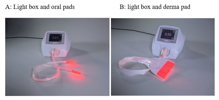
CareMin650 has been developed to improve practical use of PBM. Delivery of light, emitted by a flexible
surface (fabric made of woven optic fibres) in contact with the skin or mucosa, is accurately controlled,
reproducible and operator independent. The device consists of an electronic box generating the light, and
pads connected to the box through an optic fibre cable: either oral pads measuring 2,6*5,5 cm² emitting
light on both sides or derma pads measuring 15,6*5,5cm², emitting light on one side (Fig. 1). The dose in
J/cm² is selected on the light box that calculates automatically the length of the session to reach the
selected dose. Disposable single use sleeves are placed on the pads before applying them to the skin or
the mucosa. The main objectives of this study were to show feasibility, safety and tolerability of
CareMin650 and provide preliminary data on efficacy.
Methods
• Design and Patients
Safe PBM was a French multicentric prospective non comparative study, designed to assess CareMin650
in patients with H&NC (cohort A) or BC (cohort B), in prophylaxis (cohorts A1 and B1) or cure (cohorts A2
and B2) (Supplementary Fig. 1). Eligible patients were aged 18 years or above, had histologically proven
cancer (BC in cohorts B1 and B2, squamous cell carcinoma of oropharynx, nasopharynx, hypopharynx,
larynx or oral cavity in cohorts A1 or A2), and ECOG performance status ≤ 2. In preventive cohorts, patients
were scheduled to receive RT (on at least 50% of the oral mucosa, at a total dose of at least 40 Gy for
H&NC) and had no lesions at inclusion. In cohorts A2 and B2, patients had previously started RT and
presented with OM and/or RD lesions of grade 1 to 3. BC patients were required to have undergone tumor
resection (breast conservative surgery or mastectomy) while prior surgery was not mandatory in cohorts
A1 or A2. In all cohorts, concomitant treatment with CT and / or targeted therapies was permitted. Patients
were excluded if they had known allergy to polyurethane. In H&NC patients, specific exclusion criteria were
active bleeding or high risk of bleeding, Hb < 8g/dL, neutrophils < 1000 mm3, or platelets < 50 000/mm3. In
Page 3/15cohorts B1 and B2, patients were excluded if they had received prior irradiation to the same breast.
Consecutive patients were included in each subgroup, until the target number had been reached. All study
procedures were in accordance with the ethical standards and with the 1964 Helsinki Declaration. All
patients signed informed consent before any study procedure was implemented. The study was approved
by the Comité de Protection des Personnes (CPP) CPP Sud-Est VI and by the ANSM (Agence Nationale du
Médicament et des Produits de Santé).
• Treatment
CareMin650 started at inclusion in the study and was pursued until the end of RT. The minimal number of
sessions per week was 3, performed immediately before or after RT; however 5 sessions/week were
recommended. Any healthcare professional could administer the treatment after appropriate training. Oral
pads and derma pads delivered red light with wavelength of 650 nm, and irradiance between 10 and 50
mW/cm². Doses for prophylactic and curative treatments were 3 J/cm2 and 6 J/cm2 respectively. If OM or
RD occurred in a patient from a prophylactic cohort, the dose was increased to 6J/cm2. Conversely, if
lesions in patients from curative cohorts resolved before the end of RT, the dose was decreased to 3
J/cm2. In cohorts A1 and B1, pads were applied on irradiated areas presenting a risk of RT-related
complications. In cohorts A2 and B2, pads were applied on each lesion.
Standard OM and RD prophylaxis, including oral hygiene using soft toothbrush and bicarbonate
mouthwashes for OM, was implemented according to sites’ habits. In case of lesions, usual local care,
analgesics and corticosteroids were allowed and therapies considered necessary for the subject’s well-
being could be administered at the discretion of the investigator. Keratinocyte growth factors (palifermin)
and other PBM treatments were not allowed during the study.
• Assessments
Safety was assessed throughout the study, adverse events (AEs) were graded according to NCI CTCAE v4.
Examination of skin and oral mucosa was performed at each CareMin650 session to assess local
tolerance and detect any new lesion. In case of lesion, time of occurrence, size, location, grade according
to NCI CTCAE v3 and time to resolution (defined as lesion not requiring further treatment) were reported.
Once a week, data were collected on pain, using a Visual Analogic Scale (VAS) graded from 0 (no pain) to
100 (intolerable pain), analgesic consumption, xerostomia and in case of OM, consequences on food
intake. Quality of life was assessed using the SF-12 questionnaire, filled in at baseline and at the end of
RT. Patients’ and users’ satisfaction questionnaires were filled in at the end of RT. A follow-up visit was
performed 10 ± days after the end of treatment visit.
• Statistics
The safety and modified intention-to-treat (mITT) sets comprised all included subjects who had at least
one CareMin650 session and at least one safety or efficacy evaluation respectively. The per protocol (PP)
set comprised all subjects without major protocol violation, defined by i) less than 10 CareMin650
sessions in cohorts A1 or B1 or less than 5 sessions in cohorts A2 or B2, or ii) start of CareMin650 more
Page 4/15than 3 days after start of RT in cohorts A1 or B1. The primary endpoint of the study was the rate of device-
related AEs. Secondary endpoints included incidence and grade of lesions, pain, patients’ and users’
satisfaction.
The number of observations needed was estimated at 300 to allow detecting an undesirable effect
occurring at a frequency of 1% with a probability of 95%22. Assuming that patients would undergo at least
10 sessions on average, the total number of patients to analyze was set at 60. With an estimation of 20%
not eligible for analysis, the total number of subjects to include was 72. Analyses were only descriptive.
Quantitative variables were described by mean, standard deviation, median, minimum and maximum.
Qualitative variables were described by counts and percentages. No statistical test was performed.
Missing date were not replaced.
The study is registered with ClinicalTrials.gov, number NCT03988556
Results
Patients were recruited from July 2019 to November 2020. In total, 74 were screened, 72 were included and
analysed in the mITT set (22, 9, 23 and 18 patients in cohorts A1, A2, B1 and B2 respectively), while 58
were analysed in the PP population (17, 8, 19 and 14 patients respectively) (Fig. 2). Baseline
characteristics are summarized in Table 1. Median age was 61.4 years. Relevant comorbidities were more
frequent in cohorts A than in cohorts B, cardiovascular disorders and diabetes mellitus being the most
prevalent. H&NC were located mainly in oropharynx (41.9%) and oral cavity (32.3%) followed by
nasopharynx (9.7%), larynx (9.7%) and hypopharynx (6.5%), 71% of patients had surgery before starting RT
and 11 patients received concomitant cisplatin-based CT. In patients with BC, 70.7% of tumors were
located in the upper outer quadrant and 92% had stage I or II tumors. Median time from diagnosis to
inclusion was 3.5 months. All patients in cohorts A were treated with IMRT and/or VMAT while in cohorts
B, 51.2% were treated by RT-3D. The median number of CareMin650 sessions per week was 3.88 and the
percentage of weeks with at least 3 sessions was 87.5%, 100%, 83.3% and 100% in cohorts A1, A2, B1 and
B2 respectively.
Safety
In total, 1,312 sessions of CareMin650 were performed during the study, including 530, 156, 455 and 171
in cohorts A1, A2, B1 and B2 respectively. Nine patients reported 14 treatment-emergent adverse events,
none of which being related to the device.
OM and RD lesions, preventive setting
The PP population comprised 36 patients (17 and 19 in cohorts A1 and B1 respectively). The median
number of areas requiring application of pads was 3 in cohort A1 and 2 in cohort B1. All patients
developed some degree of OM and/or RD lesion. Median time from start of CareMin650 to first lesion was
20 days in both cohorts, ranging from 7 to 34 days in cohort A1, from 3 to 49 days in cohort B1
(Supplementary Figs. 2 and 3). Most lesions were grade 1 or 2 (Table 2). Only 1 patient developed G3 RD
Page 5/15in cohort B1 (left axilla), diagnosed after the end of RT at the follow-up visit, G3 upfront. Four patients had
G3 OM in cohort A1, after 7, 14, 19 and 25 days, mean 16.25 ± 7.63 days, median: 16.5 days. One lesion,
located on hard palate was diagnosed at grade 3 upfront. Other lesions were diagnosed at grade 1 (one
case, tongue and cheek) or at grade 2 (two cases, tongue and cheek in one patient, lips in another patient).
In the mITT population (n = 45), the maximal grade of lesions throughout the study was 0 in one patient
(cohort B1), 1 in 12 patients [3 (13.6%) in A1, 9 (39.2%) in B1], 2 in 25 patients [13 (59.1%) in A1, 12
(52.2%) in B1] and 3 in 7 patients [6 (27.3%) in A1 and 1 (4.3%) in B1]. The 2 additional patients with G3
OM had started CareMin650 on day 7 and day 5 of RT respectively.
OM and RD lesions, curative setting
At inclusion, the median number of lesions was 2 (1–3) in cohort A2 and 1 (1–4) in cohort B2. The median
time from start of RT to inclusion was 27 days (9–40) and 25 days (11–41) respectively. In the PP
population (n = 22), the maximal grade of lesions at inclusion in cohorts A2 and B2 was 1 in 62.5% and
78.6% of cases respectively, 2 in 25.0% and 21.4% of cases and 3 in 12.5% and 0% of cases. At the end of
radiotherapy visit, 15/21 patients (71.4%) had a maximal grade equal to or lower than that of inclusion
(Table 3). At the follow-up visit, lesions had disappeared in 3 patients from cohort B2 (one with G1 and 2
with G2 lesion at inclusion). In the mITT population (n = 27), results were similar with 69% of patients
showing stabilization or improvement at the end of RT and disappearance of all lesions in 4 cases at the
follow-up visit (cohort B2).
Other criteria
In 57 patients data on pain were available at inclusion and at least once during the study. Mean maximal
pain during the study was the highest in cohorts A2 (42.9 ± 26.3) and A1 (35.8 ± 28.1) while it remained
low in cohorts B1 (11.6 ± 15.5 and 11.1 ± 17.8 respectively). Data on pain are shown in Supplementary
Table 1.
Data on patient’s satisfaction were available for 64 patients. A majority of patients declared that the
application of the device was not burdensome (81.3%), provoked no discomfort (76.6%), and that the
duration of sessions was acceptable (68.8%). Overall, 87.5% reported no pain at all during applications,
while 5 (7.8%) reported slight but tolerable pain and only 3 (15.8%), all in cohort A1, found the application
quite painful. The patient preferred maintaining the device in contact with skin or mucosa himself during
the session in 89.1% of cases. Overall, most patients were very satisfied (60.3%) or satisfied (33.3%) with
the device. The application of CareMin650 was performed by physicians, nurses, residents, radiologic
technologists or clinical research assistants, depending on sites decision and organization. The
installation of the device was considered easy in all cases (very easy: 79.4%; rather easy: 20.6%). The
device was considered very handy, rather handy and rather unhandy in 69.1%, 22.1% and 8.8% of cases
respectively. Duration of sessions was assessed as rather short, acceptable and rather long in 4.4%, 73.5%
and 22.1% of cases respectively. Overall, users found the device rather satisfactory (71.6%) or very
satisfactory (23.9%) and 87.5% declared that they would like to use it in routine practice.
Page 6/15Discussion
Although PBM demonstrated efficacy and safety in a number of randomized clinical trials and meta-
analyses23–27, it is rarely used in routine practice. Indeed, treatment is time consuming, equipment is
cumbersome and unwieldy; lasers are set with various parameters (wavelength, irradiance, pulse structure,
coherence, polarization, energy, fluence) resulting in lack of standardisation. Finally, the procedure is not
fully reproducible and operator dependent as the distance from the skin or mucosa is difficult to assess
accurately. Thus, the amount of energy delivered cannot be exactly known. CareMin650 is a small and
handy device allowing reproducible and accurately controlled delivery of light thanks to direct application
of lightning tissue on skin and mucosa. This study aimed at evaluating its feasibility and safety in as
many situations as possible, leading to inclusion of 4 subgroups of patients, who underwent applications
of pads on mucosa and/or skin, on intact or damaged tissues. The study was performed at highly
experienced radiotherapy sites. Characteristics of radiotherapy reflect usual practice with IMRT/VMAT
being used in all H&NC patients while one half of patients with BC received RT-3D. Duration, total dose and
dose per fraction are consistent with current recommendations. The percentage of patients receiving
concomitant chemotherapy was quite low (33%) but not unusual.
The study protocol recommended at least 3 sessions per week, ideally 5. Compliance to treatment was
good with a median number of 3.88 sessions per week, which suggests that CareMin650 therapy is
feasible even in sites with high patient flow. Indeed, the device is easy to use as the operator only needs to
select a dose and the lightbox automatically calculates the duration of application required to deliver this
dose. Therefore, application can be performed by any healthcare professional who has been properly
trained. In this study, physicians, residents, nurses, radiologic technologists or CRAs were in charge of
device utilization.
PBM is known to have good local tolerance6,23. However, a possible concern with CareMin650 was that
direct contact of a pad on skin or mucosa could provoke pain or irritation, especially in case of pre-existing
lesion. Our results show that local tolerance was very good as no device-related adverse event relating to
local pain, irritation or unpleasant feelings has been reported during 1,312 sessions. In particular, all
patients from cohort A2 reported that application was not painful, and overall, only 3 patients (4.7%)
declared that the application was rather painful and provoked discomfort. Concerns on risks of PBM-
related proliferation of tumor cells have been raised and extensively debated due to conflicting in vitro
data. However, the use of PBM for more than 30 years, the increasing number of published data in OM
prevention in H&NC and SCT patients suggest that PBM does not influence tumor or treatment outcomes
and overall survival28. Data in H&NC patients treated with LLLT during RT without prior surgery and long-
term follow-up are very reassuring7. Specific evaluation of CareMin650 in an in vitro study showed that
irradiated cancer cells do not proliferate when illuminated29.
The study was not designed to demonstrate efficacy. However, preliminary findings can be observed. Only
one case of grade 3 RD occurred in the preventive cohorts, and it was diagnosed after the end of RT,
therefore did not occur during CareMin650 treatment.
Page 7/15In H&NC patients, grade 3 OM occurred in 4 cases (23.5%), which is lower than reported in the literature, as
incidence is usually around 50% and in any case, always exceeds 30% in the absence of efficacious
preventive treatement1,3. Importantly, these 4 cases occurred at the same investigational site, among 5
patients included. This discrepancy between sites could be explained by several hypotheses. First, a high
variability has been shown in the grading of OM lesions using CTCAEv3 criteria, with discordance rates of
34% between local investigators and central review30. In our study, lesions were graded locally, and no
training had been implemented at the beginning of the study to standardize grading. Thus, a very likely
explanation lies in the variable analysis of lesions across sites, although grading was to be performed by a
physician. The choice of CTCAEv3 for grading had been made because it was considered more accurate
than WHO grading scale or CTCAEv5. However, it is also more difficult to use and the distinction between
grade 2 and 3 appears very difficult and subject to investigator’s interpretation. Moreover, the choice of
grading scale can influence the results. In our study, among patients with food intake limitations, only 3
had G3 OM; therefore, using WHO grading scale, the number of patients with G3 OM lesions would
probably have been 3. Second, it has been observed that some lesions were reported late, although
symptoms suggesting OM had been described days or weeks earlier, leading to delay in dose increase.
This highlights the importance of early detection of lesions with dose increase to 6J/cm2 as soon as a G1
lesion appears. Finally, differences in patients’ population might partly explain a higher incidence of severe
lesions, for example different exposures to tobacco and alcohol in one site (North of France) compared to
others. Unfortunately, alcohol consumption and smoking status were not recorded in this study. In the ITT
population, 2 additional patients developed G3 OM lesions. Both had started CareMin650 several days
after the start of RT, suggesting that starting PBM on day 1 of RT is probably key for prevention of OM. No
conclusion can be drawn on the effects of CareMin650 in curative settings at this point, due to the small
sample size. However, it seems that lesions were stabilized or improved in most cases, although treatment
started at grade 2 or 3 in 27% of cases. Finally, safety and efficacy are not the only criteria for a therapy to
be implemented in clinical practice. It has to be acceptable for patients and healthcare professionals. In
this study, patients’ and users’ questionnaires showed the high rate of satisfaction towards the device.
This study has limitations. It was not a randomized controlled trial, so that no conclusion can be drawn on
efficacy. As already mentioned, no standardization training for grading had been performed which raises
questions on consistency across investigators. Some data are lacking to better interpret the results, such
as smoking habits or alcohol consumption. Finally, the size of each cohort was low, especially in curative
settings. Recruitment turned out to be easier in cohorts A1 and B1 that were almost completed when the
covid-19 pandemic started. At this time, inclusions were put on hold and ongoing treatments were
interrupted, leading to a high number of early study discontinuations. Inclusions resumed after 3 months,
in some but not all sites, at a very low rate and it was eventually decided to stop the study as the overall
target had been reached, despite the imbalance between cohorts.
Conclusion
OM and RD are frequent and disabling adverse effects of RT. Every effort should be made to reduce their
incidence and severity in order to improve patients’ quality of life and optimize supportive care. PBM is not
Page 8/15routinely used despite proven efficacy and clear recommendations, because of the lack of easy-to-use and
reliable equipment. The new CareMin650 device has shown very good safety and tolerance as well as
promising efficacy results, that will require confirmation in a larger prospective trial to allow wider
utilization in daily practice.
Declarations
Funding : No funding
Conflicts of interest/Competing interests: No Conflict of interest.
Availability of data and material: Yes.
Code availability: Yes.
Authors' contributions : All authors contribute equally to study desigh, protocol élaboration, patients
inclusion, data analyse, and manuscript writing and review.
Ethics approval : the study was approved by the « Comité de Protection des Personnes (CPP) CPP Sud-Est
VI » and by the ANSM (Agence Nationale du Médicament et des Produits de Santé).
Consent to participate : All patients approved to participate in the study, and to participate in paper’s
preparation, writing, and internal review.
Consent for publication: All authors approved paper submission to the « Journal of Supportive Care in
Cancer », and accepted the publication of this paper in this journal.
References
1. Lalla RV, Bowen J, Barasch A et al (2014) MASCC/ISOO clinical practice guidelines for the
management of mucositis secondary to cancer therapy. Cancer 120(10):1453–1461
2. Scully C, Sonis S, Diz PD (2006) Oral mucositis. Oral Dis 12(3):229–241
3. Trotti A, Bellm LA, Epstein JB et al (2003) Mucositis incidence, severity and associated outcomes in
patients with head and neck cancer receiving radiotherapy with or without chemotherapy: a
systematic literature review. Radiother Oncol 66(3):253–262
4. Elting LS, Cooksley CD, Chambers MS, Garden AS (2007) Risk, outcomes, and costs of radiation-
induced oral mucositis among patients with head-and-neck malignancies. Int J Radiat Oncol Biol
Phys 68(4):1110–1120
5. Murphy BA, Beaumont JL, Isitt J et al (2009) Mucositis-related morbidity and resource utilization in
head and neck cancer patients receiving radiation therapy with or without chemotherapy. J Pain
Symptom Manage 38(4):522–532
Page 9/156. Antunes HS, Herchenhorn D, Small IA et al (2013) Phase III trial of low-level laser therapy to prevent
oral mucositis in head and neck cancer patients treated with concurrent chemoradiation. Radiother
Oncol 109(2):297–302
7. Antunes HS, Herchenhorn D, Small IA et al (2017) Long-term survival of a randomized phase III trial of
head and neck cancer patients receiving concurrent chemoradiation therapy with or without low-level
laser therapy (LLLT) to prevent oral mucositis. Oral Oncol 71:11–15
8. Bensadoun RJ, Franquin JC, Ciais G et al (1999) Low-energy He/Ne laser in the prevention of
radiation-induced mucositis. A multicenter phase III randomized study in patients with head and neck
cancer. Support Care Cancer 7(4):244–252
9. Gautam AP, Fernandes DJ, Vidyasagar MS, Maiya AG, Vadhiraja BM (2012) Low level laser therapy for
concurrent chemoradiotherapy induced oral mucositis in head and neck cancer patients - a triple
blinded randomized controlled trial. Radiother Oncol 104(3):349–354
10. Oton-Leite AF, Elias LS, Morais MO et al (2013) Effect of low level laser therapy in the reduction of oral
complications in patients with cancer of the head and neck submitted to radiotherapy. Spec Care
Dentist 33(6):294–300
11. Gautam AP, Fernandes DJ, Vidyasagar MS, Maiya AG, Guddattu V (2015) Low level laser therapy
against radiation induced oral mucositis in elderly head and neck cancer patients-a randomized
placebo controlled trial. J Photochem Photobiol B 144:51–56
12. Elad S, Cheng KKF, Lalla RV et al. MASCC/ISOO clinical practice guidelines for the management of
mucositis secondary to cancer therapy. Cancer 2020
13. Zadik Y, Arany PR, Fregnani ER et al (2019) Systematic review of photobiomodulation for the
management of oral mucositis in cancer patients and clinical practice guidelines. Support Care
Cancer 27(10):3969–3983
14. Seite S, Bensadoun RJ, Mazer JM (2017) Prevention and treatment of acute and chronic
radiodermatitis. Breast Cancer 9:551–557
15. Wong RK, Bensadoun RJ, Boers-Doets CB et al (2013) Clinical practice guidelines for the prevention
and treatment of acute and late radiation reactions from the MASCC Skin Toxicity Study Group.
Support Care Cancer 21(10):2933–2948
16. Kole AJ, Kole L, Moran MS (2017) Acute radiation dermatitis in breast cancer patients: challenges and
solutions. Breast Cancer 9:313–323
17. Robijns J, Censabella S, Claes S et al. Prevention of acute radiodermatitis by photobiomodulation: A
randomized, placebo-controlled trial in breast cancer patients (TRANSDERMIS trial). Lasers Surg Med
2018
18. Hamblin MA, de Sousa T (2016) M. Handbook of Low-Laser Therapy. Pan Stanford Publishing
19. Huang YY, Sharma SK, Carroll J, Hamblin MR (2011) Biphasic dose response in low level light therapy
- an update. Dose Response 9(4):602–618
20. Courtois E, Bouleftour W, Guy JB et al (2021) Mechanisms of PhotoBioModulation (PBM) focused on
oral mucositis prevention and treatment: a scoping review. BMC Oral Health 21(1):220
Page 10/1521. Fekrazad R, Chiniforush N (2014) Oral mucositis prevention and management by therapeutic laser in
head and neck cancers. J Lasers Med Sci 5(1):1–7
22. Begaud B (1998) Dictionnaire de Pharmaco-épidémiologie. 3e édition. Bordeaux
23. Bjordal JM, Bensadoun RJ, Tuner J, Frigo L, Gjerde K, Lopes-Martins RA (2011) A systematic review
with meta-analysis of the effect of low-level laser therapy (LLLT) in cancer therapy-induced oral
mucositis. Support Care Cancer 19(8):1069–1077
24. Oberoi S, Zamperlini-Netto G, Beyene J, Treister NS, Sung L (2014) Effect of prophylactic low level
laser therapy on oral mucositis: a systematic review and meta-analysis. PLoS ONE 9(9):e107418
25. Clarkson JE, Worthington HV, Furness S, McCabe M, Khalid T, Meyer S. Interventions for treating oral
mucositis for patients with cancer receiving treatment. Cochrane Database Syst Rev 2010; (8):
CD001973
26. Migliorati C, Hewson I, Lalla RV et al (2013) Systematic review of laser and other light therapy for the
management of oral mucositis in cancer patients. Support Care Cancer 21(1):333–341
27. Bensadoun RJ, Nair RG (2012) Low-level laser therapy in the prevention and treatment of cancer
therapy-induced mucositis: 2012 state of the art based on literature review and meta-analysis. Curr
Opin Oncol 24(4):363–370
28. Bensadoun RJ, Epstein JB, Nair RG et al. Safety and efficacy of photobiomodulation therapy in
oncology: A systematic review. Cancer Med 2020
29. Courtois E, Guy JB, Axisa F et al. Photobiomodulation by a new optical fiber device: analysis of the in
vitro impact on proliferation/migration of keratinocytes and squamous cell carcinomas cells stressed
by X-rays. Lasers Med Sci 2020
30. Ueno T, Zenda S, Konishi T et al (2019) The post hoc analysis comparing the severity grades of
chemoradiotherapy-induced oral mucositis scored between the central and local assessors in a
multicenter, randomized controlled trial of rebamipide for head and neck cancer. Int J Clin Oncol
24(3):241–247
Tables
Table 1: Patients’ characteristics at baseline
Page 11/15Parameter A1, n=22 A2, n=9 B1, n=23 B2, n=18
(Unit)
Statistics / Category
Age (years), median (range) 63.4 58.6 59.2 58.5
(37.9 ; (36.9 ; (44.9 ; (37.8 ;
83.8) 81.7) 81.3) 86.3)
Females, n (%) 5 (22.7%) 1 (11.1%) 23 (100.0%) 18 (100.0%)
Males, n (%) 17 (77.3%) 8 (88.9%) 0 (0.0%) 0 (0.0%)
BMI (kg/m²), median (range) 25.3 22.3 25.0 24.3
(17.6 ; (17.2 ; (19.6 ; (17.5 ;
32.0) 28.1) 43.1) 33.9)
Comorbidities, n (%) 19 (86.4%) 6 (66.7%) 14 (60.9%) 8 (44.4%)
ECOG PS 0 12 (54.5%) 6 (66.7%) 20 (87.0%) 17 (94.4%)
1 9 (40.9%) 2 (22.2%) 3 (13.0%) 1 (5.6%)
2 1 (4.5%) 1 (11.1%) 0 (0.0%) 0 (0.0%)
Time from diagnosis of Mean (+/- 3.22 2.86 17.34 4.78
cancer SD)
(+/-3.86) (+/-2.54)
(months) (+/-0.35) (+/-55.32)
Min ; Max 0.95 ; 18.20 2.30 ; 3.51 1.31 ; 2.07 ; 10.89
270.92
Median 2.13 2.87 6.10 3.90
Tumor stage at I (or Tis) 4 (18.2%) 5 (55.6%) 10 (43.5%) 7 (50.0%)
diagnosis
II 8 (36.4%) 2 (22.2%) 11 (47.8%) 6 (42.9%)
III 7 (31.8%) 0 (0.0%) 2 (8.7%) 1 (7.1%)
IVa 3 (13.6%) 1 (11.1%) 0 0
IVc 0 (0.0%) 1 (11.1%) 0 0
Surgery 16 (72.7%) 6 (66.7%) 23 (100.0%) 17 (94.4%)
Chemotherapy 8 (36.4%) 3 (33.3%) 3 (13.0%) 0 (0.0%)
Total duration of RT, weeks, median 6.86 (4.86 ; 6.71 (5.00 ; 6.29 (2.86 ; 5.86 (4.71 ;
8.71) 7.29) 7.43) 7.00)
Total dose of RT (Gy), median (range) 66 (50; 70) 66 (56; 70) 66 (40; 66) 63 (50; 66)
Dose per fraction (Gy), median (range) 2.00 2.00 2.00 2.00
Page 12/15(1.59 ; (1.71 ; (2.00 ; (2.00 ;
2.50) 2.09) 2.80) 2.52)
Total number of Caremin sessions, 29 (7; 38) 14 (4; 40) 21 (4; 33) 8 (1; 24)
median (range)
Sessions of CareMin per week, median 4.06 4.33 3.29 4.00
(range)
(1.75 ; (3.00 ; (2.00 ; (1.00 ;
5.00) 5.00) 5.00) 5.00)
Table 2: Type and grade of lesions, preventive setting, per protocol population
Type of lesions A1, n=17 B1, n=19
Nb patients with OM 16 (94.1%) 0 (0%)
Nb patients with RD 11 (64.7%) 19 (100%)
OM only 6 (35.3%) 0 (0%)
RD only 1 (5.9%) 19 (100%)
OM + RD 10 (58.8%) 0 (0%)
Maximal grade, any lesion
1 3 (17.6%) 8 (42.1%)
2 10 (58.8%) 10 (52.6%)
3 4 (23.5%) 1 (5.3%)
Table 3: Maximal grades reported during the study, at the end of radiotherapy visit and at follow-up visit
compared to inclusion, curative setting, per protocol population
Page 13/15Cohort A2, n=8 Cohort B2, n=14
During treatment End of FU visit During treatment End of FU visit
RT RT
Lowest Highest Highest Highest Lowest Highest Highest Highest
max max max max max max max max
grade grade grade grade grade grade grade grade
Stable 6 2 0 4 10 11 11 2
Improved -1 1 0 2 1 3 0 2 3
Improved -2 1 0 0 0 1 0 0 1
Worsened +1 0 6 6 2 0 3 0 0
Worsened +2 0 0 0 0 0 0 0 0
Disappearance 0 0 0 0 0 0 0 3
of lesions
Missing 0 0 0 1 0 0 1 8
Figures
Figure 1
CareMin650
Page 14/15Figure 2
Disposition of patients
Supplementary Files
This is a list of supplementary files associated with this preprint. Click to download.
SupplementaryTablesandFigures.docx
Page 15/15You can also read


