Neurobehavioral and Biochemical Evidences in Support of Protective Effect of Marrubiin (Furan Labdane Diterpene) from Marrubium vulgare Linn. and ...
←
→
Page content transcription
If your browser does not render page correctly, please read the page content below
Hindawi
Evidence-Based Complementary and Alternative Medicine
Volume 2022, Article ID 4457973, 13 pages
https://doi.org/10.1155/2022/4457973

Research Article
Neurobehavioral and Biochemical Evidences in Support of
Protective Effect of Marrubiin (Furan Labdane Diterpene) from
Marrubium vulgare Linn. and Its Extracts after Traumatic Brain
Injury in Experimental Mice

          Nidhi,1 Govind Singh,1 Rekha Valecha,2 Govind Shukla,3 Deepak Kaushik,1
          Mohammad Akhlaquer Rahman,4 Rupesh K. Gautam,5 Kumud Madan,6 Vineet Mittal ,1
          and Rajeev K. Singla 7,8
          1
            Department of Pharmaceutical Sciences, Maharshi Dayanand University, Rohtak 124001, India
          2
            Department of Pharmacy, Delhi Skill and Entrepreneurship University, New Delhi 110065, India
          3
            University College of Ayurveda, Dr. S. R. Rajasthan Ayurveda University, Jodhpur 342304, India
          4
            Department of Pharmaceutics and Industrial Pharmacy, College of Pharmacy, Taif University, P.O. Box 11099,
            Taif 21944, Saudi Arabia
          5
            Department of Pharmacology, MM School of Pharmacy, MM University, Sadopur-Ambala 134007, India
          6
            Lloyd Institute of Management and Technology (Pharm.), Greater Noida, India
          7
            Institutes for Systems Genetics, Frontiers Science Center for Disease-Related Molecular Network, West China Hospital,
            Sichuan University, Chengdu 610041, Sichuan, China
          8
            iGlobal Research and Publishing Foundation, New Delhi 110059, India

          Correspondence should be addressed to Vineet Mittal; dr.vineet123@rediffmail.com and Rajeev K. Singla;
          rajeevsingla26@gmail.com

          Received 31 March 2022; Revised 27 April 2022; Accepted 6 May 2022; Published 24 May 2022

          Academic Editor: Abraham Wall Medrano

          Copyright © 2022 Nidhi et al. This is an open access article distributed under the Creative Commons Attribution License, which
          permits unrestricted use, distribution, and reproduction in any medium, provided the original work is properly cited.
          Traumatic brain injuries due to sudden accidents cause major physical and mental health problems and are one of the main
          reasons behind the mortality and disability of patients. Research on alternate natural sources could be a boon for the rehabilitation
          of poor TBI patients. The literature indicates the Marrubium vulgare Linn. and its secondary metabolite marrubiin (furan labdane
          diterpene) possess various pharmacological properties such as vasorelaxant, calcium channel blocker, antioxidant, and anti-
          edematogenic activities. Hence, in the present research, both marrubiin and hydroalcoholic extracts of the plant were evaluated for
          their neuroprotective effect after TBI. The neurological severity score and oxidative stress parameters are significantly altered by
          the test samples. Moreover, the neurotransmitter analysis indicated a significant change in GABA and glutamate. The histo-
          pathological study also supported the observed results. The improved neuroprotective potential of the extract could be attributed
          to the presence of a large number of secondary metabolites including marrubiin.

1. Introduction                                                          crush, or impact, and can lead to temporary or permanent
                                                                         impairment of cognitive, physical, and psychosocial
Injuries are a major public health problem today. Trau-                  functions [1–3]. TBIs in India have been increasing sig-
matic brain injury (TBI) is defined as damage to the brain               nificantly due to rapid motorization, industrialization,
resulting from an external mechanical force, such as that                migration, and changing value systems of Indian society.
caused by rapid acceleration or deceleration, blast waves,               Also, the Centre for Disease Control and Prevention
2                                                                  Evidence-Based Complementary and Alternative Medicine

estimated that the incidence of TBI affects a large number         2. Results and Discussion
of the population worldwide. Apart from instantaneous
deaths, the suffering and poor quality of life among               The selected medicinal plant, Marrubium vulgare L., and its
survivors is a living testimony to the impact of TBIs. The        active constituent, marrubiin, are reported to possess nu-
total volume of TBI in India is unknown, but estimates            merous pharmacological properties such as vasorelaxant,
suggest that there are more than a million trauma-related         antioxidant, antihypertensive, antispasmodic, anti-inflam-
deaths in India per year, of which 50% are TBI-related            matory, antiedematogenic, and antidiabetic properties
[4, 5]. It involves a complex cascade of changes including        [17, 25]. Hence, in the present study, the plant active and
pathological, metabolic, and gene-related changes which           hydroalcoholic extract of the herb is explored for its neu-
not only include cell damage but also contribute to the           roprotective potential against TBI. Moreover, the selected
activation of inflammatory cytokines (IL-6, TNF-α), in-            dose of plant extract/active is also investigated for protective
crease in intracellular calcium regulation, oxidative stress,     action in combination with some marketed drugs like lix-
and mitochondrial dysfunction that contributes to neu-            isenatide, celecoxib, nimodipine, and acamprosate.
rodegeneration. Other pathological changes as part of                 The powdered sample of the plant was extracted by cold
secondary injury include the neurological deficit, activa-         maceration method and a dark brown extract was obtained
tion of microglial cells, disruption of the blood-brain           with a percentage yield of 8.5 ± 1.2% (w/w). The extract was
barrier, cerebral edema, intestinal damage, and abnormal          also analyzed by HPTLC for marrubiin concentration and
nitrite, glutamate, and calcium level [6–8]. The study of         was calculated to be present in 7.1 ± 0.8% (w/w) concen-
the literature indicated that there is not any particular         tration. On the basis of literature and the result of extract
treatment strategy for TBI and research on new com-               analysis, two doses of the extract, 700 mg/kg and 1400 mg/kg,
pounds or plant metabolites is highly required to improve         were selected for the present experimental protocol. The Swiss
the quality of life [9]. Enough pieces of evidence are            albino mice were used to study the neuroprotective effect of
present in the literature which could prove the phyto-            the herb and its active principle along with selected marketed
chemicals/extracts to be effective and protective in TBI. A        drugs. TBI was induced in the animals of different groups,
huge number of herbal extracts or plant actives are also          except control, by the weight drop method.
reported to possess a significant role in neuroprotection,
reduction of inflammation, and management of oxidative             2.1. Assessment of Neurological Severity Score. After TBI, the
stress [10–14].                                                   assessment of NSS reflects the rough magnitude of motor
     The Marrubium genus (Family: Lamiaceae) has nearly           and cognitive deficiency such as difficulty in memory, at-
thirty plant species and is indigenous to Europe and Asian        tention deficits, and decrease in concentration in affected
countries. Among various species, Marrubium vulgare L. is a       animals. Early assessment of NSS is a simple and reliable
perennial herb commonly known as “white horehound” in             method to evaluate the motor ability and cognitive skills in
different regions of Europe [15]. The hydroalcoholic extract       injured rodents. Numbers of tasks were performed to check
of selected medicinal herbs was reported to possess signif-       the NSS like straight walk, exit circle, grip strength, startle
icant pharmacological properties by inhibiting the action of      reflex, beam walk, and round stick balance. NSS of 5–7
neurotransmitters such as acetylcholine, prostaglandin E,         indicated moderate injury in the experimental animals of
histamine, and bradykinin [16–19]. The various secondary          negative control (group 2) [28–30]. The NSS of animals from
metabolites of different categories such as marrubiin              different groups was presented in Figure 2.
(diterpene), arenarioside, acteoside, forsythoside B, and              The interpretation of data confirmed that plant extracts
ballotetroside (phenylpropanoids esters) have been isolated       at 1400 mg/kg significantly altered the score in the treated
and identified from the plant extracts [20–24]. The phar-          group as compared to the −ve control after the 7th day of
macological potential of marrubiin as anti-inflammatory,
                                                                  treatment protocol (p < 0.05). Further, this extract in
vasorelaxant, antioxidant, and calcium channel (L-type)
                                                                  combination with selected marketed drugs (Group 8)
blocker has been well established [25]. The other diterpenes
                                                                  significantly reduced the severity score even after the 4th
present in the plant extract like marrubinic acid and mar-
                                                                  and 7th days of treatment (p < 0.01). The assessment of NSS
rubenol also exhibited analgesic and antiedematogenic ac-
                                                                  gives a fair idea about the neuroprotective effect of plant
tivities [23]. Moreover, the various phenylpropanoid esters
                                                                  extract alone and in combination with marketed drugs.
of the herb showed significant anti-inflammatory potential
                                                                  Further, the motor coordination, mobility, and memory
due to the inhibition of the cyclooxygenase-2 (COX-2)
                                                                  potential in trauma-affected animals were evaluated by per-
enzyme [22].
                                                                  formance in the open field, rotarod, and plus-maze activities.
     On the basis of a literature study, the hydroalcoholic
extract of Marrubium vulgare L. and marrubiin, a potential
and well-tolerated diterpene (LD50 370 mg/kg) of the herb,        2.1.1. Open Field Performance Analysis. On the 7th day of the
are selected to evaluate their protective effect against TBI in    experimental protocol, the mobility/locomotor performance
experimental mice. Further, the research work is also ex-         in various animals was further analyzed by open field and
tended to evaluate the possible synergistic protective effect of   rotarod performance analysis. In this test, the number of
plant active (marrubiin)/extract with some selected mar-          lines crossed by mice from different groups was counted for
keted drugs like lixisenatide (L), nimodipine (N), acam-          at least 5 minutes and presented graphically in Figure 3(a).
prosate (A), and celecoxib (C) (Figure 1) [12, 26, 27].           Results confirmed that the trauma significantly (p < 0.001)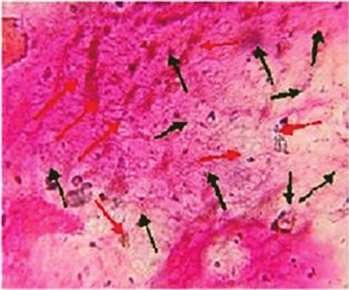
Evidence-Based Complementary and Alternative Medicine                                                                                                                           3

                                                      REASON FOR SELECTION OF DIFFERENT MARKETD DRUGS AND PLANT ACTIVE EXTRACT

                                                                                                             Celecoxib
                                                                Marrubium vulgare                                                                        Acamprosate
                                                                     extract
                                                                                        Lixisenatide
                                                                                                                                   Nimodipine
                                        Marrubiin

                                                                                                           * Cox-2 inhibitor
                                                                * inflammation                              * In inflammation
                                                                * Glutamate
                                                                  excitotoxicity                                                                     * Modulator of GABA-A
                                                2+                                                                             L-type ca2+ channel     receptor
                                    * L-type ca                                         * Activate IP3                                               * Glutamate receptor
                                      channel blocker                                   Pathway and                            blocker
                                                                                                                                                       (NMDA) antagonist
                                   * TNF-α                                              insulin release                                              * Metabotropic glutamate
                                   * Secretion of insulin                                                                                              receptor-5-antagonist

Figure 1: Pharmacological rationale for the selection of various test samples (plant active/extract) and marketed drugs for the present study.

                              35                                                                          (p > 0.05). Moreover, the extract of the selected medicinal
                                                                                                          plant at a higher dose significantly enhanced the falling
                              30                                                                          latency of experimental animals (p < 0.01). The significance
Neurological Severity Score

                              25                                                                          of the result was further improved when the selected dose
                                                                                                          (1400 mg/kg) of the extract was administered along with all
                              20                                                                          marketed drugs (p < 0.001).
                              15
                                                                                                          2.1.3. Elevated Plus Maze Study. TBI can also cause the
                              10                                                                          short-term loss in memory of the animals under the pro-
                                                                                                          tocol. A simple apparatus such as an elevated plus maze
                               5
                                                                                                          could be employed to evaluate the cognitive function in
                               0                                                                          different groups. On the seventh day of the experiment, the
                                       1        2           3    4      5           6   7         8       number of entries in the closed arm of the elevated plus maze
                                                                  Groups                                  was recorded for a minimum of 5 minutes (Figure 3(c)). Loss
                                           2 nd Day                                                       in memory potential of injured mice was indicated by the
                                           4 th Day                                                       significant reduction (p < 0.001) in the number of entries
                                           7 th Day                                                       (1.6 ± 0.32) as compared to the control group (12 ± 1.1).
Figure 2: Change in the neurological severity score on different
                                                                                                          Treatment with plant extracts/active constituents and dif-
days of experimental study (2nd, 4th, and 7th day).                                                       ferent drugs altered the memory deficit induced by the TBI.
                                                                                                          But the improvement in impaired memory function was
                                                                                                          significant in animals fed with plant extract at high doses
reduced the number of lines crossed by mice (77 ± 6.4) as                                                 (p < 0.01). Further, the synergistic neuroprotective effect was
compared to the control group (160 ± 12.4). Further, the                                                  exhibited by rodents on giving the extract along with se-
animals treated with plant extract (1400 mg/kg) enhanced                                                  lected commercial drugs.
their mobility (p < 0.05) which was further improved sig-
nificantly when this extract was supplemented with mar-
keted drugs (p < 0.01).                                                                                   2.1.4. Estimation of Biochemical Parameters. The concen-
                                                                                                          tration of various endogenous oxidative stress parameters
                                                                                                          (GSH, MDA, Catalase) was determined and was indicated in
2.1.2. Rotarod Performance Analysis. Motor coordination in                                                Figure 4. TBI significantly reduced the GSH concentration as
the experimental animals was further investigated by                                                      compared to the control group (p < 0.001) whereas the level
analysis of rotarod performance. In this experiment, the                                                  of malondialdehyde and catalase increased significantly
falling latency of animals of various groups from a rotating                                              (p < 0.01) (Figure 5). Further, the results suggested that the
rod (25 rpm) for five minutes was observed and noted                                                      plant extract alone in higher concentration and along with
(Figure 3(b)). Falling latency in the control group                                                       the marketed drugs significantly altered the oxidative stress
(43 ± 2.510) was significantly (p < 0.01) reduced in the TBI-                                             parameters (p < 0.05 and p < 0.01, respectively). Moreover,
affected group (7.6 ± 1.631). Delay in falls was also altered in                                          the total protein content in the test samples was determined
animals treated with marrubiin at different doses but the                                                 and concentration was found to be significantly altered in
change was not significant as compared to the affected group                                              treated groups (Figure 6). Also, the nitric oxide (NO)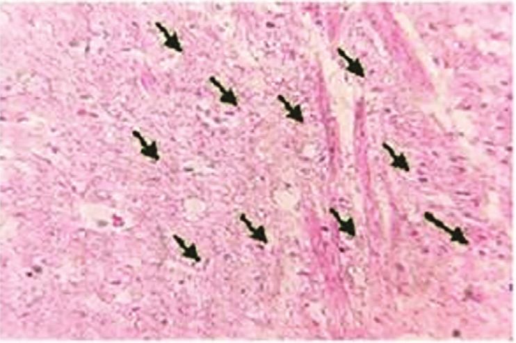
4 Evidence-Based Complementary and Alternative Medicine
200 50
180 45 ***
Number of Crossings
Falling Latency (in sec)
160 ** 40
140 * 35 ** **
120 * *
30
100 25
80 ^^^
60 20
40 15
20 10 ^^
0 5
1 2 3 4 5 6 7 8 0
Groups 1 2 3 4 5 6 7 8
Groups
(a) (b)
14
12 ***
Number of Entries
10
** **
8
*
6
*
4 *
2 ^^^
0
1 2 3 4 5 6 7 8
Groups
(c)
Figure 3: Effect of different neurobehavioral tests: (a) open field, (b) rotarod, and (c) elevated plus maze in different groups (1: control; 2:
(−ve) control (TBI); 3: marketed drugs (L + C + N + A); 4, 5: marrubiin (50,100 mg/kg); 6, 7: M. vulgare extract (700,1400 mg kg); 8:
M. vulgare extract (1400 mg/kg) + marketed drugs) (∧ p < 0.05; ∧∧ p < 0.01; ∧∧∧ p < 0.001 as compared to the control group; ∗ p < 0.05;
∗∗
p < 0.01; ∗∗∗ p < 0.001 as compared to the −ve control).
concentration was altered by test groups but the change was distinction between cytoplasm and nuclei, then it was dis-
significant with the marrubiin (50 mg/kg) and plant extract played as a red arrow in the section pictures (Figure 7).
at a higher dose (p < 0.05). Trauma due to accidents, blasts, and falls is the most
prevalent cause of injury to the brain [31]. TBI not only
causes the mortality of individuals but also produced a
2.1.5. Neurotransmitters (GABA and Glutamate) Analysis. large number of short- and long-term consequences to
The concentration of inhibitory (GABA) and excitatory brain functions. According to an estimate, TBI con-
(Glutamate) neurotransmitters was modulated significantly tributes to more than 30% of all injury-related deaths in
(p < 0.001) in the animals suffering from traumatic brain the USA, and about half of the survivors (43%) faced
injury (Figure 6). Treatment with test drugs (extract and significant chronic disabilities due to the nonavailability
plant active) at different doses enhanced the GABA con- of clinically effective treatment protocol [32, 33]. After
centration but the change was insignificant (p > 0.05) with a the TBI, there occur different primary and secondary
low dose of marrubiin and plant extract. But a higher dose of pathological signaling cascades. The effect due to primary
the extract (p < 0.01) and active constituent (p < 0.05) of the insult cannot be treated but can be prevented whereas the
herb displayed a significant change in the quantity of GABA. functional impairment and chronic disability of neurons
Further, the glutamate analysis confirmed the significant after secondary injury because of oxidative stress, exci-
reduction (p < 0.01) in its level on treatment with Mar- totoxicity, inflammation, and mitochondrial dysfunction
rubium vulgare extract at an elevated dose (1400 mg/kg). could be targeted for the improvement of patient life after
Also, there is a synergistic alteration in the effect produced injury [8, 34].
by herbal extract in combination with marketed drugs The selected medicinal plant Marrubium vulgare L. and
(p < 0.001). marrubiin (plant active) were investigated for neuro-
protective effects after TBI. After TBI, the assessment of NSS
reflects the rough magnitude of motor and cognitive defi-
2.1.6. Histopathological Study. On the 8th day of the ex- ciency such as difficulty in memory, attention deficits, and
perimental protocol, the histopathological changes in the decrease in concentration in affected animals. Improvement
cerebral cortex of brain sections from the animals of dif- in the motor ability and cognitive skills of injured rodents
ferent treated groups were observed. The nuclei (pink stain) was exhibited by the various test groups especially the plant
and cytoplasm (violet color) could be easily differentiated in extract in a higher dose and its combination with marketed
the neuronal cell of brain tissue. The intact neuronal cell drugs. The literature revealed that marrubiin itself and the
from the samples of different treated groups was depicted as plant extract are potent calcium channel blockers and the
a black arrow in the photographs, but when there is no clear significantly altered functional ability of mice might be due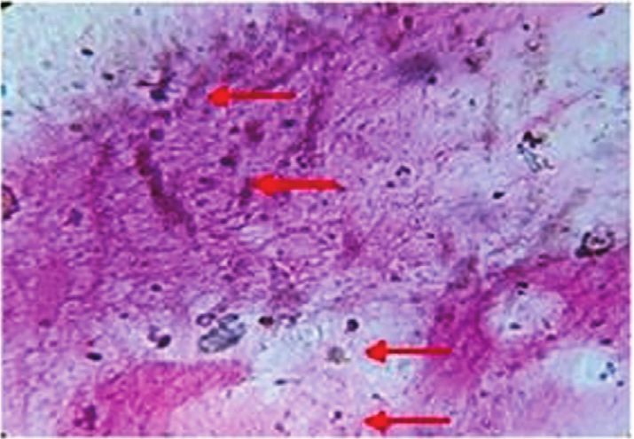
Evidence-Based Complementary and Alternative Medicine 5
0.8 16
Total Protein Content (mg/mL) ^^
0.7 14
0.6 12
**
NO (μmol/mg)
0.5 ** 10 *
** *
0.4 8 **
0.3 * 6
*
0.2 ^^^ 4
0.1 2
0 0
1 2 3 4 5 6 7 8 1 2 3 4 5 6 7 8
Groups Groups
(a) (b)
7 60
6 ^^^
50
5 **
MDA (nmol/mg)
40
GSH (μg/mg)
4 * *
* *
* 30
3 * *
*
** 20
2
1 10 ^^^
0 0
1 2 3 4 5 6 7 8 1 2 3 4 5 6 7 8
Groups Groups
(c) (d)
3
2.5
*
Catalase (μmol/mg)
2 * *
1.5
^^
1
0.5
0
1 2 3 4 5 6 7 8
Groups
(e)
Figure 4: Evaluation of various biochemical (oxidative stress) parameters: (a) total protein content, (b) nitric oxide, (c) malondialdehyde
(MDA), (d) glutathione (GSH), and (e) catalase, in different groups (1: control; 2: (−ve) control (TBI); 3: marketed drugs (L + C + N + A); 4,
5: marrubiin (50,100 mg/kg); 6, 7: M. vulgare extract (700,1400 mg kg); 8: M. vulgare extract (1400 mg/kg) + marketed drugs; ∧ p < 0.05;
∧∧
p < 0.01; ∧∧∧ p < 0.001 as compared to the control group; ∗ p < 0.05; ∗∗ p < 0.01; ∗∗∗ p < 0.001 as compared to the −ve control).
to this action [16, 35]. Also, the scientists had proved that the molecule, causes cytotoxicity due to enhanced oxidative
calcium channel blockers not only improved the behavioral stress, and the plant extract significantly altered the various
deficits but also prevent the disruption in spatial memory of stress parameters along with the NO to exert the neuro-
experimental animals [36, 37]. protective effect in the injured animals [40]. The observed
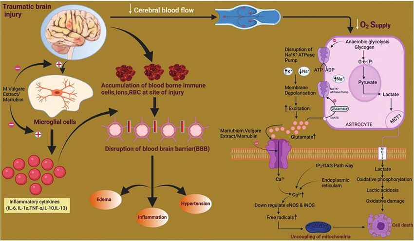
Moreover, sudden trauma to the brain triggers the results could be attributed to the blockage of Cav 1.2 and Cav
different signaling pathways in astrocytes and neurons that 1.3 (L-type) postsynaptic calcium channels localized in the
lead to the upregulation of calcium and downregulation of soma, spines, and shaft of dendrites which further help in the
endothelial nitric oxide synthase (eNOS). It further en- downregulation of enhanced calcium inflow and decrease
hanced the concentration of reactive oxidative species (ROS) the ROS production which could help in the restoration of
and leads to mitochondrial dysfunction and apoptosis the mitochondrial respiratory chain and its integrity
[38, 39]. It has also been reported that NO, which is a unique [16, 35, 37]. In past, the treatment with plant extract was also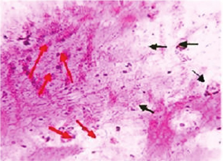
6                                                                                       Evidence-Based Complementary and Alternative Medicine

                      25                                                                    4.5
                                                                                                      ^^^
                                                                                              4
                      20                                                                    3.5

                                                                         Glutamate (μg/g)
                                                                                              3
        GABA (μg/g)

                      15                                                                                               *
                                                                                            2.5                                  *
                                                                                                                            *          **
                                                                  **                          2
                      10             **                                                                       **                            **
                                                            *                               1.5
                                                *
                       5                                                                      1
                               ^^^                                                          0.5
                       0                                                                      0
                           1    2    3     4    5     6     7     8                               1    2          3    4    5    6     7    8
                                           Groups                                                                      Groups
                                          (a)                                                                         (b)

Figure 5: Neurotransmitter: (a) GABA (inhibitory) and (b) glutamate (excitatory) analysis in different groups (1: control; 2: (−ve) control
(TBI); 3: marketed drugs (L + C + N + A); 4, 5: marrubiin (50,100 mg/kg); 6, 7: M. vulgare extract (700,1400 mg kg); 8: M. vulgare extract
(1400 mg/kg) + marketed drugs; ∧ p < 0.05; ∧∧ p < 0.01; ∧∧∧ p < 0.001 as compared to the control group; ∗ p < 0.05; ∗∗ p < 0.01; ∗∗∗ p < 0.001 as
compared to the −ve control).

                               (a)                                      (b)                                                     (c)

                               (d)                                      (e)                                                     (f )

                                                (g)                                                         (h)

Figure 6: Photomicrographs of brain sections of experimental animals from different groups (a) control; (b) (−ve) control (TBI);
(c) marketed drugs (L + C + N + A); (d, e) marrubiin (50,100 mg/kg); (f, g) M. vulgare extract (700,1400 mg kg); (h) M. vulgare extract
(1400 mg/kg) + marketed drugs) depicting intact neuron by black arrow and nonintact cells by red arrow.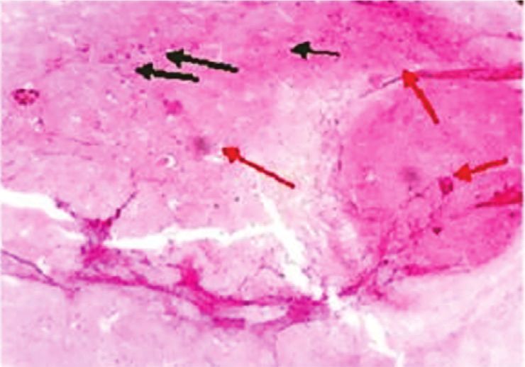
Evidence-Based Complementary and Alternative Medicine                                                                          7

       Figure 7: Proposed site of action of Marrubium vulgare extract/marrubiin to bring out the protective effect after TBI.

postulated to reduce the microglial activation which could be      metabolites including marrubiin, in the plant extract.
another reason for better neuroprotective action by affecting       Marrubiin was reported to block the L-type calcium
the inflammatory cascade after TBI [18, 41, 42].                    channels and thus bring out the vasorelaxant and anti-
     The results of the neurotransmitters analysis indicated       excitotoxicity effect of herb. Also, the inhibition of these
that there is a significant alteration in GABA (↓) and glu-         cation channels could reduce the activation of resident
tamate levels (↑) after the injury. These findings match with       microglial cells and thus lessen the release of proin-
previous investigations which enhanced the glutamate level         flammatory cytokines (IL- 1α, IL-6) [18, 41, 42]. Further, the
and excitotoxicity induced signaling cascades which resulted       previous investigations established that the different phy-
in neurodegeneration [43]. Also, it has been postulated that       tochemicals such as renarioside, acteoside, forsythoside B,
the imbalance between these neurotransmitters leads to             and ballotetroside as phenylpropanoid esters present in the
neurodegeneration in the brain [44–46]. But treatment with         extract can inhibit the cyclooxygenase-2 (COX-2) enzymes
Marrubium vulgare extract and marrubiin significantly               and thus are responsible for the anti-inflammatory action of
modulates their concentration in a positive manner. The            herb (C) [21, 24, 49]. Moreover, the hydroalcoholic extract
neuroprotective effect of the test samples implies that             was also reported to reduce the spasm induced by acetyl-
excitotoxicity due to enhanced glutamate was reduced and           choline and oxytocin at experimental conditions [50]. In
the balance between GABA and glutamate was improved to             addition, oxidative stress was considered to be as one of
bring out this action. Further, the calcium channel blockers,      significant parameters in inducing the secondary effects after
especially of L-type, were reported to reduce the necrosis in      TBI [51]. The level of GSH was reduced whereas the MDA
glutamate toxicity; therefore, the present results could be        and catalase concentration enhanced significantly after the
attributed to the inhibition of voltage-gated cation (Ca2+)        injury in experimental animals. Treatment with the plant
channels by plant active and extract [16, 35, 47]. Also, the       extract at higher dose significantly altered the oxidative
vasospasm is the obvious feature in a significant number of         stress parameters. The antioxidant potential of the herbal
traumatic cases and vasorelaxant action of the plant active/       extract could be due to the presence of total phenolic and
extract could further enhance the recovery in the victims          flavonoid content in addition to marrubiin which could also
after injury [33, 48]. Moreover, the photomicrographs of the       be a possible reason behind the fast recovery of injured
histopathology of brain sections further confirmed the              animals [17, 52–54]. Finally, TBI also reported to induce the
neuroprotective nature of plant extract at a higher dose and       oedema in the brain which is mainly responsible for the
its combination with selected marketed/commercial drugs.           significant number of mortality in the head injury cases and
     The present findings indicated that recovery after the         the selected plant extract/marrubiin also possess the anti-
injury was more significant with the herb extract as com-           oedematogenic properties [23, 55]. Hence, the improved
pared to plant active alone. Such results could be due to the      neuroprotective effect of the selected medicinal plant could
presence of pharmacologically important secondary                  also be attributed to the antioedematogenic potential along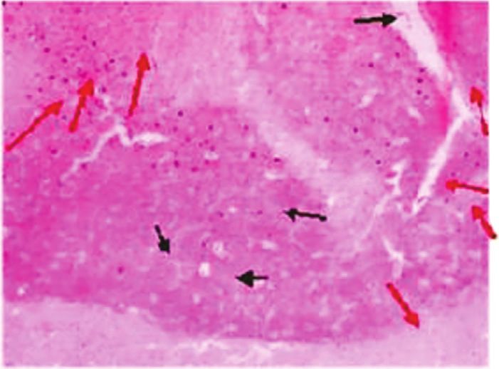
8                                                                                                     Evidence-Based Complementary and Alternative Medicine

                                                                                            IAEC APPROVAL
                                                                                                (n=40)

                                                                                                              TBI

       Control    -ve control      Marketed drug Marrubiin     Marrubiin        Marrubium       Marrubium
      (Group-1)   (Group 2)         (L+C+N+A)    50 mg/kg      100 mg/kg       Vulgare Extract Vulgare Extract
                                     (Group 3)   (Group 4)     (Group 5)         700 mg/kg      1400 mg/kg                Selected    Marketed Drug
                                                                                 (Group 6)                                  dose    + (L+C+N+A)
                                                                                                 (Group 7)
                                                                                                                               (Group 8)
                                                                                            Selected effective
                                                                                            dose of marrubiin/Extract

     1 st Day       2 nd Day            3 rd Day             4 th Day            5 th Day             6 th Day           7 th Day               8 th Day

                   Assessment                              Assessment                                                    Assessment
                        of                                      of                                                            of
                  Neurological                            Neurological                                                   Neurological               Sacrifice the Animals
                  Severity Score                          Severity Score                                                Severity Score
                                                                                                                             and
                                                                                                                        Estimation of      Histopathology        Biochemical
                                                                                                                         Behavioural                             Estimation
                                                                                                                         parameters                 Oxidative Stress           Neurotransmitter
                                                                                                                                                       Parmeter                Analysis
                           Exit            Grip         Starlex         Beam           Straight      Round
                          Circle         Strength       Reflex           Walk            Walk          Stick                                       NO GSH MDA Catalase
                                                                                                                                                                               GABA    Glutamate

                                                                                                                  Elevated    Open       Rota
                                                                                                                 Plus Maze    Field      Rod
                                                                                                                              Test       Test

                                                   Figure 8: Detailed experimental protocol for the present study.

with other discussed properties. On the basis of the above                                           3.3. Animals. For the present research, the swiss albino mice
discussion, the proposed site of action of plant extract/active                                      (25–30 g) were procured from the disease-free small animal
to bring out the protective effect in the pathological cascade                                       house of Lala Lajpat Rai University of Veterinary and
of TBI is depicted in Figure 7.                                                                      Animal Sciences (LLRUVAS), Hisar, India. The animals
                                                                                                     were housed in the group of five (n  5) in polypropylene
3. Materials and Methods                                                                             cages (29 × 22 × 14 cm) lined with proper bedding. They were
                                                                                                     acclimatized as per the standard CPCSEA guidelines for at
3.1. Plant Sample and Reagents. The selected herb, Mar-                                              least two weeks under natural light/dark cycle at 25 ± 2°C.
rubium vulgare L., was collected from the local district of                                          During this period, they have free access to standard rodent
Nainital, Uttarakhand, and identified by a botanist. A                                               feed and water ad libitum.
voucher specimen was also kept in the laboratory of the
department for future reference. All the procured chemicals
and reagents were of analytical grade. The plant active,                                             3.4. Experimental Protocol and Induction of Traumatic Brain
marrubiin, was purchased from the reliable commercial                                                Injury. TBI is followed by not only the immediate primary
source, Extrasynthese (France).                                                                      effects but also some cellular, genomic, and biochemical
                                                                                                     changes termed the secondary insult. These effects could last
                                                                                                     up to some minutes to some days [58]. Hence, the exper-
3.2. Extraction of Plant Sample and Analysis. The collected                                          imental protocol should be kept for seven days to effectively
sample of the whole plant was thoroughly washed in running                                           evaluate the protective effect of the selected plant active/
tap water and shade dried. The dried herb was powdered,                                              extract against secondary insult after TBI. Prior to the
sieved (60–80), and extracted with hydroalcoholic solvent                                            commencement of experiments, the research protocol was
(50%) by the cold maceration method. The powdered sample                                             duly approved by the institutional animal ethical committee
(100 g) was kept in a round bottom flask with solvent (1 : 15)                                       (1767/RE/S/14/CPCSEA/CAH/153–165, dated-17/12/18),
at room temperature for 10 days with occasional stirring.                                            MDU, Rohtak. After the specified time period (15 days) of
The extract obtained was concentrated, lyophilized, and                                              acclimatization of experimental animals, they were divided
stored at low temperature in an airtight container till further                                      into twelve groups (n  5). The detailed experimental pro-
study. Also, the qualitative and quantitative analysis of the                                        tocol is illustrated in Figure 8. The TBI was induced in the
extract for the marrubiin concentration was performed                                                animals of all groups except the first group (control) by the
using a validated HPTLC protocol which was previously                                                standard weight-drop method. Animals were placed in a
developed in our laboratory [56, 57].                                                                closed chamber and anesthesia was induced with 4%
Evidence-Based Complementary and Alternative Medicine 9
isoflurane. After 5–7 minutes, the mouse was removed from 3.5.2. Open Field Test. After the injury, the motor and ex-
the closed chamber and placed on the open circuit with 2% ploratory behavior of mice can be evaluated by this simple
isoflurane maintenance. The reflexes were checked by test. In this, the mice were placed in the middle of the
pressing the hind paw and observing the eye movements. In apparatus (70 × 70 × 25 cm) and the number of squares
the absence of reflexes, the mouse was placed on the foaming crossed by at least the anterior paw of animals was recorded
bed, and a midline incision was given over the head of mice for 5 min by visual inspection [61].
and exposed the skull. The metallic disc was fixed over the
exposed skull and placed properly under the metallic pipe
(1 m long). After that, 66 g weight (spherical brass ball) was 3.5.3. Rota Rod Test. The coordination in the motor skills
dropped through the upper end of the pipe to induce closed can further be evaluated by this test. The ability of the
head injury. After brain trauma, the scalp was sutured, animals to hold on to the accelerated rotating rod depicts the
neosporin powder (GlaxoSmithKline Pharmaceuticals Ltd., motor functions. In this test, the mice were placed on a
Banglore, India) was applied over the scalp, and the mouse rotating rod (5 rpm), and it was accelerated at a speed of
was returned to their cage for recovery [59]. The test drugs/ 5 rpm every 40 seconds. The animals were kept on the ro-
extracts were given to animals of different groups as per the tating rod till the rod speed reached 25 rpm. The average
protocol for seven days. The neurological severity score for latency in the fall of mice from the rod was noted for 5
each group was calculated after 24 hrs, 4th day, and 7th day, minutes [62].
and behavioral parameters were studied on the 7th day of the
experimental study. On the next day, the animals were
sacrificed by cervical dislocation and used for histological 3.5.4. Elevated Plus Maze. The learning and memory po-
and biochemical study. tential of the drug in the experimental mice can be evaluated
by the previously described methods. In this test, a wooden
plus maze with open and closed arms was kept in an elevated
3.5. Neurobehavioral Study. Immediate after the TBI, the place (25 cm from the floor). The mice were placed on the
pathological cascade is initiated and it reflects through open arm of the elevated plus maze, and no entries in the
impairment in motor coordination, balance, and locomotor closed arm and open arm were recorded for 5 min [63].
dysfunction of animals. Also, due to impact, there could be a
loss of memory in experimental mice. The magnitude of
these parameters could be assessed as per the following 3.6. Analysis of Biochemical Parameters. On the eighth day of
protocols. the experimental study, the whole brain of animals from
each group was isolated and cleaned with ice-cold normal
saline. The brain samples were homogenized with phosphate
3.5.1. Assessment of Neurological Severity Score. The neu- buffer (pH 7.4) and immediately centrifuged at 2500 rpm for
rological severity score (NSS) roughly determines the 15 min. The homogenate was used to analyze the various
magnitude of loss in motor and locomotor function. The biochemical parameters.
various tasks like exit circle, beam walk, round stick, straight
walk, startle reflex, and grip strength after 24 hrs (2nd day),
4th day, and 7th day of injury were performed to calculate it. 3.6.1. Total Protein Content. Alkaline copper solution 5 ml
One point was given for non/delayed performance and zero was added to 1 ml of brain tissue homogenate and allowed to
points for the completion of tasks. All the tasks were carried stand for 10 minutes. Further, the 0.5 ml of diluted Folin’s
out as per the standard procedures. Briefly, in the exit circle, reagent (1 : 2) was added and mixed properly. After 30
the mice were placed in the center of the circle and the time minutes, the absorbance of the prepared solution was taken
taken to exit the circular instrument was monitored for three at 750 nm in a UV-Visible spectrophotometer. The total
minutes. In beam walk, the animal was kept on the wooden protein content was determined in mg/mL [64].
beam on an elevated surface (60 cm). On the other end, a box
(20 × 25 × 24 cm) with a 10 cm opening was placed. The time
taken by the mice to reach the box was noted and compared 3.6.2. Estimation of Catalase. The catalase assay of the
with the control for the calculation of scores. For grip different samples was performed by the method described
strength, the mice were held up by the tail, and their paws by Sinha et al., [65]. In this, the small portion of brain
were touched by the anatomical forceps. If the mouse grips homogenate (0.1 mL) was mixed with 0.1 mL of phosphate
the forceps, 0 points were counted, and on failure or per- buffer (0.01 M, pH 7.4) and 0.4 ml of distilled water. Fur-
formance with very low intensity, 1 point was added. In the ther, the addition of 0.5 ml of H2O2 (2 M) initiated the
straight walk test, the mouse was placed on a clear surface reaction. The mixture was incubated for 1 min at room
and observed for the walking pattern. The impaired gait temperature and the reaction was ended by mixing 2 ml of
pattern and failure to actively explore environs by the animal potassium dichromate-acetic acid reagent. The solution was
are credited for 1 point and a normal walk earns nil. In the kept for 15 minutes in a boiling water bath. On cooling the
round stick balance test, the ability of mice to balance over a solution, the green color appeared and the absorbance was
round stick (Φ 5 mm) for 10 seconds was evaluated. The taken at 570 nm in a UV-Visible spectrophotometer. The
alertness of the mice and response to the sound of a hand concentration of catalase was expressed as μmoles/mg of
clap was evaluated in the starlet reflex test [60]. protein [66].10 Evidence-Based Complementary and Alternative Medicine
3.6.3. Estimation of Malondialdehyde (MDA). data is represented as mean ± SD (standard deviation) and
Malondialdehyde is an indicator of lipid peroxidation and the statistical significance of the data obtained in the various
was estimated as previously described [67]. The supernatant tests was carried out by the analysis of variance (ANOVA)
of tissue homogenate was mixed with various reagents like followed by the Tukey test. The different significant level of
acetic acid (1.5 ml, 20%), thiobarbituric acid (1.5 ml, 0.8%), data as compared to the control (group 1) or negative
and sodium dodecyl sulfate (0.2 ml, 8.1%). The mixture was control (group 2) is also represented in bar diagrams and
heated at 100°C for 60 min and cooled. After cooling, 5 ml of p < 0.05 is considered significant.
n-butanol: pyridine (15 : 1 v/v) and distilled water (1 ml) were
added with vigorous shaking. The solution was centrifuged
at 4000 rpm for 10 min, and the separated organic layer was 3.8. Histopathological Study. On the 8th day of the experi-
removed and absorbance was measured at 532 nm using a mental study, the mice were sacrificed and some of the brain
UV-Visible spectrophotometer (Shimadzu UV Spectro- samples from each group were dissected for histopatho-
photometer, UV-1800 Series, India). The amount of logical study. The tissue was fixed in Bouin’s fixative for 72 h.
malondialdehyde present in the sample was expressed as After following all standard procedures, the tissues were
nmol/mg protein. embedded in wax blocks and trimming of tissue (7 μm
thickness) was done using a microtome. The tissue sections
were stained with eosin and hematoxylin dyes and the
3.6.4. Nitric Oxide (NO) Assay. The accumulation of nitrite counterstaining was done with Dibutylphthalate Polystyrene
was assayed spectrophotometrically using a Greiss reagent. Xylene. The stained tissue sections were analyzed at
The supernatant (100 μl) was mixed with an equal volume of 20 × under a light microscope [72].
Greiss reagent and kept for 10 min at room temperature. The
absorbance of the reaction mixture was measured at 540 nm
against a blank solution (distilled water). The concentration 4. Conclusion
of NO in the sample was calculated by plotting the standard Increased incidences of the TBI posed a great challenge in
curve of sodium nitrite (5–30 μmol/ml) and was expressed as terms of the physical, mental, economical, and social well-
μmoles of NO/mg protein [68]. being of the victims. The secondary insult modulates not
only the various neurobehavioral functions required for
3.6.5. Measurement of Reduced Glutathione (GSH). The normal motor and cognitive functions but also induced the
reduced glutathione content in the tissue homogenate was neurotoxic cascades due to enhanced oxidative stress and
measured by the previously described method using a UV- causes cell death and necrosis. Even the long back after the
Visible spectrophotometer. The processed sample was mixed injury, the survivors of TBI, remained at the risk of in-
with an equal volume of sulfosalicylic acid (5%) and then flammatory and immune-mediated pathological cascade
centrifuged at 2000 rpm at 4°C for 10 min to separate out the leading to apoptosis and sudden death. The literature study
proteins. Take the supernatant and add phosphate buffer revealed that along with the other alternatives, several
(2 mL, pH 8.4), 5,5′-dithiobis (2-nitrobenzoic acid, (0.5 mL), herbal products (decoctions, pills, extracts) were also used
and distilled water (0.4 ml) thoroughly and take the ab- for the management of TBI. The selected medicinal plant,
sorbance at 412 nm. The concentration of GSH in the sample Marrubium vulgare L., is traditionally used as a decoction
was determined using a standard curve prepared with ref- in the Jammu and Kashmir region for many ailments. The
erence GSH (5–25 μg/ml) and expressed as μg of GSH/mg results of the present study indicated that herb extract can
protein [17, 69]. also be used as neuroprotective after a traumatic head
injury. Further, it is suggested that after establishing the
role of the herb in the reduction of proinflammatory
3.6.6. Estimation of GABA and Glutamate. The cytokines, the plant could be used for the faster recovery
Υ-aminobutyric acid (GABA) and glutamate concentration and rehabilitation of victims after TBI. Moreover, the fi-
in the brain samples of various test groups were estimated nancial burden of the secondary treatment cost for the
using HPTLC. The spots of the test and standard samples poor patients with TBI could also be reduced by the use of
were applied with the help of a Linomat 5 applicator such herbs.
(CAMAG, Switzerland) on precoated silica gel plates
(20 × 10 cm). The chromatogram was developed with
n-butanol: glacial acetic acid: water (5 : 3 : 2) as a mobile Data Availability
phase. The plate was sprayed with ninhydrin (0.2%) and The data related to the submitted manuscript could be
dried in an oven at 60–65°C for 3-4 minutes to visualize readily available to the reader on request.
desired compounds. The developed plate was scanned at
482 nm and the area under the curve (AUC) of various
components was quantitatively analyzed with the WINCAT Ethical Approval
software [70, 71].
The present study was duly approved by the Institutional
Animal Ethics Committee (IAEC), Maharshi Dayanand
3.7. Statistical Analysis. The statistical analysis of the data University, Rohtak, Haryana, India (1767/RE/S/14/
was performed by Prism Graph pad (version 9) software. The CPCSEA/CAH/153–165, dated-17/12/18).Evidence-Based Complementary and Alternative Medicine 11
Conflicts of Interest [14] S. Saito, T. Kobayashi, T. Osawa, and S. Kato, “Effectiveness of
Japanese herbal medicine yokukansan for alleviating psy-
The authors declare no conflicts of interest for the present chiatric symptoms after traumatic brain injury,” Psychogeri-
study. atrics, vol. 10, no. 1, pp. 45–48, 2010.
[15] V. Mittal and A. Nanda, “The pharmacognostical evaluation
of the Marrubium vulgare Linn collected from the pulwama
Acknowledgments district of Jammu and Kashmir state in India,” Journal of
Chemical and Pharmaceutical Research, vol. 8, pp. 7–15, 2016.
The authors want to acknowledge the Director and technical [16] S. E. Bardai, N. Morel, W. Maurice et al., “The vasorelaxant
staff of Central Instrumentation Laboratory and Central activity of marrubenol and marrubiin from Marrubium
Animal House, Maharshi Dayanand University, Rohtak, for vulgare,” Planta Medica, vol. 69, no. 1, pp. 75–77, 2003.
providing the necessary facilities to conduct the present [17] A. Nanda and V. Mittal, “Design based ultrasound-assisted
research. This research received no external funding. extraction of Marrubium vulgare linn and comparative
evaluation of extracts for furan labdane diterpene (marrubiin)
concentration and antihypertensive potential,” Current Bio-
References active Compounds, vol. 16, no. 6, pp. 924–936, 2019.
[18] V. Schlemper, A. Ribas, M. Nicolau, and V. Cechinel Filho,
[1] M. A. Flierl, P. F. Stahel, M. Kathryn, B. S. J. Morgan, “Antispasmodic effects of hydroalcoholic extract of Mar-
W. R. Smith, and E. Shohami, “Mouse closed head injury rubium vulgare on isolated tissues,” Phytomedicine, vol. 3,
model induced by a weight-drop device,” Nature Protocols, no. 2, pp. 211–216, 1996.
vol. 4, no. 9, 2009. [19] M. M. d. Souza, R. A. P. d. Jesus, V. Cechinel-Filho, and
[2] A. I. R. Maas, N. Stocchetti, and B. Ross, “Moderate and severe V. Schlemper, “Analgesic profile of hydroalcoholic extract
traumatic brain injury in adults,” Lancet Neurology, vol. 7, obtained from Marrubium vulgare,” Phytomedicine, vol. 5,
pp. 728–741, 2008. no. 2, pp. 103–107, 1998.
[3] M. Prins, T. Greco, D. Alexander, and C. C. Giza, “The [20] A. Boudjelal, C. Henchiri, L. Siracusa, M. Sari, and G. Ruberto,
pathophysiology of traumatic brain injury at a glance,” Dis- “Compositional analysis and in vivo anti-diabetic activity of
ease Models & Mechanisms, vol. 6, pp. 1307–1315, 2013. wild algerian Marrubium vulgare L. infusion,” Fitoterapia,
[4] A. I. R. Maas, “Traumatic brain injury in India: a big problem vol. 83, no. 2, pp. 286–292, 2012.
in need of data,” Neurology India, vol. 65, no. 2, pp. 257–259, [21] M. A. M. Nawwar, A. M. D. El-mousallamy, H. H. Barakat,
2017. J. Buddrus, and M. Linscheid, “Flavonoid lactates from leaves
[5] N. Marklund, “Rodent models of traumatic brain injury: of Marrubium vulgare,” Phytochemistry, vol. 28, no. 11,
methods and challenges,” Methods in Molecular Biology, pp. 3201–3206, 1989.
vol. 1462, pp. 29–46, 2016. [22] S. Sahpaz, T. Hennebelle, and F. Bailleul, “Marruboside, a new
[6] I. Cernak, “Animal models of head trauma,” American Society phenylethanoid glycoside from Marrubium vulgare L.” Nat-
for Experimental NeuroTherapeutics, vol. 2, pp. 410–422, 2005. ural Product Letters, vol. 16, no. 3, pp. 195–199, 2002.
[7] O. Kalemci, H. E. Aydin, C. Kizmazoglu, I. Kaya, H. Yılmaz, [23] H. K. Stulzer, M. P. Tagliari, J. A. Zampirolo, V. Cechinel-
and N. M. Arda, “Effects of quercetin and mannitol on Filho, and V. Schlemper, “Antioedematogenic effect of
erythropoietin levels in rats following acute severe traumatic marrubiin obtained from Marrubium vulgare,” Journal of
brain injury,” Journal of Korean Neurosurgical Society, vol. 60, Ethnopharmacology, vol. 108, no. 3, pp. 379–384, 2006.
no. 3, pp. 355–361, 2017. [24] W. K. Zapp and Josef, “Accumulation of furanic labdane
[8] A. F. Logsdon, B. P. Lucke-Wold, R. C. Turner, J. D. Huber, diterpenes in Marrubium vulgare and leonurus cardiaca,”
C. L. Rosen, and J. W. Simpkins, “Role of microvascular Planta Medica, vol. 64, no. 3, pp. 357–361, 1998.
disruption in brain damage from traumatic brain injury,” [25] O. K. Popoola, A. M. Elbagory, F. Ameer, and A. A. Hussein,
Comprehensive Physiology, vol. 5, no. 3, pp. 1147–1160, 2015. “Marrubiin,” Molecules, vol. 18, no. 8, pp. 9049–9060, 2013.
[9] A. Rana, S. Singh, R. Sharma, and A. Kumar, “Traumatic brain [26] E.-S. E.-S. Alaa, S. S. Sokkar, M. E.-S. El-Sayad, E. S. Ramadan,
injury altered normal brain signaling pathways: implications and E. Y. Osman, “Celecoxib and omega-3 fatty acids alone
for novel therapeutics approaches,” Current Neuropharma- and in combination with risperidone affect the behavior and
cology, vol. 17, no. 7, pp. 614–629, 2019. brain biochemistry in amphetamine-induced model of
[10] J. Gu, X. Zhang, Z. Fei et al., “Rhubarb extracts in treating schizophrenia,” Biomedicine & Pharmacotherapy, vol. 82,
complications of severe cerebral injury,” Chinese Medical pp. 425–431, 2016.
Journal, vol. 113, no. 6, pp. 529–531, 2000. [27] B. Mason and C. Heyser, “Acamprosate: a prototypic neu-
[11] B. Y. Hu, X. J. Liu, Q. Ren et al., “Treatment with ginseng total romodulator in the treatment of alcohol dependence,” CNS &
saponins improves the neurorestoration of rat after traumatic Neurological Disorders—Drug Targets, vol. 9, no. 1, pp. 23–32,
brain injury,” Journal of Ethnopharmacology, vol. 155, no. 2, 2012.
pp. 1243–1255, 2014. [28] C. Albert-Weißenberger, C. Várrallyay, F. Raslan,
[12] L. Li, X. Fan, X. T. Zhang et al., “The effects of Chinese C. Kleinschnitz, and A. L. Sirén, “An experimental protocol
medicines on CAMP/PKA signaling in central nervous system for mimicking pathomechanisms of traumatic brain injury in
dysfunction,” Brain Research Bulletin, vol. 132, pp. 109–117, mice,” Experimental & Translational Stroke Medicine, vol. 4,
2017. no. 1, pp. 1–5, 2012.
[13] J. Matsumoto-Miyazaki, Y. Asano, S. Yonezawa et al., [29] G. Garjan, Tahereh, M. Sharifzadeh et al., “A novel traumatic
“Acupuncture increases the excitability of the cortico-spinal brain injury model for induction of mild brain injury in rats,”
system in patients with chronic disorders of consciousness Journal of Neuroscience Methods, vol. 233, pp. 18–27, 2014.
following traumatic brain injury,” Journal of Alternative & [30] I. Khalin, N. L. A. Jamari, N. B. A. Razak et al., “A mouse
Complementary Medicine, vol. 22, no. 11, pp. 887–894, 2016. model of weight-drop closed head injury: emphasis on12 Evidence-Based Complementary and Alternative Medicine
cognitive and neurological deficiency,” Neural Regeneration neurons,” Pharmacological Reports, vol. 65, no. 3,
Research, vol. 11, no. 4, pp. 630–635, 2016. pp. 730–736, 2013.
[31] V. E. Johnson, D. F. Meaney, D. K. Cullen, and D. H. Smith, [48] A. Nelson, P. A. Nyquist, R. Alexander, and S. A. Marshall,
“Animal models of traumatic brain injury,” Handbook of “Cerebral vasospasm in traumatic brain injury,” Psychiatric
Clinical Neurology, vol. 127, pp. 115–128, 2015. Annals, vol. 43, no. 7, pp. 328–330, 2013.
[32] A. Jullienne, A. Obenaus, A. Ichkova, C. Savona-Baron, [49] C. R. A. Y. Meyre-silva, V. Schlemper, F. Campos-buzzi, and
W. J. Pearce, and J. Badaut, “Chronic cerebrovascular dys- V. Cechinel-filho, “Analgesic potential of marrubiin deriva-
function after traumatic brain injury,” Journal of Neuroscience tives, a bioactive diterpene present in Marrubium vulgare
Research, vol. 94, no. 7, pp. 609–622, 2016. (lamiaceae),” Il Farmaco, vol. 60, 2005.
[33] A. Salehi, J. H. Zhang, and A. Obenaus, “Response of the [50] S. Lodhi, G. Prakash Vadnere, V. K. Sharma, and M. Rageeb
cerebral vasculature following traumatic brain injury,” Jour- Usman, “Marrubium vulgare L.: a review on phytochemical
nal of Cerebral Blood Flow and Metabolism, vol. 37, no. 7, and pharmacological aspects,” Journal of Intercultural Eth-
pp. 2320–2339, 2017. nopharmacology, vol. 6, no. 4, pp. 429–452, 2017.
[34] D. Lozano, G. S. Gonzales-Portillo, S. Acosta et al., “Neu- [51] A. Rana, S. Singh, R. Deshmukh, and A. Kumar, “Pharma-
roinflammatory responses to traumatic brain injury,” Neu- cological potential of tocopherol and doxycycline against
ropsychiatric Disease and Treatment, vol. 11, pp. 97–106, 2015. traumatic brain injury-induced cognitive/motor impairment
[35] S. E. Bardai, B. Lyoussi, W. Maurice, and N. Morel, “Com- in rats,” Brain Injury, vol. 34, no. 8, pp. 1039–1050, 2020.
parative study of the antihypertensive activity of marrubium [52] A.-O. Nadia, I. M. Abu-Reidah, R. Quirantes-Piné, K. Madani,
vulgare and of the dihydropyridine calcium antagonist and A. Segura-Carretero, “Phytochemical profiling, in vitro
amlodipine in spontaneously hypertensive rat,” Clinical and evaluation of total phenolic contents and antioxidant prop-
Experimental Hypertension, vol. 26, no. 6, pp. 465–474, 2004. erties of Marrubium vulgare (horehound) leaves of plants
[36] A. Levy, R. M. Kong, M. J. Stillman et al., “Nimodipine growing in algeria,” Industrial Crops and Products, vol. 61,
improves spatial working memory and elevates hippocampal pp. 120–129, 2014.
acetylcholine in young rats,” Pharmacology, Biochemistry and [53] T. Hatami, S. Ahmad Emami, S. S. Miraghaee, and
Behavior, vol. 39, no. 3, pp. 781–786, 1991. M. Mojarrab, “Total phenolic contents and antioxidant ac-
[37] K. Taya, Y. Watanabe, H. Kobayashi, and M. Fujiwara, tivities of different extracts and fractions from the aerial parts
“Nimodipine improves the disruption of spatial cognition of artemisia biennis willd,” Iranian Journal of Pharmaceutical
induced by cerebral ischemia,” Physiology & Behavior, vol. 70,
Research, vol. 13, no. 2, pp. 551–558, 2014.
no. 1–2, pp. 19–25, 2000. [54] A. Kadri, Z. Zarai, I. Ben Chobba et al., “Chemical compo-
[38] E. Radi, P. Formichi, C. Battisti, and A. Federico, “Apoptosis
sition and antioxidant activity of Marrubium vulgare
and oxidative stress in neurodegenerative diseases,” Journal of
L. essential oil from tunisia,” African Journal of Biotechnology,
Alzheimer’s Disease, vol. 42, pp. S125–S152, 2014.
vol. 10, no. 19, pp. 3908–3914, 2011.
[39] J. T. Weber, “Altered calcium signaling following traumatic
[55] M. A. Abd-Elfattah Foda and A. Marmarou, “A new model of
brain injury,” Frontiers in Pharmacology, vol. 3, 2012.
diffuse brain injury in rats. Part II: morphological charac-
[40] H. Roman, J. C. Goodman, A. B. Valadka, and
terization,” Journal of Neurosurgery, vol. 80, no. 2,
C. S. Robertson, “Role of nitric oxide in cerebral blood flow
pp. 301–313, 1994.
abnormalities after traumatic brain injury,” Journal of Cere-
[56] M. Vineet and A. Nanda, “Intensification of marrubiin
bral Blood Flow and Metabolism, vol. 23, pp. 582–588, 2003.
[41] P. M. Kanyonga, M. A. Faouzi, B. Meddah, E. M. Essassi, and concentration by optimization of microwave-assisted (low
Y. Cherrah, “Assessment of methanolic extract of Marrubium CO2 yielding) extraction process for Marrubium vulgare using
vulgare for anti- inflammatory, analgesic and anti-microbi- central composite design and antioxidant evaluation,”
ologic activities,” Journal of Chemical and Pharmaceutical Pharmaceutical Biology, vol. 55, no. 1, pp. 1337–1347, 2017.
Research, vol. 3, no. 1, pp. 199–204, 2011. [57] A. Nanda and V. Mittal, “Development of a novel, rapid and
[42] C. Meyre-silva and V. Cechinel-filho, “A review of the validated HPTLC protocol for the quantitative estimation of
chemical and pharmacological aspects of the genus Mar- marrubiin from the extract of Marrubium vulgare linn,”
rubium,” Current Pharmaceutical Design, vol. 16, Journal of Advances in Medical and Pharmaceutical Sciences,
pp. 3503–3518, 2010. vol. 10, no. 3, pp. 1–8, 2016.
[43] J.-h. Yi and A. S. Hazell, “Excitotoxic mechanisms and the role [58] A. Kunz, U. Dirnagl, and P. Mergenthaler, “Acute patho-
of astrocytic glutamate transporters in traumatic brain in- physiological processes after ischaemic and traumatic brain
jury,” Neurochemistry International, vol. 48, pp. 394–403, injury,” Best Practice & Research Clinical Anaesthesiology,
2006. vol. 24, no. 4, pp. 495–509, 2010.
[44] J. W. Błaszczyk and H. De Jesus-cortes, “Parkinson’s disease [59] A. Marmarou, M. A. Abd-Elfattah Foda, W. Van den Brink,
and neurodegeneration: GABA-collapse hypothesis,” Fron- J. Campbell, H. Kita, and K. Demetriadou, “A new model of
tiers in Neuroscience, vol. 10, pp. 1–8, 2016. diffuse brain injury in rats. Part I: pathophysiology and
[45] V. S. Kokhan, K. Petr, A. Oleg, V. Belov, and M. V. Gulyaev, biomechanics,” Journal of Neurosurgery, vol. 80, no. 2,
“Cortical glutamate/GABA imbalance after combined radi- pp. 291–300, 1994.
ation exposure: relevance to human deep-space missions,” [60] J. Zhao, H. Chen, M. Zhang et al., “Early expression of serum
Neuroscience, vol. 416, pp. 295–308, 2019. neutrophil gelatinase-associated lipocalin (NGAL) is associ-
[46] R. M. Guerriero, C. C. Giza, and R. Alexander, “Glutamate ated with neurological severity immediately after traumatic
and GABA imbalance following traumatic brain injury,” brain injury,” Journal of the Neurological Sciences, vol. 368,
Current Neurology and Neuroscience Reports, vol. 1, 2015. pp. 392–398, 2016.
[47] K. Sendrowski, M. Rusak, P. Sobaniec et al., “Study of the [61] E. Choleris, A. W. Thomas, M. Kavaliers, and F. S. Prato, “A
protective effect of calcium channel blockers against neuronal detailed ethological analysis of the mouse open field test:
damage induced by glutamate in cultured hippocampal effects of diazepam, chlordiazepoxide and an extremely lowEvidence-Based Complementary and Alternative Medicine 13
frequency pulsed magnetic field,” Neuroscience & Bio-
behavioral Reviews, vol. 25, no. 3, pp. 235–260, 2001.
[62] J. Barot and B. Saxena, “Therapeutic effects of eugenol in a rat
model of traumatic brain injury: a behavioral, biochemical,
and histological study,” Journal of Traditional and Comple-
mentary Medicine, vol. 11, no. 4, pp. 318–327, 2021.
[63] V. Mittal, S. K. Sharma, P. Jalwal, A. Hooda, and J. Mor,
“Plumbago zeylanica roots: a potential source for improve-
ment of learning and memory,” International Journal of
Pharma Bio Sciences, vol. 1, no. 2, 2010.
[64] O. H. Lowry, N. J. Rosebrough, A. L. Farr, and R. J. Randall,
“Protein measurement with the folin phenol reagent,” Journal
of Biological Chemistry, vol. 193, no. 1, pp. 265–275, 1951.
[65] A. K. Sinha, “Colrimetric assay of catalase,” Analytical Bio-
chemistry, vol. 47, pp. 389–394, 1972.
[66] C. Govindasamy, K. S. Al-Numair, M. A. Alsaif, and
K. P. Viswanathan, “Influence of 3-hydroxymethyl xylitol, a
novel antidiabetic compound isolated from casearia esculenta
(roxb.) root, on glycoprotein components in streptozotocin-
diabetic rats,” Journal of Asian Natural Products Research,
vol. 13, no. 8, pp. 700–706, 2011.
[67] H. Ohkawa, N. Ohishi, and K. Yagi, “Assay for lipid peroxides
in animal tissues by thiobarbituric acid reaction,” Analytical
Biochemistry, vol. 95, no. 2, pp. 351–358, 1979.
[68] L. C. Green, D. A. Wagner, J. Glogowski, L. Paul,
J. S. W. Skipper, and S. R. Tannenbaum, “Analysis of nitrate,
nitrite, and [15N] nitrate in biological fluids,” Analytical
Biochemistry, vol. 126, no. 1, pp. 131–138, 1982.
[69] G. L. Ellman, “Tissue sulfhydryl groups,” Archives of Bio-
chemistry and Biophysics, vol. 82, no. 1, pp. 70–77, 1959.
[70] J. S. Sancheti, M. F. Shaikh, P. F. Khatwani, S. R. Kulkarni, and
S. Sathaye, “Development and validation of a HPTLC method
for simultaneous estimation of L-glutamic acid and c-ami-
nobutyric acid in mice brain,” Indian Journal of Pharma-
ceutical Sciences, vol. 75, no. 6, pp. 716–721, 2013.
[71] C. S. Babu, K. S. Kesavanarayanan, P. Kalaivani, V. Ranju, and
M. Ramanathan, “A simple densitometric method for the
quantification of inhibitory neurotransmitter gamma-ami-
nobutyric acid (GABA) in rat brain tissue,” Chromatography
Research International, vol. 2011, Article ID 732409, 5 pages,
2011.
[72] A. Sivaraman, R. Sanchez-Salas, E. Barret et al., “Prostate
histoscanning true targeting guided prostate biopsy: initial
clinical experience,” World Journal of Urology, vol. 33, no. 10,
pp. 1475–1479, 2015.You can also read