NERVOUS SYSTEM-SYSTEMIC CROSSTALK IN SARS-COV-2/COVID-19: A UNIQUE DYSHOMEOSTASIS SYNDROME
←
→
Page content transcription
If your browser does not render page correctly, please read the page content below
REVIEW
                                                                                                                                               published: 27 August 2021
                                                                                                                                         doi: 10.3389/fnins.2021.727060

                                            Nervous System-Systemic Crosstalk
                                            in SARS-CoV-2/COVID-19: A Unique
                                            Dyshomeostasis Syndrome
                                            Harnadar Anand 1 , Victoria Ende 2 , Gurinder Singh 2 , Irfan Qureshi 1,3 , Tim Q. Duong 4,5 and
                                            Mark F. Mehler 1,6,7,8,9,10,11,12*
                                            1
                                              The Saul R. Korey Department of Neurology, Albert Einstein College of Medicine, Bronx, NY, United States, 2 Renaissance
                                            School of Medicine at Stony Brook University, Stony Brook, NY, United States, 3 Biohaven Pharmaceuticals, New Haven, CT,
                                            United States, 4 Department of Radiology, Albert Einstein College of Medicine and Montefiore Medical Center, Bronx, NY,
                                            United States, 5 Department of Physiology and Biophysics, Albert Einstein College of Medicine, Bronx, NY, United States,
                                            6
                                              Dominick P. Purpura Department of Neuroscience, Albert Einstein College of Medicine, Bronx, NY, United States,
                                            7
                                              Department of Psychiatry and Behavioral Sciences, Albert Einstein College of Medicine, Bronx, NY, United States, 8 Institute
                                            for Brain Disorders and Neural Regeneration, Albert Einstein College of Medicine, Bronx, NY, United States, 9 Rose F.
                                            Kennedy Center for Intellectual and Developmental Disabilities, Albert Einstein College of Medicine, Bronx, NY, United States,
                                            10
                                               Einstein Cancer Center, Albert Einstein College of Medicine, Bronx, NY, United States, 11 Gottesman Institute for Stem Cell
                                            Biology and Regenerative Medicine, Albert Einstein College of Medicine, Bronx, NY, United States, 12 Center
                                            for Epigenomics, Albert Einstein College of Medicine, Bronx, NY, United States

                                            SARS-CoV-2 infection is associated with a spectrum of acute neurological syndromes.
                          Edited by:        A subset of these syndromes promotes higher in-hospital mortality than is predicted
               Vitor Engracia Valenti,
                                            by traditional parameters defining critical care illness. This suggests that deregulation of
     São Paulo State University, Brazil
                                            components of the central and peripheral nervous systems compromises the interplay
                       Reviewed by:
                     Andi Wangzhou,         with systemic cellular, tissue and organ interfaces to mediate numerous atypical
               University of California,    manifestations of COVID-19 through impairments in organismal homeostasis. This
        San Francisco, United States
                         Jaro Hrenak,
                                            unique dyshomeostasis syndrome involves components of the ACE-2/1 lifecycles, renin-
        Insel Gruppe AG, Switzerland        angiotensin system regulatory axes, integrated nervous system functional interactions
               *Correspondence:             and brain regions differentially sculpted by accelerated evolutionary processes and more
                    Mark F. Mehler
                                            primordial homeostatic functions. These biological contingencies suggest a mechanistic
       mark.mehler@einsteinmed.org
                                            blueprint to define long-term neurological sequelae and systemic manifestations such as
                   Specialty section:       premature aging phenotypes, including organ fibrosis, tissue degeneration and cancer.
         This article was submitted to
                                            Therapeutic initiatives must therefore encompass innovative combinatorial agents,
           Autonomic Neuroscience,
               a section of the journal     including repurposing FDA-approved drugs targeting components of the autonomic
            Frontiers in Neuroscience       nervous system and recently identified products of SARS-CoV-2-host interactions.
           Received: 17 June 2021
           Accepted: 30 July 2021           Keywords: autonomic nervous system, COVID-19 sequelae, evolutionary processes, premature aging,
                                            combinatorial therapeutics
         Published: 27 August 2021
                             Citation:
           Anand H, Ende V, Singh G,
 Qureshi I, Duong TQ and Mehler MF
                                            INTRODUCTION
    (2021) Nervous System-Systemic
 Crosstalk in SARS-CoV-2/COVID-19:
                                            Coronavirus Disease 2019 (COVID-19) is a systemic disease that impacts multiple organ systems
A Unique Dyshomeostasis Syndrome.           and is caused by the severe acute respiratory syndrome coronavirus 2 (SARS-CoV-2) virus (Zhu
          Front. Neurosci. 15:727060.       et al., 2020). Like some other coronaviruses, SARS-CoV-2 is a zoonotic virus that has likely
     doi: 10.3389/fnins.2021.727060         jumped from animal species to humans. The potential rapid viral evolution that occurs by

Frontiers in Neuroscience | www.frontiersin.org                                    1                                           August 2021 | Volume 15 | Article 727060
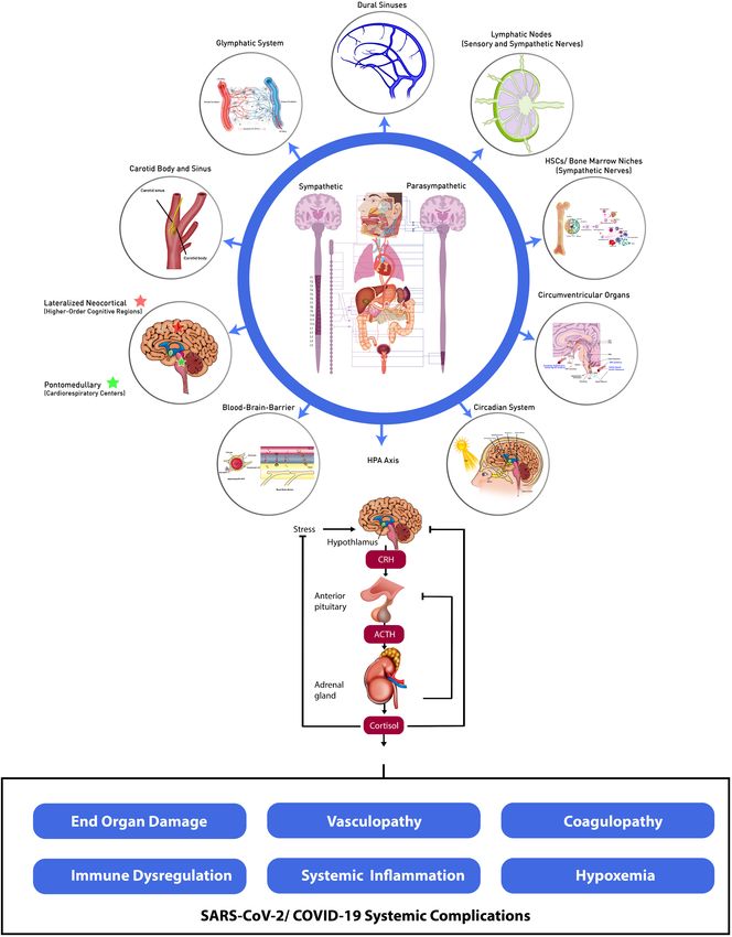
Anand et al.                                                                                            COVID-19: A Unique Dyshomeostasis Syndrome

transitioning between poorly defined intermediate hosts may                 mediated by the CNS and PNS give rise to the spectrum
engender novel forms of pathogen-host interactions (Zhang and               of unanticipated, novel, and multifactorial somatic organ,
Holmes, 2020). Moreover, the human central nervous system                   tissue and cellular damage observed in COVID-19. These
has also undergone accelerated evolutionary innovations in the              include often severe and frequently broad-based end-organ
hominid-to-human lineage (Mattick and Mehler, 2008; Qureshi                 dysfunction, and biologically complex forms of vasculopathy,
and Mehler, 2012, 2014). While these evolutionary mechanisms                coagulopathy, hypoxemia and immune deregulation and
have facilitated rapid change in the human neocortex, other                 systemic inflammation amongst other systemic and life-
areas of the brain involved in more caudal midline homeostatic              threatening complications (Perico et al., 2020; Nie et al., 2021).
functions, which are often also preferential viral targets, represent       We postulate that SARS-CoV-2-induced deregulation of the
more primordial evolutionary centers. The interplay between                 central and peripheral nervous systems subvert cardinal systemic
the potential rapid evolution of SARS-CoV-2 and regions of                  homeostatic functions through interference amongst dynamic
the human brain that have experienced rapid and differential                neural and systemic organ, tissue and cellular interfaces and
evolution like the neocortex may be a mechanism underlying the              SARS-CoV-2-mediated cellular signaling pathways, modulatory
unique profiles of damage caused by SARS-CoV-2.                             interactions and associated multidimensional effector networks
   It is increasingly evident that SARS-CoV-2 is not                        (Qureshi and Mehler, 2013; Figure 1). Characterizing such
only neurotropic, but is associated with a much broader                     intricate nervous system-systemic crosstalk and associated
spectrum of acute and atypical neurological syndromes and                   viral-host evolutionary adaptations is therefore essential for
manifestations than prior infections, particularly those involving          identifying novel measures to address the morbidity and
β-coronaviruses (Bohmwald et al., 2018; Desforges et al.,                   mortality of the acute and critical care illness as well as the
2019; Ellul et al., 2020; Iadecola et al., 2020; Koralnik and               long-term sequelae of SARS-CoV-2 infection.
Tyler, 2020; Paterson et al., 2020). Severe acute neurological
events observed in COVID-19 have included ischemic
stroke, intracranial hemorrhage, diffuse encephalopathy,                    EVOLUTIONARY ADAPTATIONS
encephalitis and neuromuscular disorders (Alquisiras-Burgos
et al., 2020; Lee et al., 2020; MacLean et al., 2020; Conklin               Several mechanisms by which the human neocortex has
et al., 2021). Neurocognitive symptoms and dysfunction of                   undergone accelerated evolution have been elucidated (Figure 2).
various severities have also become increasingly recognized                 These innovations in regional nervous system form and function
as potential consequences of SARS-CoV-2 infection (Levine                   may promote higher-order cognitive and behavioral repertoires
et al., 2020; Graham et al., 2021; Taquet et al., 2021). The                while simultaneously enhancing the vulnerability for age-related
occurrence of severe neurologic dysfunction suggests that less              human brain disorders such as degenerative dementias and
obvious neuropathologic processes are likely present among                  accelerated aging phenotypes (Mattick and Mehler, 2008). For
patients who do not exhibit overt neurologic disease but may                example, numerous transcription factors interact with cis-acting
exhibit differential degrees of systemic involvement. This                  genomic elements such as enhancers and super-enhancers to
raises the important question of the potential mechanistic                  control the gene expression of the human forebrain (Nord
interrelationships between nervous system and systemic                      et al., 2015). Degrees of adenosine to inosine RNA editing
homeostasis in mediating the pathogenesis and progression                   (Mehler and Mattick, 2007) of transcripts and proximal promoter
of COVID-19.                                                                associated regions with higher levels of chromatin interactivity
   In recent large acute retrospective incidence studies, higher            are also enriched for human-specific genes (Song et al., 2020).
in-hospital mortality has been associated with the early presence           Additionally, the emergence of individual human-specific genes
of a subset of neurological syndromes seen with COVID-                      such as NOTCH2NL, SRGAP2, TBC1D3, and ARHGAP11B
19 (Eskandar et al., 2020; Chou et al., 2021). The most                     may be central to increased gyrification and to the preferential
commonly observed central nervous system manifestations                     activation of basal progenitors, which are thought to give rise to
included stroke, encephalopathy, seizures and neuro-COVID-19                an expansion of the population of human neocortical neurons
complex (Chou et al., 2021). These neurological complications               and to contribute to a greater sophistication of neural network
were observed in 82% of hospitalized patients (Chou et al.,                 connections and functional properties (Florio et al., 2015; Ju et al.,
2021). Among these manifestations, altered arousal, orientation,            2016; Rincic et al., 2016; Fiddes et al., 2018). The expansion of the
attention, concentration (encephalopathy) and stroke conferred              human neocortex has also been linked to evolutionary changes in
a significantly higher risk of mortality, independent of overall            neural stem cells forming the outer subventricular zone (Lui et al.,
disease severity measures as assessed by a novel integrative                2011) and to a delay in the neuroepithelial cell differentiation
COVID-19 severity rating scale (Altschul et al., 2020). These               process (Benito-Kwiecinski et al., 2021).
observations suggest that the nervous system writ large plays                  Current studies generally agree that SARS-CoV-2 likely
a preeminent role in the course and outcomes of SARS-CoV-                   emerged via zoonotic spillover first from bats (Lei and Zhang,
2 infection.                                                                2020; Hedman et al., 2021). Other mammals have been proposed
   In this review, we provide emerging evidence that SARS-                  as intermediate hosts; for example, minks are susceptible to
CoV-2 targets widely distributed components of the central and              natural SARS-CoV-2 infection and can transmit the virus to
peripheral nervous systems (CNS and PNS, respectively). We                  humans (Hedman et al., 2021), and mutational analysis of the
propose that disruption of cardinal homeostatic mechanisms                  SARS-CoV-2 receptor-binding domains suggests that increased

Frontiers in Neuroscience | www.frontiersin.org                         2                                     August 2021 | Volume 15 | Article 727060
Anand et al.                                                                                                           COVID-19: A Unique Dyshomeostasis Syndrome

  FIGURE 1 | Dynamic neural-systemic interactions mediating SARS-CoV-2 infection. CSF, cerebrospinal fluid; ISF, interstitial fluid; HSCs, hematopoietic stem cells;
  SFO, subfornical organ; PVN, paraventricular nucleus; PBN, parabrachial nucleus; DMV, dorsal motor nucleus of the vagus; NTS, nucleus tractus solitarius; RVLM,
  rostral ventrolateral medulla; OVLT, organum vasculosum of the lamina terminalis; MPO, myeloperoxidase; AVP, arginine vasopressin; HPA,
  hypothalamic–pituitary–adrenal; CRH, corticotrophin-releasing hormone; ACTH, adrenocorticotropic hormone.

Frontiers in Neuroscience | www.frontiersin.org                                    3                                          August 2021 | Volume 15 | Article 727060
Anand et al.                                                                                                         COVID-19: A Unique Dyshomeostasis Syndrome

  FIGURE 2 | Accelerated evolution of human brain and SARS-CoV-2: potential mechanisms underlying systemic dyshomeostasis. Evolutionary adaptations
  mediating innovations in human brain form and function (upper panel). Parallel evolutionary adaptations in SARS-CoV-2 (lower panel). Created using biorender.com.
  A, Adenosine; I, inosine; ADAR, adenosine deaminases acting on RNA; oSVZ, outer subventricular zone; TF, transcription factor; ORF, open reading frame; RdRp,
  RNA-dependent RNA-polymerase; E, envelope protein; N, nucleocapsid protein.

human susceptibility may emerge in pangolins (Lei and Zhang,                          inflection points (Blanco-Melo et al., 2020; Lucas et al., 2020;
2020; Li et al., 2020a). These contingencies suggest that SARS-                       Meckiff et al., 2020; Su et al., 2020; Liu et al., 2021; Rodda et al.,
CoV-2 exhibits novel evolutionary trajectories, responds to                           2021). Moreover, the exceptional versatility of the lifecycle of
selective pressures, generates genomic innovations and gives                          the ACE2 receptor, SARS-CoV-2 molecular constituents, entry
rise to innate immune dysregulation, thereby promoting greater                        factors, functional adaptations and downstream signaling axes
transmissibility and more complex and interrelated clinical                           help to ensure rapid and efficient environmental innovations
syndromes (Zeberg and Paabo, 2020; Zhang and Holmes,                                  including linking targeted co-morbidities to cardinal viral-host
2020). Modifications in SARS-CoV-2 cellular tropism elicit                            mediated signaling pathways, age- and gender-dependent disease
deregulated and more severe and atypical nervous system and                           phenotypes and seminal evolutionary mechanisms, including
systemic pathologies in the acute phase of illness and the                            formation of novel ACE2 isoforms through co-option of intronic
contextual alterations to give rise to long-term neural and                           retroelements as promoters and alternative exons (Anand et al.,
systemic sequelae (Jacob et al., 2020; Pellegrini et al., 2020).                      2020; Konno et al., 2020; Maremanda et al., 2020; Shang et al.,
SARS-CoV-2 therefore dramatically modifies the host-mediated                          2020; Singh et al., 2020; Wang et al., 2020b).
transcriptome and epitranscriptome, long-distance molecular
signaling pathways, profiles of multi-organ cellular involvement,
activation of premature aging pathways, and regenerative                              SARS-CoV-2 NEUROTROPISM
responses that define acute COVID-19 infection (Kim et al.,
2020; Shen et al., 2020; Delorey et al., 2021). Such systemic                         CNS and PNS Interrelationships
consequences and pathogenic mechanisms give rise to a plethora                        SARS-CoV-2 infects the nervous system (NS) through multiple
of imbalanced immunological and inflammatory reactions that                           points of entry, leading to the severity and unique characteristics
sculpt disease progression, severity and the trajectory of clinical                   of systemic involvement of the virus in mediating cumulative

Frontiers in Neuroscience | www.frontiersin.org                                   4                                         August 2021 | Volume 15 | Article 727060
Anand et al.                                                                                           COVID-19: A Unique Dyshomeostasis Syndrome

disease burden and excessive morbidity and frequently mortality.            respiratory infection (Zhou et al., 2017). Infection via GI
The nervous system is composed of the CNS and the PNS. The                  inoculation led to higher viral loads in the CNS through feedback
PNS is further divided into the somatic and autonomic nervous               regulation via vagal efferents in these studies (Zhou et al.,
systems. The autonomic nervous system is then segregated                    2017). Moreover, recent work using murine models has identified
into the parasympathetic, sympathetic, and enteric divisions.               a novel taxonomy involving twelve enteric neuron classes,
These PNS pathways provide important communication routes                   distinguished by unique transcription factors, communication
to systemic organs, tissues and cell types and their molecular              features, and functionality (Morarach et al., 2021). The enteric
effectors for also defining NS viral tropism (Qureshi and Mehler,           neuron classes develop a distinct diversification pattern through
2013; Shahriari et al., 2020; Zahalka and Frenette, 2020).                  post-mitotic differentiation assisted by spatiotemporal defined
    Other coronaviruses have been known to directly invade the              landmarks (Morarach et al., 2021). These observations help
brainstem (Porzionato et al., 2020), where the presence of the              distinguish the ENS from other branches of the NS and suggest
major receptor for SARS-CoV-2, angiotensin converting enzyme                potential new profiles of viral neurotropism embedded within the
2 (ACE2), has been documented. Previous studies have suggested              three-dimensional body axes. The relatively high concentration
that SARS-CoV-2 may spread to the CNS directly, by invasion                 of ACE2 receptor and viral replication potential within the GI
through the cribriform plate into the olfactory epithelium and              tract, along with frequently noted GI symptoms of COVID-19,
olfactory tract (Baig et al., 2020). The olfactory bulb has direct          emphasize the significance of ENS neurotropism (Esposito et al.,
neuronal connections to the amygdala, entorhinal area, and                  2020). In totality, the PNS will likely prove to be a key and under-
hippocampus (Serrano et al., 2021). Additionally, a recent neuro-           appreciated feature of SARS-CoV-2 neurotropism, inclusive
anatomical study of patients who died with COVID-19 found                   of all three autonomic branches and via direct and indirect
that 20% of patients studied had SARS-CoV-2 RNA in one or                   target interactions.
more regions of the CNS, including the olfactory bulb, amygdala,
entorhinal area, temporal and frontal neocortex, and dorsal
medulla (Serrano et al., 2021). In this study, the olfactory bulb           Cell Autonomous and Non-cell
was the only brain region with viral RNA in more than one                   Autonomous Interplay
subject and had the strongest PCR signal of brain regions studied           The ACE2 receptor is found in neurons, glial cells, astrocytes,
(Serrano et al., 2021), suggesting that the CNS may be accessed             endothelial cells, and those of the lung parenchyma alike (Doobay
through this route.                                                         et al., 2007; Miller and Arnold, 2019). This route is likely
    In addition to modulating basal homeostatic functions, the              cell autonomous in nature, with direct target infection via a
sympathetic nervous system (SNS) is responsible for “fight or               requisite entry factor (e.g., the ACE2 receptor). While there is no
flight” reactions. In such situations where the body is under               consensus on which cells are directly infected, there is evidence
stress, the SNS acts to mitigate this through global release of             for direct neurotropism of several of these cell types through the
adrenaline and cortisol (Hanoun et al., 2015). The hypothalamus             presence of contiguous active virions and intracellular viral RNA.
and pituitary gland, which express the ACE2 receptor, largely               For example, a human induced pluripotent stem cell-derived
mediate the hormonal stress regulatory activity of the SNS via              BrainSphere model has been employed to demonstrate SARS-
the hypothalamic-pituitary-adrenal (HPA) axis and may thus                  CoV-2 infectivity of neuronal cells (Bullen et al., 2020). Similarly,
represent cardinal central viral targets (Pal, 2020). The nucleus           Wang et al. have shown that human induced pluripotent stem
tractus solitarius (NTS) of the brainstem, which acts as a sensory          cell-derived ApoE4 neurons and astrocytes are susceptible to
integrative center for several autonomic functions, projects                SARS-CoV-2 infection (Wang et al., 2021). Moreover, post-
onto the hypothalamus and also expresses the ACE2 receptor                  mortem brain tissue analysis of a COVID-19 patient revealed the
(Porzionato et al., 2020). Additionally, ACE2 is expressed in the           presence of viral particles in brain endothelial cells, indicating
carotid body, which can mediate local sympathetic activation                possible direct neurotropism (Bullen et al., 2020; Paniz-Mondolfi
to modulate optimal blood oxygenation and blood pressure                    et al., 2020). Alternatively, SARS-CoV-2 neurotropism can exert
regulation (Porzionato et al., 2020).                                       its actions non-cell autonomously through the use of soluble
    The enteric nervous system (ENS), a branch of the PNS,                  mediators; extracellular vesicles; inflammatory and immune cell
is located in the walls of the gastrointestinal (GI) tract, where           signaling; non-traditional neurotransmitter, neuropeptide and
it plays a key role in gut homeostasis, systemic metabolism                 ion channel signaling; via the lymphatics; and by microvascular
and immunity. The parasympathetic NS closely interacts with                 damage (Hanoun et al., 2015; Solomos and Rall, 2016; Benveniste
this branch to control the basal state and digestive functions.             et al., 2017; Maryanovich et al., 2018; Merad and Martin, 2020;
Esposito et al. argue that SARS-CoV-2 can directly invade the               Rhea et al., 2021). Because a relatively low rate of SARS-CoV-2
ENS or the parasympathetic NS, and the virus can spread                     RNA is detected in the brain tissue of patients who died from
along the vagus nerve and its innervations of visceral organs,              COVID-19, there is a developing consensus that neurological
which can potentially provide another route of viral tropism                manifestations associated with COVID-19 are due, in large
(Esposito et al., 2020; Tassorelli et al., 2020). The parasympathetic       part, to non-cell autonomous effects (Serrano et al., 2021). For
pathway can then facilitate entry into the brainstem through                example, of twenty COVID-19 patients studied in one neuro-
vagus nerve synapses onto the NTS in the medulla oblongata (Li              anatomical survey, only two had unequivocal neuropathological
et al., 2020c). In rat studies of MERS-CoV, enteric involvement             findings, and only one of those two had detectable SARS-CoV-2
was shown to cause neurological symptoms and to precede                     RNA in the brain (Serrano et al., 2021).

Frontiers in Neuroscience | www.frontiersin.org                         5                                    August 2021 | Volume 15 | Article 727060
Anand et al. COVID-19: A Unique Dyshomeostasis Syndrome
Neurotropism can be the result of blood-borne viruses encephalopathy and demyelination (Domingues et al., 2020;
subverting the blood-brain barrier (BBB) (Iadecola et al., 2020). Virhammar et al., 2020).
SARS-CoV-2 has been shown to cross the BBB in a murine model
(Rhea et al., 2021). Using the S1 subunit of the SARS-CoV-2
spike protein as a proxy for uptake, viral entry was observed MOLECULAR AND CELLULAR
throughout the mouse brain parenchyma and in endothelial MECHANISMS OF VIRAL ENTRY AND
cells (Rhea et al., 2021). These results implicate direct viral SIGNAL TRANSDUCTION
invasion of the BBB in SARS-CoV-2 mediated neurological
dysregulation. ACE2 and other viral proteases are expressed on Angiotensin Converting Enzyme 2/1
endothelial cells of the vasculature (Xiao et al., 2020). Because
lung tissue infected with SARS-CoV-2 shows patterns of damage (ACE2/1)
characteristic of pulmonary fibrosis, viral blood-borne tropism The ACE2 receptor plays a pivotal role as the initiator of
may be facilitated in COVID-19 through upregulated ACE2 in SARS-CoV-2 cellular entry in multiple organs (Xiao et al.,
these arterial vascular cells (Guo et al., 2020). Immune cells 2020). When the viral spike glycoprotein interacts with ACE2,
reaching the CNS may also promote neurotropism, particularly SARS-CoV-2 is endocytosed (Liu et al., 2020a). ACE2 is a key
via macrophages and monocytes (Merad and Martin, 2020). protein for the renin-angiotensin system (RAS), which plays
Although several autopsy studies have not revealed widespread a central role in the homeostatic regulation of electrolyte and
immune cell infiltration, the potential contributions of infected fluid balance, blood pressure, arterial oxygenation, and end
innate and acquired immune cells to neurotropism may be organ function, particularly those mediated by the renal and
consequential (Iadecola et al., 2020). cardiovascular systems (Li et al., 2017; Miller and Arnold, 2019).
Once in the circulation, the virus may easily avoid the BBB The RAS is divided into vasopressor and vasoprotective axes
by targeting circumventricular organs in the brain. These CNS (Li et al., 2017). The multiple arms of this complex pathway
sites have areas of fenestrated capillaries where the endothelial ultimately result in the dynamic interplay of two main enzymes:
border does not represent a fixed boundary to molecular entry Angiotensin Converting Enzyme 1 (ACE1) and ACE2. ACE1
(Iadecola et al., 2020). The presence of ACE2 receptors in such converts angiotensin I (Ang I) to angiotensin II (Ang II), while
delimited CNS locations support this potential route of entry ACE2 converts Ang I and Ang II to angiotensin (1-7) (Ang
(Chigr et al., 2020). Furthermore, inflammation resulting from (1-7)) and angiotensin (1-9) (Ang (1-9), respectively (Miller
reactive oxygen species and free radicals produced by SARS- and Arnold, 2019). The vasopressor axes are comprised of
CoV-2 infection can damage the BBB, facilitating neurotropism the classic angiotensinogen/renin/ACE1/Ang II axis and the
via the circulation in other locations lacking fenestrations prorenin/renin axis (Li et al., 2017). The vasoprotective axes,
(Li et al., 2020c). Similarly, cytokines, which are elevated which are dependent on ACE2/ Ang (1-7)/Ang (1-9)/Mas
during COVID-19 infection, are known to cross the BBB and receptors, counteract detrimental effects of the vasopressor axes
contribute to neuroinflammation observed in SARS-CoV-2 brain (Li et al., 2017). When SARS-CoV-2 is endocytosed, the ACE2
neuropathological studies (Azizi and Azizi, 2020; Iba et al., 2020; enzyme is endocytosed with it, shifting the balance of the
Rhea et al., 2021). RAS toward the pro-inflammatory vasopressor axes (Li et al.,
Although the BBB largely segregates the brain from the 2017; Gheblawi et al., 2020). Such disruption of RAS balance,
systemic circulation, toxic wastes and metabolites must still which has previously been implicated in the development of
be removed from the brain. A waste removal system, termed cardiovascular disease, renal disease, and hypertension, amongst
“glymphatics” for its dependence on glial cells, is facilitated other systemic derangements, may be a driver of COVID-19
by a CSF and interstitial fluid transport system (Benveniste systemic dyshomeostasis (Li et al., 2017).
et al., 2017). In this system, CSF is transported within a
peri-vascular network that helps drain waste from the brain Protease Cofactors and Alternative
parenchyma (Benveniste et al., 2017). In addition to glymphatics, Pathways
a true lymphatic vasculature that drains interstitial fluid has Proteases help to orchestrate neurotropism by assisting the ACE2
been described in the brain’s meningeal layer (Solomos and receptor interaction with SARS-CoV-2. For epithelial cells, these
Rall, 2016). Furthermore, the brain dural sinuses are another proteases may even assist viral invasion through non-ACE2-
location of potential neuro-immune interactions. At the dural mediated routes, via CD147-spike protein and CD26 expressed
sinus, accumulated CNS-derived antigens in the CSF are ubiquitously (Radzikowska et al., 2020; Wang et al., 2020a). ACE2
taken up by dural antigen-presenting cells that introduce receptors are further modulated by being endocytosed following
the antigens to patrolling T cells (Rustenhoven et al., 2021). binding to SARS-COV-2 (Gheblawi et al., 2020; Hirano and
Circulating lymphocytes and viral proteins in these systems Murakami, 2020). Transmembrane protease serine 2 (TMPRSS2)
might be potential routes of both neuroinvasion and subsequent is a serine protease on the plasma membrane that mediates spike
transport back to the periphery (Solomos and Rall, 2016). protein activation and promotes SARS-CoV-2 entry via direct
Other coronaviruses have previously been documented to fusion, thereby subverting endocytic entry (Bailey and Diamond,
be present in the CSF (Nath, 2020), and now SARS-CoV- 2021). ACE2 can exist as a transmembrane bound protein in
2 has been observed in the CSF of COVID-19 patients vascular endothelial cells, or as a circulating form, once cleaved
with rare neurological presentations like acute necrotizing by TMPRSS2 or transmembrane protease serine 4 (TMPRSS4)
Frontiers in Neuroscience | www.frontiersin.org 6 August 2021 | Volume 15 | Article 727060Anand et al. COVID-19: A Unique Dyshomeostasis Syndrome
(Xiao et al., 2020). ACE2 is subsequently shed, giving rise to the including SARS-CoV-2 (Ng et al., 2020; Onabajo et al., 2020).
circulating form, while TMPRSS2 and TMPRSS4 simultaneously The novel truncated ACE2 isoform does not appear to bind the
facilitate the endocytosis of SARS-CoV-2 (Xiao et al., 2020). SARS-CoV-2 spike protein or act as a peptidase, suggesting that
The SARS-CoV-2 spike protein has been shown to interact interferon-stimulated induction may not play a role in promoting
with the soluble form of ACE2 or a soluble ACE2-vasopressin SARS-CoV-2 cellular entry (Ng et al., 2020; Onabajo et al., 2020).
complex extracellularly, and then may enter cells via endocytosis
mediated by the angiotensin II type I receptor (AT1 R) or
arginine-vasopressin receptor 1B (AVPR1B), respectively (Yeung NERVOUS SYSTEM CONTRIBUTIONS
et al., 2021). Additionally, the soluble form of ACE2 preserves TO SYSTEMIC DYSHOMEOSTASIS
the viral binding site and may therefore facilitate viral tropism of
cells where the tissue-bound form of ACE2 is poorly expressed Several key molecular factors are closely involved in stress-
(Yeung et al., 2021). mediated dysregulation, including catecholamines: adrenaline,
Furin is a pro-protein convertase found in many tissues, noradrenaline, dopamine (DA); peptide hormones and associated
including the brain, neuroendocrine organs, the GI tract and factors: arginine vasopressin (AVP), Ang II; and steroid
liver, with few differences in expression levels (Wu et al., 2020). hormones, including cortisol (Goldstein, 2020). This broad-
The SARS-CoV-2 spike protein contains a unique furin cleavage based stress-mediated bioactive factor deregulation leads to
site not found in other β-coronaviruses (Wu et al., 2020). Furin activation of the sympathetic nervous system which, in turn,
cleavage facilitates stronger viral receptor binding and membrane increases Ang II, depletes ACE2 and subsequently decreases the
fusion. These may contribute to the high infectivity profiles and protective action of Ang (1-7) (Xiao et al., 2011; Porzionato et al.,
the multi-organ involvement, especially where ACE2 is present at 2020). These mechanisms are associated with typically observed
lower levels of expression (Wu et al., 2020). For example, furin laboratory values and manifestations observed in COVID-
produced in intestinal cells could provide an avenue for viral 19 infection: hyperglycemia, hyponatremia, electrocardiogram
entry into the CNS via the myenteric nerve plexus. Moreover, abnormalities, cytokine storm, heart failure, acute kidney injury,
loss of furin is an effective measure to prevent viral infection acute respiratory distress syndrome (ARDS), clotting disorders,
(Johnson et al., 2021). Other proteases like cathepsin L, cathepsin and emotional stress (Porzionato et al., 2020).
B, trypsin, factor X, elastase, and Coronavirus 3CL protease
have also been implicated in SARS-CoV2 binding (Gheblawi Distributed Renin-Angiotensin System
et al., 2020; Macchiagodena et al., 2020). CRISPR-Cas-9 screening The Ang II peptide of the RAS can act on a few receptor
has also identified several genes necessary for the synthesis of types, the most notable being AT1 R to effect change, including
glycosaminoglycans, which are negatively charged polymers that increased sympathetic tone (Xia and Lazartigues, 2010; Miller
likely increase infectivity by attracting exposed positive charges and Arnold, 2019). AT1 R is a major mediator of RAS in
on viruses (Bailey and Diamond, 2021). raising blood pressure through several homeostatic mechanisms
Interferon modulation of ACE2 receptors can lead to (Miller and Arnold, 2019). AT1 R is localized at circumventricular
increased degrees of neurotropism (Merad and Martin, 2020; organ sites and other cardinal integrative regulatory centers
Ziegler et al., 2020). Interferons are downstream inflammatory of the brain like the hypothalamus and medulla (Doobay
products of IL-1, IL-6, and tumor necrosis factor (TNF), key et al., 2007). Circulating Ang II can mediate activation of the
inflammatory molecules released by immune cells in multiple SNS by acting on circumventricular organs and the carotid
tissues affected by COVID-19 (Ziegler et al., 2020). Severe body (Porzionato et al., 2020). This system is triggered during
COVID-19 infection and death following infection have been times of blood loss, hyponatremia, renal hypotension, general
associated with elevated inflammatory markers and chemokines SNS activation and infection. Notably, an overactive SNS is
(Merad and Martin, 2020). The resulting interferons enhance implicated in many comorbidities associated with mortality
viral invasion as Ziegler et al. demonstrated that ACE2 receptors in COVID-19 (Porzionato et al., 2020). Ang II interacts with
represent the translation product of interferon-stimulated genes the pro-inflammatory AT1 R, and downstream products of
in human barrier tissue epithelial cells (Ziegler et al., 2020). these processes activate STAT3 and NF-κB transcription factors
Smoking and chronic obstructive pulmonary disease (COPD) (Hirano and Murakami, 2020). This promotes IL-6 production,
are associated with the increased presence of endocytic vacuoles which upregulates NF-κB and STAT3 with production of other
implicated in SARS-CoV-2 endocytosis (Eapen et al., 2021). pro-inflammatory cytokines (Hirano and Murakami, 2020).
Patients with this pathogenic profile may be more susceptible ACE2 allows Ang (1-7) to act on Mas receptors to countervail
to viral entry and associated profiles of neurotropism. With AT1 R actions (Miller and Arnold, 2019), since the Mas
endocytosis and downregulation of surface ACE2 proteins, the receptor mediates vasodilation and anti-proliferative effects
pro-inflammatory axes of the RAS can prevail, namely through (Xia and Lazartigues, 2010). ACE2 overexpression promotes
Ang II and the AT1 R (Gheblawi et al., 2020). Interferon- a protective phenotype in cardiovascular disease states in
stimulated ACE2 and ACE2 endocytosis both can modulate viral transgenic mouse models (Alenina and Bader, 2019). ACE2 in the
entry. However, a novel truncated isoform of the ACE2, termed hypothalamus has powerful antihypertensive and sympathetic
deltaACE2 or MIRb-ACE2, has recently been discovered (Ng nervous system dampening effects, with ACE2 overexpression
et al., 2020; Onabajo et al., 2020). This truncated ACE2 isoform increasing Ang (1–7) relative to Ang II, thereby providing
has been observed to be induced by interferons and viruses, beneficial effects in mouse models displaying brain injury
Frontiers in Neuroscience | www.frontiersin.org 7 August 2021 | Volume 15 | Article 727060Anand et al. COVID-19: A Unique Dyshomeostasis Syndrome
and stroke (Alenina and Bader, 2019). ACE1/ACE 2 receptor as homeostatic regulators of alveolar organization and function
interactions work to effectively mediate the effects of Ang II and following injury, since they have been described to remodel the
Ang (1-7). Unfortunately, with SARS-COV-2 infection, AT1 R postnatal mice alveolus via distinct intracellular signaling factors
activity frequently prevails. (Zepp et al., 2021).
ACE2-Mediated Cellular Tropism and Liver Injury
Organ System Damage Although the exact mechanisms of liver damage in COVID-
As mentioned, the SARS-CoV-2 spike protein interacts with the 19 remain to be elucidated, direct tropism via the ACE2
ACE2 receptor in epithelial cells, which line a variety of tissues receptor expressed in hepatocytes, secondary hepatic damage
including the lung, heart, and kidney. SARS-CoV-2 infection as a result of systemic inflammation, and the hepatotoxicity of
thus potentially contributes to intricate profiles of end organ COVID-19 therapies contribute to organ dysfunction (Han et al.,
damage via interacting neural and ACE2-RAS deregulation of 2021a). Because the liver plays an important role in regulating
cardinal homeostatic processes. The involvement of and damage inflammation by producing acute phase proteins, complement
to the CNS affects different systemic organ systems which proteins, and several cytokines, liver damage in COVID-19 could
then exert feedback regulation to the CNS to exacerbate the potentially contribute to central homeostatic dysregulation of
neurological and overall clinical patterns of COVID-19 infection immune function (Robinson et al., 2016).
(Robinson et al., 2016; Batlle et al., 2020; Garvin et al., 2020;
Kidney Injury
Hundt et al., 2020; Lei et al., 2020). Recent autopsy results of
COVID-19 patients reflect dysregulation of key molecular factors The kidney regulates blood pressure and blood volume through
involved in hypoxia, coagulation, and fibrosis in multiple organs, the RAS. Acute kidney injury (AKI) is frequently observed in
highlighting the extent of aberrant local regulatory processes and hospitalized COVID-19 patients (Batlle et al., 2020; Benedetti
systemic homeostasis (Nie et al., 2021). et al., 2020; Farouk et al., 2020; Sharma et al., 2020). As
with COVID-19-associated liver injury, the mechanisms of AKI
Lung Injury are likely multifactorial. ACE2 is strongly expressed in the
Pulmonary edema as a result of SARS-CoV-2 infection may kidney, suggesting direct SARS-CoV-2 renal tropism (Batlle et al.,
occur as a result of the activation of the bradykinin 1 and 2 2020; Benedetti et al., 2020; Farouk et al., 2020). However,
receptors (B1R and B2R, respectively) in lung epithelial cells. the presence of SARS-CoV-2 RNA and viral particles in the
Normally, ACE2 plays a protective role by preventing fluid kidney has not been consistently reported in the literature
accumulation through inactivation of the B1R ligand known as (Farouk et al., 2020; Sharma et al., 2020). The non-cell
bradykinin (BK). Bradykinin lowers blood pressure and promotes autonomous influences of hyperinflammation, hypoxemia, and
capillary leakage. Furthermore, elevated D-dimer levels in these hypercoagulability may contribute to the development of AKI
patients are most likely reflective of the leakage of plasma (Batlle et al., 2020; Benedetti et al., 2020; Farouk et al., 2020).
contents. A similar mechanism leading to pulmonary edema Moreover, ACE2 expression has been found to be downregulated
has been suggested to play an important role in ARDS and in AKI (Batlle et al., 2020). This may lead to higher ACE1
COVID-19 due to increased hydrostatic pressures as a result expression and lower levels of Ang (1-7), which could promote
of increased Ang II. Increased Ang II has been experimentally a hyperinflammatory and hypercoagulable state in the kidney
shown to not alter hydrostatic pressure; however, increased (Batlle et al., 2020). Because the causes of COVID-19 AKI are
bradykinin has been proposed to increase hydrostatic pressure. likely multifaceted, the disruption of RAS homeostasis in the
ACE1 inactivates bradykinin, which is an activating ligand of kidney may create a vicious cycle that exacerbates renal and
B2R, while ACE2 inactivates nine-arginine bradykinin (Arg9- systemic damage.
BK) and bradykinin without nine-arginine (des-Arg9-BK) which
are activating ligands of B1R (van de Veerdonk et al., 2020). Cardiorespiratory Failure and Hypoxemia
In bronchoalveolar samples of COVID-19 patients, the BK SARS-CoV-2 has been hypothesized to infect the CNS through
precursor, kininogen, was expressed while remaining undetected the cribriform plate, travel in a retrograde fashion through
in control specimens. Furthermore, BK degradative enzymes peripheral olfactory nerves to the brain stem (inclusive of
were downregulated compared to those present in control the medulla) and to target and destroy the medullary centers
patients (Garvin et al., 2020). There is significant upregulation responsible for cardiorespiratory control, such as the Pre-
of the genes encoding the enzymes responsible for production of Botzinger complex. This has been thought to result in the
different forms of hyaluronic acid. The corresponding enzymes compromise of the respiratory centers (Gandhi et al., 2020).
responsible for its degradation, hyaluronidases, are conversely Similar neurotropic viruses, like the avian coronavirus, have been
downregulated. Hyaluronic acid promotes avid water absorption reported to track to nuclei of the medulla: the NTS and the
leading to the formation of a hydrogel within the lungs. The nucleus ambiguus (NA), sites that receive information from the
combination of increased vascular permeability due to increased chemoreceptors of the respiratory tract and lungs and innervate
bradykinin levels as well as increased hyaluronic acid production local resident smooth muscle, glands, and vessels (Costa et al.,
leads to hydrogel formation impairing the diffusion of oxygen 2014; Li et al., 2020b).
and carbon dioxide within the lungs of patients with COVID- In SARS-CoV-2 infection, there is a proposed loss of
19. Interestingly, the alveolar type I cells in the lung may act homeostatic control by the medulla resulting in cardiorespiratory
Frontiers in Neuroscience | www.frontiersin.org 8 August 2021 | Volume 15 | Article 727060Anand et al. COVID-19: A Unique Dyshomeostasis Syndrome
compromise: inadequate blood flow, respiration and elevated fibrinogen, D-dimer levels, Von Willebrand factor
oxygenation, leading to a multifaceted impairment of alveolar (VWF), factor VIII and inflammatory cytokines that can induce
gas exchange. In healthy individuals, peripheral chemoreceptors, a generalized thrombotic disorder (Iba et al., 2020). SARS-CoV-
present at highest concentrations in the carotid body and aortic 2 infected macrophages express tissue factor on their cell surface
arch, monitor the flux of arterial oxygen levels. When there promoting coagulation. Factor VIII and VWF released by SARS-
is arterial oxygen desaturation, an excitatory signal is sent CoV-2 infected endothelial cells also contribute to increased
from the glomeruli here, most directly to the NTS, integrating coagulation. Thrombosis may occur due to these multifaceted
at the rostral ventrolateral medulla (Costa et al., 2014). In a prothrombotic modifications in cell signaling (Iba et al., 2020).
hypoxic state with low oxygen reserves, the body will aggressively However, unlike other coagulopathies, CAC is not marked
work to increase blood flow to high impact organs, promote by thrombocytopenia and does not show increased partial
vasoconstriction in other peripheral locations, and increase thromboplastin time, distinguishing it from similar disorders of
respiratory rate as well as inspiratory/expiratory force, in an sepsis-induced coagulopathy (Iba et al., 2020).
attempt to efficiently deploy oxygen (Costa et al., 2014; Tassorelli CAC is notable for endothelial damage as SARS-CoV-2
et al., 2020). There is likely an inability to adequately detect directly infects vascular endothelial cells via ACE2 receptors
and respond to changes in oxygenation when SARS-CoV-2 with the help of TMPRSS2 cleavage. Damaged endothelial cells
infection compromises medullary and carotid body oxygen then fail to produce nitric oxide, thus allowing adhesion of
sensing and set-point regulation as well as sensory nerve-related leukocytes and platelets and migration of inflammatory cells
respiratory muscle function. The often profound hypoxemia, into the vessel wall (Iba et al., 2020). In the event of infection,
frequently without premonitory symptoms, is characteristic of endothelial cell damage results in tissue factor release, especially
COVID-19 patients facing imminent respiratory compromise in the brain, activating thrombin, the final serine protease in
and is indicative of homeostatic deregulation. the coagulation cascade (Festoff and Citron, 2019). Because
Interestingly, increases in peripheral hypoxic chemosensitivity thrombin exerts pro-inflammatory effects in addition to its role
have been ascribed to overstimulation of the sympathetic nervous in coagulation, it plays a key role in the interactions mediating
system (Porzionato et al., 2020). A hyperactive sympathetic the coagulation-inflammatory nexus (Festoff and Citron, 2019).
nervous system due to higher hypoxic chemosensitivity has Thrombin cleaves the protease-activated receptor on endothelial
been implicated in chronic obstructive pulmonary disorder and cells to gain access through the BBB to the CNS, where it can then
metabolic syndrome, comorbidities that enhance the severity of lead to neuroinflammation (Festoff and Citron, 2019).
COVID-19 (Porzionato et al., 2020). The result of this robust coagulation, inflammation, and
endothelial damage, including active viral-mediated endotheliitis,
Gastrointestinal Dysfunction is profound degrees of vasculitis, thrombosis, and stroke affecting
Acute GI symptoms of COVID-19 include nausea, vomiting, large arterial and venous vessels as well as the entire brain
and diarrhea (Esposito et al., 2020; Li et al., 2020c). Viral and body microvasculature, including arterioles, venules and
neurotropism of the area postrema and in the NTS of the capillary beds. This results in complex impairments in oxygen
medulla oblongata of the brainstem may disrupt their normal exchange and unique microinfarctions and mechanosensitive
homeostatic roles. The NTS is particularly known to be microbleeds with dramatic CNS microglial activation, active
targeted by other coronaviruses, emphasizing its potential role innate immune sensing and neuroinflammation (Azizi and
in the acute GI symptoms frequently seen in COVID-19 Azizi, 2020; Iba et al., 2020; Thakur et al., 2021). Microscopic
(Porzionato et al., 2020). examination of the brains of patients who have died from
The ENS regulates gut homeostasis through interactions COVID-19 has indicated multifocal microvascular ischemic
between enteric glial cells, gut epithelium, and gut-associated and hemorrhagic parenchymal injury, neuroinflammation and
lymphoid tissue (GALT) (Esposito et al., 2020). Enteric glial cells microglial activation without cell autonomous viral effects
serve as antigen-presenting cells to the GALT, and their activation targeting neurons, astrocytes or oligodendrocytes (Lee et al.,
is characterized by IL-6 release, contributing to inflammation in 2020; Thakur et al., 2021).
COVID-19 (Esposito et al., 2020). Furthermore, their activation
by viruses has been implicated as a key step in neurological
immune priming that leads to later neurological impairments
Neural-Immune, System Inflammatory
(Esposito et al., 2020). Deregulation of these components of and Hematopoietic-Mediated
the nervous system may therefore be responsible for the Dysfunction
observed GI dysfunction. An unusual inflammatory syndrome characterized by fever,
inflammation, and multisystem organ injury has been associated
COVID-19 Associated Coagulopathy with a subset of COVID-19 patients, including children
Specific forms of neurological dysfunction in COVID-19 may (Whittaker et al., 2020). Elevated fractions of mononuclear
be related, among other pathological processes, to characteristic phagocytes, particularly inflammatory monocyte-derived
features of the newly-defined COVID-19 associated coagulopathy macrophages, have also been observed in COVID-19 patients
(CAC). Disruption of interactions between the coagulation (Liao et al., 2020; Merad and Martin, 2020). Overactivation of
cascade and inflammation has been observed in SARS-CoV- these mononuclear phagocytes may contribute to the cytokine
2 infected patients; CAC is unique in that it presents with storm that is characteristic of COVID-19, and the cytokine profile
Frontiers in Neuroscience | www.frontiersin.org 9 August 2021 | Volume 15 | Article 727060Anand et al. COVID-19: A Unique Dyshomeostasis Syndrome of COVID-19 hyperinflammation resembles that of macrophage example, activation of the parasympathetic NS has been shown activation syndrome (Merad and Martin, 2020). Elevated to mediate an anti-inflammatory immune cell response via cytokines, namely IL-6 and tumor necrosis factor (TNF), inhibition of tumor necrosis factor release by macrophages released by macrophages in severe SARS-CoV-2 infection, not (Porzionato et al., 2020). only promote an inflammatory milieu, but may also stimulate The circadian control of the immune system represents upregulation of the ACE2 receptor, further driving SARS-CoV-2 an additional axis that may be disrupted by COVID-19. The regional cellular tropism (Merad and Martin, 2020). “master clock” of the circadian system is the suprachiasmatic Hyperinflammation and activation of mononuclear nucleus of the hypothalamus (Scheiermann et al., 2013). The phagocytes may also contribute to the process of HPA axis and SNS modulate local circadian rhythms in body hypercoagulation observed in COVID-19. In the absence tissues through hormonal and autonomic neural regulation of direct vascular damage, the initiation of coagulation is of peripheral clock components (Scheiermann et al., 2013). dependent on increased expression of the pro-thrombotic The SNS can act as a local regulator of the body clocks molecule, coagulation factor III, on mononuclear cells via because the SNS directly innervates tissues and drives cyclical pro-inflammatory cytokines (Merad and Martin, 2020). An noradrenaline release from nerve varicosities (Scheiermann inflammatory state inhibits anticoagulant pathways, further et al., 2013). A large complement of circulating hematopoietic promoting a severe systemic hypercoagulable state. For example, cells, lymphocytes, hormones, and cytokines have been shown antiphospholipid antibodies, which are characteristically present to exhibit circadian oscillations (Scheiermann et al., 2013). in several autoimmune conditions, activate the complement and In murine models, the acute inflammatory response exhibits coagulation cascades to contribute to thrombosis and to mediate circadian rhythms, likely due to circadian-influenced leukocyte proinflammatory signaling in innate immune and vascular migration and phagocytic activity (Scheiermann et al., 2013). endothelial cells (Muller-Calleja et al., 2021). Müller-Calleja et al. Furthermore, several diseases in humans display circadian recently described a process by which endothelial protein C dysregulation as part of their presentation. Myocardial infarction receptors act in complex with the lipid lysobisphosphatidic acid and ischemic stroke, documented consequences of COVID- to activate pathogenic antiphospholipid antibodies, representing 19, are examples of such diseases, where a surge in SNS a mechanistic link between coagulation and innate immune activity during the morning hours is thought to contribute signaling (Muller-Calleja et al., 2021). to cardiovascular complications (Scheiermann et al., 2013; The autonomic nervous system plays a crucial homeostatic Choudry et al., 2020). It is conceivable that SARS-CoV-2 role through its pro-inflammatory and anti-inflammatory effects neurotropism, both in the central and peripheral nervous on immune cells. An innate immune response activates sensory systems, could therefore be adversely impacting the modulation neurons that release neuropeptides such as calcitonin-gene- of the circadian system over the immune system, thereby related peptide and substance P, the downstream products of contributing to some of the observed COVID-19-related acute which promote inflammation (Hanoun et al., 2015). When phase complications. the HPA axis detects inflammation via afferent nerves or Furthermore, the peripheral nervous system also dynamically inflammatory markers, it exerts an anti-inflammatory effect regulates bone remodeling and bone marrow effector functions. by stimulating glucocorticoids and dopamine release from the Hematopoietic stem cells (HSCs) in the bone marrow are adrenal glands (Hanoun et al., 2015; Maryanovich et al., 2018). activated in response to infection to maintain homeostasis Both major branches of the autonomic nervous system (Maryanovich et al., 2018). The SNS has been documented contribute to neural regulation of the immune response. to regulate the microenvironment in the bone marrow, where The SNS releases noradrenaline that exerts distinct actions sympathetic nerve fibers help to create niches essential for through specific subtypes of adrenergic receptors. Noradrenaline normal HSC egress and differentiation of individual blood has pro-inflammatory effects at low concentrations and anti- elements as well as behavioral effects during stress (Maryanovich inflammatory effects at high concentrations (Hanoun et al., et al., 2018). Sensory nerves also reach the bone marrow, 2015). Chronic inflammation alters the expression pattern of and sensory neuropeptides have been suggested to directly adrenergic receptor subtypes and shifts local innervation toward contribute to regulating HSC activity (Maryanovich et al., pro-inflammatory sensory nerves (Hanoun et al., 2015); it 2018). Many of the unique systemic manifestations of COVID- is possible that COVID-19 causes this type of shift as well 19 affect the derivatives of HSCs like platelets, red blood (Porzionato et al., 2020). Furthermore, in murine models, cells, and immune cells, potentially suggesting that HSCs increased sympathetic stimulation led to noradrenaline-mediated represent another end organ effector system at the cellular level vascular inflammation and increased output of neutrophils and whose components are differentially regulated and impaired inflammatory monocytes (Porzionato et al., 2020). While there by SARS-CoV-2. is no definitive evidence for direct parasympathetic innervation Bidirectional neuro-immune communications involving of the immune system, inflammatory cytokines activate afferent the lymph nodes represent another axis where the function branches of the parasympathetic NS to send signals to the of sensory neurons may be disrupted. Recent work by Huang NTS and activate efferent branches of the parasympathetic et al. further elucidates previously described communications NS (Hanoun et al., 2015; Maryanovich et al., 2018). The between the PNS and the immune system (Huang et al., 2021). parasympathetic NS can then counteract an inflammatory They find that lymph nodes are innervated by heterogeneous state via acetylcholine release (Hanoun et al., 2015). For populations of sensory and sympathetic neurons which Frontiers in Neuroscience | www.frontiersin.org 10 August 2021 | Volume 15 | Article 727060
Anand et al.                                                                                            COVID-19: A Unique Dyshomeostasis Syndrome

demonstrate inflammation-induced plasticity (Huang et al.,                   hemorrhage, the latter frequently presenting as a persistent
2021). These sensory neurons are largely skewed toward                       encephalopathy without additional lateralizing neurological signs
peripheral lymph nodes, suggesting that these neurons can                    representing the potential progression of a classical delirium
act to monitor the homeostatic status of the lymphatic system                into a syndrome of mild to moderate protracted cognitive
(Huang et al., 2021). When activated, these neurons can                      disability (Hess et al., 2020). Additionally, demyelinating lesions
modulate activity-dependent gene transcription in immune                     of the CNS have been described as a sequela as a result of
cells (Huang et al., 2021). SARS-CoV-2 neurotropism could be                 inflammation-induced glial cell activation (Brun et al., 2020;
differentially targeting sensory neurons in this heterogeneous               Zanin et al., 2020). Similarly, neuronophagia and microglial
population, thereby contributing to the maladaptive immune                   nodules have been found in the cerebellar dentate nuclei of
response. Dysregulation of the interactions of the PNS with                  a COVID-19 patient (Al-Dalahmah et al., 2020). These may
the immune system may therefore be contributing to the                       be the result of microglial cell activation by cytokines, which
hyperinflammatory state of COVID-19 and its concomitant and                  drives phagocytosis of hypoxic neurons (Al-Dalahmah et al.,
protean systemic manifestations.                                             2020). ApoE4, a genetic risk factor for neural injury responses
                                                                             in Alzheimer’s disease and other degenerative dementias and
                                                                             chronic traumatic encephalopathy, has been shown in glial cells
LONG-TERM NEUROLOGICAL                                                       to exacerbate neurodegeneration and is associated with more
SEQUELAE                                                                     severe COVID-19 (Wang et al., 2021). SARS-CoV-2 has been
                                                                             shown to preferentially target and induce death of human
There is increasing evidence of a “Long COVID” syndrome,                     induced pluripotent stem cell-derived ApoE4 astrocytes (Wang
in which some COVID-19 patients, regardless of initial disease               et al., 2021). These results may help explain why a subset of
severity, experience manifestations of the illness beyond acute              COVID-19 patients have neurodegenerative manifestations and
COVID-19 infection (Al-Aly et al., 2021; Boldrini et al., 2021).             accelerated aging phenotypes (Wang et al., 2021).
A profile of patients with “Long COVID” reveals fatigue,
post-exertional malaise, and cognitive dysfunction as the most
common symptoms of this syndrome reported up to 6 months                     SYSTEMIC VIRAL SEQUELAE
from the acute period of illness (Davis et al., 2020). Many of
these patients experience prolonged multisystem involvement                  The inability to return to normal homeostasis after acute
and significant disability (Davis et al., 2020). This is analogous to        infection may result in an unprecedented level of stress and
a prolonged post-concussive syndrome associated with elements                injury, which has profound implications for the long-term
of traumatic brain injury. Such a clinical course is compatible              sequelae of COVID-19. In this section, we explore how this
with the lack of regenerative responses described in COVID-19                unique pathology may lead to organ fibrosis, tissue degeneration,
(Delorey et al., 2021).                                                      and systemic cancer.
    Given the unprecedented range and severity of acute                         Multiple cellular responses must be coordinated to restore
neurological COVID-19 syndromes already described in the                     homeostasis (You et al., 2021). Severe stress conditions
literature, the consequences of prolonged aberrant, viral-                   adversely impact the differentiation and tissue regenerative
mediated neural regulation of systemic processes are likely to be            functions of HSCs (Cho et al., 2013). In human stress and
profound. Many of the observed long-term sequelae are likely                 disease environments, transcriptional and post-transcriptional
due to a disruption of the bi-directional interactions of the                mechanisms disrupt tissue-specific stem and progenitor cell
nervous system and the immune system detailed above, which                   homeostasis (Puisieux et al., 2018). For example, dysregulation
promotes a pro-inflammatory, hypoxemic, hypercoagulable state.               of the mitochondrial unfolded protein stress response impairs
This sustained detrimental environment is known to promote                   HSC proliferation, and additional degrees of extreme stress
long-term neurological syndromes including demyelinating                     via the endoplasmic reticulum (ER) stress response induces
disorders, degenerative dementias, movement disorders, delayed               HSC apoptosis and associated regenerative responses (Puisieux
immune-mediated encephalopathies, neuromuscular disorders,                   et al., 2018). Moreover, sympathetic hyperactivation, a feature
neuropsychiatric conditions and unusual cognitive disorders                  of COVID-19, has been observed to deregulate HSC niches
frequently seen with prior delayed effects of head and spinal                (Maryanovich et al., 2018).
cord injuries, rare focal neurovascular syndromes, and more                     Stress and injury also have influences at the level of inter-
remote effects of viral encephalitides (Tavassoly et al., 2020).             organelle communications, where, for example, stress to the
These post-acute sequelae may be associated with alterations in              ER has been identified as a hallmark of hyperinflammation,
cortical circuit hyperconnectivity, impaired excitation-inhibition           protein misfolding, and cell death (You et al., 2021). The
coupling, excitotoxicity, and immune memory dysfunction (Brun                unfolded protein response is central to the resolution of ER stress
et al., 2020; Domingues et al., 2020; Zanin et al., 2020).                   programs because it modulates newly-described transcriptional
    The resulting pathological substrate, including COVID-19                 regulators like QRICH1 that dictates cell fate responses (You
coagulopathy, vasculopathy, neuroinflammation, and immune                    et al., 2021). In situations of unresolved, sustained stress, a
dysregulation, increases the risk of COVID-19 related strokes,               dysregulated unfolded protein response can ultimately induce
including atypical presentations indicative of multiple large                cell dysfunction and death (You et al., 2021). In C. elegans
vessel involvement or diffuse microvascular ischemia and                     models, dopaminergic and serotonergic neurons have been

Frontiers in Neuroscience | www.frontiersin.org                         11                                    August 2021 | Volume 15 | Article 727060
You can also read