Molecular systematics of Keratinophyton: the inclusion of species formerly referred to Chrysosporium and description of four new species - IMA Fungus
←
→
Page content transcription
If your browser does not render page correctly, please read the page content below
Labuda et al. IMA Fungus      (2021) 12:17
https://doi.org/10.1186/s43008-021-00070-2                                                                                     IMA Fungus

 RESEARCH                                                                                                                                            Open Access

Molecular systematics of Keratinophyton:
the inclusion of species formerly referred to
Chrysosporium and description of four new
species
Roman Labuda1,2* , Andreas Bernreiter2, Doris Hochenauer2, Alena Kubátová3, Hazal Kandemir4,5 and
Christoph Schüller2,6

  Abstract
  Four new Keratinophyton species (Ascomycota, Pezizomycotina, Onygenales), K. gollerae, K. lemmensii, K. straussii, and
  K. wagneri, isolated from soil samples originating from Europe (Austria, Italy, and Slovakia) are described and
  illustrated. The new taxa are well supported by phylogenetic analysis of the internal transcribed spacer region (ITS)
  region, the combined data analysis of ITS and the nuclear large subunit (LSU) rDNA, and their phenotype. Based on
  ITS phylogeny, within the Keratinophyton clade, K. lemmensii is clustered with K. durum, K. hubeiense, K. submersum,
  and K. siglerae, while K. gollerae, K. straussii and K. wagneri are resolved in a separate terminal cluster. All four new
  species can be well distinguished from other species in the genus based on phenotype characteristics alone. Ten
  new combinations are proposed for Chrysosporium species which are resolved in the monophyletic Keratinophyton
  clade. A new key to the recognized species is provided herein.
  Keywords: Chrysosporium, Keratinophilic fungi, Keratinolysis, One fungus = one name, New taxa

INTRODUCTION                                                                            species which all have sexual morphs. Only recently, the
Keratinophyton is a genus of microscopic fungi (Ascomy-                                 polyphyletic status of Aphanoascus s. lat. has been re-
cota, Onygenales, Onygenaceae) comprising species that                                  solved by Sutton et al. (2013) who established the genus
live mostly on the remains of hair and feather in soil as                               Keratinophyton encompassing and redisposing six spe-
saprotrophs (Cano and Guarro 1990; Hubka et al. 2016;                                   cies, namely K. durum, K. hispanicum, K. multisporum,
Sutton et al. 2013; Vidal et al. 2000). Formerly, they were                             K. punsolae, K. saturnoideum and the type species K.
classified in Aphanoascus mainly based on the presence                                  terreum. Ascospores of Keratinophyton species are char-
of ascomata (cleistothecia) composed of a membranous                                    acterized by a conspicuous equatorial rim and pitted
peridium (Cano and Guarro 1990; Cano et al. 2002). In a                                 wall, while Aphanoascus species have reticulate
review employing a phenotypic and phylogenetic ap-                                      ascospores without a rim (Sutton et al. 2013). Within
proach, Cano et al. (2002) accepted 18 Aphanoascus                                      Keratinophyton, only K. multiporum is related to a
                                                                                        Malbranchea asexual morph (Cano and Guarro 1990),
* Correspondence: roman.labuda@vetmeduni.ac.at
                                                                                        while the remaining known species have a Chrysospor-
1
 Department for Farm Animals and Veterinary Public Health, Institute of                 ium asexual morph. In addition to the above mentioned
Food Safety, Food Technology and Veterinary Public Health; Unit of Food                 species, the monophyletic Keratinophyton clade cur-
Microbiology, University of Veterinary Medicine Vienna, Veterinaerplatz 1,
1210 Vienna, Austria
                                                                                        rently encompasses at least 11 species known only as
2
 Research Platform Bioactive Microbial Metabolites (BiMM), Konrad Lorenz                asexual morphs (Cano and Guarro 1990; Hubka et al.
Strasse 24, 3430 Tulln a.d. Donau, Austria                                              2016; Sharma and Shouche 2017; Liang et al. 2009; van
Full list of author information is available at the end of the article

                                         © The Author(s). 2021 Open Access This article is licensed under a Creative Commons Attribution 4.0 International License,
                                         which permits use, sharing, adaptation, distribution and reproduction in any medium or format, as long as you give
                                         appropriate credit to the original author(s) and the source, provide a link to the Creative Commons licence, and indicate if
                                         changes were made. The images or other third party material in this article are included in the article's Creative Commons
                                         licence, unless indicated otherwise in a credit line to the material. If material is not included in the article's Creative Commons
                                         licence and your intended use is not permitted by statutory regulation or exceeds the permitted use, you will need to obtain
                                         permission directly from the copyright holder. To view a copy of this licence, visit http://creativecommons.org/licenses/by/4.0/.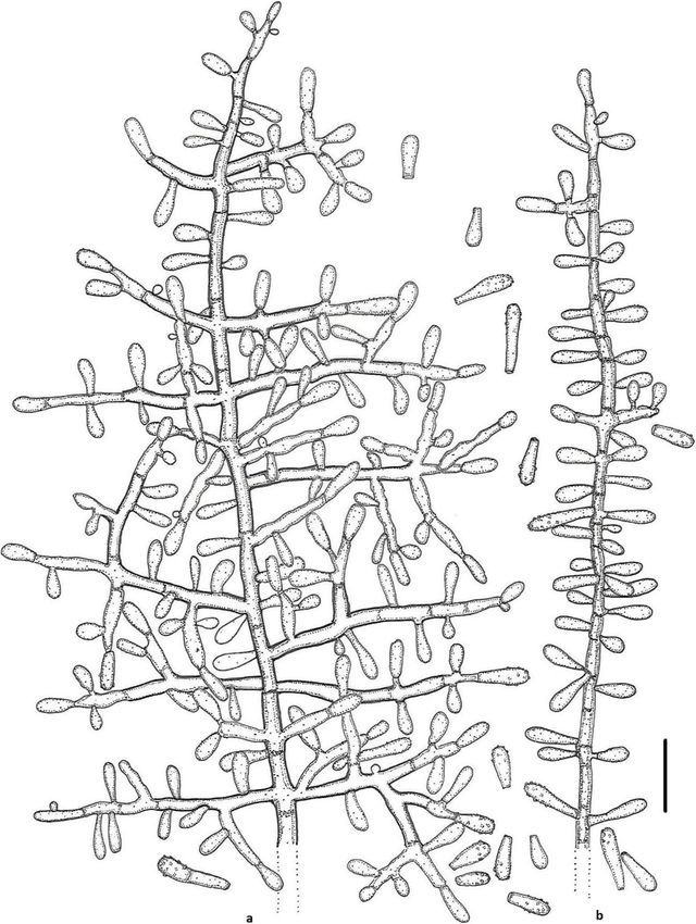
Labuda et al. IMA Fungus   (2021) 12:17                                                                            Page 2 of 21

Oorschot 1980; Vidal et al. 2000; Vidal et al. 2002;             Agrobiotechnology (IFA Tulln, Austria) in August 2015.
Zhang et al. 2016; Zhang et al. 2017). Sharma and                All three samples were taken from the surface layer (3–
Shouche (2017) introduced a new species, Keratinophy-            5 cm deep), dried, and stored in plastic bags in a fridge
ton turgidum, based on the morphology of its                     (5–8 °C) until the time of analysis (August 2015 and July
chrysosporium-like aleurioconidia and ITS locus phyl-            2019). Isolation of the keratinophilic fungi was per-
ogeny. The same authors stated that all species in this          formed as described previously (Javoreková et al. 2012).
monophyletic clade which have a Chrysosporium asexual            Each sample was divided into 10 subsamples. The sub-
morph require redisposing in the genus Keratinophyton.           samples (20 g each) were poured into Petri dishes and
   The presence of this large group of ubiquitous and            soaked with antibiotic solution containing 0.5 g cyclo-
keratinolytic species is rather common especially in areas       heximide and 0.1 g chloramphenicol. Sterile defatted
with high animal activity that results in transfer of the        horse hair fragments (10 pieces of ca 2.0 cm per plate)
keratinous material (fur, hairs, etc.) to the soil (Papini       were used as baits. The Petri dishes were then incubated
et al. 1998; Vidal et al. 2000). The following reports con-      at laboratory temperature (23–25 ± 1 °C), under ambient
firm their world-wide distribution and occurrence in dif-        daylight, for a period of 2–3 months and remoistened
ferent habitats usually associated with soil environments,       with sterile deionized water when necessary. The Petri
e.g. soil in city parks (Papini et al. 1998; Vidyasagar et al.   dishes were checked weekly for the presence of fungi,
2005), flower pots (Singh et al. 2009), sand in children’s       and isolates were cultured on Sabouraud 4% dextrose
sandpits (Labuda et al. 2008), mud (Zaki et al. 2005),           agar (SDA; Merck, Darmstadt, Germany) supplemented
poultry farms (Anbu et al. 2004; Cano and Guarro                 with 0.5 g cycloheximide and 0.05 g chloramphenicol.
1990), marshy meadows, salt pans, desert, cultivated or          Pure cultures were then transferred onto potato dextrose
uncultivated soils (Cano and Guarro 1990; Chmel and              agar [PDA; Van Waters and Rogers (VWR) Inter-
Vláčilíková 1977; Deshmukh 2004; Deshmukh et al.                 national, Leuven, Belgium]. The preliminary identifica-
2008; Han et al. 2013; Javorekova et al. 2012; Zhang             tion of the resulting keratinophilic fungi was carried out
et al. 2016; Zhang et al. 2017) and river sediments (Ulfig       based on their phenotypic characteristics according to
et al. 1997; Vidal et al. 2000; Vidal et al. 2002). In gen-      van Oorschot (1980) and Vidal et al. (2000, 2002).
eral, these fungi are rarely reported as animal pathogens,
and in fact, only two species C. echinulatum and C. pan-
nicola (formerly known as C. evolceanui) have been in-           Morphological analysis
volved in mycoses (Hajsig et al. 1974; Cabanes et al.            For phenotypic determination, the strains were trans-
2014; Hubka et al. 2016).                                        ferred (three-point inoculation with a needle) to PDA,
   During a microbiological survey of environmental              Malt Extract Agar (MEA; Merck, Darmstadt, Germany),
samples (soil and compost) in July 2019, several interest-       and SDA, and incubated for 14 d in the dark at 25 °C.
ing Chrysosporium asexual morphs were isolated. These            Christensen’s urea agar (Sigma-Aldrich, St Louis, MO,
isolates were phenotypically similar to those previously         USA) was used for additional physiological and bio-
isolated from the same samples in August 2015 by one             chemical characteristics (25 °C, 14 d, in the dark). Corn
us (R.L.). These isolates were designated BiMM-F76,              Meal Agar (CMA; Oxoid, Basingstoke, UK), Potato Car-
BiMM-F77 (also strain RL-07, isolated in July 2019),             rot Agar (PCA) (Samson et al. 2010) and Emerson YpSs
BiMM-F78 (also strains RL-05 and RL-06, isolated in              agar (Atlas 1946) were used for stimulation of sexual
July 2019), and BiMM-F250. All strains were further              reproduction (at 20 °, 25 °, and 28 °C, for up to 3 months
characterized in terms of morphology, physiology, and            in the dark).
molecular phylogeny. Phylogenetically informative se-               Colony size (mm), colony structure and characteristics
quences were obtained from the internal transcribed              were noted after 14 d (on PDA, MEA, SDA, PYE, YpSs,
spacer (ITS) region and the nuclear large subunit (LSU)          CMA, and PCA). However, the cultivation was extended
rDNA. Overall, the resulting data revealed that these iso-       up to 3 months to observe and record changes in pig-
lates represent novel species of the genus Keratinophy-          mentation of the colonies as well as to determine the
ton, and they were illustrated for the first time in this        onset of sexual reproduction. In order to determine the
paper.                                                           optimal and minimum/maximum temperatures for
                                                                 growth, PDA, MEA and SDA plates were incubated at 5
MATERIALS AND METHODS                                            °, 8 °, 10 °, 12 °, 15 °, 18 °, 20 °, 25 °, 28–32 °, 35 °, and
Sample collection and isolation of the fungi                     37 °C, and the growth rate was measured on the 14th
A sample of a garden soil in Vieste (Italy) was collected        day of cultivation. For comparative descriptions of the
in July 2004, one of a forest soil in Tatranská Lomnica          macroscopic and microscopic characteristics, PDA was
(The Slovak Republic) in August 2011, and one of com-            used according to Vidal et al. (2002), Hubka et al. (2016)
post from an agricultural base at the Institute of               and Sharma and Shouche (2017).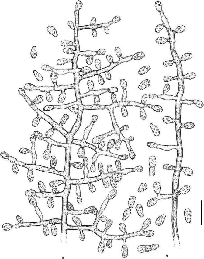
Labuda et al. IMA Fungus   (2021) 12:17                                                                       Page 3 of 21

   For determination of microscopic traits, PDA was used      with about 20% degradation of the hair; 3 = degradation
after 14–18 d. Conidiophore and conidia formation were        of cuticle and cortex, with about 50% degradation of the
observed in situ under low magnification (50–100x). De-       hair; 4 = degradation of cuticle and cortex, with about
tails of conidiophores, conidia (aleurioconidia) and other    80% degradation of the hair. The photomicrographs of
microscopic structures, such as width of hyphae, were         the hairs were taken using a Motic BA 310 microscope
observed in Melzer’s reagent and lactic acid with cotton      with Motic Image Plus 3.0 software. The final micro-
blue. Photomicrographs were taken in Melzer’s reagent         scopic pictures were black-and-white inverted.
and lactic acid with cotton blue using phase and
Nomarski contrast optics on an Olympus BX51 micro-            DNA extraction, PCR amplification and sequencing
scope with Olympus DP72 camera and QuickPHOTO                 DNA was extracted using a standard cetyltrimethyl am-
Micro 3.0 software. Photographs of the colonies were          monium bromide (CTAB) procedure, as described previ-
taken with a Sony DSC-RX100.                                  ously (Doyle and Doyle 1987). The internal transcribed
   Scanning electron microscopy (SEM) was performed           spacer (ITS) region was amplified with primers ITS1-F
on a JEOL JSM-6380 LV microscope (JEOL, Tokyo,                (Gardes and Bruns 1993) and ITS4 (White et al. 1990)
Japan). Fungal samples were prepared according to a           using Taq-polymerase. The D1/D2 domains of the large-
simplified method (Samson et al. 1979). Pieces of col-        subunit (28S) rRNA gene (LSU) were amplified and se-
onies (ca. 3 × 5 mm) growing on PDA were fixed in 6%          quenced using the primer pair ITS1/TW14 (White et al.
glutaraldehyde overnight in the refrigerator (ca. 20 h),      1990; Mori et al. 2000). All reactions were performed in
then dehydrated in 2-methoxyethanol for 10 min. This          an Eppendorf Gradient MasterCycler (Eppendorf, Ham-
was followed by critical point drying and gold coating in     burg, Germany). Conditions for amplification of ITS and
a BAL-TEC SCD 050 Sputter Coater. The samples were            LSU domains: 95 °C for 5 min; 35 cycles of 95 °C for 30 s,
observed with spot size 35–39 and accelerating voltage        54 °C for 30 s, 72 °C for 90 s, and finally 5 min at 72 °C.
20–23 kV.                                                     The PCR products were sequenced with the same
   Dried fungarium specimens deposited as holotypes in        primers used for the PCR amplifications (Microsynth,
the collections of the Mycological Department, National       Balgach, Switzerland). All sequences obtained in this
Museum in Prague, Czech Republic (PRM); ex-type cul-          study were deposited in GenBank nucleotide database
tures were deposited in the Bioactive Microbial Metabo-       (Table 1).
lites (BiMM) Fungal Collection, UFT- Tulln in Austria
and in the Culture Collection of Fungi in Prague (CCF).       Phylogenetic analysis
                                                              For phylogenetic analysis, sequences were aligned with
Keratinolytic activity                                        ClustalX (Larkin et al. 2007). Phylogenetic analysis based
Keratinolytic activity was tested by placing a few steril-    on ITS locus was performed using GTR + I + G4 + F
ized blond hairs of a 5 y old child on a PDA plate 1 cm       model with 1000 bootstrap replicates on IQ-TREE web
away from the point of inoculation (van Oorschot 1980).       server (Trifinopoulos et al. 2016) and ITS-LSU com-
Ability to digest keratin was observed after 21 d of incu-    bined data phylogeny was constructed using MRBAYES
bation at 25 °C in the dark. In addition, a hair perfor-      v3.2.7adev (Ronquist and Huelsenbeck 2003) with de-
ation test was also performed following de Hoog et al.        fault settings on the CIPRES portal (http://www.phylo.
(2020) using 25 mL water containing 2–3 drops 10%             org/). Ctenomyces serratus (type species CBS 187.61)
yeast extract (YEW). The hairs were examined micro-           was used as an outgroup. TREEVIEW v1.6.6 (Page 1996)
scopically after 14 and 21 d of the inoculation at 25 °C in   and iTOL v6 (Letunic and Bork 2019) were used to dis-
the dark. At the end of the incubation period, a few          play and edit phylogenetic trees.
pieces of hair were taken out from the testing media
(PDA and YEW). The overgrowing fungus was deacti-             RESULTS
vated with 70% ethanol and then removed from the hair         Morphological analyses and keratin degradation
surface mechanically in a stream of a tap water. The de-      The results of the morphological analyses are given for
gree of hair digestion-degradation (keratinolytic activity)   each novel species under the Taxonomy section below.
was assessed in the light microscope under 100x and           Temperature dependent growth of the new Keratinophy-
400x magnification. For the observation and micropho-         ton species on PDA, MEA and SDA after 14 d are pro-
tography of the hairs, water was used as mounting fluid.      vided in Table S1a–c. Briefly, K. lemmensii grew better
Intensity of degradation of the hair was estimated on a       than the other three new species on the same type of
scale of 0 to 4 (Marchisio et al. 1994): 0 = no degrad-       media and at the same incubation temperatures. All spe-
ation; 0–1 = light degradation on the cuticle; 1 = moder-     cies showed good growth at 20–25 °C on all three media.
ate degradation on the cuticle and/or rare formation of         Ability to digest keratin after 21 d was observed in all
boring hyphae; 2 = degradation of cuticle and cortex,         four new species on both testing media (PDA and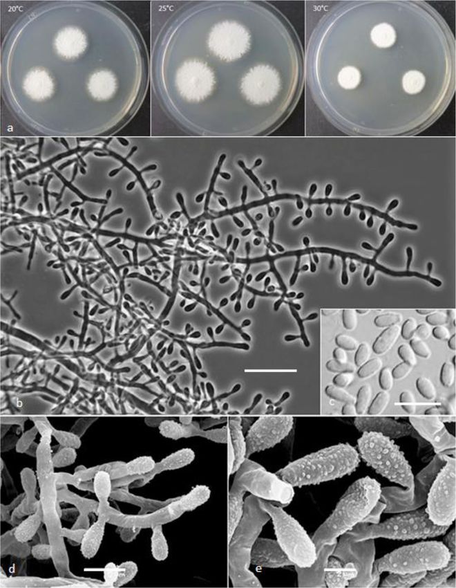
Labuda et al. IMA Fungus          (2021) 12:17                                                                                                      Page 4 of 21

Table 1 List of the strains included in the study
Species name                                     Straina                   Source                                          GenBank accession numbers
                                                                                                                           ITS                     LSU
A. canadensis                                    UAMH 4574                 Carnivore dung, Canada                          AJ439435                –
A. clathratus                                    IMI 329400                Arable soil, Spain                              AJ439436                –
A. cubensis                                      FMR 4220                  Soil of tobacco field, Cuba                     AJ439432                –
                                                              T
A. foetidus                                      CBS 453.75                Myomys daltoni coat, Nigeria                    KT155907                KT155252
A. fulvescens                                    NBRC 30411                Soil of rice paddy field, Japan                 JN943432                JN941547
                                                            T
A. keratinophilus                                IFM 55159                 Pasture land soil, Papua New Guinea             NR165936                NG064030
A. mephitalis                                    IMI 151084T               Dung of wolf, Canada                            AJ439439                AY176725
A. orissae                                       CBS 340.89                Soil in animal husbandry, Kuwait                AJ390393                –
A. pinarensis                                    FMR 4221                  Forest soil, Cuba                               AJ439433                –
                                                              T
A. reticulisporus                                CBS 392.67                Soil, New Zealand                               MH859002                MH870704
A. verrucosus                                    NBRC 32381T               Arable soil, Spain                              NR131309                NG057011
K. clavisporum (C. clavisporum)                  G80.1T                    Plant root soil, China                          KY026601                –
K. durum                                         CBS 118.85T               Soil, Nepal                                     MH861856                AB075345
                                                            T
K. echinulatum (C. echinulatum)                  CCF 4652                  Sole of the foot, Czechia                       LT548276                LT548276
K. fluviale (C. fluviale)                        FMR 6005T                 River sediments, Spain                          AJ005367                MT875000
K. gollerae                                      BiMM F250T                Forest soil, Slovakia                           MN633084                MT874997
K. hispanicum                                    CBS 456.90T               Beach soil, Spain                               KT155910                MT875003
K. hubeiense (C. hubeiense)                      EM66601T                  Soil under the chicken feather, China           KJ849227                –
K. lemmensii                                     BiMM F76T                 Compost soil, Austria                           MN633082                MT874998
K. linfenense (C. linfenense)                    GZAC H31    T
                                                                           Rhizosphere soil, China                         NR158289                –
K. minutisporosum (C. minutisporosum)            IMI 379912T               River sediments, Spain                          KT155616                MT875001
                                                              T
K. pannicola (C. pannicola)                      CBS 116.63                Soil, India                                     AJ005368                MH869834
K. punsolae                                      IMI 334818T               Arable soil, Spain                              AJ439440                –
K. qinghaiense (C. qinghaiense)                  GZUIFR Chry 11T           Farmland soil, China                            JX868607                –
K. saturnoideum                                  CBS 628.88T               Arable soil, Spain                              NR077135                AB075347
                                                                  T
K. siglerae (C. siglerae)                        UAMH 6541                 Garden soil, Spain                              AJ131684                MT875002
K. straussii                                     BiMM F78T                 Garden soil, Italy                              MN633081                MT874996
                                                                 T
K. submersum (C submersum)                       CBS 101575                River sediments, Spain                          NR157445                NG064180
K. terreum                                       CBS 342.64T               Lawn soil, India                                KT155876                KC989709
                                                                 T
K. turgidum                                      CBS 142596                Barber shop soil, India                         KY290503                KY962732
K. wagneri                                       BiMM F77T                 Forest soil, Slovakia                           MN633083                MT874999
Ct. serratus                                     CBS 187.61T               Soil, Australia                                 NR144890                AY176733
a
 BiMM, Bioactive Microbial Metabolites Unit, UFT-Tulln, Austria; UAMH, University of Alberta Microfungus Collection and Herbarium; IMI, CAB International
Biosciences, Egham, UK; FMR, Facultat de Medicina in Ciències de la Salut, Reus, Spain; CBS (Westerdijk Fungal Biodiversity Institute), Utrecht, The Netherlands;
NBRC, NITE Biological Resource Centre, Japan; IFO, Institute for Fermentation, Osaka, Japan; G, EM, and GZUIFR strains, The Institute of Fungus Resource, Guizhou
University, China; A, Aphanoascus; K, Keratinophyton; C, Chrysosporium; Ct, Ctenomyces; T, ex-type culture. Data in bold generated in the present study

YEW). However, a value of attack intensity on the hair                              informative sites. ITS phylogeny indicated the presence
according to the scale of Marchisio et al. (1994) differed                          of six terminal clusters in the monophyletic Keratino-
substantially amongst the species. It was very strong in                            phyton clade with high bootstrap support and low inter-
K. gollerae and K. straussii (=4), moderate in K. wagneri                           specific sequence divergence (Fig. 1a). Keratinophyton
(=2), and weak in K. lemmensii (= 0–-1) (Fig. 10).                                  saturnoideum and K. minutisporosum formed a basal
                                                                                    branch to the clade. Isolate BiMM-F76 (K. lemmensii sp.
Phylogenetical analysis                                                             nov.) was close to K. durum (with 99% ITS and 95%
The phylogenetic tree of ITS dataset (n = 32) was 551 bp                            LSU similarity) and clustered also with K. hubeiense and
in length which had 286 variable and 200 parsimony-                                 K. submersum. In addition, K. straussii sp. nov., K.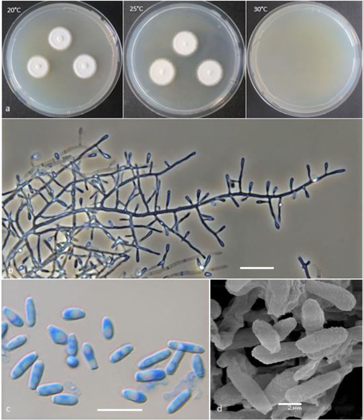
Labuda et al. IMA Fungus      (2021) 12:17                                                                                            Page 5 of 21

 Fig. 1 a Maximum Likelihood (ML) tree based on ITS sequence for the new taxa of Keratinophyton is compared with available sequences of the
 other related species as well as their conidium size, presence of intercalary conidia and ability to grow at 37 °C. Numbers at nodes indicate
 bootstrap values. Ctenomyces serratus was used as outgroup. A sequence for K. multiporum was not available for the study. T, ex-type culture.
 New species are shown in bold. b Bayesian interference tree based on combination of ITS and LSU rDNA sequences for new taxa of
 Keratinophyton together with available sequences of the other related species. Numbers at nodes indicate bootstrap values. Ctenomyces serratus
 was used as outgroup. New species are shown in bold

gollerae sp. nov., and K. wagneri sp. nov., represented by                 and LSU sequences (n = 22) was 1094 bp length and in-
the ex-type cultures BiMM-F78, BiMM-F250 and                               cluded 354 variable and 224 parsimony-informative sites.
BiMM-F77, respectively, were resolved in a separate ter-                   According to a combined data set analysis, four clusters
minal cluster-lineage. A concatenated phylogeny of ITS                     were found in the Keratinophyton clade with K.
Labuda et al. IMA Fungus   (2021) 12:17                                                                     Page 6 of 21

saturnoideum as a basal branch (Fig. 1b). Differently           Type: Slovak Republic: Tatranská Lomnica, from for-
from the ITS phylogeny, K. durum was placed in a dif-        est soil, Jul. 2019, R. Labuda (PRM 952499 – holotype;
ferent cluster from K. submersum and K. lemmensii in         BiMM-F250 = CCF 6360 – ex-type cultures). ITS se-
the concatenated loci phylogeny (Fig. 1b).                   quence, GenBank MN633084; LSU sequence, GenBank
                                                             MT874997.
TAXONOMY                                                        Description: Sexual morph not observed on any of
The phylogenetic analyses strongly supported the re-         the media used. Asexual morph on PDA. Vegetative
cent distinct classification of the species previously       mycelium of hyaline, septate, smooth-walled, sparsely
classified as Chrysosporium and only known from              to pronouncedly branched hyphae, often at right an-
asexual morphs into two phylogenetically different           gles, 1.0–5.0 μm diam. Racquet hyphae present. Co-
genera, Aphanoascus and Keratinophyton (Sharma and           nidia (aleurioconidia), hyaline, white in mass, thin-
Shouche 2017; Sutton et al. 2013). Species described         walled, mostly smooth to finely roughened, some also
in Chrysosporium which were resolved in a monophy-           verrucose (light microscope) and irregularly ornamen-
letic clade with Keratinophyton are therefore com-           ted with minute warts (SEM). Terminal and lateral
bined into Keratinophyton in the present paper and           conidia born on main fertile hyphae or from side
provided together with four new Keratinophyton spe-          branches of variable length, sessile or on short pro-
cies. The main distinguishing phenotypic characteris-        trusions, occasionally only very slightly swollen and of
tics of the four new species were compared with              variable length, solitary, 1–3 (− 5) per conidiogenous
those in the other members of the genus that are also        cell, obovate to clavate, mostly 1-celled, (3.5–)5.0–
unable to produce ascomata (Table 2).                        7.0(− 10.0) x (1.5–)2.0–2.5(− 3.0) μm (mean = 5.2 ±
  Keratinophyton clavisporum (Zhang, Han & Liang)            0.9 × 2.2 ± 0.2 μm, n = 120). Intercalary conidia not ob-
Labuda & Bernreiter, comb. nov.                              served. Chlamydospores not observed.
  MycoBank: MB833653                                            Culture characteristics: Colonies on PDA 20–22 mm
  Basionym: Chrysosporium clavisporum Y.W. Zhang,            diam at 25 °C, after 14 d, powdery to downy (mealy),
Y.F. Han & Z.Q. Liang - Phytotaxa 303: 177; 2017.            with abundant sporulation, white to creamy, flat, umbo-
  Type: GZUIFR-G80.1; isolated from plant root soil by       nate at the centre, with regular colony margin sub-
Y. Luo, China. For detailed description of the species,      mersed into agar, reverse white to slightly yellowish, no
see the Zhang et al. (2017).                                 pigment or exudate produced. At 30 °C, no growth (ger-
  Keratinophyton echinulatum (Hubka, Mallátová,              mination only). Colonies on SDA 23–25 mm diam at
Čmoková & Kolařík) Labuda & Bernreiter, comb. nov.           25 °C, after 14 d, morphology similar to when on PDA
  MycoBank: MB833636                                         with more floccose colony margin and more yellowish
  Basionym: Chrysosporium echinulatum Hubka, Mallá-          colonies, with dark yellow reverse. At 30 °C, no growth
tová, Čmoková & M. Kolařík - Persoonia 36: 410; 2016.        (no germination). Colonies on MEA 14–16 mm diam at
  Type: CCF 4652 = CBS 141178 = UAMH 11824; from             25 °C, after 14 d, morphology similar to PDA with more
sole of the foot by N. Mallátová, Czechia. For detailed      floccose colonies and with yellow reverse. At 30 °C, no
description of the species, see the Hubka et al. (2016).     growth (no germination). Colonies on CMA and PCA
  Keratinophyton fluviale (Vidal & Guarro) Labuda &          attaining 15–20 mm diam at 25 °C, after 21 d, white,
Bernreiter, comb. nov.                                       granular, with good sporulation, reverse yellowish. No
  MycoBank: MB8333637                                        ascomata observed after prolonged incubation (3
  Basionym: Chrysosporium fluviale Vidal & Guarro -          months). The optimum temperature for growth on PDA,
Mycol. Res. 104: 245; 2000.                                  SDA and MEA 15–25 °C (Table S1a–c). Minimum
  Type: CBS 100809 = FMR 6005 = IMI 378764, isolated         growth (microcolonies to 1 mm in diam) at 10 °C. Ger-
from river sediments, by P. Vidal, Spain. For detailed de-   mination of the conidia observed at 8 °C. The maximum
scription of the species, see the Vidal et al. (2000).       temperature for growth on PDA 29 °C, while 27 °C and
  Keratinophyton gollerae Labuda, Bernreiter, Kubá-          28 °C on MEA and SDA, respectively (microcolonies to
tová, Schüller & Strauss, sp. nov.                           1 mm diam). Keratinolytic activity very strong (Fig. 10b),
  (Figs. 2 and 3)                                            with hair attack intensity = 4. Urease activity negative
  MycoBank: MB833633                                         (after 14 d of incubation).
  Etymology: Named in honour of Sabine Strauss-Goller,          Diagnosis: Keratinophyton gollerae molecularly can be
Department of Applied Genetics and Cell Biology, Fun-        distinguished from other Keratinophyton species by ITS
gal Genetics and Genomics Laboratory, University of          locus analysis. Combination of the following phenotypic
Natural Resources and Life Sciences, Vienna (BOKU),          features can be used to differentiate this fungus from
Austria, an expert in the fungal genetics and indoor         other species in the genus: (1) obovoid-clavate and
mould analyses.                                              smooth to finely roughened conidia, (2) No growth at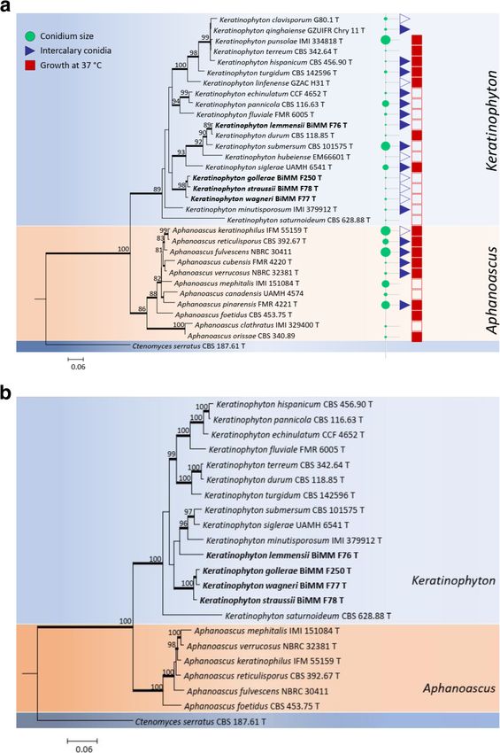
Table 2 Comparison of the key phenotypic characteristics of Keratinophyton species
Species            Growth at 30 °C     Colony color, growth/ reverse on PDA     Conidial shape                    Conidial               Conidial surface       Intercalary       References
                   on PDAa             at 25 °C, after 14db                                                       dimensions                                    conidia
                                                                                                                  (μm)
K. gollerae sp.    None                White to creamy, 20–22 mm/white to       Obovoid to clavate                5.0–7.0 × 2.0–2.5      Smooth to finely       Absent            This study
                                                                                                                                                                                                      Labuda et al. IMA Fungus

nov.                                   yellowish                                                                                         roughened
K. lemmensii       Present (good)      White, 28–35 mm/lemon yellow             Clavate to filiform               3.0–40 μm (1- to 2-    Smooth                 Present           This study
sp. nov.                                                                                                          celled)
K. straussii sp.   Present (good)      White to creamy, 24–28 mm/white to       Obovoid to clavate                4.5–5.0 × 2.5–3.0      Verrucose              Absent            This study
nov.                                   yellowish
                                                                                                                                                                                                      (2021) 12:17

K. wagneri sp.     Present             White to yellowish, 25–30 mm/white to    Obovoid to clavate                4.0–8.0 × 2.5–4.0      Verrucose              Absent            This study
nov.               (restricted)        yellowish
K. clavisporum     Present             White, 53 mm (26 °C)/red-brown           Clavate to long -ellipsoidal      5.0–10 × 2.5–5.0       Smooth                 Absent            Zhang et al. 2017
                   (restricted)e
K. echinulatum     Present (good)      Yellow to pale orange yellow, 28–45 mm/ Obovoid to clavate                 4.5–7.0 × 2.5–4.0      Echinulate             Present           Hubka et al. 2016
                                       orange yellow
K. fluviale        Present (good)      White to yellowish white, 60–70 mm       Obovate, clavate, nearly          3.5–15 × 2.0–3.0 (1-   Verrucose              Present (very     Vidal et al. 2000
                                       (30 °C)/brownish orange                  ellipsoidal or pyriform           and 2-celled)                                 rare)
K. qinghaiense     Present (good)d     White to yellowish, 30 mm (7 days)/      Clavate to cylindrical            3.6–13 × 1.8–3.6       Smooth                 Present           Han et al. 2013
                                       yellowish
K. hubeiense       Present             Grey white to white, 65–67 mm/reverse    Obovoid to ellipsoidal            2.2–4.3 × 1.6–3.2      Smooth                 Absent            Zhang et al. 2016
                   (restricted)e       yellowish
K. linfenense      Present (good)      White to cream, 72 mm (30 °C)/white to   Ellipsoidal to fusiform, also     3.2–5.4 x-1.4–2.2      Smooth                 Absent            Liang et al. 2009
                                       light yellow                             clavate
K.             Present (good)          White to yellowish white, 55–70 mm/      Pyriform or subglobose, also      3.0–4.0 (−11) × 1.5–   Verrucose              Present (very     Vidal et al. 2002
minutisporosum                         white                                    clavate                           3.5                                           rare)
K. pannicola       Present (good)      White to pale yellow, 20–38 mm /pale     Obovoid to clavate                6.0–11 × 3.5–4.5       Verrucose              Present (less     van Oorschot
                                       brownc                                                                                                                   abundant)         1980
K. siglerae        Present (good)      Griseous orange, 15–20 mm (21 d)/ pale   Cylindrical to clavate            5.0–30 × 2.0–3.5       Smooth to slightly     Present           Cano and Guarro
                                       brown                                                                      1-and 2-celled         verrucose                                1994
K. submersum       Present             Yellowish white, 50–60 mm/yellowish      Clavate, also pyriform, obovoid   4.0–35 × 2.5–5.0       Smooth to verrucose-   Present (in old   Vidal et al. 2002
                   (restricted)        white                                    and subglobose                    (1- to 4-celled)       thick-walled           cultures)
K. turgidum        Present (good)      White, 50–55 mm (SGA at 28 °C)/pale      Pyriform to oval                  5.0–7.0 × 3.5–5.0      Smooth                 Present           Sharma and
                                       brown                                                                                                                                      Shouche 2017
a
  if not stated other medium
b
  if not stated otherwise
c
 PYE, Phytone yeast extract agar
d
  Yanfeng Han personal communication
                                                                                                                                                                                                      Page 7 of 21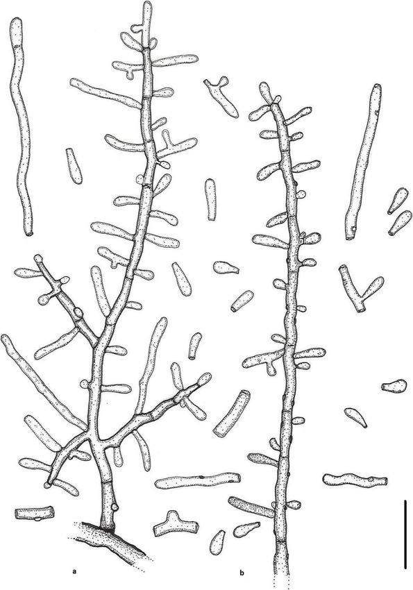
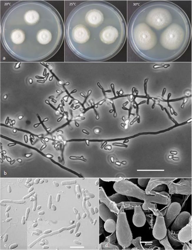
Labuda et al. IMA Fungus       (2021) 12:17                                                                                            Page 8 of 21

 Fig. 2 Keratinophyton gollerae (BiMM-F250). a Colonies on PDA (after 14 d) at 20 °C, 25 °C and 30 °C. b Conidiophores with aleurioconidia. c
 Aleurioconidia and arthroconidia (on PDA, after 14 d). d Scanning electron microscopy (SEM) of aleurioconidia (on PDA, after 14 d). Bars = 20 μm
 (b), 10 μm (c), 2 μm (d)

30 °C, and (3) yellowish colonies with dark yellow re-                      slower growth at 25 °C on PDA. Moreover, in compari-
verse at 25 °C on SDA.                                                      son with K. straussii, K. gollerae grows substantially fas-
  Notes: Based on a search of NCBI GenBank nucleotide                       ter at 15 °C (on PDA and SDA) and its conidia
database, the closest hit for K. gollerae using the ITS se-                 germinate at 8 °C (see Table S1a–c).
quence is K. minutisporosum (as Chrysosporium minutis-                        Keratinophyton hubeiense (Zhang, Han & Liang)
porosum CBS 101577; GenBank acc. KT155616), with                            Labuda & Bernreiter, comb. nov.
identity = 487/543 (90%) and gaps 11/543 (2%). Pheno-                         MycoBank: MB833638
typically, K. gollerae can be readily distinguished from                      Basionym: Chrysosporium hubeiense Yan W. Zhang,
the K. minutisporum by its smooth to finely roughened                       Y.F. Han & Z.Q. Liang - Phytotaxa 270: 213; 2016.
larger conida (5–7 × 2–2.5 μm vs. 3–4 × 1.5–3.5 μm),                          Type: GZAC EM66601, isolated from soil under the
dark yellow colony reverse at 25 °C on PDA. Based on                        chicken feather by Y.R. Wang, China. For detailed de-
ITS phylogeny (Fig. 1a), K. gollerae formed a cluster to-                   scription of the species, see the Zhang et al. (2016).
gether with K. straussii and K. wagneri, and it can be dif-                   Keratinophyton lemmensii Labuda, Bernreiter, Kubá-
ferentiated by its inability to grow at 30 °C, narrower and                 tová & Schüller, sp. nov.
mostly smooth to finely roughened conidia, and its                            (Figs. 4 and 5)
Labuda et al. IMA Fungus     (2021) 12:17                                                                                        Page 9 of 21

 Fig. 3 Line drawing of micromorphology of Keratinophyton gollerae (BiMM-F250). a, b Conidiophores with young and mature aleurioconidia on
 PDA (after 14 d). a Branched conidiophore. b Unbranched conidiophore with sessile aleurioconidia. Bar = 10 μm

  MycoBank: MB833632                                                    consisting of hyaline, smooth-walled, septate, sparsely
  Etymology: Named in honour of Marc Lemmens, De-                       branched hyphae, 1.5–5.0 μm diam. Racquet hyphae
partment of Plant Protection, University of Natural Re-                 present. Conidia aleuroconidia, hyaline, white in mass,
sources and Life Sciences, Vienna (BOKU), Austria, an                   thin-walled, smooth to sparsely irregularly ornamented
expert in fungal plant pathology.                                       with minute warts (SEM); terminal and lateral conidia
  Type: Austria: Tulln and der Donau, from compost                      born on main fertile hyphae as sessile or on short protru-
soil at IFA Tulln, Aug. 2015, R. Labuda (PRM 952498 –                   sions, solitary, 1–3 (− 5) per conidiogenous cell, obovate to
holotype; BiMM-F76 = CCF 6359 – ex-type cultures).                      clavate, 1-celled, (3.0–)4.5–6.5(− 7.5) x (1.5–)2.0–2.5(−
ITS sequence GenBank MN633082; LSU sequence Gen-                        4.0) μm (mean = 4.9 ± 0.8 × 2.4 ± 0.4 μm, n = 120), and fili-
Bank MT874998.                                                          form, often sinusoidal, 1- to 2-celled, 25–35(− 40) μm long
  Description: Sexual morph not observed on any of the                  conidia also present. Intercalary conidia (arthroconidia)
media used in the present study. Vegetative mycelium                    present, 10–15 μm long. Chlamydospores not observed.
Labuda et al. IMA Fungus       (2021) 12:17                                                                                           Page 10 of 21

 Fig. 4 Keratinophyton lemmensii (BiMM-F76). a Colonies on PDA (after 14 d) at 20 °C, 25 °C and 30 °C. b Conidiophores with aleurioconidia. c
 Aleurioconidia and arthroconidia (on PDA, after 14 d). d Scanning electron microscopy (SEM) of aleurioconidia (on PDA, after 14 d). Bars = 20 μm
 (b), 10 μm (c), 2 μm (d)

  Culture characteristics: Colonies on PDA 28–35 mm                         after 14 d, white, flat, floccose with good sporulation,
diam at 25 °C, after 14 d, floccose, with good sporulation,                 with pale yellow reverse. Colonies on MEA 20–25 mm
white, flat, slightly elevated (umbonate) at the centre,                    diam at 25 °C after 14 d, morphology similar to PDA, ex-
with irregular margin, reverse lemon yellow, soluble pig-                   udate absent, and pale-yellow reverse. At 30 °C, 18–20
ment bright yellow, a few small clear to yellow-orange                      mm diam after 14 d, white, floccose and radially sulcate,
exudate droplets produced. At 30 °C, 38–45 mm diam                          with good sporulation, and with pale yellow reverse. Col-
after 14 d, white, flat, floccose and radially sulcate with                 onies on CMA and PCA, 45–50 mm diam at 25 °C, after
good sporulation only at the centre, and with lemon yel-                    21 d, white, flat and spread with poor sporulation, re-
low reverse. Colonies on SDA 28–35 mm diam at 25 °C,                        verse white. No ascomata observed after prolonged incu-
after 14 d, morphology similar to PDA, without exudate                      bation (3 months). The optimum temperature on PDA,
and with pale yellow reverse. At 30 °C, 30–32 mm diam                       SDA and MEA 25–30 °C (Table S1a–c). Minimum
Labuda et al. IMA Fungus      (2021) 12:17                                                                                       Page 11 of 21

 Fig. 5 Line drawing of micromorphology of Keratinophyton lemmensii (BiMM-F76). a, b Conidiophores with young and mature aleurioconidia,
 including arthroconidia on PDA (after 14 d). a Branched conidiophore. b Unbranched conidiophore with sessile aleurioconidia. Bar = 10 μm

growth (1–2 mm diam) at 8 °C. The maximum                                phylogeny (K. durum, K. hubeiense, K. submersum, and
temperature for growth 32 °C (microcolonies to 1 mm                      K. siglerae) by the combination of the following features:
diam). Keratinolytic activity very weak (Fig. 10a), with                 (1) presence of long filiform often sinusoidal uni- to
hair attack intensity = 0–1. Urease activity positive (after             bicellular conidia (to 40 μm), (2) white, moderately fast
3 d of incubation).                                                      growing colonies (28–35 mm diam, on PDA at 25 °C),
  Diagnosis: This species molecularly can be distin-                     (3) production of lemon yellow pigment on PDA at
guished from other Keratinophyton species by ITS locus                   25 °C, (4) minimum 8 °C and maximum 32 °C growth
analysis. Phenotypically, K. lemmensii is unique and dif-                temperature, (5) very weak keratin digestion after 21 d.
fers from the relatives in the same clade based on ITS                   Presence of filiform often sinusoidal conidia and
Labuda et al. IMA Fungus   (2021) 12:17                                                                      Page 12 of 21

abundant arthroconidia, production of bright yellow pig-        Basionym: Chrysosporium submersum P. Vidal &
ment on PDA and good growth at 30 °C.                         Guarro - Stud. Mycol. 47: 200; 2002.
  Notes: Based on a search of NCBI GenBank nucleotide           Type: CBS 101575 = IMI 379911 = FMR 6088, isolated
database, the closest hit for K. lemmensii using the ITS      from river mouth sediment by P. Vidal, Spain. For de-
sequence was K. durum (FMR5651; GenBank acc.                  tailed description of the species, see Vidal et al. (2002).
AJ439434; identities = 568/577 (98%), gaps 0/577 (0%).          Keratinophyton straussii Labuda, Bernreiter, Kubá-
However, K. lemmensii can be directly distinguished           tová & Schüller, sp. nov.
from K. durum by its asexual morph also by the pres-            (Figs. 6 and 7)
ence of numerous arthroconidia which are completely             MycoBank: MB833634
missing in the latter species (Cano and Guarro 1990;            Etymology: Named in honour of Joseph Strauss, Head
Currah 1985).                                                 of the Department of Applied Genetics and Cell Biology,
  Keratinophyton linfenense (Liang, Liang & Han)              founder of the Fungal Genetics and Genomics Labora-
Labuda & Bernreiter, comb. nov.                               tory, University of Natural Resources and Life Sciences,
  MycoBank: MB833639                                          Vienna (BOKU), Austria, and an expert in fungal genet-
  Basionym: Chrysosporium linfenense Z.Q. Liang, J.D.         ics, epigenetics and functional genomics.
Liang & Y.F. Han - Mycotaxon 110: 67; 2009.                     Type: Italy: Vieste, from garden soil, Aug. 2015, R.
  Type: GZUXIFR H31, isolated from rhizosphere soil           Labuda (PRM 952500 – holotype; BiMM-F78 = CCF
by G. Don, China. For detailed description of the species,    6361 – ex-type cultures). ITS sequence, GenBank
see Liang et al. (2009).                                      MN633081; LSU sequences, GenBank MT874996.
  Keratinophyton minutisporosum (Vidal & Guarro)                Description: Sexual morph not observed on any of the
Labuda & Bernreiter, comb. nov.                               media used. Asexual morph on PDA. Vegetative
  MycoBank: MB833640                                          mycelium of hyaline, septate, smooth-walled, sparsely to
  Basionym: Chrysosporium minutisporosum P. Vidal &           pronouncedly branched hyphae, usually at right angles,
Guarro - Stud. Mycol. 47: 205; 2002.                          1.5–4.0 μm diam. Racquet hyphae present. Conidia
  Type: CBS 101577 = IMI 379912 = FMR 6096 isolated           (aleurioconidia), hyaline, white to yellowish in mass,
from river mouth sediment by P. Vidal, Spain. For de-         thin-walled and regularly ornamented with minute warts
tailed description of the species, see Vidal et al. (2002).   (SEM) and coarsely roughened (light microscope). Ter-
  Keratinophyton pannicola (Corda) Labuda & Bern-             minal and lateral conidia born on main fertile hyphae or
reiter, comb. nov.                                            from side branches of variable length, sessile or on short
  MycoBank: MB8333643                                         protrusions, commonly slightly swollen, length variable,
  Basionym: Capillaria pannicola Corda - Icon. Fung.1:        solitary, 1–3 (5) per conidiogenous cell, obovate to cla-
10; 1837.                                                     vate, 1-celled, (3.5–)4.5–5.0(− 6.5) x (2.0–)2.5–3.0(− 3.5)
  ≡ Sporotrichum pannicola (Corda) Rabenh. - Deutschl.        μm (mean = 4.9 ± 0.4 × 2.6 ± 0.2 μm, n = 120), very rarely
Krypt.-Fl. 1: 78; 1844.                                       2- to 3-celled, to 12 μm large aleurioconida also present.
  ≡ Chrysosporium pannicola (Corda) Oorschot & Stal-          Intercalary conidia not observed. Chlamydospores not
pers - Stud. Mycol. 20: 43; 1980.                             observed.
  Synonym: Trichophyton evolceanui H.S. Randhawa &              Culture characteristics: Colonies on PDA 24–28 mm
R.S. Sandhu - Mycopath. Mycol. Appl. 20: 232; 1963.           diam at 25 °C, after 14 d, powdery to downy (mealy),
  ≡ Chrysosporium evolceanui (Randhawa & Sandhu)              with abundant sporulation, white to very slightly creamy
Garg - Sabouraudia 4: 262; 1966.                              yellowish, flat, slightly elevated (umbonate) remaining
  Type: CBS 116.63 = ATCC 22400 = IHEM 4436 = IMI             powdery at the centre, with irregular margin, reverse
147545 = NCPF 489 = RV 26475 = UAMH 1275, isolated            white with slightly yellowish centre, no pigment or exud-
from soil by Randhawa & Sandhu, India.                        ate produced. At 30 °C, 15–20 mm diam after 14 d,
  Keratinophyton siglerae (Cano & Guarro) Labuda &            white to creamy yellowish, flat, powdery to downy
Bernreiter, comb. nov.                                        (mealy) with very good sporulation, and with white to
  MycoBank: MB833641                                          yellowish reverse. Colonies on SDA 16–20 mm diam at
  Basionym: Chrysosporium siglerae Cano & Guarro -            25 °C, after 14 d, morphology as on PDA with dark yel-
Mycotaxon 51: 75; 1994.                                       low reverse. In age (after 5 wk) yellow pigment produced
  Type: UAMH 6541 = FMR 3066 = IMI 336467, isolated           and colony reverse becoming bright reddish yellow to
from garden soil, Spain. For detailed description of the      orange. At 30 °C, 15–20 mm diam after 14 d, white to
species, see Cano and Guarro (1994).                          creamy yellowish, umbonate, with strong sporulation,
  Keratinophyton submersum (Vidal & Guarro) Labuda            and with yellowish reverse. Colonies on MEA 18–20 mm
& Bernreiter, comb. nov.                                      diam at 25 °C, after 14 d, morphology as on PDA with
  MycoBank: MB833642                                          more floccose and yellowish. At 30 °C, 5–10 mm diam
Labuda et al. IMA Fungus       (2021) 12:17                                                                                           Page 13 of 21

 Fig. 6 Keratinophyton straussii BiMM-F78. a Colonies on PDA (14 d old) at 20 °C, 25 °C and 30 °C. b Conidiophores with aleurioconidia. c
 Aleurioconidia (on PDA, after 14 d). d, e Scanning electron microscopy (SEM) of conidiogenous cells and aleurioconidia (on PDA, after 14 d).
 Bars = 20 μm (b), 10 μm (c), 5 μm (d), 2 μm (e)

after 14 d, slightly umbonate, floccose to granular, with                   very strong (Fig. 10c), with hair degradation intensity =
very good sporulation white to yellowish, and with yel-                     4. Urease activity negative (after 14 d of incubation).
low reverse. Colonies on CMA and PCA 18–20 mm                                 Diagnosis: Keratinophyton straussii molecularly can be
diam at 25 °C, after 21 d, white, granular, good sporula-                   distinguished from other Keratinophyton species by ITS
tion, reverse yellowish. No ascomata observed after pro-                    locus analysis. Phenotypically, it can be differentiated by
longed     incubation     (3 months).   The     optimum                     combination of the ability to grow at 30 °C, white to
temperature for growth on PDA, SDA and MEA 20–                              creamy colonies with white to yellowish revers at 25 °C
25 °C (Table S1a–c). Minimum growth (microcolonies to                       on PDA and conidia morphology (obovoid to clavate
1–2 mm diam) at 10 °C. No germination of the spores at                      and verrucose) (Table 2).
8 °C. The maximum temperature for growth 32 °C                                Additional material examined: Italy: Vieste, from gar-
(microcolonies to 1–2 mm diam). Keratinolytic activity                      den soil, isolated from different sub-samples, July 2019,
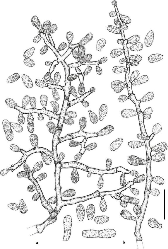
Labuda et al. IMA Fungus      (2021) 12:17                                                                                       Page 14 of 21

 Fig. 7 Line drawing of micromorphology of Keratinophyton straussii (BiMM-F78). a, b Conidiophores with young and mature aleurioconidia on
 PDA (14 d old). a Branched conidiophore. b Unbranched conidiophore with sessile aleurioconidia. Bar = 10 μm

R. Labuda RL-05 ITS sequence, MT898644; LSU se-                          between K. gollerae and K. straussii, see under K. gol-
quence, MT898648); ibid., RL-06 (ITS sequence,                           lerae. Additional strains RL-05 and RL-06 grew relatively
MT898645; LSU sequence, MT898649).                                       better (to 5 mm larger diam) than the ex-type culture at
  Notes: Based on a search of the NCBI GenBank nu-                       30 °C.
cleotide database, the closest hit for K. straussii using                  Keratinophyton qinghaiense (Han, Liang & Liang)
the ITS sequence was K. minutisporosum (as Chrysospor-                   Labuda & Bernreiter, comb. nov.
ium minutisporosum CBS 101577; GenBank acc.                                MycoBank: MB833655
KT155616), with identity = 489/543 (90%) and gaps 10/                      Basionym: Chrysosporium qinghaiense Y.F. Han, J.D.
543 (1%). Two species can be differentiated from each                    Liang & Z.Q. Liang - Mycosystema 32: 607, 2013.
other based on growth rate and colony reverse at 25 °C                     Type: GZAC GZUIFR-Chry 11, from farmland soil by,
on PDA (Table 2). Additionally, K. straussii differs from                Y.F. Han, China.
K. wagnerii by its ability to grow at 30 °C and strong ker-                Keratinophyton wagneri Labuda, Bernreiter, Kubá-
atinolytic activity. For the morphological differences                   tová & Schüller, sp. nov.
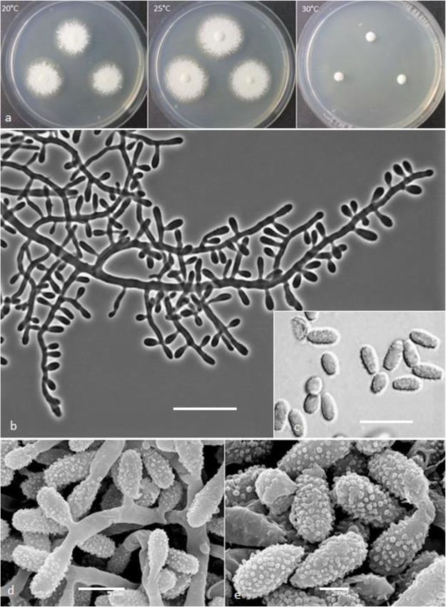
Labuda et al. IMA Fungus       (2021) 12:17                                                                                           Page 15 of 21

  (Figs. 8 and 9)                                                           sequence, GenBank MN633083; LSU sequence, Gen-
  MycoBank: MB 833635.                                                      Bank MT874999.
  Etymology: Named in honour of Martin Wagner, Head                            Description: Sexual morph not observed on any of the
of the Unit for Food Microbiology and Head of Institute                     media used. Asexual morph on PDA. Vegetative myce-
for Food Safety, Food Technology and Veterinary Public                      lium hyaline, septate, smooth-walled, sparsely to pro-
Health, University of Veterinary Medicine, Vienna                           nouncedly branched hyphae, 2.0–6.0 μm diam. Racquet
(Austria), an expert in veterinary microbiology.                            hyphae present. Conidia (aleurioconidia), hyaline, white
  Type: Slovak Republic: Tatranská Lomnica, from for-                       to yellowish in mass, thin-walled and regularly ornamen-
est soil, Aug. 2015, R. Labuda (PRM 952501 – holotype;                      ted with minute warts (SEM) and coarsely roughened
BiMM-F77 = CCF 6362 – ex-type cultures). ITS                                (light microscope). Terminal and lateral conidia born on

 Fig. 8 Keratinophyton wagneri (BiMM-F77). a Colonies on PDA (after 14 d) at 20 °C, 25 °C and 30 °C. b Conidiophores with aleurioconidia. c
 Aleurioconidia (on PDA, after 14 d). d, e Scanning electron microscopy (SEM) of conidiogenous cells and aleurioconidia (on PDA, after 14 d).
 Bars = 20 μm (b), 10 μm (c), 5 μm (d), 2 μm (e)
Labuda et al. IMA Fungus     (2021) 12:17                                                                                      Page 16 of 21

 Fig. 9 Line drawing of micromorphology of Keratinophyton wagneri (BiMM-F77). a, b Conidiophores with young and mature aleurioconidia on
 PDA (after 14 d). a Branched conidiophore. b Unbranched conidiophore with sessile aleurioconidia. Bar = 10 μm

main fertile hyphae or from side branches of variable                      Culture characteristics: Colonies on PDA 25–30 mm
length, sessile or on short protrusions, occasionally swol-             diam at 25 °C, after 14 d, powdery to downy (mealy),
len and of variable length, solitary, 1–4 (− 10) per coni-              with abundant sporulation, white to slightly yellowish,
diogenous cell, obovate to clavate, single celled, (4.0–)               flat, slightly elevated (umbonate) and more floccose at
5.5–6.5 (− 8.0) x (2.5–) 3.0–3.5(− 4.0) μm (mean = 5.7 ±                the centre, margin irregular, reverse white with slightly
0.4 × 3.2 ± 0.2 μm, n = 120), rarely 2-celled, up to 12 μm              yellowish centre, no pigment or exudate produced. At
large ones also present. Intercalary conidia not observed.              30 °C, 4–8 mm diam after 14 d, white, floccose with poor
Chlamydospores not observed.                                            sporulation, and with yellowish reverse. Colonies on
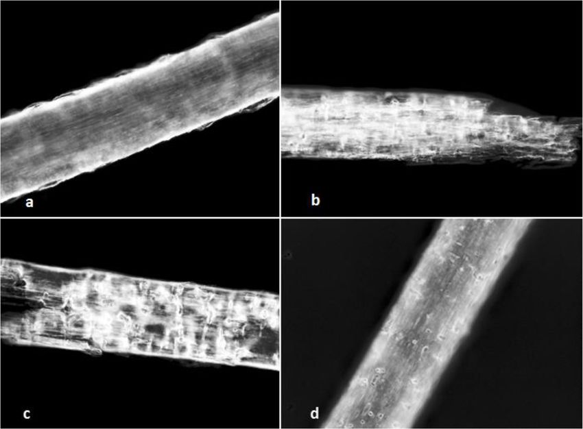
Labuda et al. IMA Fungus        (2021) 12:17                                                                                                 Page 17 of 21

SDA 14–18 mm diam at 25 °C, after 14 d, morphology                              different sub-sample, July 2019, R. Labuda, RL-07 (RL;
similar to PDA. In age, yellowish brown (amber) pig-                            ITS sequence, MT903275; LSU sequence, MT903309).
ment produced and colony reverse becoming dark red-                               Notes: Based on a search of the NCBI GenBank nu-
dish brown (after 4 wk). At 30 °C, no growth or only                            cleotide database, the closest hit for K. wagneri using the
microcolonies. Colonies on MEA 18–22 mm diam at                                 ITS sequence was K. minutisporosum (as Chrysosporium
25 °C, after 14 d, morphology as on PDA but more                                minutisporosum CBS 101577; GenBbank: KT155616);
yellowish. At 30 °C, no growth or only micro-colonies                           with identity = 486/541 (90%) and gaps 11/541 (2%).
produced. Colonies on CMA and PCA 20–25 mm diam                                 Morphologically, K. wagneri can be separated from C.
at 25 °C, after 21 d, white to yellowish, granular, good                        minutisporsoum by its larger conidia (4.0–8.0 × 2.5–
sporulation, reverse yellowish. Pinkish pigment after 3–4                       4.0 μm vs. 3.0–4.0 × 1.5–3.5 μm) and growth rate at 25
wk. on PCA (in both tested strains). No ascomata ob-                            °C on PDA after 14d (25–30 mm vs. 55–70 mm). Kerati-
served after prolonged incubation (3 months). The pti-                          nophyton straussii and K. wagneri seem to be very simi-
mum temperature for growth on PDA, SDA and MEA                                  lar, however, they can be distinguished by: (1) size of
20–25 °C (Table S1a–c). Minimum growth (1–2 mm                                  conidia (av. = 4.9 × 2.5 μm vs. 5.7 × 3.2 μm), (2) growth
diam) at 10 °C, and germination of a majority of the co-                        at 30 °C on PDA (15–20 mm vs 3–4 mm diam), (3)
nidia at 8 °C. The maximum temperature for growth                               morphology of conidiogenous cells (commonly vs. non–
31 °C (1–3 mm diam). Keratinolytic activity weak to                             to occasionally swollen), (4) colony pigmentation on
moderate (Fig. 10d), with hair attack intensity = 2. Ure-                       SDA after prolonged incubation (bright orange vs. dark
ase activity negative (after 14 d of incubation).                               brown), and (5) keratinolytic ability after 3 wk. (very
  Diagnosis: K. wagneri molecularly can be distinguished                        strong vs moderate). In addition, the production of a
from other Keratinophyton species by ITS locus analysis.                        pinkish pigment on PCA after 3–4 wk. (at 20 °C and
Phenotypically, it can be differentiated by combination                         25 °C) has been observed only in K. wagneri. Moreover,
of the growth rate at 30 °C and conidia size (4.0–8.0 ×                         conidia of this species are more coarsely roughed (warty)
2.5–4.0 μm) and morphology (obovoid to clavate, verru-                          than those in K. straussii (Fig. 8c–e).
cose) (Table 2).                                                                  All four new species are readily distinguished from the
  Additional material examined: Slovak Republic:                                other taxa in the genus Keratinophyton, based on pheno-
Tatranská Lomnica, from forest soil, isolated from a                            typical characteristics such as growth at high

 Fig. 10 Hair perforation in vitro – keratinolysis; detail of a child’s hair after colonization by the fungus on PDA (after 21 d) at 25 °C. a
 Keratinophyton lemmensii (BiMM-F76). b Keratinophyton gollerae (BiMM-F250). c Keratinophyton straussii (BiMM-F78). d Keratinophyton wagneri
 (BiMM-F77). Intensity of attack on the hair estimated on a scale of 0 to 4 (Marchisio et al. 1994). a 0–1 = light attack to cuticle. b, c 4 = cuticle and
 cortex attack with about 80% destruction. d 2 = cuticle and cortex attack with about 20% destruction
Labuda et al. IMA Fungus           (2021) 12:17                                                                                      Page 18 of 21

temperature and/or conidial morphology (Cano and                       KEY TO SPECIES OF KERATINOPHYTON (Continued)
Guarro 1990; Cano and Guarro 1994; Currah 1985). The
most important species-specific phenotypic distinguish-                (10)   reverse white to light yellow at 25 °C on PDA
                                                                              rhizosphere soil, China
ing characteristics are found as morphology of conidia
(shape, surface and dimensions) and growth rate at 30 °C                      Conidia verrucose, obovoid to clavate; colony     straussii
                                                                              reverse white with slightly yellowish centre at
after 14 d on PDA.                                                            25 °C on PDA; from garden soil, Italy
                                                                       12     Conidia echinulate, obovoid to clavate; colony    echinulatum
KEY TO SPECIES OF KERATINOPHYTON                                       (10)   reverse orange yellow at 25 °C on PDA; from
This key is modified from that of Cano et al. (2002). The                     sole of the foot, Czechia
given data for source and origin represent the type                           Conidia verrucose                                 13
strains of the related species.                                               Conidia smooth, or smooth to verrucose            15
                                                                       13     Conidia 3–4 μm wide; colony reverse white at minutisporosum
1     Ascomata developed                                2              (12)   25 °C on PYE agar; from river sediments, Spain
      Ascomata not developed                            6                     Conidia obovoid to clavate; colony reverse        14
2     Ascospores 7.5–8.5 × 4.5–5 μm; from arable        saturnoideum          different colour than white on PYE agar
(1)   soil, Spain                                                      14     Conidia more than 3 μm wide; colony reverse       pannicola
      Ascospores smaller                                3              (13)   pale brown at 25 °C on PYE agar; from soil,
                                                                              India
3     Ascospore with broad equatorial rim               4
(2)                                                                           Conidia up to 3 μm wide; colony reverse           fluviale
                                                                              brownish orange at 25 °C on PYE agar; from
      Ascospore with narrow equatorial rim              5                     river sediments, Spain
4     Ascospores discoid; daily growth 3–4 mm at        hispanicum     15     Conidia smooth                                    16
(3)   28 °C on PYE agar and reverse uncoloured;                        (12)
      from beach soil, Spain
                                                                              Conidia smooth to verrucose                       18
      Ascospores with pitted equatorial rim;         durum
      cruciform in lateral view; daily growth 2–3 mm                   16     Conidia pyriform to oval, 5–7 × 3.5–5 μm;         turgidum
      at 28 °C on PYE and reverse cream coloured;                      (15)   from barber shop soil, India
      from soil, Nepal                                                        Conidia smaller                                   17
5     Ascospores lenticular, 5–6 × 2.5–3.5 μm;      terreum            17     Conidia ellipsoidal, clavate to cylindrical;      qinghaiense
(3)   pronounced radial ridges at 37 °C on PYE agar                    (16)   racquet hyphae absent; colony reverse
      and reverse uncoloured; from lawn soil, India                           yellowish at 25 °C on PDA; from farmland soil,
      Ascospores with conoid poles, 4–4.5 × 2–2.5       punsolae              China
      μm; ridges absent at 37 °C on PYE agar and                              Conidia obovate to clavate; racquet hyphae        lemmensii
      reverse uncoloured; from arable soil, Spain                             present; colony reverse lemon yellow at 25 °C
6     No or restricted ( 1cm in diam) at 30 °C on           10             18     Conidia cylindrical to clavate, 5–30 × 2–3.5      siglerae
      PDA                                                              (15)   μm; colony reverse initially uncoloured and
                                                                              later pale brown at 25 °C on PDA; from
7     Conidia smooth; racquet hyphae present            8                     garden soil, Spain
(6)
                                                                              Conidia 4–35 × 2.5–5 μm; colony reverse           submersum
      Verrucose conidia; racquet hyphae absent;         wagneri               yellowish white at 25 °C on PDA; from river
      forest soil, Slovakia                                                   sediments, Spain
8     Conidia obovoid to ellipsoidal, 2.2–4.3 × 1.6–    hubeiense
(7)   3.2 μm; reverse yellowish on PDA at 25 °C;
      from soil under the chicken feather, China                       DISCUSSION
      Conidia larger                                    9              Phylogeny
                                                                       Phylogenetic reconstruction using ITS sequences
9     Conidia clavate to long-ellipsoidal; colony re- clavisporum
(8)   verse brown in centre and light yellow in mar-                   resulted in clustering of a new species, Keratinophyton
      gin at 25 °C on PDA; from plant root soil,                       lemmensii, with K. durum (as Aphanoascus durus; Cano
      China                                                            and Guarro 1990), K. hubeiense (as Chrysosporium
      Conidia obovoid to clavate; colony reverse        gollerae       hubeiense; Zhang et al. 2016) and K. submersum (as
      white to slightly yellowish at 25 °C on PDA;
      from forest soil, Slovakia
                                                                       Chrysosporium submersum; Vidal et al. 2002), and
                                                                       forming a sister clade to K. siglerae (as Chrysosporium
10    Intercalary conidia absent                        11
(6)                                                                    siglerae; Cano and Guarro 1994). The other three novel
                                                                       species, K. gollerae, K. straussii, and K. wagneri, were
      Intercalary conidia present                       12
                                                                       resolved in a separate terminal clade (Fig. 1a). Its sister
11    Conidia smooth, ellipsoidal or fusiform; colony   linfenense
                                                                       clade encompasses K. clavisporum (as Chrysosporium
Labuda et al. IMA Fungus (2021) 12:17 Page 19 of 21
clavisporum; Zhang et al. 2017), K. quinghaense (as of a dog in former Yugoslavia (Hajsig et al. 1974; van
Chrysosporium quinghaense; Han et al. 2013), K. Oorschot 1980) and from a case of keratomycosis in a
linfenense (as Chrysosporium linfenense; Liang et al. horse (Grahn et al. 1993).
2009), and K. turgidum (Sharma and Shouche 2017). In her review on Chrysosporium and related genera in
Based on the phylogeny and as a result of the Onygenaceae, Sigler (2003) stated that some reports
abandoning of separate names for morphs of the same concerning Chrysosporium species as etiological agents
fungus (May et al. 2019), species previously described in must be viewed with caution, in case the isolated fungus
Chrysosporium require redisposing in the genus has neither been identified to species level nor
Keratinophyton. In our study we confirmed ten species documented well enough to confirm the aetiology. In
required transfer. The monophyletic genus the follow-up list of medically relevant species provided
Keratinophyton is now extended and includes 25 species by Sigler (2003), no species is mentioned as being cur-
including ten species known from sexual morphs rently affiliated within the genus Keratinophyton, while
(Sutton et al. 2013; and this paper) and 15 species which K. pannicola (as C. pannicola) is included in the Atlas of
are currently known only from asexual morphs Clinical Fungi (de Hoog et al. 2020) as a concern in skin
(including the recently described K. turgidum (Sharma infections. Even though the keratinophilic fungi were
and Shouche 2017). The species known only from the considered as potential pathogens by several researchers
asexual morphs can be distinguished by particular (Rippon 1982; Papini et al. 1998); they rarely cause infec-
combinations of their morphological traits (colony tions. Therefore, soil is proposed as an epidemiological
colour and growth rate, growth response at higher/lower and probably also an evolutionary link, that relates geo-
temperatures, as well as morphology of conidia) and philic, zoophilic, and anthropophilic keratinophilic fungi
differences in the ITS regions (Fig. 1a, Table 2). (Papini et al. 1998). Interestingly, during a mycological
investigation of the soil samples in the present study, a
Ecology and distribution high prevalence of geophilic dermatophytes such as
Almost all known Keratinophyton species have been Nannizzia gypsea from Italy (collected in 2004), a co-
isolated from soil or soil-like substrates, such as river occurrence of Arthroderma uncinatum with Aphanoas-
sediments, compost and sand (Table 1; Cano and cus keratinophilus (as Chrysosporium keratinophilum)
Guarro 1990; Sharma and Shouche 2017; Labuda et al. from the Slovak Republic (collected in 2011), and
2008; Liang et al. 2009; van Oorschot 1980; Vidal et al. Arthroderma terrestre along with abundant A. uncina-
2000; Vidal et al. 2002). Hubalek (2000) provided a list tum from Austria (collected in 2015) were noted (data
of keratinolytic fungi associated with free-living mam- not shown).
mals and birds of which Keratinophyton pannicola (as As the members Keratinophyton are considered as
Chrysosporium evolceanui) has been isolated from a var- typical soil-borne fungi (Cano and Guarro 1990; Cano
iety of animals, different species of rodents in Australia, et al. 2002; Sutton et al. 2013) and there is no solid evi-
Czechia, Germany, the UK, and the former Yugoslavia; a dence of pathogenicity, it is likely that previously re-
rabbit in Canada; and from birds in Australia (Queens- ported animal-associated cases reflect environmental
land), Czechia, India, and the former Yugoslavia. Kerati- transmissions from soil to the animals during activities
nophyton durum (as Aphanoascus durus) has been in contact with soil. The ability of these fungi to persist
isolated from a hedgehog in Ivory Coast, and K. terreum and survive in the soil was observed also during the
(as Aphanoascus terreus) has been found associated with present study, as in case of K. straussii, the type strain
a variety of rodents in Czechia, Germany, India, Nigeria, was isolated 11 years after sampling in 2004, and two
Romania, and the former Yugoslavia and, and further more strains (RL-05 and RL-06) representing the same
birds in Australia (Queensland) Czechia, India, the USA, taxon were isolated in a repeated study even 15 years
and the former Yugoslavia (Hubalek 2000). To the best after the sampling. Likewise, a second strain (RL-07)
of our knowledge, there is only a single report of a hu- used for the description of K. wagneri and the type of K.
man clinical isolate belongs to K. echinulatum (CCF gollerae (BiMM-F250) were both isolated 8 years after
4652 = CBS 141178) from the sole of the foot of a 35- the samples were collected.
year-old woman in Czechia (Hubka et al. 2016). How- The degree of keratin degradation by the novel strains
ever, Hubka et al. (2016) indicated that the etiological described here varied. It was very strong in both K.
significance of this fungus was unclear, and they con- gollerae and K. straussii compared to other tested
cluded that the infection was actually caused by another strains, attacking the cuticle and cortex of hairs with
dermatophyte, which was not isolated or was overgrown about 50–80% degradation. In addition to keratin
by K. echinulatum. A few other cases have been pub- degradation, keratinolytic fungi share common
lished in a small range of animals including Keratinophy- properties with dermatophytes (Marchisio et al. 1994;
ton pannicola (as Chrysosporium pannicola) from skin Mitola et al. 2002). Even though some of these fungi canYou can also read