Molecular Mechanisms and Regulation of Mammalian Mitophagy - MDPI
←
→
Page content transcription
If your browser does not render page correctly, please read the page content below

cells
Review
Molecular Mechanisms and Regulation of Mammalian Mitophagy
Vinay Choubey * , Akbar Zeb                      and Allen Kaasik
                                          Department of Pharmacology, Institute of Biomedicine and Translational Medicine, University of Tartu, Ravila 19,
                                          50411 Tartu, Estonia; akbar@ut.ee (A.Z.); allen.kaasik@ut.ee (A.K.)
                                          * Correspondence: vinay.choubey@ut.ee

                                          Abstract: Mitochondria in the cell are the center for energy production, essential biomolecule synthe-
                                          sis, and cell fate determination. Moreover, the mitochondrial functional versatility enables cells to
                                          adapt to the changes in cellular environment and various stresses. In the process of discharging its cel-
                                          lular duties, mitochondria face multiple types of challenges, such as oxidative stress, protein-related
                                          challenges (import, folding, and degradation) and mitochondrial DNA damage. They mitigate all
                                          these challenges with robust quality control mechanisms which include antioxidant defenses, pro-
                                          teostasis systems (chaperones and proteases) and mitochondrial biogenesis. Failure of these quality
                                          control mechanisms leaves mitochondria as terminally damaged, which then have to be promptly
                                          cleared from the cells before they become a threat to cell survival. Such damaged mitochondria are
                                          degraded by a selective form of autophagy called mitophagy. Rigorous research in the field has
                                          identified multiple types of mitophagy processes based on targeting signals on damaged or super-
                                          fluous mitochondria. In this review, we provide an in-depth overview of mammalian mitophagy
                                          and its importance in human health and diseases. We also attempted to highlight the future area of
                                          investigation in the field of mitophagy.

                                Keywords: PINK1; PARKIN; mitophagy; autophagy; FUNDC1; BNIP3; cardiolipin; Parkinson’s

                                          disease; quality control
Citation: Choubey, V.; Zeb, A.;
Kaasik, A. Molecular Mechanisms
and Regulation of Mammalian
Mitophagy. Cells 2022, 11, 38.
                                          1. Introduction
https://doi.org/10.3390/cells
11010038                                       Mitochondria in the cell are double-membraned organelles, which hold a central role
                                          in energy production [1], essential biomolecule synthesis [2], calcium buffering [3] and
Academic Editors:
                                          importantly, in pro-survival or pro-apoptotic signaling [4]. These functions are carried out
Nadine Camougrand and
                                          by over 1200 proteins [5], although the exact composition of mitochondrial proteins can
Ingrid Bhtia-Kissova
                                          vary greatly depending on the cell stage, type and environment [5]. Due to the functional
Received: 29 November 2021                importance of mitochondria, any disturbance in this proteome will leave a profound impact
Accepted: 20 December 2021                on cell fate and could result in diseases ranging from neurodegenerative disorders and
Published: 23 December 2021               heart diseases to diabetes and cancer [6–11].
Publisher’s Note: MDPI stays neutral
                                               Owing to their role in energy production by the electron transport chain, mitochon-
with regard to jurisdictional claims in   dria are the primary site of reactive oxygen species (ROS) generation [8]. Excessive ROS
published maps and institutional affil-   production damages mitochondria further, creating a vicious circle [12]. Although ROS are
iations.                                  important signaling molecules [13], they are detrimental to the cell when in excess [11,12,14].
                                          To control the ROS accumulation, mitochondria are equipped with different types of antiox-
                                          idant systems, such as mitochondrial superoxide dismutases (SOD), thioredoxin reductase
                                          and glutathione peroxidase [15]. The second potential threat to mitochondrial health
Copyright: © 2021 by the authors.         arises from mitochondrial plasticity, which demands a constant change in the mitochon-
Licensee MDPI, Basel, Switzerland.        drial proteome to adapt to cellular needs [16,17]. Under such fluctuating conditions, the
This article is an open access article    mitochondrial proteostasis is maintained by a robust mitochondrial import system collab-
distributed under the terms and           orating with mitochondrial proteases and chaperones [5,16,18–20]. This system not only
conditions of the Creative Commons
                                          delivers the functional proteins to mitochondria but also prevents the accumulation of non-
Attribution (CC BY) license (https://
                                          functional or non-desirable proteins under given conditions. Besides, the ROS defense and
creativecommons.org/licenses/by/
                                          proteostasis system, mitochondrial dynamics is also involved in the mitochondrial quality
4.0/).

Cells 2022, 11, 38. https://doi.org/10.3390/cells11010038                                                         https://www.mdpi.com/journal/cells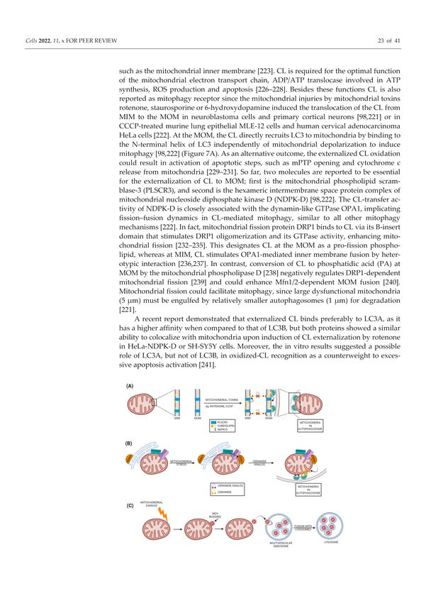
Cells 2022, 11, 38                                                                                                    2 of 38

                     control, as slightly damaged mitochondria may fuse with healthy ones or may separate
                     from the damaged mitochondrial part via fission [21,22]. These multilayered mitochondrial
                     quality control systems, including ROS defense, proteostasis and mitochondrial dynamics,
                     all work in concert to preserve mitochondrial function and normal cell physiology.
                          However, if the damage to mitochondria overwhelms the capacity of the quality
                     control systems, then these mitochondria should be removed from mitochondrial network
                     by mitophagy, the selective degradation of mitochondria in lysosomes by autophagy
                     (Figure 1) [23,24].

                     Figure 1. Autophagy Introduction: Autophagy is one of the two main cellular degradative systems
                     along with the ubiquitin proteasome system. The word autophagy (meaning self-eating in Greek) was
                     first coined by Christian de Duve in 1963. During this process, the cytoplasmic components (cargo)
                     including organelles and macromolecules are delivered to lysosomes, where the collection of different
                     acidic hydrolases cause their degradation. This degradation enables the cells to recycle building blocks
                     as well as serves as temporary energy source under conditions like starvation. Based on the route of
                     cargo delivery to lysosomes, the autophagy can be divided into three main types: macroautophagy,
                     microautophagy and chaperon-mediated autophagy. Macroautophagy is the major type of autophagy,
                     during which a cytoplasmic portion containing proteins and organelles is engulfed by autophagosome,
                     which is then fused with lysosome to degrade the engulfed cytoplasmic materials by lysosomal
                     enzymes. In microautophagy, a portion of the cytoplasm is directly delivered to the lysosomes for
                     degradation by lysosomal membrane invagination. Contrary to above two, the chaperon-mediated
                     autophagy specifically degrades proteins containing a specific motif (KFERQ). This involves the
                     formation of a complex between chaperons (Hsc70) and target proteins and then subsequent delivery
                     to lysosome via binding to lysosomal associated membrane protein 2A (LAMP2A).

                           Lysosomal mitochondria were first observed in mammalian cells by early electron
                     microscopy studies by De Duve and Wattiaux in 1966 [25] but this was first referred to as
                     selective mitochondrial autophagy or mitophagy by J. J. Lemasters in 2005 [26]. Mitophagy
                     removes old, superfluous or dysfunctional mitochondria before they become a threat for
                     cells. Since that initial observation, extensive studies have identified different mechanisms
                     of mitophagy activated under different kinds of stresses, such as oxidative damage, hypoxia,
                     mitochondrial depolarization and mitochondrial DNA (mtDNA) damage [23,24,27–34].
                     Based on the targeting signals on damaged or superfluous mitochondria that initiate
                     mitophagy, this process can be divided into:
Cells 2022, 11, 38                                                                                       3 of 38

                     â    Ubiquitin-dependent mitophagy
                          •    PARKIN dependent (PINK1-PARKIN pathway)
                          •    PARKIN independent but ubiquitin dependent mitophagy:
                     â    Ubiquitin-independent or receptor based mitophagy
                          •    Apoptosis related proteins as mitophagy receptors or inhibitor
                          •    Other mitophagy receptors
                     â    Lipid based mitophagy
                          •    Cardiolipin based
                          •    Sphingolipid Based
                     â    Micromitophagy

                     2. Ubiquitin-Dependent Mitophagy
                         Ubiquitin-dependent mitophagy relies on ubiquitin as a signal on the surface of
                     damaged or superfluous mitochondria. Ubiquitin-marked mitochondria will then recruit
                     autophagic machinery that leads to their degradation by mitophagy. Ubiquitination of
                     mitochondrial proteins is achieved by different pathways that lead to the initiation of
                     mitophagy, but below are described only the most well-studied ones.

                     2.1. PARKIN Dependent (PINK1-PARKIN Pathway)
                          The landmark studies from Richard Youle’s lab on the PINK1-PARKIN pathway rev-
                     olutionized the field of mitophagy [35,36], which made this pathway the best-studied
                     mitophagy pathway among all others. This pathway primarily depends on the mito-
                     chondrial serine/threonine protein kinase PINK1 and the cytosolic E3 ubiquitin ligase
                     PARKIN [29,37]. Earlier studies in Drosophila found that PARKIN and PINK1 were essential
                     for mitochondrial function and work in the same pathway [38,39]. Later studies established
                     the roles of PINK1 and PARKIN in mitochondrial biology more precisely [29,37,40]. PINK1
                     and PARKIN were found to monitor diverse aspects of mitochondrial health, ranging from
                     mitochondrial quality control and mitochondrial dynamics to mitochondrial biogenesis [29].
                     Thus, not surprisingly, mutations in PINK1 and PARKIN genes (resulting in mitochondrial
                     dysfunction) are implicated in several neurodegenerative diseases such as Parkinson’s
                     disease (PD), Alzheimer’s disease (AD) and Multiple Lateral Sclerosis (MLS) [29,41]
                          PINK1 utilizes the canonical presequence-driven mitochondrial import pathway to
                     monitor mitochondrial health [29,37,42]. Under basal conditions, PINK1 is imported
                     into the polarized mitochondria through the mitochondrial translocases of the outer and
                     inner membranes (TOM and TIM), with the help of its positively charged amino-terminal
                     mitochondrial targeting sequence [37,42] (Figure 2). Following the import of PINK1, it is
                     cleaved twice when it is in the inner mitochondrial membrane. The first cleavage by the
                     matrix processing peptidase (MPP) removes the mitochondrial targeting sequence, while
                     the second cleavage occurs in the transmembrane domain between Ala103 and Phe104 by
                     the inner membrane protease Presenilin-Associated Rhomboid-Like protein (PARL) [37,42]
                     (Figure 2). As PARL is involved in the cleavage of PINK1 and PGAM5, it is now called the
                     PINK1/PGAM5-associated rhomboid-like protease [42–44].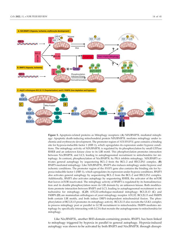
Cells 2022, 11, 38                                                                                                   4 of 38

                     Figure 2. Mitochondrial stress leads to accumulation of PINK1 and PARKIN on mitochondria.
                     Newly translated mitochondrial kinase PINK1 has two fates depending on mitochondrial status.
                     (A) Under unstressed and polarized conditions, the full-length PINK1 containing positively charged
                     mitochondria target sequence (MTS, 1-34 amino acids) at its N-terminus is imported into mitochondria
                     with the help of transporters of the outer and inner membranes (TOM and TIM). The MTS is cleaved
                     off by mitochondrial processing peptidase (MPP) followed by cleavage in the transmembrane domain
                     (TMD) of PINK1 between Ala103 and Phe104, by mitochondrial inner membrane protease PARL.
                     The sequential cleavages generate a shorter PINK1 with the N-terminal Phe104. Two degradation
                     possibilities are proposed for the cleaved PINK1 (1) widely recognized rapid degradation by the
                     proteasome via the N-end rule ubiquitination machinery (2) the second possibility that ubiquitination
                     and degradation of cleaved PINK1 occur at mitochondrial-endoplasmic reticulum (ER) interface by
                     components of the ER-associated degradation pathway, such as the E3 ligases GP78 and HRD1. (B) In
                     contrast to (A), under stressed and depolarized conditions, the import of full-length PINK1 is arrested
                     and it forms a complex with TOM, most likely as a dimer. The PINK1 dimerization is proposed to
                     facilitate PINK1’s autophosphorylation and activation. The mitochondrial outer membrane (MOM)
                     stabilizes full-length PINK1 which phosphorylates pre-existing ubiquitins (Ub) or/and autoinhibited
                     PARKIN. There appears to be no particular order in which PINK1 would phosphorylate Ub or
                     PARKIN first. In both cases, PARKIN translocates to mitochondria and self-associates. The activated
                     PARKIN starts to conjugate ubiquitin to MOM proteins, which are then phosphorylated by PINK1.
                     This forms a positive feedback loop that amplifies the initial signal, resulting in extensive PARKIN
                     recruitment and ubiquitination of MOM proteins. (MOMP and MIMP: mitochondrial outer/inner
                     membrane protein).

                          Coming back to PINK1 import, PARL-cleavaged PINK1 is released into the cytoplasm
                     and degraded rapidly by the proteasome via the N-end rule ubiquitination machinery,
                     keeping the basal levels of PINK1 low [45]. However, it has been suggested that N-end rule
                     might not be the primary mechanism of PINK1 degradation. It might also be degraded
                     through a proteasome-dependent mechanism relying on the polyubiquitination of the
                     mature 52-kDa form of PINK1 preferential at K137. This hypothesis is supported by
                     evidence that the bulk of ubiquitinated (Ub)-PINK1 is mitochondrially anchored rather
                     than cytosolic. Secondly, the N-terminal phenylalanine (F104) of PINK1 was not detected in
                     the cytosol but in the mitochondrial outer membrane (MOM) where it would be inaccessible
                     to the cytosolically localized N-end rule ubiquitination machinery [46]. Later, the same
                     group proposed that 52 kDa PINK1 localizes at the mitochondrial–endoplasmic reticulum
                     (ER) interface, where components of ER-associated degradation pathway, such as the
                     E3 ligases GP78 and HRD1, catalyze PINK1 ubiquitination and promote its proteasomal
Cells 2022, 11, 38                                                                                             5 of 38

                     degradation to maintain PINK1 content low in healthy mitochondria [47]. Contrary to
                     the PINK1 degradation mechanism, it is largely accepted that the full-length PINK1 is
                     stabilized on the MOM upon mitochondrial membrane potential dissipation [36], excessive
                     mitochondrial reactive oxygen species (ROS) generation [48,49] and/or mitochondrial
                     protein aggregation [50,51]. The mitochondrial depolarization or bioenergetic deficit leads
                     to the inhibition of the TIM23-mediated import of PINK1 via indirect modulation by the
                     adenine nucleotide translocator (ANT) [52]. In another study, PINK1 import arrest under
                     mitochondrial depolarization was not found solely dependent on Tim23 inactivation but
                     also by an actively regulated “tug of war” between Tom7 and OMA1 [53]. Irrespective of
                     the mechanism of PINK1 import arrest, it is generally accepted that PINK1 forms a 700 kDa
                     complex with a translocase of the outer membrane on depolarized mitochondria after its
                     import is arrested [54] (Figure 2). In this MOM complex, PINK1 molecules likely exist as
                     dimers, however, the role of dimeric PINK1 in its kinase activity remains to be established.
                     PINK1 dimerization is proposed to facilitate PINK1’s autophosphorylation on Ser228 and
                     Ser402 residues in its kinase domain [55,56].
                           After stabilization on MOM and autophosphorylation, PINK1 affects PARKIN in
                     two ways. First, it phosphorylates pre-existing ubiquitins already conjugated to MOM
                     proteins at Ser65. PARKIN’s high affinity for phosphorylated ubiquitin (pSer65–Ub) drives
                     its translocation from the cytosol to mitochondria [57,58] as well as partially activates its
                     ubiquitination activity [59]. This partially activated pSer65–Ub-bound PARKIN, ubiquiti-
                     nates MOM proteins further, providing more substrate for PINK1. This generates more
                     pSer65–Ub which attracts a second wave of PARKIN from the cytosol to mitochondria
                     (Figure 2). PINK1 also phosphorylates pSer65–Ub-bound PARKIN at Ser65 in the ubiquitin-
                     like domain, activating its autoinhibited E3 ubiquitin ligase activity nearly 4400-fold [60–62],
                     which further drives ubiquitin chain formation on the MOM proteins. The binding of
                     pSer65-Ub to pSer65-PARKIN is 19 times stronger than to unphosphorylated PARKIN,
                     thus further favoring the retention of fully active pSer65-PARKIN on damaged mitochon-
                     dria [63]. However, there appears no particular order to it. PINK1 could phosphorylate first
                     PARKIN, since PARKIN can be phosphorylated and activated by PINK1 without its initial
                     encounter with pSer65-Ub; additionally, ubiquitin could be phosphorylated independently
                     of PARKIN. In both scenarios, activated PARKIN conjugates further ubiquitins to MOM
                     proteins, which are then phosphorylated by PINK1. This forms a positive feedback loop
                     that amplifies the initial signal, resulting in extensive PARKIN recruitment and ubiquiti-
                     nation [57,59,62] (Figure 2). Interestingly, PINK1 kinase activity is sufficient for PARKIN
                     recruitment, as the forced expression of PINK1 on peroxisomes or lysosomes leads to
                     PARKIN translocation to respective organelles [54].
                           The recruitment of PARKIN to mitochondria leads to the ubiquitination and fur-
                     ther proteasomal degradation of multiple MOM proteins [64] such as Mfn1/2 [65–67],
                     Miro1/2 [64,68], VDAC [69,70], TOMs [71] and mitochondrial hexokinase [64]. The ex-
                     traction of ubiquitinated protein for degradation from MOM is accomplished by p97,
                     an AAA+ ATPase which accumulates in mitochondria along with the proteasome in a
                     PARKIN-dependent manner [66,72]. Another possible way of proteasome accumulation
                     onto mitochondria is the direct interaction between PARKIN’s ubiquitin-like domain and
                     the Rpn13 subunit of the 26S proteasome [73]. This interaction attracts the proteasome
                     to mitochondria and facilitates the proteasomal degradation of certain MOM proteins
                     and PARKIN itself [73]. Ubiquitination and proteasomal degradation possibly occur in a
                     bi-phasic manner [74]. The primary targets of the first phase are MOM proteins, which
                     lead to the rupture of MOM [71]. Rupture of MOM opens the door for the second phase
                     of ubiquitination of proteins localized inside mitochondria (Figure 3) [71,74]. A total of
                     36 MOM substrates of PARKIN have been identified with high confidence [64]. Interest-
                     ingly, some mitochondrial substrates were earlier considered not only as substrates but also
                     as PARKIN receptors, but it was later suggested that phosphorylated ubiquitin is the main
                     receptor for PARKIN at mitochondria [58]. Nevertheless, we recently demonstrated that
                     Miro proteins are not only the substrates for PINK1/PARKIN-dependent degradation, but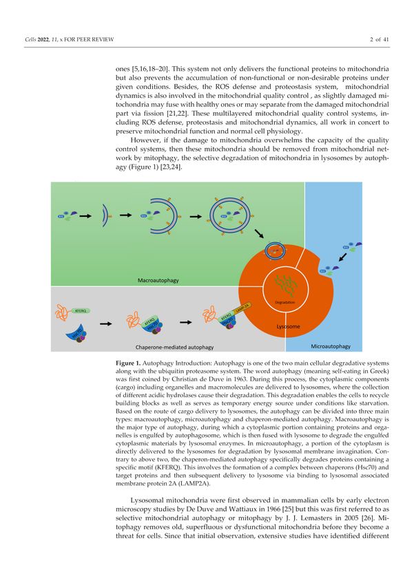
Cells 2022, 11, 38                                                                                                6 of 38

                     they can also function as a calcium-dependent docking site and safety switch for PARKIN
                     recruitment [75].

                     Figure 3. Three fates of PARKIN ubiquitinated mitochondrial proteins. The PARKIN ubiquitinated
                     mitochondrial proteins could have different fates (A). The ubiquitinated proteins could undergo
                     deubiquitination by deubiquitinases (DUBs), such as mitochondrially localized ubiquitin carboxyl-
                     terminal hydrolase 30 (USP30). The deubiquitination would remove the polyubiquitin chain, con-
                     jugated by PARKIN (shown in Figure 2), and would restore its past status. (B) The ubiquitinated
                     proteins could undergo extraction and degradation by the ubiquitin–proteasome system. This leads
                     to the rupture of MOM, which exposes the inner membrane proteins followed by their ubiquitination.
                     (C) As the third possibility, the PARKIN-conjugated polyubiquitin chains could initiate the delivery
                     of mitochondria to autophagosomes via the recruitment of the autophagy cargo receptors such as
                     Optineurin (OPTN) and NDP52. The autophagy receptors bind at one end polyubiquitin chains
                     located on mitochondria, and, on another end, they bind LC3 protein, located on autophagosomal
                     membranes (Phagophore). Thus, the autophagy receptors facilitate the delivery of mitochondria to
                     the autophagosome for their further degradation in lysosomes after autophagosomal fusion with
                     lysosomes. (MOMP and MIMP: mitochondrial outer/inner membrane protein).

                           It is relevant to note that translocated PARKIN forms ubiquitin chains on MOM
                     proteins with linkage types that are characteristic of both autophagy and proteasomal
                     degradation [76–78]. PARKIN has been shown to a the Lys48-linked polyubiquitin chains
                     onto several MOM proteins [78] and typically, Lys48 polyubiquitin chains consist of a mini-
                     mum of four ubiquitin moieties target proteins for degradation via the proteasome [79,80].
                     Fast proteasomal removal of mitofusins prevents damaged mitochondria from fusing with
                     the healthy mitochondrial network and segregates them for mitophagy degradation [78].
                     Similarly, Miro proteins involved in mitochondrial motility, are removed by the proteaso-
                     mal degradation, leading to mitochondrial movement arrest to facilitate mitophagy [81].
                     However, whether these events are really prerequisites for mitophagy is currently uncertain
                     and demands further investigation.
                           In addition to Lys48 polyubiquitin chains, PARKIN also as polyubiquitin chains that
                     are linked through Lys63, Lys6 or Lys11 to MOM proteins [62]. The overall abundance of
                     Lys6 linked polyubiquitin chains does not increase with proteasome inhibition, suggesting
                     that Lys6 chains do not lead to proteasomal degradation [82]. However, the functional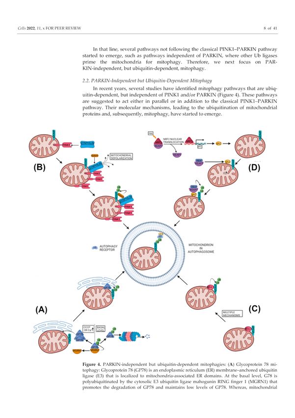
Cells 2022, 11, 38                                                                                          7 of 38

                     relevance of different types of polyubiquitin chains in mitophagy is so far not conclu-
                     sively established.
                           PARKIN-mediated ubiquitination can also be affected by the deubiquitinating en-
                     zymes (DUBs) (Figure 3A). In fact, mitochondrially localized ubiquitin carboxyl-terminal
                     hydrolase 30 (USP30) deubiquitinase was found to reverse the PARKIN-dependent ubiqui-
                     tination of TOMM20 and Miro1 [83]. Thus, the deubiquitinating enzymes oppose PINK1-
                     PARKIN mitophagy [83] and it has been suggested that this might prevent the degradation
                     of healthy mitochondria [84]. However, the polyubiquitination and deubiquitination pro-
                     cess is a wasteful process and cells should know how to avoid that and proceed with
                     mitophagy. The answer is provided by an extensive study demonstrating that Ser65 phos-
                     phorylated ubiquitin dimers are particularly resistant for the cleavage by 31 different
                     DUBs [85]. This suggests that at mitochondria, PINK1-dependent phosphocapping of ubiq-
                     uitin is making ubiquitinated MOM proteins DUB-resistant. An alternative explanation
                     would be that PARKIN activation outpaces the ubiquitin chain removal to an extent that
                     would overcome deubiquitinase (USP30)-mediated antagonization [84].
                           As mentioned above, PARKIN-ubiquitinated MOM proteins could undergo either
                     proteasome mediated degradation (Figure 3B) or disassembly of ubiquitin chains by DUBs
                     (Figure 3A). As the third possibility, the PARKIN-ubiquitinated MOM proteins can also
                     trigger the recruitment of the autophagy cargo receptors to mitochondria (Figure 3C). The
                     autophagy receptors bind at one end to the ubiquitinated cargo (via their ubiquitin binding
                     domains, UBD) and at other end (via their LC3-interacting region, LIR) to Microtubule
                     Associated Protein-Light Chain 3 (MAP-LC3 or just LC3) that is localized on the autophago-
                     somal membranes [86]. Thus, autophagy receptors facilitate the delivery of ubiquitinated
                     cargo to the autophagosome for autophagic degradation [86]. In mammalian cells, five
                     autophagy receptors have been linked to ubiquitin dependent mitophagy: p62, AMBRA1,
                     Nuclear Domain 10 Protein 52 (NDP52), Optineurin (OPTN) and TAX1BP1 [87]. However,
                     p62 was found to be required for the perinuclear clustering of depolarized mitochondria,
                     but not for mitophagy [88]. More importantly a knock-out study of five autophagy recep-
                     tors (p62, Neighbor of BRCA1 gene 1 (NBR1), NDP52, OPTN and TAX1BP1) revealed that
                     NDP52 and OPTN could rescue mitophagy redundantly in these Penta KO HeLa cells [89].
                     The study additionally showed that the compensatory ability of OPTN for NDP52 during
                     mitophagy required TANK binding Kinase 1 (TBK1) [89]. TBK1 was shown to phospho-
                     rylate OPTN at multiple sites in a PINK1- and PARKIN-dependent manner, leading to
                     the increased affinity of OPTN for K63-Ub chains and ATG8 (yeast equivalent of LC3)
                     proteins [90,91]. Importantly, mutations in TBK1 and OPTN have been genetically linked
                     with amyotrophic lateral sclerosis. These mutations in TBK1 and OPTN often disrupt their
                     respective proteins association suggesting the significance of their association in removing
                     autophagy cargo [92–94]. Moreover, the autophagy cargo receptors, i.e., OPTN and NDP52,
                     promote the biogenesis of phagophores in close proximity to mitochondria by recruiting
                     the autophagy-initiating factors ULK1 (unc-51-like autophagy activating kinase 1), DFCP1
                     (double FYVE-domain containing protein 1) and WIPI1 (WD repeat domain, phospho-
                     inositide interacting 1) upstream to LC3 recruitment [89]. After the engulfment of targeted
                     mitochondria by the autophagosome, it fuses with acidic hydrolases containing lysosomes
                     for the complete degradation of mitochondria [95].
                           Interestingly, PINK1 and PARKIN play not only key roles in mitophagy degradation,
                     but also regulate mitochondrial biogenesis (discussed later in the review).
                           Though the PINK1–PARKIN mitophagy pathway is the most studied mitophagy
                     pathway, it has been often criticized for the mostly studied in in vitro with PARKIN overex-
                     pression in non-neuronal immortalized cells in the presence of mitochondrial toxins, or in
                     conditions that are far from physiological [96–99]. Moreover, recent in vivo studies indicate
                     that PINK1 and PARKIN are not critical for basal mitophagy in various tissues, including
                     the brain [100,101]. Therefore, recent studies have been focused on the identification of the
                     alternative mitophagy pathways.
Cells 2022, 11, 38 8 of 38
In that line, several pathways not following the classical PINK1–PARKIN pathway
started to emerge, such as pathways independent of PARKIN, where other Ub ligases prime
the mitochondria for mitophagy. Therefore, we next focus on PARKIN-independent, but
ubiquitin-dependent, mitophagy.
2.2. PARKIN-Independent but Ubiquitin-Dependent Mitophagy
In recent years, several studies have identified mitophagy pathways that are ubiquitin-
dependent, but independent of PINK1 and/or PARKIN (Figure 4). These pathways are
suggested to act either in parallel or in addition to the classical PINK1–PARKIN pathway.
Their molecular mechanisms, leading to the ubiquitination of mitochondrial proteins and,
subsequently, mitophagy, have started to emerge.
Figure 4. PARKIN-independent but ubiquitin-dependent mitophagies: (A) Glycoprotein 78 mi-
tophagy: Glycoprotein 78 (GP78) is an endoplasmic reticulum (ER) membrane–anchored ubiquitin
ligase (E3) that is localized to mitochondria-associated ER domains. At the basal level, G78 is polyubiq-
uitinated by the cytosolic E3 ubiquitin ligase mahogunin RING finger 1 (MGRN1) that promotes the
degradation of GP78 and maintains low levels of GP78. Whereas, mitochondrial stresses such as CCCP
or higher cytosolic Ca2+ lead to GP78 accumulation at the mitochondria-associated ER domain trig-
gering PARKIN-independent but ubiquitin-dependent mitophagy. (B) PINK1-SYNPHILIN1-SIAH1
mitophagy: The protein SYNPHILIN1 localizes to mitochondria with the help of the mitochondrial
kinase PINK1, since SYNPHILIN1 has the affinity towards full length as well as the cleaved form
of PINK1. Mitochondrial localization of SYNPHILIN1 causes mitochondrial depolarization, leadingCells 2022, 11, 38 9 of 38
to the stabilization of full-length PINK1 onto MOM which further attracts SYNPHILIN1 to the
mitochondria. Parallel to PINK1, SYNPHILIN1 also binds to E3 ubiquitin ligase seven in absentia ho-
molog 1 (SIAH1), thus recruiting it to mitochondria. SIAH1 at mitochondria polyubiquitinates MOMP,
which leads to recruitment of autophagosomes to mitochondria via the autophagy receptor, and LC3
for mitophagy. (MOMP: mitochondrial outer membrane protein) (C) MUL1 based mitophagy: The
mitochondrial E3 ubiquitin ligase 1 (MUL1) induces ubiquitin-dependent but PARKIN-independent
mitophagy by multiple proposed mechanisms, which are poorly understood and lack consensus.
(D) p62/SQSTM based mitophagy: p62/SQSTM based mitophagy was observed with the Keap1-Nrf2
PPI inhibitor HB229 (PMI). PMI inhibited Keap1-Nrf2 interaction and led to the nuclear translocation
of Nrf2. Nuclear translocation of Nrf2 upregulated the expression of p62, which accumulated in mito-
chondria. Mitochondrial p62 could induce mitophagy either by increasing mitochondrial superoxide
or by anchoring the Keap1–RBX1 complex on mitochondria or both. The Keap1–RBX1 complex, being
the E3 ubiquitin ligase, polyubiquitinates MOMP, which leads to recruitment of autophagosomes
to mitochondria via autophagy receptor and LC3 for mitophagy. (MOMP: mitochondrial outer
membrane protein).
2.2.1. Glycoprotein 78 Mitophagy
Glycoprotein 78 (GP78) is an endoplasmic reticulum (ER) membrane–anchored ubiq-
uitin ligase (E3) that is a key component of the ER-associated degradation (ERAD) and is
found localized to the mitochondria-associated ER domain [102]. Over-expression of GP78
has been found to ubiquitinate Mfn1 and Mfn2, inducing their proteasomal degradation,
leading to mitochondrial fragmentation [103]. Moreover, over-expressed GP78 was found
to induce mitophagy upon mitochondrial depolarization in COS-7 cell lines by recruiting
LC3 to the GP78-positive ER domains, closely associated with depolarized mitochondria.
The GP78 induced mitophagy was dependent on GP78’s ubiquitin ligase activity, and the
autophagy protein Atg5 and Mfn1, but it was PARKIN-independent since it occurred in
PARKIN-null HeLa cells, as well as in PARKIN knockdown HEK293 cells [103]. Interest-
ingly, GP78 activity is regulated by the cytosolic E3 ubiquitin ligase mahogunin RING
finger 1 (MGRN1), which ubiquitinates GP78 in trans through noncanonical K11 linkages,
promoting the degradation of GP78 [102]. This maintains constitutively low levels of GP78
in healthy cells and downregulates mitophagy. Whereas, mitochondrial stresses by CCCP
or higher cytosolic Ca2+ perturb the interaction between MGRN1 and GP78, leading to
GP78 accumulation and triggering PARKIN-independent mitophagy [104] (Figure 4A).
Interestingly, ERAD machinery containing GP78 has been proposed to regulate PINK1
levels in human and monkey cell lines also [47].
2.2.2. PINK1-SYNPHILIN1-SIAH1 Mitophagy
Another reported ubiquitin-dependent but PARKIN-independent mitophagy pathway
involves the PINK1, SYNPHILIN1 and seven in absentia homolog 1 (SIAH1) [105]. In this
pathway, PINK1 recruits SYNPHILIN1 to the mitochondria, as SYNPHILIN1 can interact
with the full-length and cleaved form of PINK1 in rat brain tissues and cultured cells [105].
Though SYNPHILIN1 was observed to preferentially interact with cleaved PINK1, the
authors suggested that this could be due to the nature of the antibody used [105]. The
PINK1-mediated localization of SYNPHILIN1 to mitochondria causes depolarization of the
mitochondria that leads to stabilization of uncleaved PINK1 at the organelle. This further
promotes translocation of SYNPHILIN1 to the mitochondria, which in turn recruits the E3
ubiquitin ligase SIAH1 to the mitochondria as SYNPHILIN1 has the ability to interact with
SIAH1 also. The SIAH1 subsequently ubiquitinates mitochondrial proteins that results in
the recruitment of the autophagosome marker LC3 and the lysosome marker Lamp1 to the
mitochondria for mitophagy [105] (Figure 4B). This PINK1–SYNPHILIN1–SIAH1 induced
mitophagy did not depend on an exogenous depolarizing agent or on PINK1-mediated
phosphorylation of SYNPHILIN1or ubiquitin, as well as did not involve PARKIN. Al-
though this pathway was independent of PINK1 kinase activity, PD-related PINK1 mutants
(G309D, A168P and L347P) decreased this mitophagy by more than 50%, pointing thatCells 2022, 11, 38 10 of 38
not only the PINK1–PARKIN pathway but alternative mitophagy pathways could also be
affected by PINK1 mutations [105]. Since this pathway is independent of PINK1 kinase
activity, it may represent a possible new drug target in disease cases involving PINK1
kinase domain mutants. Besides SIAH1, the other members of the SIAH family have been
found to regulate different aspects of mitochondrial biology. For example, SIAH2 has been
shown to regulate mitochondrial dynamics by controlling the degradation of Mfn1 and
Drp1 in cortical neurons under hypoxia [106]. Moreover, SIAH2 negatively regulates mito-
chondrial biogenesis by ubiquitination and subsequent proteasomal degradation of nuclear
respiratory factor 1 (NRF1), a crucial factor for mitochondrial biogenesis [107] under hy-
poxic microenvironments [108]. Whereas, PARKIN, another E3 ubiquitin ligase, promotes
mitochondrial biogenesis by degradation of the repressor of PGC-1α, PARIS [109,110].
2.2.3. MUL1-Based Mitophagy
The mitochondrial E3 ubiquitin ligase 1 (MUL1), also known as mitochondrial-anchored
protein ligase (MAPL) [111] or mitochondrial ubiquitin ligase activator of NF-κB (MU-
LAN) [112], has been reported to be involved in mitophagy induction [113–115]. However,
the mechanisms proposed for MUL1-mediated mitophagy are poorly understood and lack
consensus (Figure 4C).
MUL1 is a MOM-embedded protein, with its RING finger facing the cytoplasm and its
intermembrane domain located in the intermembrane space (IMS). MUL1 is a multifunc-
tional protein but its major biological functions are ubiquitination and sumoylation. Like
other ubiquitin ligases, MUL1 should interact with E2-conjugating enzymes for ubiquitina-
tion. Currently, four E2-conjugating enzymes (Ube2E2, Ube2E3, Ube2G2 and Ube2L3) are
identified as specific interactors of MUL1 [114]. Among them, the Ube2E3 was implicated
in the induction of mitophagy in HEK293 cells treated with CCCP. In this case, MUL1
was found to bind GABARAP (GABA receptor-associated protein), a member of the Atg8
family that plays a major role in mitophagy [114]. The binding with GABARAP requires an
LC3-interacting region (LIR), located in the RING finger domain of MUL1, as well as the
presence of Ube2E3, suggesting a plausible mechanism of MUL1-induced mitophagy [114].
Another study demonstrated a role of MUL1 in selenite-induced mitophagy, which
was ULK1- and Atg5-dependent but was PARKIN-independent [115]. The study proposed
that under normal conditions, MUL1 monitors the MOM quality and prevents ULK1 from
initiating mitophagy. However, under stress conditions or a higher intermembrane spatial
ROS, ULK1 translocates to the mitochondria to initiate mitophagy. Similarly, selenite
promoted the partial translocation of ULK1 to mitochondria, where it interacted with
MUL1, which, in turn, ubiquitinated ULK1 for proteasomal degradation, making ULK1
a novel substrate of MUL1 [115]. However, the mechanism proposed was unclear, as
MUL1-enhanced mitophagy paradoxically promoted ULK1 degradation. Additionally,
two highly conserved cysteine residues in MUL1 were proposed to play an important
role in MUL1-induced mitophagy by ROS and selenite, since treatment with anti-oxidants
prevented the mitophagy induction [115].
As an E3 ubiquitin ligase, MUL1 ubiquitinates many functional and signaling proteins,
such as mitofusin2 (Mfn2), Akt, p53 and ULK1, leading to mostly their degradation [116].
On the other hand, MUL1 promotes the sumoylation of DNM1L/Drp1 which stabilizes it
and leads to mitochondrial fission [116]. This, together with the reduction in mitochondrial
fusion via ubiquitination and degradation of Mfn2, results in an overall fragmented mi-
tochondrial morphology [112], creating an environment favoring mitophagy. This ability
of MUL1 to regulate Mfn2 has been proposed as a mitophagy-inducing mechanism in
Omi/HtrA2(−/−) mouse embryonic fibroblasts (MEFs) treated with CCCP [113]. The study
also suggested Omi/HtrA2 protease as a negative regulator of MUL1, since it accumulated
in Omi/HtrA2(−/−) MEFs and in different tissues of motor neuron degeneration-2 (mnd2)
mutant mice [113]. Moreover, MUL1-mediated Mfn2 degradation was attributed to the
suppression of PINK1 or PARKIN mutant phenotypes in Drosophila and mouse neurons.
In contrast, double mutants of MUL1, with either PINK1 or PARKIN, aggravates severeCells 2022, 11, 38 11 of 38
phenotypes [117]. The study suggested that MUL1 functions in a pathway parallel to
the PINK1–PARKIN pathway and could compensate for the loss of PINK1 or PARKIN in
Drosophila and mammals [117].
Another recent study supported the mitophagy-inducing role of MUL1 by demonstrat-
ing the stabilization of PINK1 and its subsequent mitophagy in mammalian cells treated
with the anticancer drug gemcitabine [118]. This mitophagy did not require mitochondrial
depolarization and took place even in PARKIN-deficient HeLa cells [118]. Interestingly,
stabilization of PINK1 in this study required MUL1 but the mitophagy mechanism was
again unclear [118].
Contrary to the abovementioned studies, one recent report observed that MUL1 resists
PARKIN translocation, as well as mitophagy, in response to mild/chronic mitochondrial
stress [119]. This resistance was observed under early stress conditions to allow the recov-
ery of stressed mitochondria instead of their mitophagy in non-dividing and post-mitotic
neurons [119]. The study proposed MUL1 with Mfn2 forms a checkpoint that maintains the
integrity of neuronal mitochondrial morphology and interplay between mitochondria and
endoplasmic reticulum (ER). MUL1-deficient neurons trigger a biphasic mitochondrial re-
sponse to mild stress. In the first phase, stabilized Mfn2 leads to transient hyper-perfusion
and also antagonizes Mito–ER contacts. In the later phase, the disturbance of commu-
nication between Mito–ER increases intracellular Ca2+ , which leads to the activation of
calcineurin, Drp1 and PARKIN-mediated mitophagy. In contrast to deficiency, overexpres-
sion of MUL1 suppresses PARKIN translocation and mitophagy [119].
2.2.4. SQSTM1 Based Ubiquitin Based Mitophagy
The autophagy adapter p62/SQSTM1 was earlier demonstrated to connect mito-
chondria to autophagosomes by binding to polyubiquitinated MOM proteins and LC3
simultaneously [69,120]. However, later studies suggested that p62 is required only for the
perinuclear clustering of depolarized mitochondria, but not for mitophagy [88]. In recent
times, new roles of p62/SQSTM1 have started to emerge in the mitophagy pathway.
To that end, a Keap1-Nrf2 (kelch-like ECH-associated protein 1-Nuclear factor-erythroid
factor 2-related factor 2) PPI inhibitor HB229 (PMI) was shown to activate p62/SQSTM1
dependent mitophagy by up-regulating p62/SQSTM1 gene expression via Nrf2 activa-
tion [121,122]. The upregulated p62/SQSTM1 translocated to the mitochondria and
enhanced polyubiquitination of the MOM proteins that, in turn, recruited LC3 for mi-
tophagy [122]. Interestingly, PMI was found not to cause depolarization or damage to
mitochondria; instead, it was found to increase mitochondrial superoxide production.
This superoxide production was suggested to play an important role in PMI induced mi-
tophagy [122,123] (Figure 4D). Moreover, PMI induced mitophagy was observed, even
in the cells lacking a fully functional PINK1–PARKIN pathway, but not in Nrf2−/− and
p62/SQSTM1−/− MEFs [122]. The PMI was also not found to alter the expression of
other mitophagy receptors in SH-SY5Y cells [123]. However, the study did not reveal the
specific E3 ubiquitin ligase involved in the polyubiquitination of the MOM proteins after
p62/SQSTM1 mitochondrial translocation.
A possible link between p62/SQSTM1 and polyubiquitination of the MOM proteins
came from an interesting report suggesting that during mitophagy, p62/SQSTM1 can itself
regulate the polyubiquitination of mitochondrial proteins via Keap1 and its associated
proteins [124]. In this study, mitophagy intermediates were studied from multiple organs
of dynamin-related GTPase (mediates mitochondrial division) Dnm1l/Drp1 KO mice [124].
In these organs (brain, heart and liver), the loss of Dnm1l/Drp1 enlarged mitochondria
and halted mitophagy with mitochondria having accumulated p62/SQSTM1, ubiquiti-
nated proteins and LC3 on their surfaces. In contrast, an additional KO of p62/Sqstm1
dramatically decreased the mitochondrial ubiquitination in dnm1l KO hepatocytes, sug-
gesting p62/SQSTM1 was responsible for polyubiquitination of the MOM proteins [124].
The p62/SQSTM1 was proposed to influence ubiquitination, by associating with Keap1
along with its companions cullin-RING ubiquitin ligase with the E3 ligase RBX1 [124].Cells 2022, 11, 38 12 of 38
Both KEAP1 and RBX1 are recruited to the mitophagy intermediates by p62/SQSTM1 in
dnm1l KO hepatocytes, and knockdown of Rbx1 also decreased mitochondrial ubiquiti-
nation (Figure 4D). Moreover, their data suggested that recruitment of p62/SQSTM1 to
mitochondria and the polyubiquitination of mitochondrial proteins were independent
of PINK1 as well as PARKIN, as their KO did not affect that. Unlike PMI studies, in
this case p62/SQSTM1-driven mitophagy was independent of the Nrf2 (NFE2L2) path-
way [121,122,124].
In contrast to the abovementioned studies, there are reports suggesting that p62/SQSTM1
is dispensable for mitophagy. For example, CCCP or oligomycin and antimycin-induced
mitophagy was found to be unaffected by loss of p62/SQSTM1 in neuroepithelial stem cells
(NESC) and in differentiated neurons derived from reprogrammed fibroblasts, obtained
from patients carrying nonsense mutations at the 50 end of p62/SQSTM1 [125]. Similarly,
another study suggested p62/SQSTM1 was not essential for mitophagy in iPSC-derived
p62/SQSTM1-KO neurons [126]. In addition, as mentioned earlier, p62/SQSTM1 was found
to be required only for the perinuclear clustering of depolarized mitochondria, but not for
mitophagy [88].
However, further studies on p62/SQSTM1 are very important with respect to hu-
man health since deficiency or loss of p62/SQSTM1 could lead to amyotrophic lateral
sclerosis [127–129], frontotemporal dementia [130] and childhood- or adolescence-onset
neurodegenerative disorders [131,132]. Very importantly, the pathogenic mechanism that
contributes to p62/SQSTM1-related neurodegeneration remains poorly understood.
3. Ubiquitin-Independent or Receptor-Based Mitophagy Pathways
In addition to Ub-driven mitophagy, several mitophagy mechanisms have now been re-
ported which target mitochondria to autophagosomes independently of mitochondrial ubiq-
uitination. This kind of mitophagy depends on autophagy receptors which are capable to
interact directly with LC3 and/or GABARAP (GABA-receptor-associated protein) through
typical or atypical LC3 Interacting Region (LIR) motifs such as BNIP3 (BCL2/adenovirus
E1B 19 kDa protein interacting protein 3) [133], NIX (NIP3-like protein X)/BNIP3L [134],
FUNDC1 (FUN14 domain-containing 1) [135], BCL2L13 (BCL2 like 13) [136] and FKBP8
(FKBP prolyl isomerase 8) [137] in mammals. These receptors are mostly located on the
MOM and depend on their LIR for mitochondrial clearance. The receptor-mediated mi-
tophagy components are regulated by transcriptional or post-transcriptional modification
in response to different mitochondrial stresses. It is proposed that receptor-mediated mi-
tophagy operates at high rate under basal level or chronic stress situations, whereas the
PINK1–PARKIN pathway compensates for acute, chemical-insult-mediated mitochondrial
dysfunction [95,101].
3.1. Apoptosis Related Proteins as Mitophagy Receptors or Inhibitors
The BCL-2 family proteins are well-known for their major regulatory role in apop-
tosis [138], but in recent times their key role in mitophagy pathways have also been
recognized [139]. Ubiquitination-independent mitophagy in several instances was found
to be regulated either by pro-apoptotic proteins such as BNIP3, BNIP3L and BCL2L13,
belonging to the BCL2 family [133,134,136], or by an anti-apoptotic protein, FK506 binding
protein 8 (FKBP8), belonging to FKBP family [137,140].
3.1.1. BNIP3 and Nix/BNIP3L in Mitophagy
BCL2/adenovirus E1B 19 kDa protein-interacting protein 3 (BNIP3) and BNIP3-like
(BNIP3L) or Nip3-like protein X (NIX), belong to the BH3-only domain proteins of the
BCL2 family, which are localized at MOM and are proapoptotic proteins [141].
Nix/BNIP3L anchors at MOM by its carboxy-terminus, whereas its amino terminus
faces the cytoplasm, which contains an LC3-interacting region (LIR, SWxxL) capable of inter-
acting with GABARAP and LC3 [142]. Nix/BNIP3L was reported to be primarily involved
in stress sensing and in the induction of cell death when cellular stress prevailed [141].Cells 2022, 11, 38 13 of 38
Besides that, Nix/BNIP3L plays a key role during erythrocyte maturation by eliminating all
the mitochondria by Ulk1-dependent but Atg5- and Atg7-independent mitophagy, where
LIR–LC3 interactions guide the delivery of mitochondria to the autophagosome [143–146].
Additionally, Nix/BNIP3L has now been shown to induce mitophagy in several different
type of cells, such as in neurons during ischemic stroke or cerebral ischemia [147,148].
Another possible mechanism proposed that oxidative phosphorylation (OXPHOS) stimula-
tion induces Nix/BNIP3L-dependent mitophagy through mitochondrial translocation of a
small GTPase RHEB (Ras homolog, Binds and activates mTORC1), which then promotes
interactions between Nix/BNIP3L and LC3 to induce mitophagy [149]. Besides RHEB, the
phosphorylation of Nix/BNIP3L (at serine 34 and 35) in close proximity to the LIR also
promoted the Nix/BNIP3L–LC3 complex formation and enhanced the autophagosomal
recruitment of mitochondria [150]. On the other hand, the phosphorylation of Nix/BNIP3L
at Serine 212 by PKA inhibits mitophagy [151] (Figure 5A).
Like Nix/BNIP3L, another BH3-domain-containing protein, BNIP3, has been linked
to mitophagy triggered by hypoxia in parallel to general autophagy. Hypoxia-induced
autophagy was shown to be activated by both BNIP3 and Nix/BNIP3L through disrupting
BCL-2-BECLIN1 interactions via binding to BCL-2 [152]. Additionally, BNIP3 could also ac-
tivate autophagy by preventing the activation of the mTOR by sequestering the RHEB, the
activator of mTOR that leaves mTOR inactivated [153]. BNIP3 shares 56% amino acid iden-
tity with Nix/BNIP3L [154] and both are upregulated by hypoxia-inducible factor 1 (HIF-1)
that initiates LC3-dependent mitophagy during hypoxia [155,156]. Similar to Nix/BNIP3L,
the LIR activity of BNIP3 requires phosphorylation of its serine 17 and 24, bordering the
LIR domain by an unknown kinase and homodimerization of BNIP3 [157] (Figure 5B). Even
after many similarities, the NIX/BNIP3L could not fully compensate for BNIP3 depletion
to trigger mitophagy and cell death in response to ischemic stroke [147]. Besides inducing
ubiquitin-independent mitophagy, both BNIP3 [158,159] and BNIP3L [160] can participate
in ubiquitin-dependent mitophagy also by promoting the mitochondrial translocation of
PARKIN and facilitating PARKIN-mediated mitophagy. In fact, Nix/BNIP3L has been
shown to be a part of the PINK1–PARKIN pathway, as it was ubiquitinated by PARKIN,
which recruited the mitophagy receptor NBR1 for mitophagy [161]. These studies sug-
gested that mitophagy pathways could crosstalk with each other. However, evidence from
cellular [162] and human studies [163] suggest that the PINK1–PARKIN pathway may
not to be required for NIX/BNIP3L mediated mitophagy. This is evident from the study
showing that NIX/BNIP3L induced mitophagy preserved the mitochondrial function in an
asymptomatic homozygous PARKIN mutation carrier (lacking functional PARKIN) that
protected the carrier from developing PD by her eighth decade [163]. Moreover, genetic
and pharmacological induction of NIX/BNIP3L can restore mitophagy in skin fibroblasts
from PD patients carrying mutations in PINK1 or PARKIN [163]. More importantly, studies
have demonstrated upregulation of Nix/BNIP3L and BNIP3 as a potential drug target
in models of neurodegenerative diseases such as AD and PD. In that context, the NAD+
precursor restored the cognitive function in a Caenorhabditis elegans-based AD model by
the activation of neuronal mitophagy mediated by BNIP3 equivalent DCT-1 [164]. Simi-
larly, the NAD+ precursor rescued mitochondrial defects by upregulating Nix/BNIP3L
expression in PD patient-isolated iPSC-derived neurons [165]. In agreement with neuro-
protective role of Nix/BNIP3L, decreased Nix/BNIP3L mediated mitophagy was found
to be detrimental for synaptic density and memory function in hippocampal neurons and
in mice exposed to stress-related glucocorticoids, which are major etiological factors in
the development of neurodegenerative diseases [166]. In contrast, NIX/BNIP3L upreg-
ulation by phorbol 12-myristate 13-acetate (PMA) pretreatment improved synaptic and
cognitive function in glucocorticoids (corticosterone)-exposed mice [166]. These studies
suggest the activation of alternative mitophagy pathways could be potential drug targets
in neurodegenerative diseases.Cells 2022, 11, 38 14 of 38
Figure 5. Apoptosis-related proteins as Mitophagy receptors: (A) NIX/BNIP3L mediated mitophagy:
Apoptotic death-inducing mitochondrial protein NIX/BNIP3L mediates mitophagy under ischemia
and erythrocyte development. The promoter region of NIX/BNIP3L gene contains a binding site
for hypoxia-inducible factor 1 (HIF-1), which upregulates its expression under hypoxic conditions.
The mitophagy activity of NIX/BNIP3L is regulated by its phosphorylation by small GTPase RHEB
and an unknown kinase close to its LIR motif. This phosphorylation promotes interaction between
Nix/BNIP3L and LC3, leading to autophagosomal recruitment to mitochondria for mitophagy.
In contrast, phosphorylation of Nix/BNIP3L by PKA inhibits mitophagy. NIX/BNIP3 activates
general autophagy by sequestering BCL-2 from the BCL-2 and BECLIN1 complex. (B) BNIP3-
mediated mitophagy: Like NIX/BNIP3L, BNIP3 also induces mitophagy under hypoxic and ischemic
conditions. The promoter region of the BNIP3 gene also contains the binding site for hypoxia-
inducible factor 1 (HIF-1), which upregulates its expression under hypoxic conditions. BNIP3
also activates general autophagy by sequestering BCL-2 from the BCL-2 and BECLIN1 complex.
Additionally, BNIP3 also activates autophagy by sequestering RHEB, the activator of the mTOR that
leaves mTOR inactivated. The mitophagy activity of BNIP3 is regulated by its homodimerization
and its double phosphorylation nears its LIR domain by an unknown kinase. Both modifications
promote interaction between BNIP3 and LC3, leading to autophagosomal recruitment to mitochondria
for mitophagy. (C,D) ATG32-orthologue-mediated mitophagy: BCL2L13 (C) and FKBP8 (D) are
mammalian orthologues of yeast mitophagy receptor ATG32. BCL2L13 and FKBP8 both contain LIR
motifs, and both induce DRP1-independent mitochondrial fission. The phosphorylation of BCL2L13
promotes its mitophagy activity. BCL2L13 also recruits the ULK1 complex to process mitophagy
post or parallel to LC3B recruitment to mitochondria. FKBP8 mediates mitophagy by specifically
interacting with LC3A that recruits the autophagosome to mitochondria for mitophagy.Cells 2022, 11, 38 15 of 38
3.1.2. BCL2L13 in Mitophagy
BCL2-like 13 (BCL2L13) was discovered as a mitophagy receptor while screening
for the mammalian ortholog of yeast Atg32; it is an essential mitophagy receptor in
yeast [167,168]. Mitophagy protein Atg32 is a transmembrane protein embedded in the
MOM that interacts with ubiquitin-like protein Atg8 through its Atg8-interacting motif
(AIM) for the recruitment of autophagosomes to mitochondria [167]. In the process, Atg32
cooperates with scaffold protein, Atg11, that probably recruits the core Atg proteins to
mitochondria undergoing mitophagy [167]. Similarly, BCL2L13 is also located on the
MOM with its single transmembrane region and has the ability to bind to microtubule-
associated protein 1A- or 1B-light chain 3B (LC3B), a mammalian ortholog of Atg8, through
an LC3-interacting region (LIR) containing a WXXI motif, on its N-terminus facing the
cytoplasm [168]. Moreover, exogenous BCL2L13 expression has the ability to partially
restore mitophagy defects in Atg32-deficient yeast, indicating that BCL2L13 is indeed a
functional mammalian ortholog of Atg32 [168]. Similar to yeast Atg32, in mammalian cells,
the expression of BCL2L13 increased upon mitochondrial depolarization, which induced
mitochondrial fragmentation and mitophagy in HEK293 cells [168]. In fact, BCL2L13 shares
several molecular characteristics with its yeast counterpart Atg32, such as mitochondrial
localization, WXXL or WXXI motifs, acidic amino acid clusters, and single membrane-
spanning topology. Moreover, the phosphorylation of Ser272 on BCL2L13 stimulated the
binding of BCL2L13 to LC3, similar to the case of Atg32 at Ser114 [168,169]. However,
the mitophagy machinery, which Atg32 uses in yeast, may not be used by its mammalian
counterpart BCL2L13, as evident from the differential requirement of Atg11 and Atg13.
Atg11 was essential for Atg32-mediated mitophagy, whereas Atg11 was not required in
BCL2L13-mediated mitophagy in yeast [167,170]. On the contrary, Atg13 is not essential
for Atg32-mediated mitophagy, but it is indispensable in BCL2L13-mediated mitophagy in
yeast [167,170]. Notably, BCL2L13 is able to promote mitochondrial fragmentation in DRP1-
depleted cells, as well as mitophagy in PARKIN-deficient cells, indicating that the ubiquiti-
nation of mitochondrial proteins is not involved in BCL2L13-mediated mitophagy [168].
However, the molecular mechanism by which BCL2L13 coordinates mitochondrial fission
and mitophagy was not described [168]. Later, the same group proposed that the ULK1
complex (composed of ULK1, ATG13, FIP200 and ATG101) which is involved in starvation-
induced autophagy, is vital for BCL2L13-mediated mitophagy in mammalian cells [170].
Their study suggested that the BCL2L13 recruits the ULK1 complex to process mitophagy
post or parallel to LC3B recruitment to mitochondria (Figure 5C). The interaction of LC3
with ULK1, as well as with BCL2L13, is important for mitophagy [170]. Recently, a specific
role of BCL2L13 in the maintenance of mitochondrial quality via increased mitochondrial
turnover synchronized with increased fusion and decreased fission was shown in humans
as the long-term effects of chronic training or exercise [171].
3.1.3. FKBP8 in Mitophagy
Like BCL2L13, FK506-binding protein 8 (FKBP8) was also identified as a mitophagy
receptor during the screening for mammalian orthologs of yeast Atg32 [137]. The FKBP8
was able to promote stress-induced mitophagy in a PARKIN-independent manner [137].
FKBP8 is a MOM protein belonging to the FKBP family and, unlike other members
of the FKBP family, it exhibits peptidylprolyl isomerase activity upon binding to calmod-
ulin [172]. Besides that, FKBP8 also plays a role in anchoring the proteasome onto the
mitochondria and it inhibits apoptosis by binding to BCL-2 [140,173]. The mitophagy
receptor FKBP8 contains an LIR motif (like other mitophagy receptors) at its N-terminus
and mediates mitophagy by interacting with LC3A that recruits autophagosome to mi-
tochondria [137]. Overexpression of FKBP8 promotes mitochondrial fission and TOM20
degradation [137]. Moreover, the coexpression of FKBP8 with LC3A in HeLa cells in-
duced PARKIN-independent mitophagy without depolarization [137]. Interestingly, in
this setting, the majority of FKBP8 escaped the mitophagy-associated degradation, as
shown earlier during CCCP-induced PARKIN-dependent mitophagy to prevent unwantedCells 2022, 11, 38 16 of 38
apoptosis [174]. Later investigations revealed that FKBP8 played an important role also
in iron depletion- and hypoxia-induced mitophagy in mammalian cells [175] (Figure 5D).
Under these stress conditions, FKBP8 played a dual role in mitochondrial fragmentation
and mitophagy, with the help of its LIR motif-like sequence (LIRL) and the LIR motifs,
respectively. FKBP8-induced mitochondrial fragmentation was independent of Drp1,
BNIP3 and NIX but required OPA1, to which FKBP8 binds with its N-terminal LIRL motif,
thus facilitating mitochondrial fragmentation and mitophagy [175]. However, the study
found that FKBP8 knockdown did not affect mitochondrial fragmentation triggered by the
treatment with CCCP, but it did affect mitochondrial fragmentation under hypoxic condi-
tions [175], suggesting that mitochondrial fragmentation depends on different mediators in
a stress-dependent manner.
3.1.4. BCL-XL as an Inhibitor of Mitophagy
The prosurvival members of the BCL-2 family (e.g., BCL-XL and MCL-1) have been
found to suppress different mitophagy pathways, in sharp contrast to proapoptic BCL-2
family proteins (BNIP3 and Nix). BCL-2 proteins (e.g., BCL-XL and MCL-1) suppressed mi-
tophagy through the inhibition of PARKIN translocation to depolarized mitochondria [176],
thereby blocking PARKIN-dependent ubiquitination of mitochondrial substrates and down-
stream events. Interesting, this suppressive effect of BCL-XL and MCL-1 was found to be
BECLIN-1-independent. BECLIN-1, the mammalian ortholog of yeast Atg6, has a functional
BH3 domain. Therefore, BCL-2, BCL-XL, BCL-W and MCL-1 all bind to BECLIN-1 and
prevent it from forming the initiation complex, which inhibits autophagy [177,178], partly
explaining the mitophagy suppressive effect. The suppressive effect of BCL-2 proteins was
mainly attributed to the continuous retrotranslocation of PARKIN from mitochondria to
the cytosol, that blocked the feed-forward amplification loop of the PINK1–PARKIN path-
way [176]. Similarly, another antiapoptotic protein BCL-B suppresses mitophagy in hepatic
stellate cells by inhibiting the phosphorylation of PARKIN, as well as by directly binding to
phospho-PARKIN. Suppression of mitophagy in this case also inhibited apoptosis [179].
Later studies also demonstrated that BCL-XL could inhibit PINK1/PARKIN-dependent
mitophagy by preventing the accumulation of PARKIN on mitochondria via two ways: first
by directly binding to PARKIN in the cytoplasm to prevent the translocation of PARKIN
from the cytoplasm to mitochondria. Secondly, by binding to PINK1 on mitochondria to
inhibit the signal for PARKIN translocation [180]. Contrary to PARKIN accumulation on
mitochondria, the mitochondrial localization of YFP-PINK1 was not inhibited by BCL-XL
under CCCP treatment [180].
BCL-XL not only affects the PINK1-PARKIN mitophagy pathway, but it can regu-
late FUNDC1 mitophagy also. It was found that BCL-XL interacted with PGAM5 to
inhibit dephosphorylation of FUNDC1 and subsequent mitophagy [181]. Later, the same
group found that the reciprocal interaction of PGAM5 with FUNDC1 and BCL-XL, was
controlled by PGAM5 multimerization, which serves as a molecular switch between mitofis-
sion/mitophagy and apoptosis [182].
Taken together, it appears that pro-apoptotic signaling proteins (BNIP3, Nix) stimulate
mitophagy whereas anti-apoptotic BCL-2 family members (e.g., BCL-XL, MCL-1) suppress
mitophagy. However, such a hypothesis could be challenged, for example, by the FKBP8,
which is an anti-apoptotic protein but stimulates mitophagy [137].
3.2. Other Mitophagy Receptors
3.2.1. FUNDC1
One of the most prominent mitophagy receptors is FUN14 domain containing 1
(FUNDC1), which is located on the outer mitochondrial membrane with its three trans-
membrane domains. FUNDC1 is reported to regulate mitochondrial clearance in differ-
ent mitochondrial stresses or physiological demands, such as in mitochondrial
uncoupling or hypoxia-mediated mitophagy and paternal mitochondrial clearance in
C. elegans [135,183,184]. Like other mitophagy receptors described above, FUNDC1 alsoYou can also read