Mid-Liver Stage Arrest of Plasmodium falciparum Schizonts in Primary Porcine Hepatocytes
←
→
Page content transcription
If your browser does not render page correctly, please read the page content below
ORIGINAL RESEARCH
                                                                                                                                           published: 17 February 2022
                                                                                                                                      doi: 10.3389/fcimb.2022.834850

                                             Mid-Liver Stage Arrest of
                                             Plasmodium falciparum Schizonts
                                             in Primary Porcine Hepatocytes
                            Edited by:       Saskia C. van der Boor 1*, Geert-Jan van Gemert 1, Alex E. J. Hanssen 2,
             Tania F. De Koning-Ward,        Youri M. van Waardenburg 1, Matthew B. B. McCall 1, Teun Bousema 1,
            Deakin University, Australia     Johannes H. W. de Wilt 3, Robert W. Sauerwein 1,4 and Annie S. P. Yang 1*
                       Reviewed by:          1Radboudumc Center for Infectious Diseases, Department of Medical Microbiology, Radboud University Medical Center,
                          Olivier Silvie,
                                             Nijmegen, Netherlands, 2 Animal Research Facility, Radboud University Medical Center, Nijmegen, Netherlands, 3 Department
   U1135 Centre d’Immunologie et de
                                             of Surgery, Radboud University Medical Center, Nijmegen, Netherlands, 4 TropIQ Health Sciences, Nijmegen, Netherlands
     Maladies Infectieuses (INSERM),
                                France
                Vanessa Zuzarte-Luis,        During co-evolution Plasmodium parasites and vertebrates went through a process of
     Global Biosciences Center, SGS,
                               Portugal
                                             selection resulting in defined and preferred parasite-host combinations. As such,
                     Josh Ryan Beck,         Plasmodium falciparum (Pf) sporozoites can infect human hepatocytes while seemingly
  Iowa State University, United States
                                             incompatible with host cellular machinery of other species. The compatibility between
                 *Correspondence:
                                             parasite invasion ligands and their respective human hepatocyte receptors plays a key role
             Saskia C. van der Boor
  Saskia.vanderboor@radboudumc.nl            in Pf host selectivity. However, it is unclear whether the ability of Pf sporozoites to mature
                   Annie S. P. Yang          in cross-species infection also plays a role in host tropism. Here we used fresh
         Annie.yang@radboudumc.nl
                                             hepatocytes isolated from porcine livers to study permissiveness to Pf sporozoite
                    Specialty section:       invasion and development. We monitored intra-hepatic development via
          This article was submitted to      immunofluorescence using anti-HSP70, MSP1, EXP1, and EXP2 antibodies. Our data
                      Parasite and Host,
                a section of the journal
                                             shows that Pf sporozoites can invade non-human hepatocytes and undergo partial
               Frontiers in Cellular and     maturation with a significant decrease in schizont numbers between day three and day
                 Infection Microbiology      five. A possible explanation is that Pf sporozoites fail to form a parasitophorous vacuolar
       Received: 13 December 2021            membrane (PVM) during invasion. Indeed, the observed aberrant EXP1 and EXP2 staining
         Accepted: 24 January 2022
        Published: 17 February 2022          supports the presence of an atypical PVM. Functions of the PVM include the transport of
                                 Citation:   nutrients, export of waste, and offering a protective barrier against intracellular host
                        van der Boor SC,     effectors. Therefore, an atypical PVM likely results in deficiencies that may detrimentally
        van Gemert G-J, Hanssen AEJ,
 van Waardenburg YM, McCall MBB,
                                             impact parasite development at multiple levels. In summary, despite successful invasion of
               Bousema T, de Wilt JHW,       porcine hepatocytes, Pf development arrests at mid-stage, possibly due to an inability to
         Sauerwein RW and Yang ASP           mobilize critical nutrients across the PVM. These findings underscore the potential of a
       (2022) Mid-Liver Stage Arrest of
  Plasmodium falciparum Schizonts in         porcine liver model for understanding the importance of host factors required for Pf mid-
          Primary Porcine Hepatocytes.       liver stage development.
Front. Cell. Infect. Microbiol. 12:834850.
       doi: 10.3389/fcimb.2022.834850        Keywords: malaria, hepatocyte, Plasmodium falciparum, schizont, porcine (pig) model

Frontiers in Cellular and Infection Microbiology | www.frontiersin.org             1                                      February 2022 | Volume 12 | Article 834850
van der Boor et al.                                                                                           Pf Development in Porcine Hepatocytes

INTRODUCTION                                                                 MATERIALS AND METHODS
Plasmodium falciparum (Pf) is the causative agent for most of the            Ethics Statement
mortality and morbidity associated with malaria. The disease                 Primary human hepatocytes were obtained as remnant surgical
begins when a human is bitten by an infectious mosquito and                  material from patients undergoing partial hepatectomy at the
parasites (sporozoites) are injected into the skin and travel to the         Radboud University Medical Center (Radboudumc). Human
liver. In the liver, sporozoites traverse or transmigrate through            samples were anonymized, and general approval for use of
multiple hepatocytes. Eventually, the parasites undergo                      remnant surgical material was granted in accordance to the
productive invasion of a final hepatocyte, resulting in the                   Dutch ethical legislation, as described in the Medical Research
parasite being surrounded by a parasitophorous vacuole made                  (Human Subjects) Act and confirmed by the Committee on
up of the parasitophorous vacuolar membrane (PVM) of host                    Research involving Human Subjects, in the region of Arnhem-
origin (Vaughan and Kappe, 2017). The PVM gets modified by                    Nijmegen, the Netherlands. Primary porcine hepatocytes were
the parasite as it develops and matures over the subsequent                  obtained as remnant surgical material from minipigs (Sus scrofa
period of seven days, presumably to support the acquisition of               domesticus) undergoing surgical procedures for research and
host nutrients needed for parasite growth and to act as barrier              training purposes and were provided by the Animal Research
against intracellular host defenses (Nyboer et al., 2018).                   Facility of the Radboudumc.
    A comprehensive understanding of the key processes required
for successful Pf invasion and maturation within the hepatocyte is
                                                                             Viability Assay
lacking, including the molecular mechanisms that define host
                                                                             The viability of porcine hepatocytes was determined by
specificity (Mota et al., 2001; Mello-Vieira et al., 2020). Although
                                                                             measuring their overall mitochondrial enzymatic activity using
key host receptors such as cluster of differentiation 81 (CD81)
                                                                             3-(4,5-dimethylthiazol-2-yl)-2,5-diphenyltetrazolium bromide
(Silvie et al., 2003), class B scavenger receptor type 1 (SR-B1)
                                                                             (MTT). MTT was dissolved in phosphate buffered saline (PBS)
(Rodrigues et al., 2008), Ephrin A2 (Kaushansky et al., 2015), and
                                                                             at a concentration of 5 mg/mL and was added to each well.
highly sulfated heparin sulfate proteoglycans (HSPGs) (Coppi et al.,
                                                                             Hepatocytes were incubated for four hours at 37°C.
2007) have been identified as playing a role, their reciprocal
                                                                             Subsequently, the MTT solution was removed, and dimethyl
interacting partners in the parasite remain largely unknown.
                                                                             sulfoxide (DMSO) was added to the culture for five minutes to
Similarly, while there is an abundant number of parasite proteins
                                                                             dissolve formazan crystals. Next, the amount of converted MTT
implicated in sporozoite entry into hepatocytes such as
                                                                             was determined by measuring the extinction of the well. The
circumsporozoite protein (CSP) [reviewed in (Sinnis and Coppi,
                                                                             analysis was performed with the iMarkTM Microplate Reader
2007)], apical merozoite antigen 1 (AMA1), merozoite apical
                                                                             (Bio-Rad). Extinction was measured at a wavelength of 570 nm.
erythrocyte-binding ligand (MAEBL) (Yang et al., 2017), and the
                                                                             The reference wavelength was set at 630 nm.
thrombospondin-related anonymous protein (TRAP) (Ejigiri et al.,
2012), many of the corresponding host ligands remain to be
identified. It is unclear why Pf sporozoites show exclusive human             Generation of Sporozoites for
tropism, with natural infection appearing limited to humans (Liu             In Vitro Infection
et al., 2016; Sato, 2021), unlike their rodent malaria counterparts          Pf NF135.C10 asexual stages were generated as described
P. yoelii (Py) and P. berghei (Pb) (Prudencio et al., 2011). A               previously (Yang et al., 2021). This clone originates from a
prevailing theory is the narrow pre-defined window of parasite-               clinical isolate in Cambodia (Teirlinck et al., 2013). Briefly,
host selectivity. For instance, Silvie et al. (2003) showed that Pf          asexual and sexual blood stages were cultured in a semi-
sporozoites are unable to achieve invasion in mouse hepatocytes              automated culture system. Sporozoites were produced by
while Py sporozoites can invade both human and mouse                         feeding Anopheles stephensi (Sind-Kasur Nijmegen strain)
hepatocytes (Silvie et al., 2003). While this makes logical sense as         using standard membrane feeding of cultured gametocytes.
mice are very evolutionary distant from humans, it remains unclear           Salivary glands were dissected by hand, collected in William’s
whether Pf can infect and mature in non-human hepatocytes from               B medium [William’s E medium with Glutamax (Thermo Fisher,
a more closely related species such as pigs, which have been used as         32551-087), supplemented with 1× insulin/transferrin/selenium
general surgical models, as pharmacological models, in                       (Thermo Fisher, 41400-045), 1 mM sodium pyruvate (Thermo
xenotransplantation research, in gene modification research, and              Fisher, 11360-070), 1× MEM-NEAA (Thermo Fisher,
in toxicology and immunological studies (Wernersson et al., 2005;            11140-035), 2.5 µg/ml Fungizone (Thermo Fisher 15290-018),
Walters and Prather, 2013; Meier et al., 2015). Identifying potential        200 U/ml penicillin/streptomycin (Thermo Fisher 15140-122),
hidden host reservoirs for malaria parasites would be critically             and 1.6 µM dexamethasone (Sigma-Aldrich D4902-100MG)],
relevant for elimination of the disease in the future. Additionally, a       and homogenized in a custom-made glass grinder. Sporozoites
better understanding of the molecular mechanism behind                       were counted in a Bürker-Türk counting chamber using phase-
sporozoite invasion and maturation is key to the identification of            contrast microscopy. All sporozoites were supplemented with
novel treatment targets. Here, we investigated whether Pf                    heat inactivated human serum at 10% of the total volume prior to
sporozoites can invade and mature in porcine hepatocytes.                    hepatocyte infection.

Frontiers in Cellular and Infection Microbiology | www.frontiersin.org   2                               February 2022 | Volume 12 | Article 834850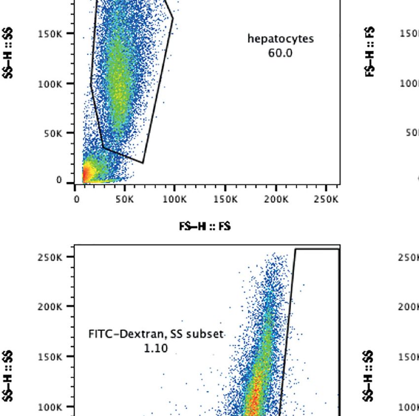
van der Boor et al.                                                                                           Pf Development in Porcine Hepatocytes

Isolation and Infection of Primary Human                                     LSM880 microscope with Airyscan using a 63x (oil) objective
and Porcine Hepatocytes                                                      and 2x zoom.
The isolation and infection of fresh hepatocytes has been
described elsewhere (Walk et al., 2017; Yang et al., 2021).                  Traversal Assay
Briefly, viable hepatocytes were suspended in complete                        A master mix was prepared including 10% heat inactivated
William’s B medium without serum. Hepatocytes were seeded                    human serum, 100 mg/ml FITC dextran, sporozoites (1:1
into 96-well flat-bottom plates at a seeding density of 62.500                infection), and William’s B medium, and was added to each
hepatocytes per well and cultured at 37°C in an atmosphere of                well. Controls included master mix containing no sporozoites,
5% CO2 with daily media refreshment.                                         and master mix containing cytochalasin D (1:100 dilution). Cells
    Two days after plating, sporozoites were added to the wells at           were spun at 3000 revolutions per minute for ten minutes and
a 1:1 ratio. Media was refreshed after three hours to remove non-            stored at 37°C for two hours. Subsequently, cells were washed
invaded sporozoites and refreshed daily throughout the time-                 with PBS. Trypsin was added per well and cells were kept at 37°C
course. After time-course completion, cells were fixed with 4%                for five minutes. Finally, 10% fetal bovine serum in PBS was
paraformaldehyde (Thermo Fisher Scientific: catalogue number                  added to each well. Traversal data was obtained by flow
28906) for ten minutes.                                                      cytometry (Beckman Coulter Gallios 10-color), and analysis
                                                                             was performed using FlowJo software (version 10.0.8, Tree Star).
Immunofluorescence Assay
After each incubation step, cells were washed thrice with PBS.               Statistical Analysis
First, fixed cells were permeabilized with 1% triton x-100 for ten            Three biological replicates were used for fresh porcine
minutes at room temperature. Subsequently, cells were incubated              hepatocytes, and three biological replicates were used for
with 0.1 M glycine to avoid nonspecific binding of antibodies to              human h e p ato c yt es, o f w hi ch tw o r ep li ca te s we r e
free aldehyde groups. Cells were then incubated with 3% bovine               cryopreserved human hepatocytes and one was fresh. Each
serum albumin for forty-five minutes. Next, cells were incubated              biological replicate had at least two technical replicates. All
with primary antibodies for one hour at room temperature: rabbit             statistical tests were performed using Prism 9 (version 9.2.0,
anti-Pf HSP70 (Heat Shock Protein, 1:75 dilution), mouse anti-Pf             GraphPad Software, California USA).
MSP1 (Merozoites Surface Protein, 1:100 dilution), mouse anti-Pf
EXP1 (Exported Protein 1, 1:1000 dilution), mouse anti-Pf EXP2
(Exported Protein 2, 1:1000 dilution) or goat anti-Plasmodium
berghei (Pb) UIS4 (Upregulated in Infectious Sporozoites 4, 1:300            RESULTS
dilution). Cells were subsequently incubated with secondary
antibodies (1:200 dilution) for one hour at room temperature in              Traversal of Porcine Hepatocytes by
the dark: goat anti-rabbit Alexa 594 for PfHSP70, donkey anti-               Plasmodium falciparum Sporozoites
mouse Alexa 647 for anti-PfMSP1 staining, goat anti-mouse Alexa              Fresh porcine hepatocytes were isolated based on human
488 for anti-PfEXP1 and anti-PfEXP2 staining, and donkey anti-               protocols as described previously Yang et al. (2021) (Yang
goat Alexa 594. Subsequently, nuclei were stained with chromatin-            et al., 2021). Different seeding densities were compared, and
specific 4’,6-diamidino-2-phenylindole (DAPI, 1:200 dilution) and             the viability of each density was assessed using brightfield
incubated for one hour in the dark. Finally, 0.1% sodium azide was           microscopy and mitochondrial enzymatic activity (MTT) from
added. Cells were stored at 4°C.                                             day of invasion up to day five (Figures 1A, B). For all seeding
                                                                             densities, metabolic activity increased post-plating (up to
Microscopy                                                                   seventy-two hours post-invasion) and remained steady as
Monolayer viability was assessed by brightfield microscopy at                 measured by MTT (Figure 1B). After day seven, the viability
400x magnification. Following immunofluorescent staining,                      of the culture decreased with clumping of cells observed by
parasite invasion into hepatocytes was visualized with a Leica               brightfield microscopy. An optimal seeding density of 62.500 was
DMI6000B inverted epifluorescent high content microscope. For                 chosen, as at higher densities the host cells tended to clump
each well, a tile size of 9x9 was obtained using a 20x objective.            instead of forming monolayers (image not shown). This is in line
Tiles were manually counted based on PfHSP70 and DAPI                        with the seeding density used for primary human hepatocytes
positivity in FIJI. Schizonts were characterized using the region            (Yang et al., 2021).
of interest (ROI) tool to determine the cross-sectional surface                  Both human (freshly isolated and cryopreserved) and porcine
area, circumference, and raw integrated density (RawIntDen) of               (fresh) hepatocytes were subsequently infected with NF135.C10
each manually encircled schizont. The RawIntDen values give                  sporozoites two days after plating. Traversal assays were
the sum of all pixel values in the ROI. The RawIntDen value was              performed for three hours post-invasion and a significant
corrected for background noise. Background noise was                         presence of FITC dextran-positive cells were detected,
calculated by measuring the total intensity of a whole image                 indicating NF135.C10 sporozoites were able to traverse porcine
divided by the area of the image. The final intensity of the                  hepatocytes, albeit at a lower rate than in human hepatocytes.
parasite was calculated by subtracting the background value from             The percentage of traversed hepatocytes was lower in porcine
the measured value. Confocal images were obtained with a Zeiss               donors compared to human donors (Figures 1C, D).

Frontiers in Cellular and Infection Microbiology | www.frontiersin.org   3                               February 2022 | Volume 12 | Article 834850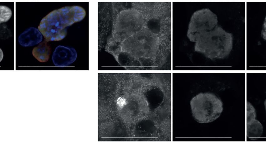
van der Boor et al.                                                                                                                  Pf Development in Porcine Hepatocytes

                      A                                                                    B

                      C                                                                    D

 FIGURE 1 | (A) Microscopic images of freshly seeded porcine hepatocytes at 62.500, 75.000 and 90.000 hepatocytes per well on days two, three and five post-
 invasion. (B) Viability of freshly isolated porcine hepatocytes. Porcine hepatocytes were seeded at densities of 62.500, 81.250 and 100.000 hepatocytes per well.
 MTT levels are depicted of uninfected control hepatocytes on day three, day five, and day seven post-invasion. Mean MTT reading and standard deviation of four
 technical replicates of two biological replicates. The extinction was measured at a wavelength of 570 nm. The reference wavelength was set at 630 nm. (C, D)
 Traversal of NF135.C10 sporozoites in fresh human and porcine hepatocytes three hours post-invasion of three biological donors, each with three technical
 replicates. (C) Fluorescence activated cell sorting gating strategy used to study traversal. Top left panel: selection of hepatocytes. Top right panel: selection of single
 cells. Bottom left panel: hepatocytes incubated in FITC-Dextran only (negative control). Bottom right panel: hepatocytes incubated with FITC-Dextran and
 sporozoites. For both bottom panels, the y-axis shows the degree of FITC-positivity in the cells. For all panels, each dot represents a cell/event measured. (D) Mean
 percentage (± SD) of traversed porcine and human hepatocytes.

Plasmodium falciparum Sporozoite                                                              In the porcine cells, there was a significant decrease in
Development in Primary Porcine                                                            schizont numbers from day three post-invasion onwards with
Hepatocytes                                                                               no remaining schizonts left on day seven, indicating that
To assess whether infection of porcine hepatocytes was possible,                          parasites were unable to survive during this period. While a
liver cells from porcine and human donors were infected with                              decrease in the number of intracellular schizonts was also
NF135.C10 sporozoites. The number of intracellular schizonts                              observed in human hepatocytes, intracellular schizonts could
was determined by immunofluorescence assay using DAPI and                                  still be detected on day seven post-invasion (Figure 2B).
HSP70 staining on day three, five, and seven post-invasion                                 However, some degree of parasite nuclear division was
(Figure 2). NF135.C10 schizonts of comparable size were                                   observed in schizonts developing in porcine host hepatocytes,
detected in human and porcine hepatocytes on day three,                                   as illustrated by DAPI staining (Figure 2E). The integrated
illustrating the permissiveness of porcine hepatocytes to                                 HSP70 signal intensity per schizont surface area remained
support Pf development (Figure 2A). The number of                                         relatively constant between day three and five post-invasion for
intracellular schizonts was significantly lower in porcine                                 both human and porcine hepatocytes, suggesting that the health
compared to human hepatocytes (Figure 2B) (p=0.0005 on                                    of these parasites remained stable (Figure 2C). As expected, the
day three and p=0.0074 on day five).                                                       signal was higher for parasites developing in the human

Frontiers in Cellular and Infection Microbiology | www.frontiersin.org                4                                        February 2022 | Volume 12 | Article 834850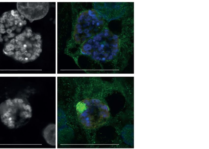
van der Boor et al.                                                                                                          Pf Development in Porcine Hepatocytes

                      A                                                              B

                      C                                                           D

                      E

 FIGURE 2 | (A) Schizont size. Mean sizes (±SD) of NF135.C10 schizonts on day three, day five and day seven post-invasion in primary porcine and human
 hepatocytes based on immunofluorescence staining with HSP70. Per technical replicate, all or up to seventy-five schizonts were measured for each of the biological
 replicates (three human and three porcine donors). A two-way RM ANOVA was performed on the median of each replicate. Mean size (±SD) of NF135.C10
 schizonts per technical replicate and circumference data is shown in Supplementary Figure 1 and Supplementary Table 1 (B) Schizont number. Mean number
 (±SD) of NF135.C10 schizonts on day three, day five and day seven post-invasion in primary porcine and human hepatocytes based on immunofluorescence
 staining with HSP70 and DAPI. All schizonts were counted per technical replicate of each of the biological replicates. A two-way RM ANOVA was performed on the
 median of each replicate. The mean number (±SD) of NF135.C10 schizonts per technical replicate is shown in Supplementary Figure 2 (C) HSP70 signal. The
 median HSP70 signal (±IQR) measured per schizont on day three and day five post-invasion is shown, corrected for noise and schizont size. All or up to fifty
 schizonts were measured per technical replicate of the three biological replicates. A two-way RM ANOVA was performed on the median of each replicate. (D) MSP1
 signal. Median MSP1 signal (±IQR) measured per schizont on day three and day five post-invasion, corrected for noise and schizont size. All or up to fifty schizonts
 were measured per technical replicate of three biological replicates. A two-way RM ANOVA was performed. (E) Expression of typical liver stage protein markers in
 porcine hepatocytes visualized by confocal microscopy including GAPDH, HSP70, MSP1 and DAPI. For comparative human images, please refer to (Yang et al.,
 2021). Objective 63×; zoom 2×; scale bar 25 µm. For panels A, C and D, the total number of schizonts analyzed per condition is shown in Supplementary Table 2.
 RawIntDen: raw integrated density. GAPDH, glyceraldehyde 3-phosphate dehydrogenase; HSP70, heat shock protein 70; IQR, interquartile range; MSP1, merozoite
 surface protein; NS, not significant; P.I., post-invasion; IQR, interquartile range. *P < 0.05; **P < 0.005; ***P < 0.001; ****P < 0.0001.

Frontiers in Cellular and Infection Microbiology | www.frontiersin.org           5                                      February 2022 | Volume 12 | Article 834850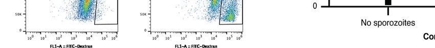
van der Boor et al.                                                                                                        Pf Development in Porcine Hepatocytes

hepatocytes as it is the ideal host for the parasite. Furthermore, a                supplementary material and showed a similar trend
large increase in the MSP1 signal intensity per schizont size, a                    (Figure S1).
marker of maturation, was only observed in schizonts present in
human hepatocytes (Figure 2D), indicating the ability of these                      Developing Schizonts Show Aberrant
schizonts to mature in the human host but not in the porcine                        EXP1 and EXP2 Expression
host. Finally, parasites developing in the porcine host displayed                   As shown by the disappearance of schizonts from day five to day
typical liver stage markers such as HSP70, GAPDH and                                seven post-invasion as well as the steady MSP1 signal, it appears
MSP1 (Figure 2E).                                                                   that schizonts cannot mature properly in the porcine hepatocyte
   While schizonts developing in the porcine and human                              system. The integrity of the PVM was next assessed using
hepatocytes did not differ in size on day three post-invasion,                      antibodies against two established PVM proteins, EXP1 and 2
there was a significant difference on day five post-invasion                          (van Dijk et al., 2005; Labaied et al., 2007; Ploemen et al., 2012;
(p=0.0038) (Figure 2A). Schizonts developing in human                               Annoura et al., 2014; Kalanon et al., 2016; Manzoni et al., 2017;
hepatocytes increased in size from day three to day five                             Yang et al., 2021). EXP1 and EXP2 have been identified as crucial
(p=0.0008) and from day five to day seven (p=0.0006). In                             PVM components for nutrient uptake and protein export in blood
contrast, no significant difference in size was found from day                       stages while a defined role in liver stage development remains
three to day five in surviving schizonts in porcine hepatocytes                      elusive (Charnaud et al., 2018; Garten et al., 2018; Mesén-Ramı́rez
(p=0.1137), indicating again that maturation is hindered in                         et al., 2019; Nessel et al., 2020). In porcine hepatocytes, there was
non-human host cells. Circumference data is shown in                                no apparent increase in EXP1 signal (Figure 3A). Although not

                      A                                                     B

                      C                                                     D

 FIGURE 3 | Median EXP signal (±IQR) measured per schizont in porcine hepatocytes on day three and day five post-invasion, corrected for noise and schizont size.
 All or up to seventy-five schizonts were measured per technical replicate of three biological replicates. A two-way RM ANOVA was performed on the median of each
 replicate. (A) EXP1 signal in porcine hepatocytes. (B) EXP1 signal in human hepatocytes. (C) EXP2 signal in porcine hepatocytes. (D) EXP2 signal in human
 hepatocytes. For all panels, the total number of schizonts analyzed per condition is shown in Supplementary Table 2. CHH, cryopreserved human hepatocytes;
 EXP, exported protein; FHH, fresh human hepatocytes; FPH, fresh porcine hepatocytes; NS, not significant. *P < 0.05; **P < 0.005; ****P < 0.0001.

Frontiers in Cellular and Infection Microbiology | www.frontiersin.org          6                                     February 2022 | Volume 12 | Article 834850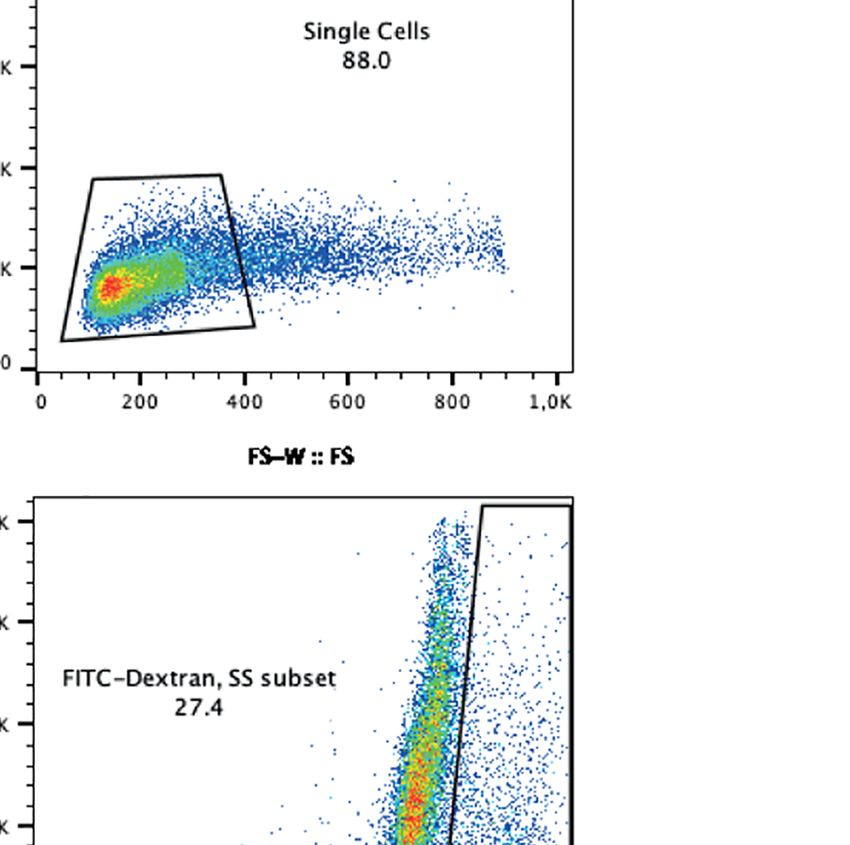
van der Boor et al.                                                                                                            Pf Development in Porcine Hepatocytes

significant, there was an increasing trend in EXP1 signal intensity                    schizonts developing in human hepatocytes with a typical
observed from day three to day five for schizonts developing in                        pattern increased over time, although this difference was
human hepatocytes for some donors (Figure 3B). With regards to                        not significant.
the EXP2 signal, there was a decrease in signal in schizonts                             Additionally, fresh porcine hepatocytes were infected with
developing in porcine hepatocytes from day three to day five                           the rodent Pb, showing that UIS4 localized to the PVM and is
whereas an increase was observed over time for schizonts in                           expressed throughout liver stage development in Pb (Mueller
human hepatocytes (Figures 3C, D).                                                    et al., 2005). Unlike porcine hepatocytes infected with Pf, Pb
    Two main staining patterns of EXP1 and EXP2 were observed                         schizonts developing in porcine hepatocytes all showed a
and classified as typical (circle surrounding HSP70 and DAPI                           typical staining pattern for UIS4 (Figure S3B), suggestive for
staining) and atypical (Figure 4A and Figure S3A). The majority                       an intact PVM. Finally, schizonts developing in porcine
of schizonts developing in porcine hepatocytes showed atypical                        hepatocytes with atypical EXP1 and EXP2 signal did not
EXP1 and EXP2 staining, compared to schizonts developing in                           show a difference in schizont area (determined by measuring
human hepatocytes that showed mostly a typical EXP1 and                               HSP70 signal) compared to schizonts with a typical EXP1 and
EXP2 staining pattern (Figures 4B, C). Schizonts developing                           EXP2 signal (Figure S4), whereas in human hepatocytes
in porcine hepatocytes showed an increasing trend of atypical                         schizonts with a typical pattern appeared to be non-
EXP1 and EXP2 staining over time, whereas the proportion of                           significantly larger.

                      A

                      B                                                                   C

 FIGURE 4 | (A) Representative immunofluorescence images of typical liver stage protein markers visualized through confocal microscopy including EXP1 and EXP2,
 HSP70, and DAPI, classified as a typical (top) or atypical (bottom) staining pattern. Images shown were obtained from infected porcine hepatocytes. Objective 63×;
 zoom 2×; scale bar 25 µm. (B, C) Mean percentage of human and porcine schizonts with a typical or atypical EXP1(B) or EXP2 (C) staining pattern. Porcine
 hepatocytes are depicted on the left and human hepatocytes on the right of each bar plot. All or up to seventy-five schizonts were measured per technical replicate
 of three biological replicates. Results per biological replicate are shown in Supplementary Figure 4. For panels B and C, the total number of schizonts analyzed per
 condition is shown in Supplementary Table 2. EXP, exported protein; HSP, heat shock protein; PHH, primary human hepatocytes; PPH, primary porcine
 hepatocytes.

Frontiers in Cellular and Infection Microbiology | www.frontiersin.org            7                                      February 2022 | Volume 12 | Article 834850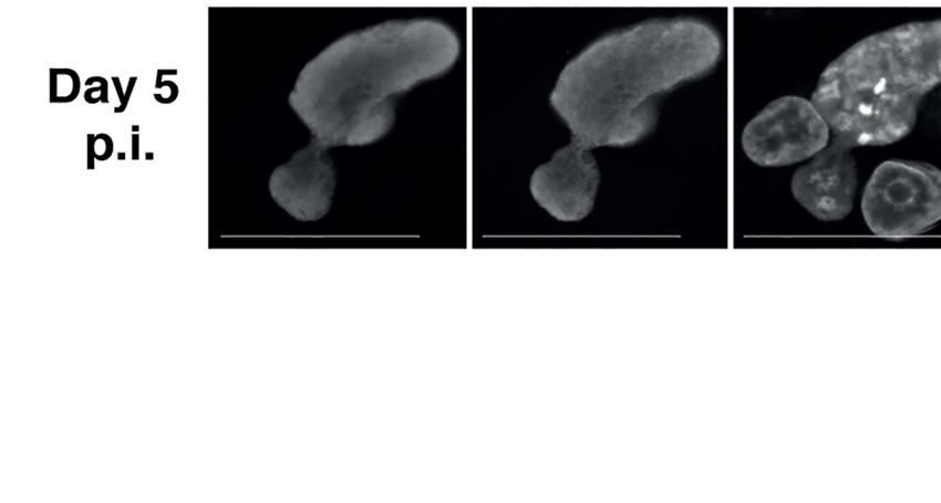
van der Boor et al.                                                                                                Pf Development in Porcine Hepatocytes

DISCUSSION                                                                     combination thereof, including refractory porcine hepatocytes
                                                                               for productive invasion, the inability of parasites to maintain the
Here, we show that Pf is capable of traversing, invading and                   PVM, as well as host factors that may compromise the integrity
partially maturing in non-human porcine hepatocytes.                           of the PVM. We find that sporozoites do show characteristics of
Incomplete maturation of schizonts may be due to an aberrant                   early transformation into normal liver-stage schizonts, as
PVM, supported by the increasingly atypical EXP1 and EXP2                      illustrated by the increase in nuclear material and presence of
expression pattern observed over time. Additionally, the aberrant              nuclear division. However, the increasingly aberrant expression
EXP1 and EXP2 staining suggest that parasite nutrient uptake                   of EXP1 and EXP2 supports the failure of schizonts to complete
may be impaired, which could hamper further maturation of                      maturation up to day seven. The lack of fluorescently tagged
the parasites.                                                                 EXP1/2 prevented live-imaging and we therefore cannot exactly
    Pf shows a remarkably narrow host range compared to other                  pinpoint the timing of the loss of EXP1/2 from the PVM over
Plasmodium species and appears to uniquely infect primates in                  time. This loss may be due to deteriorating parasite health or
vivo and in vitro (Collins et al., 1994; Rathore et al., 2003). This           active degradation by the host. Alternatively, aberrant EXP1 and
selectivity is partially explained by different invasion                       EXP2 expression of sporozoites in porcine hepatocytes could
requirements between Plasmodium species, as well as                            affect proper parasite development due impaired nutrient
incompatibility of invasion proteins between Pf and the non-                   scavenging from the host environment.
human host hepatocyte (i.e. a lock and key model), including                       Mello-Vieira et al. (2020) show that EXP2 is required for liver
CSP and CD81 (Rathore et al., 2003; Silvie et al., 2003; Silvie                invasion and maturation and for establishing blood stage
et al., 2006). As Pf is successful in invading porcine hepatocytes,            infection in a mouse model, which corroborates with our
albeit at a lower rate, it suggests that compatibility of parasite             findings of its importance in maturation (Mello-Vieira et al.,
invasion ligands with their host counterparts is not the only                  2020). Furthermore, the EXP1 C-terminal region exposed to the
requirement needed for parasite survival within a hepatocyte.                  host cytoplasm interacts with host Apolipoprotein A (ApoH)
    A key finding is the abrupt mid-liver stage arrest in parasite              which is pivotal for successful liver-stage development in Pb,
development coinciding with the increasingly aberrant EXP1 and                 although it is unclear whether it performs the same role in Pf (Sá
EXP2 expression, established markers for PVM quality. The PVM                  et al., 2017; Mesé n-Ramı́rez et al., 2019; Wolanin et al., 2019).
forms a barrier with the host environment and is extensively                   EXP1 is continuously trafficking to the PVM throughout the first
modified by the parasite to provide the appropriate machinery to                days of the liver stage (Sá et al., 2017). As the parasite matures, its
scavenge sufficient host nutrients and export proteins, whilst                  survival becomes increasingly dependent on its ability to
simultaneously offering protection from the hepatocytes’                       scavenge lipids and nutrients from its host cell, for which it
defense mechanisms (Spielmann et al., 2012). Studies with Pb                   requires a functional PVM (Agop-Nersesian et al., 2018). The
and Py show that complete cytosolic development of schizonts                   combined findings in rodent models may be a potential
lacking an intact PVM is possible but generally schizonts only                 explanation for the inability of Pf to fully mature in
develop up to early-liver stage at twenty-four hours post-invasion             porcine hepatocytes.
(van Dijk et al., 2005; Silvie et al., 2006; Ploemen et al., 2012).                An aberrant PVM additionally exposes the developing
Sporozoites lacking SLARP/SAP1 (sporozoite Asparagine-rich                     parasites to the hostile host environment and host cell defense
protein) arrest early (twenty-four hours post-invasion) and fail               mechanisms. Although host responses to the invading parasites
to express amongst others effector proteins P52, UIS4 and EXP1                 may vary between parasite species, targeting intracellular
(Aly et al., 2008). These proteins are critical for the formation of a         parasites is a conserved defense strategy of infected hepatocytes
PVM and lead to near-complete developmental arrest during                      (Wacker et al., 2017). During parasite infection, diverse host
early development (Aly et al., 2008). Sporozoites with P52/p36                 autophagy pathways can be activated in hepatocytes (reviewed in
deletions are incapable of productively invading hepatocytes                   (Agop-Nersesian et al., 2018)). This includes marking the PVM
resulting in near-complete attenuation during early liver stage                with the autophagy marker LC3, which activates the host cell
in Pb and Py, as well as Pf (Ishino et al., 2005; van Dijk et al., 2005;       autophagy machinery to eliminate intracellular parasites. The
Labaied et al., 2007; VanBuskirk et al., 2009; Ploemen et al., 2012;           observed aberrant PVM may be the result of active host
Manzoni et al., 2017). Finally, the function of B9 remains more                destruction of the PVM: the parasite proteins involved in PVM
controversial in different Plasmodia species: while required in Pb             maintenance in human hepatocytes may be incompatible to
for productive hepatocyte invasion, this is not the case for Pf                those in porcine hepatocytes. This could impact the parasites’
invasion into human hepatocytes despite structural similarity                  ability to secure host resources and protect itself against
(Annoura et al., 2014; Fernandes et al., 2021). As schizonts are               host degradation.
detected in porcine hepatocytes on day three and (to a lesser                      To our knowledge, Pf infection of porcine hepatocytes has not
amount) on day five, this challenges this prevailing theory that a              been reported before. Our findings are confined to an in vitro
functional, intact, PVM is required for progression beyond early-              system and it is not clear whether a similar mechanism of
stage Pf infection.                                                            invasion and maturation occurs in vivo. Additionally, the cause
    An important limitation is that we do not understand the                   of an impaired PVM remains unknown. The aberrant PVM
mechanism behind the atypical PVM in porcine hepatocytes.                      could make the parasite susceptible to or may be a direct
This abnormal event could be due to several factors or a                       response to intracellular host defence mechanisms.

Frontiers in Cellular and Infection Microbiology | www.frontiersin.org     8                                  February 2022 | Volume 12 | Article 834850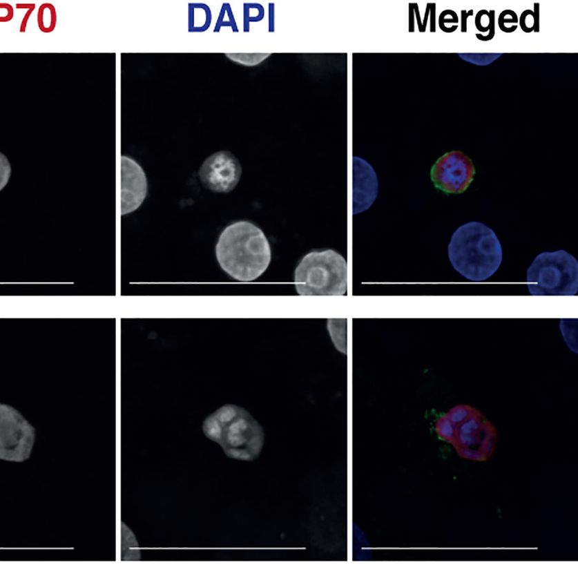
van der Boor et al.                                                                                                                        Pf Development in Porcine Hepatocytes

Furthermore, these results underline the importance of EXP1                                      contributed and reviewed the manuscript and approved the
and EXP2 in Pf liver stage development and maturation,                                           submitted version.
although their exact roles are not explored here. Future studies
with PfEXP-1 and EXP-2 knockdown parasites, as well as other
PVM proteins, may be able to delineate the key processes
involved in parasite maturation.
                                                                                                 FUNDING
    Altogether, our findings highlight the importance of                                          This study was funded by the Department of Medical
understanding species-specific intracellular host factors                                         Microbiology, Radboud University Medical Center. AY is
involved in Pf liver-stage development. The partial                                              funded by the Dutch Research Council (NWO) talent scheme
development of Pf sporozoites in porcine hepatocytes                                             veni (VI.Veni.192.171).
underscores its potential as a platform to study early to mid-
stage Pf liver-stage development, and to identify essential Pf and
host proteins required for the formation and maintenance of the
PVM. Additionally, the model may provide a novel platform to                                     ACKNOWLEDGMENTS
study host factors and nutrients required for full Pf liver-
                                                                                                 We are grateful for M. van de Vegte-Bolmer, R. Stoter, R.
stage development.
                                                                                                 Heutink, J. Klaasen, A. Pouwelsen, L. Pelser-Posthumus and J.
                                                                                                 Kuhnen of the Malaria Unit at the Radboud University Medical
                                                                                                 Center for their contribution in parasite, mosquito and
DATA AVAILABILITY STATEMENT                                                                      sporozoite production. We would also like to thank the
                                                                                                 Microscopic Imaging Center (MIC) of the Radboud University
The raw data supporting the conclusions of this article will be                                  for access to their facilities. Additionally, we would like to thank
made available by the authors when possible and upon request                                     R. Woestenink of the Radboudumc Technology Center of Flow
                                                                                                 cytometry for his help in single cell analysis. We thank T. Kooij,
                                                                                                 N. Proellochs, A. van der Starre, and T. Roos for providing
                                                                                                 critical feedback on the project.
AUTHOR CONTRIBUTIONS
The study was conceptualized and designed by SB, AY, and RS.
SB, AY, YW, GJG, and AH performed the experiments. JW                                            SUPPLEMENTARY MATERIAL
coordinated the collection of fresh human liver segments. SB,
AY, RS, TB, and MM were involved in conceptualization of the                                     The Supplementary Material for this article can be found online
data. SC and AY analyzed the data and made figures for the                                        at: https://www.frontiersin.org/articles/10.3389/fcimb.2022.
article. SB and AY wrote the first manuscript. All authors                                        834850/full#supplementary-material

                                                                                                    Sporozoite Surface is Essential for Gliding Motility and Sporozoite
REFERENCES                                                                                          Infectivity. PloS Path. 8, 7. doi: 10.1371/journal.ppat.1002725
Agop-Nersesian, C., Niklaus, L., Wacker, R., and Theo Heussler, V. (2018). Host                  Fernandes, P., Loubens, M., Marinach, C., Coppé e, R., Grand, M., Andre, T. -P.,
    Cell Cytosolic Immune Response During Plasmodium Liver Stage                                    et al. (2021). Plasmodium Sporozoites Require the Protein B9 to Invade
    Development. FEMS Microbiol. Rev. 42, 3. doi: 10.1093/femsre/fuy007                             Hepatocytes. bioRxiv. doi: 10.1101/2021.10.25.465731
Aly, A. S., Mikolajczak, S. A., Rivera, H. S., Camargo, N., Jacobs-Lorena, V.,                   Garten, M., Nasamu, A. S., Niles, J. C., Zimmerberg, J., Goldberg, D. E., and Beck,
    Labaied, M., et al. (2008). Targeted Deletion of SAP1 Abolishes the Expression                  J. (2018). EXP2 Is a Nutrient-Permeable Channel in the Vacuolar Membrane of
    of Infectivity Factors Necessary for Successful Malaria Parasite Liver Infection.               Plasmodium and is Essential for Protein Export via PTEX. Nat. Microbiol. 3,
    Mol. Microbiol. 69, 1. doi: 10.1111/j.1365-2958.2008.06271.x                                    10. doi: 10.1038/s41564-018-0222-7
Annoura, T., van Schaijk, B. C., Ploemen, I. H., Sajid, M., Lin, J. W., Vos, M. W.,              Ishino, T., Chinzei, Y., and Yuda, M. (2005). Two Proteins With 6-Cys Motifs are
    et al. (2014). Two Plasmodium 6-Cys Family-Related Proteins Have Distinct                       Required for Malarial Parasites to Commit to Infection of the Hepatocyte. Mol.
    and Critical Roles in Liver-Stage Development. FASEB J. 28, 5. doi: 10.1096/                    Microbiol. 58, 5, 1264–1275. doi: 10.1111/j.1365-2958.2005.04801.x
    fj.13-241570                                                                                 Kalanon, M., Bargieri, D., Sturm, A., Matthews, K., Ghosh, S., Goodman, C. D.,
Charnaud, S. C., Jonsdottir, T. K., Ploemen, P. R., Sajid, H. E., Lin, B. K., Vos, B.,              et al. (2016). The Plasmodium Translocon of Exported Proteins Component
    et al. (2018). Spatial Organization of Protein Export in Malaria Parasite Blood                 EXP2 is Critical for Establishing a Patent Malaria Infection in Mice. Cell.
    Stages. Traffic 19, 8. doi: 10.1111/tra.12577                                                    Microbiol. 18 (3), 399–412. doi: 10.1111/cmi.12520
Collins, W. E., Galland, G. G., Sullivan, J. S., and Morris, C. L. (1994). Selection of          Kaushansky, A., Douglass, A. N., Arang, N., Vigdorovich, V., Dambrauskas, N.,
    Different Strains of Plasmodium Falciparum for Testing Blood-Stage Vaccines                     Kain, H. S., et al. (2015). Malaria Parasites Target the Hepatocyte Receptor
    in Aotus Nancymai Monkeys. Am. J. Trop. Med. Hyg 51, 2. doi: 10.4269/                           Epha2 for Successful Host Infection. Science 350 (6264), 1089–1092.
    ajtmh.1994.51.224                                                                               doi: 10.1126/science.aad3318
Coppi, A., Tewari, R., Bishop, J. R., Bennett, B. L., Lawrence, R., Esko, J. D., et al.          Labaied, M., Harupa, A., Dumpit, R. F., Coppens, I., Mikolajczak, S. A., Kappe, S.
    (2007). Heparan Sulfate Proteoglycans Provide a Signal to Plasmodium                            H., et al. (2007). Plasmodium Yoelii Sporozoites With Simultaneous Deletion
    Sporozoites to Stop Migrating and Productively Invade Host Cells. Cell.                         of P52 and P36 are Completely Attenuated and Confer Sterile Immunity
    Host. Microbe 2, 5. doi: 10.1016/j.chom.2007.10.002                                             Against Infection. Infect. Immun. 75 (8), 3758–3768. doi: 10.1128/iai.00225-07
Ejigiri, I., Ragheb, D. R., Pino, P., Coppi, A., Bennett, B. L., Soldati-Favre, D., et al.       Liu, W., Sundararaman, S. A., Loy, D. E., Learn, G. H., Li, Y., Plenderleith, L. J.,
    (2012). Shedding of TRAP by a Rhomboid Protease From the Malaria                                et al. (2016). Multigenomic Delineation of Plasmodium Species of the

Frontiers in Cellular and Infection Microbiology | www.frontiersin.org                       9                                       February 2022 | Volume 12 | Article 834850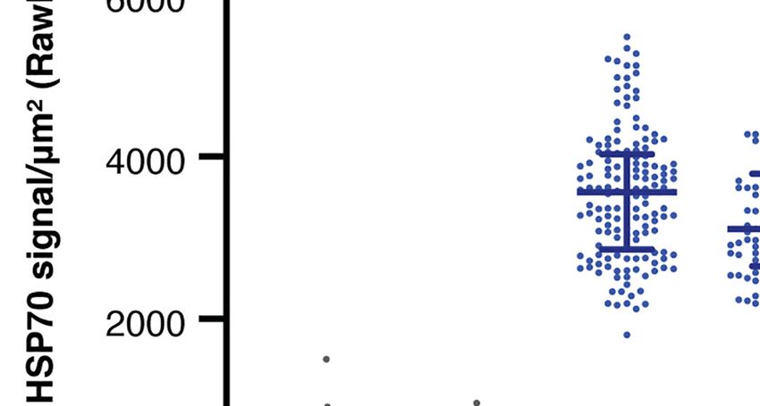
van der Boor et al.                                                                                                                                Pf Development in Porcine Hepatocytes

     Laverania Subgenus Infecting Wild-Living Chimpanzees and Gorillas. Genome                        Membrane. Int. J. Med. Microbiol. 302, 4–5. doi: 10.1016/j.ijmm.
     Biol. Evol. 8 (6), 1929–1939. doi: 10.1093/gbe/evw128                                            2012.07.011
Manzoni, G., Marinach, C., Topçu, S., Briquet, S., Grand, M., Tolle, M., et al.                   Teirlinck, A. C., Roestenberg, M., van de Vegte-Bolmer, M., Scholzen, A.,
     (2017). Plasmodium P36 Determines Host Cell Receptor Usage During                                Heinrichs, M. J., Siebelink-Stoter, R., et al. (2013). NF135.C10: A New
     Sporozoite Invasion. eLife 6, e25903. doi: 10.7554/eLife.25903                                   Plasmodium Falciparum Clone for Controlled Human Malaria Infections.
Meier, R. P. H., Navarro-Alvarez, N., Morel, P., Schuurman, H. J., Strom, S.,                         J. Infect. Dis. 207 (4), 656–660. doi: 10.1093/infdis/jis725
     Bühler, L. H., et al. (2015). Current Status of Hepatocyte Xenotransplantation.               VanBuskirk, K. M., O'Neill, M. T., De La Vega, P., Maier, A. G., Krzych, U.,
     Int. J. Surg. 23 (Pt B), 273–279. doi: 10.1016/j.ijsu.2015.08.077                                Williams, J., et al. (2009). Preerythrocytic, Live-Attenuated Plasmodium
Mello-Vieira, J., Enguita, F. J., de Koning-Ward, T. F., Zuzarte-Luı́s, V., Mota, M.                  Falciparum Vaccine Candidates by Design. Proc. Natl. Acad. Sci. U. S. A.
     M., et al. (2020). Plasmodium Translocon Component EXP2 Facilitates                              106, 31. doi: 10.1073/pnas.0906387106
     Hepatocyte Invasion. Nat. Commun. 11, 1. doi: 10.1038/s41467-020-19492-4                      van Dijk, M. R., Douradinha, B., Franke-Fayard, B., Heussler, V., van Dooren,
Mesé n-Ramı́rez, P., Bergmann, B., Tran, T. T., Garten, M., Stäcker, J., Naranjo-                    M.W., van Schaijk, B., et al. (2005). Genetically Attenuated, P36p-Deficient
     Prado, I., et al. (2019). EXP1 is Critical for Nutrient Uptake Across the                        Malarial Sporozoites Induce Protective Immunity and Apoptosis of Infected
     Parasitophorous Vacuole Membrane of Malaria Parasites. PloS Biol. 17, 9.                         Liver Cells. Proc. Natl. Acad. Sci. U. S. A. 102, 34. doi: 10.1073/pnas.0500925102
     doi: 10.1371/journal.pbio.3000473                                                             Vaughan, A. M., and Kappe, S. H. I. (2017). Malaria Parasite Liver Infection and
Mota, M. M., Pradel, G., Vanderberg, J. P., Hafalla, J. C., Frevert, U.,                              Exoerythrocytic Biology. Cold Spring Harb. Perspect. Med. 7, 6. doi: 10.1101/
     Nussenzweig, R. S., et al. (2001). Migration of Plasmodium Sporozoites                           cshperspect.a025486
     Through Cells Before Infection. Science 291 (5501), 141–144 . doi: 10.1126/                   Wacker, R., Eickel, N., Schmuckli-Maurer, J., Annoura, T., Niklaus, L., Khan, S.
     science.291.5501.141                                                                             M., et al. (2017). LC3-Association With the Parasitophorous Vacuole
Mueller, A. K., Camargo, N., Kaiser, K., Andorfer, C., Frevert, U., Matuschewski,                     Membrane of Plasmodium Berghei Liver Stages Follows a Noncanonical
     K., et al. (2005). Plasmodium Liver Stage Developmental Arrest by Depletion of                   Autophagy Pathway. Cell. Microbiol. 19, 10. doi: 10.1111/cmi.12754
     a Protein at the Parasite-Host Interface. Proc. Natl. Acad. Sci. U. S. A. 102, 8.             Walk, J., Reuling, I. J., Behet, M. C., Meerstein-Kessel, L., Graumans, W., van
     doi: 10.1073/pnas.0408442102                                                                     Gemert, G. J., et al. (2017). Modest Heterologous Protection After Plasmodium
Nessel, T., Beck, J. M., Rayatpisheh, S., Jami-Alahmadi, Y., Wohlschlegel, J. A.,                     Falciparum Sporozoite Immunization: A Double-Blind Randomized
     Goldberg, D. E., et al. (2020). EXP1 is Required for Organisation of EXP2 in the                 Controlled Clinical Trial. BMC Med. 15 (1), 168. doi: 10.1186/s12916-017-
     Intraerythrocytic Malaria Parasite Vacuole. Cell Microbiol. 22, 5. doi: 10.1111/                 0923-4
     cmi.13168                                                                                     Walters, E. M., and Prather, R. S. (2013). Advancing Swine Models for Human
Nyboer, B., Heiss, K., Mueller, A. K., and Ingmundson, A. (2018). The                                 Health and Diseases. Mo. Med. 110 (3), 212–215.
     Plasmodium Liver-Stage Parasitophorous Vacuole: A Front-Line of                               Wernersson, R., Schierup, M. H., Jørgensen, F. G., Gorodkin, J., Panitz, F.,
     Communication Between Parasite and Host. Int. J. Med. Microbiol. 308, 1.                         Staerfeldt, H. H., et al. (2005). Pigs in Sequence Space: A 0.66X Coverage Pig
     doi: 10.1016/j.ijmm.2017.09.008                                                                  Genome Survey Based on Shotgun Sequencing. BMC Genomics 6, 70.
Ploemen, I. H., Croes, H. J., van Gemert, G. J., Wijers-Rouw, M., Hermsen, C. C.,                     doi: 10.1186/1471-2164-6-70
     Sauerwein, R. W., et al. (2012). Plasmodium Berghei Dp52&P36 Parasites                        Wolanin, K., Fontinha, D., Sanches-Vaz, M., Nyboer, M., Heiss, B., Mueller, K.,
     Develop Independent of a Parasitophorous Vacuole Membrane in Huh-7 Liver                         et al. (2019). A Crucial Role for the C-Terminal Domain of Exported Protein 1
     Cells. PloS One 7, 12. doi: 10.1371/journal.pone.0050772                                         During the Mosquito and Hepatic Stages of the Plasmodium Berghei Life
Prudencio, M., Mota, M. M., and Mendes, A. M. (2011). A Toolbox to Study Liver                        Cycle. Cell Microbiol. 21 (10), e13088. doi: 10.1111/cmi.13088
     Stage Malaria. Trends Parasitol. 27, 12. doi: 10.1016/j.pt.2011.09.004                        Yang, A. S. P., Lopaticki, S., O'Neill, M. T., Erickson, S. M., Douglas, D. N.,
Rathore, D., Hrstka, S. C., Sacci, J. B. Jr, De la Vega, P., Linhardt, R. J., Kumar, S.,              Kneteman, N. M., et al. (2017). AMA1 and MAEBL are Important for
     et al. (2003). Molecular Mechanism of Host Specificity in Plasmodium                              Plasmodium Falciparum Sporozoite Infection of the Liver. Cell. Microbiol.
     Falciparum Infection: Role of Circumsporozoite Protein. J. Biol. Chem. 278,                      19, (9). doi: 10.1111/cmi.12745
     42. doi: 10.1074/jbc.M306250200                                                               Yang, A. S. P., van Waardenburg, Y. M., van de Vegte-Bolmer, M., van Gemert, G.
Rodrigues, C. D., Hannus, M., Prudêncio, M., Martin, C., Gonçalves, L. A.,                          A., Graumans, W., Wilt, J. H. W., et al. (2021). Zonal Human Hepatocytes are
     Portugal, S., et al. (2008). Host Scavenger Receptor SR-BI Plays a Dual Role                     Differentially Permissive to Plasmodium Falciparum Malaria Parasites. EMBO
     in the Establishment of Malaria Parasite Liver Infection. Cell. Host. Microbe 4,                 J. 40 (6), e106583. doi: 10.15252/embj.2020106583
     3. doi: 10.1016/j.chom.2008.07.012
Sá , E., Cunha, C., Nyboer, B., Heiss, K., Sanches-Vaz, M., Fontinha, D., et al.                  Conflict of Interest: Author RS was employed by company TropIQ Health
     (2017). Plasmodium Berghei EXP-1 Interacts With Host Apolipoprotein H                         Sciences.
     During Plasmodium Liver-Stage Development. Proc. Natl. Acad. Sci. U.S.A.
     114, 7. doi: 10.1073/pnas.1606419114                                                          The remaining authors declare that the research was conducted in the absence of
Sato, S. (2021). Plasmodium—A Brief Introduction to the Parasites Causing                          any commercial or financial relationships that could be construed as a potential
     Human Malaria and Their Basic Biology. J. Physiol. Anthropol. 40, 1.                          conflict of interest.
     doi: 10.1186/s40101-020-00251-9
Silvie, O., Greco, C., Franetich, J. F., Dubart-Kupperschmitt, A., Hannoun, L., van                Publisher’s Note: All claims expressed in this article are solely those of the authors
     Gemert, G. J., et al. (2003). Hepatocyte CD81 is Required for Plasmodium                      and do not necessarily represent those of their affiliated organizations, or those of
     Falciparum and Plasmodium Yoelii Sporozoite Infectivity. Nat. Med. 9, 1.                      the publisher, the editors and the reviewers. Any product that may be evaluated in
     doi: 10.1038/nm808                                                                            this article, or claim that may be made by its manufacturer, is not guaranteed or
Silvie, O., Rubinstein, E., Franetich, J. F., Prenant, M., Belnoue, E., Ré nia, L., et al.        endorsed by the publisher.
     (2006). Expression of Human CD81 Differently Affects Host Cell Susceptibility
     to Malaria Sporozoites Depending on the Plasmodium Species. Cell. Microbiol.                  Copyright © 2022 van der Boor, van Gemert, Hanssen, van Waardenburg, McCall,
     8, 7. doi: 10.1111/j.1462-5822.2006.00697.x                                                   Bousema, de Wilt, Sauerwein and Yang. This is an open-access article distributed under
Sinnis, P., and Coppi, A. (2007). A Long and Winding Road: The Plasmodium                          the terms of the Creative Commons Attribution License (CC BY). The use, distribution or
     Sporozoite’s Journey in the Mammalian Host. Parasitol. Int. 56, 3. doi: 10.1016/              reproduction in other forums is permitted, provided the original author(s) and the
     j.parint.2007.04.002                                                                          copyright owner(s) are credited and that the original publication in this journal is cited, in
Spielmann, T., Montagna, G. N., Hecht, L., and Matuschewski, K. (2012).                            accordance with accepted academic practice. No use, distribution or reproduction is
     Molecular Make-Up of the Plasmodium Parasitophorous Vacuolar                                  permitted which does not comply with these terms.

Frontiers in Cellular and Infection Microbiology | www.frontiersin.org                        10                                            February 2022 | Volume 12 | Article 834850
You can also read