Management of Ludwig's Angina at a Tertiary Care Hospital in Western Region of India - Cureus
←
→
Page content transcription
If your browser does not render page correctly, please read the page content below
Open Access Original
                               Article                                                  DOI: 10.7759/cureus.23311

                                                Management of Ludwig's Angina at a Tertiary
                                                Care Hospital in Western Region of India
Review began 02/14/2022
                                                Bhagirath D. Parmar 1 , Krupal J. Joshi 2 , Ankur D. Modi 3 , Gavendra P. Dave 4 , Raji S. Desai 4
Review ended 02/28/2022
Published 03/19/2022                            1. Department of Otorhinolaryngology-Head and Neck Surgery, C U Shah Medical College and Hospital,
© Copyright 2022                                Surendranagar, IND 2. Department of Community and Family Medicine, All India Institute of Medical Sciences, Rajkot,
Parmar et al. This is an open access article    IND 3. Department of General Surgery, C U Shah Medical College and Hospital, Surendranagar, IND 4. Department of
distributed under the terms of the Creative     Otorhinolaryngology-Head and Neck Surgery, Sir Takhatsinhji (T) Hospital and Government Medical College,
Commons Attribution License CC-BY 4.0.,         Bhavnagar, IND
which permits unrestricted use, distribution,
and reproduction in any medium, provided
the original author and source are credited.    Corresponding author: Krupal J. Joshi, dr.krupaljoshi@gmail.com

                                                Abstract
                                                Introduction
                                                Ludwig’s angina is cellulitis of submandibular space, submental space, and sublingual space. The main
                                                causative factors include dental infections (dental caries with atypical periodontitis, pericoronitis, and
                                                dental procedures). Other predisposing conditions include poor dental hygiene, dental caries, malnutrition,
                                                diabetes mellitus, AIDS, and various other immunocompromised states. It presents as an acute onset and
                                                spreads very rapidly causing bilateral diffuse neck swelling, edema of floor of mouth, pain, fever, trismus,
                                                foul-smelling pus discharge, difficulty in swallowing, airway edema, and tongue displacement creating a
                                                compromised airway with stridor. So it requires early diagnosis and aggressive management.

                                                Material and methods
                                                Clinical data of all patients with clinical diagnosis of Ludwig’s angina managed at the Department of
                                                Otorhinolaryngology-Head and Neck Surgery, Sir Takhatsinhji (T) General Hospital and Government Medical
                                                College, Bhavnagar, India, from 2015 to 2019 were analyzed retrospectively in this study.

                                                Result
                                                Over the review period, 30 cases were diagnosed as Ludwig’s angina, out of which 12 (40%) were males and
                                                18 (60%) were females; male to female ratio was 1:1.5. The age of the patients ranged from six months to 64
                                                years, with a mean age of 38.86 years. Fever, neck swelling, and neck pain were present in all patients.

                                                In 16 patients, incision and drainage were done under general anesthesia while the rest five patients
                                                required only local anesthesia. In six patients (20%), for maintenance of airway, tracheostomy was required.
                                                The most common complication was necrotizing fasciitis and death followed by septicemia. Mortality was
                                                observed in three patients (10%) in this study.

                                                Conclusion
                                                Despite improved outcomes compare to pre-antibiotic era, Ludwig's angina still remains a potentially life-
                                                threatening disease in ENT at present. Dental caries, uncontrolled diabetes mellitus, and malnutrition are
                                                commonly associated conditions. With early diagnosis, close airway observation, aggressive intravenous
                                                antibiotic treatment, and timely surgical intervention, morbidity, and mortality can be reduced.

                                                Categories: Otolaryngology, Infectious Disease, Dentistry
                                                Keywords: incision and drainage, grodinsky criteria, tracheostomy, surgical decompression, submental and
                                                submaxillary space, floor of mouth cellulitis, dental caries

                                                Introduction
                                                Angina is from a Greek word, "Anchone," meaning strangulation. Ludwig’s angina was first described by
                                                German physician Wilhelm Frederick Von Ludwig in 1836. Cellulitis is an infectious inflammation of the
                                                cellular adipose tissue located in the apo-neurotic spaces. Ludwig's angina is cellulitis of sub-mandibular
                                                space, sub-mental space, and sublingual space. Initially, swelling develops unilaterally and then spreads
                                                bilaterally. Edema and cellulitis of supra-omohyoid space may develop then after causing compression of
                                                airway and dysphagia [1]. Infection may spread to retro-pharyngeal space, para-pharyngeal space, and in
                                                some cases up to mediastinum resulting in difficulty in breathing. The possible ways of infection spread are
                                                destruction of facial planes, aspirations of infective particles, or septic embolism to pulmonary vasculature
                                                [2].

                                                The incidence of Ludwig’s angina is nowadays less compared to pre-antibiotic era because of awareness

                               How to cite this article
                               Parmar B D, Joshi K J, Modi D, et al. (March 19, 2022) Management of Ludwig's Angina at a Tertiary Care Hospital in Western Region of India.
                               Cureus 14(3): e23311. DOI 10.7759/cureus.23311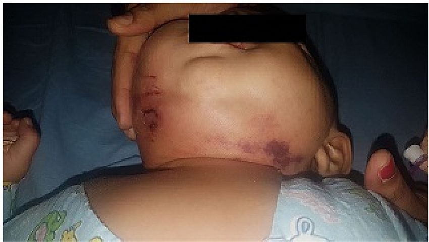
regarding dental health and effective antibiotic therapy [3]. The origin is generally from the carious second
                                                 and third molar. In fact, the roots of these teeth penetrate the mylohyoid ridge, such that any abscess or
                                                 dental infection has direct access to the sub-maxillary space. The main causative factors include dental
                                                 infections (dental caries with atypical periodontitis and pericoronitis). Other predisposing conditions
                                                 include poor dental hygiene, malnutrition, other infections like sialadenitis, floor of mouth trauma,
                                                 mandible fracture, diabetes mellitus, AIDS, and various immuno-compromised states [4]. The commonly
                                                 underlying microorganisms are Streptococcus sp., Staphylococcal aureus, Hemophilus influenza, and some
                                                 anaerobes like Bacteroids sp., Fusobacterium, etc. [5].

                                                 It presents as an acute onset and spreads very rapidly causing bilateral diffuse neck swelling, edema of the
                                                 floor of mouth, pain, fever, trismus, difficulty in swallowing, airway edema, and tongue displacement
                                                 causing compromised airway, stridor, foul-smelling pus discharge. So it requires early diagnosis and
                                                 aggressive management. Diagnosis is mainly clinical and supported with radiological investigation to know
                                                 the extent of spread and to discover dental origin. Plain x-ray neck is used to see the extent of soft tissue
                                                 swelling, gas formation, and airway compression [6]. Ultrasonography is also helpful to access involvement
                                                 of various spaces, size of collection, and also help in monitoring of prognosis. Culture and sensitivity of
                                                 collected pus/fluid help in the choice of proper antibiotics. It is managed initially with empirical broad-
                                                 spectrum intravenous antibiotics with anti-inflammatory drugs. Primary dental causes or associated factors
                                                 are addressed simultaneously. In severe cases, surgical decompression along with maintenance of secured
                                                 airway by tracheostomy may require. If not managed properly and aggressively, it can lead to fatal
                                                 complications.

                                                 Grodinsky in 1939, proposed four criteria, to distinguish Ludwig's angina from other deep neck abscesses [5].
                                                 According to the criteria, the infection must (1) occur bilaterally in more than one space, (2) produce
                                                 serosanguinous infiltration with or without pus, (3) involve connective tissue, fascia, and muscles but not
                                                 glandular structures, and (4) spread by continuity, not by lymphatics.

                                                 Materials And Methods
                                                 Objectives
                                                 This study aimed to evaluate, find out, and compare various clinical features, underlying etiology and
                                                 associated factors, management protocol, and complications in patients of Ludwig's angina who present
                                                 within/after 72 hours (early/late presentation) to the hospital.

                                                 Study design
                                                 This study is a retrospective analysis of 30 cases of Ludwig’s angina managed in the Department of
                                                 Otorhinolaryngology-Head and Neck Surgery, Sir Takhatsinhji (T) Hospital and Government Medical
                                                 College, Bhavanagar, India, from 2015 to 2019. Collected data of patients like age, sex, socioeconomic status,
                                                 predisposing condition, time interval of presentation, presenting signs and symptoms, radiological and
                                                 pathological investigations, treatment protocol, complications, outcomes, and follow-up states were
                                                 evaluated.

                                                 Inclusion and exclusion criteria
                                                 All the patients of age group between six months to 65 years irrespective of their gender were included in
                                                 the study. Patients must fulfill Grodinsky’s criteria to satisfy the inclusion criteria of the study. The patients
                                                 who did not admit and not fulfilled all four Grodinsky’s criteria were excluded from this study.

                                                 Statistical analysis
                                                 Data were collected, compiled, and analyzed using SPSS version 21.0 (Chicago, IL: IBM Corp.). For
                                                 the analysis, we used measurements of central tendency like mean, standard deviation and median,
                                                 confidence interval. For qualitative data analysis, we used chi-square test and Fisher's exact test to find out
                                                 the association between two variables like complication, etiology, management protocol, early presentation,
                                                 and late presentation.

                                                 Results
                                                 Over the review period, 30 cases were diagnosed as Ludwig’s angina, out of which 12 (40%) were males and
                                                 18 (60%) were females; male to female ratio was 1:1.5. The age of the patients ranged from six months to 64
                                                 years, with a mean age of 38.86±7.80 years. Twenty-one (70%) patients were from lower socioeconomic class
                                                 whereas seven (23.33%) patients were from middle and two (6.66%) from upper socioeconomic class. Out
                                                 of 30 patients, nine were presented early to the hospital while the rest 21 patients presented late to the
                                                 hospital. Duration of stay ranged from seven days to 60 days with a mean stay duration of 23 days.

                                                 Association of presenting complaints with early and late presentations is shown in Table 1. Fever, neck
                                                 swelling, and neck pain were present in all patients. Dental infections and diabetes mellitus were the two
                                                 most common underlying etiologies observed.

2022 Parmar et al. Cureus 14(3): e23311. DOI 10.7759/cureus.23311                                                                                                     2 of 9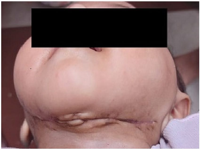
Number of                          Symptoms in early            Symptoms in late            Chi-square/Fisher's         p-
               Symptoms                                    Percentage
                                        patients                           presentation                 presentation                exact test                  Value

               Neck swelling            30                 100%            9                            21

               Neck pain                30                 100%            9                            21

               Fever                    30                 100%            9                            21

               Dysphagia                25                 83.33%          4                            21

               Dental pain              18                 63.33%          12                           6

                                                                                                                                    16.71                       0.05
               Trismus                  13                 43.33%          5                            8

               Muffled voice            11                 36.66%          0                            11

               Fetid breath             8                  26.66%          2                            6

               Respiratory distress     6                  20%             0                            6

               Swelling in the floor
                                        4                  13.33%          2                            2
               of mouth

              TABLE 1: Association of presenting symptoms with early and late presentations.
              Fisher's exact test was used to find out the association between symptoms with early and late presentations. The test value was 16.71 with a p-value of
              0.05. Here, the p-value is less than 0.05, so the null hypothesis was rejected. It means there is an association of symptoms with early and late
              presentations (more than one symptom was present in all patients).

                                                   Association of etiology with early and late presentations is mentioned in Table 2. In 11 patients, caries of
                                                   second and/or third molar root with exposed pulp was observed. In two patients, infection developed after
                                                   tooth extraction. Three mentally retarded children (out of which two were male and one female) with poor
                                                   oral hygiene developed infection. Two patients were admitted to medicine department for diabetic
                                                   ketoacidosis and then were referred to ENT department for Ludwig’s angina. Details of bacterial organisms
                                                   isolated in culture are shown in Table 3. In 24 patients, pus/serous fluid was sent for microbiological culture.
                                                   The most common organism found was Streptococcus followed by Escherichia coli.

2022 Parmar et al. Cureus 14(3): e23311. DOI 10.7759/cureus.23311                                                                                                       3 of 9
Present in Etiology in Chi-
Etiology in late p-
Etiologies number of Percentage early square/Fisher's
presentation Value
patients presentation exact test
Dental infection 19 63.33% 7 12
Diabetes mellitus 11 36.77 1 10
Salivary-gland infection/sialoadenitis 8 26.66% 6 2
Malnutrition 6 20% 1 5
Oral laceration 3 10% 2 1
7.095 0.52
Sharp foreign-body ingestion like
2 6.66% 0 2
fishbone
Chronic renal failure 2 6.66% 0 2
Insect bite 1 3.33% 1 0
Application and ingestion of some
1 3.33% 1 0
home remedies/herbal products
TABLE 2: Association of etiology with early and late presentations.
Fisher's exact test was used to find out the association between etiology with early and late presentations. The test value was 7.095 with a p-value of
0.52. Here, the p-value is more than 0.05, so the null hypothesis was accepted. It means there is no association of etiology with early and late
presentations (more than one underlying causative factor was present in one patient).
Organisms Number of patients Percentage
Streptococcus 11 45.83%
Escherichia coli 5 20.83%
Staphylococcus 3 12.5%
Pseudomonas 1 4.16%
No growth 4 16.55%
Surgical-decompression 24 80%
Conservative medical management 6 20%
TABLE 3: Bacterial organisms isolated in culture.
Association of management protocol with early and late presentations is illustrated in Table 4. In 21 cases,
incision and drainage were done externally while in three cases, it was being done intra-oral route. In
external route, incision was given from one angle of mandible to another for better drainage of
serosanguinous fluid/pus with surgical decompression of gas. Frank pus was found in eight patients and
serosanguinous fluid was found in 16 patients. In 16 patients, incision and drainage were done under
general anesthesia while the rest five patients required only local anesthesia. In six patients (20%), for
maintenance of airway, tracheostomy was required. A soft tissue neck x-ray was done in all patients except
one pregnant female. In eight patients, contrast CT (computed tomography) scan neck was done to access
the extent of infection spread. Ultrasonography was done in all cases for both diagnosis and prognosis.
2022 Parmar et al. Cureus 14(3): e23311. DOI 10.7759/cureus.23311 4 of 9In early In late Chi-square/Fisher's exact p-
Management protocol
presentation presentation test Value
Conservative medical management 02 04
Surgical-decompression (Incision and drainage) 07 11
1.77 0.62
Surgical-decompression (Incision and drainage) +
00 06
tracheostomy
TABLE 4: Association of management protocol with early and late presentations.
Fisher's exact test was used to find out the association between management protocol with early and late presentations. The test value was 1.77 with a p-
value of 0.62. Here, the p-value was more than 0.05, so the null hypothesis was accepted. It means there is no association of complication with early and
late presentations.
Association of complications with early and late presentations is shown in Table 5. The most common
complication was necrotizing fasciitis (16.66%). Death (10%) and septicemia (10%) were the second common
complications in this study. Mortality was observed in three patients (10%) in this study. Sudden death
was observed 12 hours after surgical decompression in one diabetic middle-aged male patient
who presented with diabetic ketoacidosis and the complications observed were septicemia followed by
disseminated intravascular coagulation (DIC) and cardiac arrest. Another old-age female patient having
some undiagnosed brain tumor died one month after drainage because of septicemic shock, DIC, and cardiac
arrest. One younger pregnant female patient (with seven months amenorrhea) presented on the fourth
day with a history of ingestion and application of some home remedies/herbal products and started
deteriorating. In spite of aggressive intravenous antibiotics, tracheostomy, incision and drainage (I&D), and
lower segment caesarian section, she ultimately died on the 11th day because of septicemia and
mediastinitis. Delayed wound healing was observed in six patients (20%) because of uncontrolled diabetes
mellitus, multiple dental infections, and poor nutritional status. In eight patients secondary suturing was
required.
Number of Complications observed in Complications observed in Fisher's p-
Complications
patients early presentation late presentation exact test Value
Necrotizing fasciitis 5 (16.66%) 2 3
Death 3 (10%) 0 3
Septicemia 3 (10%) 1 2
1.51 0.91
Disseminated intravascular
2 (6.67%) 0 2
coagulation (DIC)
Mediastinitis 1 (3.33%) 0 1
Cardiac arrest 1 (3.33%) 0 1
TABLE 5: Association of complications with early and late presentation.
Fisher's exact test was used to find out the association between complications with early and late presentation. The test value was 1.51 with a p-value of
0.91. Here, the p-value was more than 0.05, the null hypothesis was accepted. It means there is no association of complication with early and late
presentations.
Discussion
Ludwig’s angina has been reported as a rare clinical condition and mortality observed in the pre-antibiotic
era was 50% [7]. However, with the advent of current antimicrobial therapies and awareness regarding
dental health, mortality has reduced to around 10% [8,9]. The male-to-female ratio of treated patients in the
present study was 1:1.5, which is different from other studies, in which ratio of male patients was higher.
Linas et al. and Fakir et al. showed male-to-female ratio of 1.3:1 and 2.57:1, respectively [10,11]. In this
study, the mean age of patients is 38.86 years, which is a little lower than the mean age of 43.18 years found
in the study held by Linas et al. [10]. Ludwig’s angina was unusual in children. But in this study, it was found
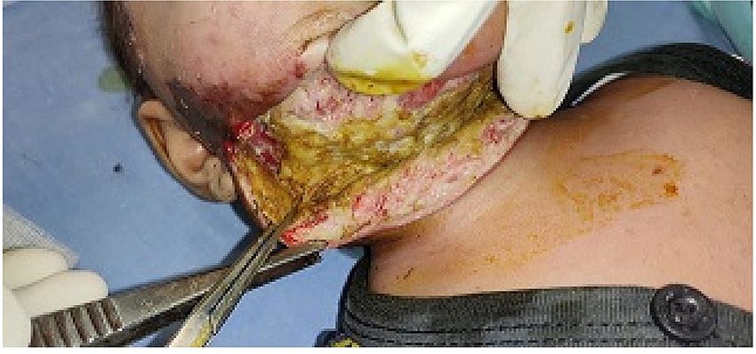
in four children, including one female infant patient (six months old). Photos of clinical presentation,
surgical decompression, and healed wound after decompression of this female infant are shown in Figures 1,
2022 Parmar et al. Cureus 14(3): e23311. DOI 10.7759/cureus.23311 5 of 92, 3, respectively. Like Fakir et al., in this study, 21 patients (70%) were from lower socioeconomic classes
which may be due to poor oral hygiene [11].
FIGURE 1: Ludwig's angina presentation in the six-month-old patient.
FIGURE 2: Surgical decompression from one angle of mandible to
another.
2022 Parmar et al. Cureus 14(3): e23311. DOI 10.7759/cureus.23311 6 of 9FIGURE 3: Healed wound post decompression and after secondary
suturing.
The trend of late presentation of patients to hospital was observed in this study. This trend was also
reported by Ugboko et al. in Southwest Nigeria [12]. One should expect that patient report to health system
early because Ludwig’s angina is a rapidly progressing disease with life-threatening complications. Patients
who present early were managed mostly with conservative management and some with surgical intervention
with low risk. Late presentation was mostly seen in unemployed, neglected, mentally retarded, old patients,
or else, in patients who didn’t come until they developed difficulty in swallowing and/or breathing. In
delayed presented patients, even after very aggressive management prognosis was poor.
In this study, dental infection from the mandibular molars was the most common causative factor of
Ludwig’s angina (80%), while diabetes mellitus was the second most common associated factor constituting
36.67% of total patients which was like the Fakir et al. [11]. According to Tschiassny, attachments of roots of
second and third lower molars extend below the mylohyoid line and close to inner cortex of mandible leading
to submaxillary space involvement, while attachment of roots of other lower teeth remains above the line
and close to outer cortex of mandible, therefore sublingual space involvement found more in their
infections [13]. The cause of cellulitis in the female infant, as shown in Figure 1, was unknown insect bite.
Etiology like tonsillar infection, HIV-AIDS, autoimmune conditions, etc. were not observed in this study.
Neck swelling, neck pain, fever, dental problems, and dysphagia were common clinical features observed in
this study which was similar to the case series of Fakir et al. [11]. More than one symptom was present in all
patients in this study, while according to Ovassapian et al., pain in floor of mouth and anterior neck,
dysphagia, odynophagia, and respiratory distress were the most common presenting symptoms [14]. Other
common symptoms observed in this study were swelling in floor of mouth, fetid breath, muffled voice,
trismus, and respiratory distress. In all patients, on palpation diffuse, firm, non-pitting, indurated, tender
swelling was found in submental and bilateral submaxillary space in supraomohyoid neck which may extend
further in the lower neck in some cases.
Treatment protocol in various past studies and the current study was almost the same. Prasad et al.
described four principles of management - (1) sufficient airway management, (2) early and aggressive
injectable broad-spectrum antibiotics, (3) incision and drainage and removal of primary cause if found any
(i.e., odontogenic cause), (4) adequate hydration and nutrition support [15].
Initial empirical antimicrobial therapy targeted against Gram-positive organisms and oral anaerobes.
Intravenous amoxicillin+clavulinic acid or cefoperazone+sulbactam, with intravenous
metronidazole/clindamycin and aminoglycosides like gentamicin/amikacin were given empirically to all
patients initially in this study, followed by antibiotics as per culture and sensitivity report. In some previous
studies, intravenous steroids were used to relieve soft tissue swelling and edema [16]. Systemic
corticosteroid was also used in eight patients in this study.
2022 Parmar et al. Cureus 14(3): e23311. DOI 10.7759/cureus.23311 7 of 9Polymicrobial type of flora is commonly seen in Ludwig’s angina. Streptococcus, Staphylococcus, and
Bacteroides species are the most commonly found organisms followed by some Gram-negative organisms
like Klebsiella, Haemophilus influenzae, Pseudomonas aeruginosa, and Proteus species [17]. Gram-positive
organisms like Streptococcus and Staphylococcus and Gram-negative organisms like Escherichia coli were
commonly isolated from pus culture in this study which is similar to the case series of Fakir et al. [11].
Diagnosis in Ludwig’s is mostly based on clinical findings though the contrast-enhanced CT scan is more
helpful in the prediction of drainable pus and extension of spread in deep neck abscesses [18]. Most of the
patients in this study were from lower and middle socioeconomic class and were non-affording for CT scans,
so CT scan was done only in six patients. Instead of CT scan, ultrasonography and plain soft tissue x-ray
were done for most of the cases, which are more cost-effective and gave information regarding site and size
of the collection, extent of spread, and site of pus collection where maximum fluctuation was palpable. Only
clinical findings have low efficacy in the prediction of site of drainage while combined with CT scan finding,
the accuracy is 89% and sensitivity and specificity is 95% and 89%, respectively [19].
Airway monitoring and protection have been identified as the most important aspect of the management of
Ludwig’s angina [20,21]. All patients were managed in the intensive care unit. Our management of the airway
included placement of patients in cardiac position with the use of oropharyngeal airway and tracheostomy
under local anesthesia, whenever indicated. Endotracheal intubation with flexible fiberoptic bronchoscopy
is another method used to secure airway. In this study, six patients (20%) required tracheostomy, and this
rate is almost double than the case series in Fakir et al. [11]. Many patients with co-morbidities presented
late with diffuse swelling, and at the time of surgical decompression, they required tracheostomy.
The indications of surgical decompression are clinically palpable fluctuation or crepitation of gas, serous or
pus collection in ultrasonography, radiological evidence of air in soft tissue, and no clinical improvement in
48 hours after starting antimicrobial therapy [22]. Out of total 30 patients, 24 (80%) required surgical
decompression in this study, which was similar to the case series of Fakir et al. [11]. Timely surgical
decompression reduces complications and thus mortality also.
Sepsis, pneumonia, asphyxia, empyema, pericarditis, mediastinitis, and pneumothorax are possible
complications of Ludwig’s angina [14]. The most common complication reported in this study were
necrotizing fasciitis (16.66%) followed by septicemia (10%) and death (10%). While according to the case
series of Fakir et al., the most common complications are necrotizing fasciitis (8%) and septicemia (8%)
followed by mediastinitis (6%) [11]. Many case series reflect mortality in Ludwig’s angina resulting from
mediastinitis, severe sepsis, and laryngeal spasm in past [8,22]. In the current study, septicemia found the
most common complication which leads to mortality followed by DIC and/or cardiac arrest. Mediastinitis-
related mortality was also observed in one pregnant female in this study. This study has some major
limitations. It is a single-center, retrospective study with limited cases. In future, it can be done on multi-
centric basis with more cases of Ludwig's angina.
Conclusions
Despite improved outcomes with higher antibiotics and available advanced surgical facilities, Ludwig's
angina still remains a potentially life-threatening disease in ENT. Dental caries, uncontrolled diabetes
mellitus, and malnutrition are commonly associated conditions. With early diagnosis, close airway
observation, aggressive intravenous antimicrobial treatment, and timely surgical intervention, one can
reduce morbidity and mortality.
Additional Information
Disclosures
Human subjects: Consent was obtained or waived by all participants in this study. Animal subjects: All
authors have confirmed that this study did not involve animal subjects or tissue. Conflicts of interest: In
compliance with the ICMJE uniform disclosure form, all authors declare the following: Payment/services
info: All authors have declared that no financial support was received from any organization for the
submitted work. Financial relationships: All authors have declared that they have no financial
relationships at present or within the previous three years with any organizations that might have an
interest in the submitted work. Other relationships: All authors have declared that there are no other
relationships or activities that could appear to have influenced the submitted work.
References
1. Har-El, Aroesty, Shaha and Lucente: Changing trends in deep neck abscess. A retrospective study of 110
patients. Oral Surg Oral Med Oral Pathol. 1994, 77:446-50. 10.1016/0030-4220(94)90221-6
2. Lee KC, Tami TA, Echavez M, Wildes TO: Deep neck infections in patients at risk for acquired
immunodeficiency syndrome. Laryngoscope. 1990, 100:915-9. 10.1288/00005537-199009000-00001
3. Fritsch DE, Klein DG: Ludwig's angina. Heart Lung. 1992, 21:39-47.
4. Otolaryngology-Head and Neck Surgery . Cummings C, Fredrickson JM, Harker LA, Krause CJ, Schuller DE
(ed): Mosby Inc., St. Louis, Missouri; 1993.
2022 Parmar et al. Cureus 14(3): e23311. DOI 10.7759/cureus.23311 8 of 95. Grodinsky M: Retropharyngeal and lateral pharyngeal abscesses: an anatomic and clinical study . Ann Surg.
1939, 110:177-99. 10.1097/00000658-193908000-00003
6. Mendelson MH, Gurtman A, Szabo S, et al.: Pseudomonas aeruginosa bacteremia in patients with AIDS . Clin
Infect Dis. 1994, 18:886-95. 10.1093/clinids/18.6.886
7. Doldo G, Albanese I, Macheda S, Caminiti G: Ludwig angina: a disease of the past century. Case report.
[Article in Italian]. Minerva Anestesiol. 2001, 67:811-4.
8. Hartmann RW Jr: Ludwig's angina in children . Am Fam Physician. 1999, 60:109-12.
9. Furst IM, Ersil P, Caminiti M: A rare complication of tooth abscess--Ludwig's angina and mediastinitis . J
Can Dent Assoc. 2001, 67:324-7.
10. Linas Z, Ruta R, Jurate R, Rimante S: Retrospective analysis of cellulitis of the floor of the mouth .
Stomatologija. 2010, 12:23-7.
11. Fakir AY, Bhuyan AH, Uddin M, Rahman HM, Al-Masum SH, Khan AF: Ludwig’s angina: a study of 50 cases .
Bangladesh j. otorhinolaryngol. 2009, 14:51-6. 10.3329/bjo.v14i2.3281
12. Ugboko VI, Ndukwe KC, Oginni FO: Ludwig’s angina: an analysis of sixteen cases in a suburban Nigerian
tertiary facility. Afr J Oral Health. 2005, 2:16. 10.4314/ajoh.v2i1-2.56993
13. Tschiassny K: Ludwig’s angina, an anatomic study of the role of the lower molar teeth in its pathogenesis .
Am J Orthod Dentofacial Orthop. 1944, 30:133-45. 10.1016/S0096-6347(44)90258-9
14. Ovassapian A, Tuncbilek M, Weitzel EK, Joshi CW: Airway management in adult patients with deep neck
infections: a case series and review of the literature. Anesth Analg. 2005, 100:585-9.
10.1213/01.ANE.0000141526.32741.CF
15. Prasad V, Das CP, Mohanty D, Rout MR, Prasanna EV: Ludwig’sangina, treatment protocol and observation
at atertiary care hospital, a case series. Indian J Appl Res. 2017, 7:100-01.
16. Barakate MS, Jensen MJ, Hemli JM, Graham AR: Ludwig's angina: report of a case and review of
management issues. Ann Otol Rhinol Laryngol. 2001, 110:453-6. 10.1177/000348940111000511
17. Bansal A, Miskoff J, Lis RJ: Otolaryngologic critical care. Crit Care Clin. 2003, 19:55-72. 10.1016/s0749-
0704(02)00062-3
18. Miller WD, Furst IM, Sàndor GK, Keller MA: A prospective, blinded comparison of clinical examination and
computed tomography in deep neck infections. Laryngoscope. 1999, 109:1873-9. 10.1097/00005537-
199911000-00029
19. Mehrotra M, Mehrotra S: Decompression of Ludwig angina under cervical block . Anesthesiology. 2002,
97:1625-6. 10.1097/00000542-200212000-00040
20. Bast YD, Appoloni O, Firket C, Capello M, Rocmans P, Vincent JL: Ludwig’s angina. Rev Med Brux. 2000,
21:137-41.
21. Hasan W, Leonard D, Russell J: Ludwig's angina-a controversial surgical emergency: how we do It . Int J
Otolaryngol. 2011, 2011:10.1155/2011/231816
22. Braimah RO, Taiwo AO, Ibikunle AA: Ludwig’s angina: analysis of 28 cases seen and managed in Sokoto,
Northwest Nigeria. Saudi Surg J. 2016, 4:77-83. 10.4103/2320-3846.183700
2022 Parmar et al. Cureus 14(3): e23311. DOI 10.7759/cureus.23311 9 of 9You can also read


