Single-Center Evaluation of Treatment Success Using Two Different Protocols for MRI-Guided Transurethral Ultrasound Ablation of Localized Prostate ...
←
→
Page content transcription
If your browser does not render page correctly, please read the page content below
ORIGINAL RESEARCH
                                                                                                                                         published: 27 October 2021
                                                                                                                                     doi: 10.3389/fonc.2021.782546

                                              Single-Center Evaluation of
                                              Treatment Success Using Two
                                              Different Protocols for MRI–Guided
                                              Transurethral Ultrasound Ablation
                                              of Localized Prostate Cancer
                                              Gencay Hatiboglu 1*, Valentin Popeneciu 1, David Bonekamp 2, Mathieu Burtnyk 3,
                                              Robert Staruch 3, Florian Distler 1, Jan Philipp Radtke 1, Johann Motsch 4,
                                              Heinz Peter Schlemmer 2, Sascha Pahernik 1 and Joanne Nyarangi-Dix 1
                                              1 Department of Urology, University of Heidelberg, Heidelberg, Germany, 2 German Cancer Research Center Deutsches

                                              Krebsforschungszentrum (DKFZ), Heidelberg, Germany, 3 Profound Medical Inc., Mississauga, ON, Canada, 4 Department of
                                              Anaesthesiology, University of Heidelberg, Heidelberg, Germany
                            Edited by:
                      Viktor Grünwald,
 Universitätsklinikum Essen, Germany
                                              Objectives: To assess differences in 24-month oncologic and functional outcomes in
                       Reviewed by:
                                              men with low to intermediate-risk prostate cancer treated with MRI-guided transurethral
                       Claudia Kesch,         ultrasound ablation (TULSA) using intentionally conservative versus intensified treatment
 Universitätsklinikum Essen, Germany
                                              parameters.
                         Dogu Teber,
      Städtisches Klinikum Karlsruhe,         Patients and Methods: Patients from a single center involved in two multicenter trials
                             Germany
                                              were included in this analysis. This included 14 of 30 patients with Gleason 3 + 3 from a
                 *Correspondence:
                   Gencay Hatiboglu
                                              Phase I study using intentionally conservative treatment parameters, and 15 of 115
          gencay.hatiboglu@med.uni-           patients with Gleason ≤ 3 + 4 from a pivotal study using intensified parameters. Follow-up
                       heidelberg.de
                                              data compared across these cohorts included 12-month biopsy and MRI for all patients,
                                              and 24-month PSA, micturition and quality of life (IIEF, IPSS, IPSS-QOL). The prognostic
                   Specialty section:
         This article was submitted to        value of baseline parameters and PSA kinetics on 12-month histological recurrence was
              Genitourinary Oncology,         evaluated by logistic regression.
               a section of the journal
                 Frontiers in Oncology        Results: 12-month biopsy revealed clinically significant residual disease in 4 (29%) and 2
      Received: 24 September 2021             (14%) patients from the Phase I and pivotal studies, respectively. PSA nadir was 0.7 ng/ml
        Accepted: 11 October 2021
                                              for Phase I and 0.5 ng/ml for pivotal study patients. Patient age at diagnosis, use of MRI
       Published: 27 October 2021
                                              fusion/systematic prostate biopsy, number of obtained cores at initial biopsy, PSA course,
                             Citation:
            Hatiboglu G, Popeneciu V,         and PSA nadir were identified as prognostic factors for treatment success. All but one
 Bonekamp D, Burtnyk M, Staruch R,            patient from each cohort maintained erection firmness sufficient for penetration. No cases
       Distler F, Radtke JP, Motsch J,
       Schlemmer HP, Pahernik S and
                                              of pad use were reported at 24 months. There were no Grade 4 or higher adverse events,
  Nyarangi-Dix J (2021) Single-Center         and no late toxicity related to the procedure.
     Evaluation of Treatment Success
Using Two Different Protocols for MRI–        Conclusion: Two-year follow-up demonstrated the efficacy of TULSA for the treatment of
      Guided Transurethral Ultrasound         localized prostate cancer, and the durability of PSA and functional outcomes. Intensifying
Ablation of Localized Prostate Cancer.
             Front. Oncol. 11:782546.
                                              treatment parameters in the pivotal trial had no impact on safety or functional outcomes
      doi: 10.3389/fonc.2021.782546           through 24 months, while reducing the recurrence rate for clinically significant disease.

Frontiers in Oncology | www.frontiersin.org                                        1                                     October 2021 | Volume 11 | Article 782546
Hatiboglu et al. TULSA: Treatment Success Using Two Protocols
Careful patient selection by MRI fusion/systematic prostate biopsy and adequate follow-
up through routine 12-month biopsy are recommended.
Keywords: TULSA, success, outcome, phase 1 clinical studies, pivotal
INTRODUCTION biopsy-proven PCa (clinical stage T1c–T2a, N0, M0), PSA ≤10
ng/ml, and Gleason score 3 + 3 between March 2013 and March
Men with early stage low – or intermediate risk prostate cancer 2014. Recruitment of men with Gleason score 3 + 4 was allowed
face different therapy options for treatment. Although, standard at one of the three sites in the Phase I study, but not at our
therapy like active surveillance, radical prostatectomy or institution. The multicenter TACT pivotal trial (NCT02766543)
radiotherapy provide excellent oncological and functional enrolled 115 men ≥45 years (clinical stage T1c-T2b, N0, M0),
results in experienced centres (1), some patients seek for PSA ≤15 ng/ml), and Gleason score on biopsy of 3 + 3 or 3 + 4.
alternative treatment options with less procedure-related side All men in the pivotal trial were treated between September 2016
effects. MRI-guided transurethral ultrasound ablation (TULSA) and February 2018.
of prostate tissue is an emerging technology for thermal For this single-center analysis, only patients treated at the
coagulation of diseased prostate tissue that has the advantages University of Heidelberg (Germany) were included: 14 patients
of intraoperative MRI-based treatment planning and automated treated in Phase I and 15 patients treated in the pivotal trial.
treatment control based on real-time MRI thermometry (2). A Both trials were approved by the institutional ethics board
Phase I trial in patients with localized prostate cancer (PCa) and written informed consent was obtained from all study
demonstrated clinical feasibility and safety (3). As feasibility participants. All patients at our institution were treated by the
rather than oncological effect was the main purpose of that same surgeons.
evaluation, treatment parameters were intentionally conservative
by sparing 3 mm of prostatic tissue within the capsule, leaving TULSA Procedure
10% of the prostate volume untreated, resulting in residual Men in both studies were treated with the TULSA-PRO system
disease in up to 55% of patients at 12-month prostate biopsy (Profound Medical Inc., Mississauga, Canada) (3, 5–8). The
(3). For the subsequent pivotal study of treatment efficacy, treatment was performed under general anesthesia in a 3T MRI
treatment parameters were intensified to achieve complete unit. A robotic positioning system (PS) was used to control the
whole-gland ablation (4). linear and rotational motion of the 10-element transurethral
As a center that enrolled and treated patients in both ultrasound applicator (UA) within the prostate under MRI-
multicenter trials, we previously presented a single-center guidance, with each element emitting high-intensity, directional
comparison of the initial 6-month safety outcomes between ultrasound. A 3-mm safety margin was preserved towards the apical
patients in the Phase I and pivotal trials (5). By comparing sphincter in both studies. Treatment plans were defined according
patients treated by the same physicians in both trials, the to high-resolution MRI images which were acquired continuously
surgeon’s impact on treatment outcome could be excluded. We during treatment to provide real-time MRI thermometry feedback
demonstrated that there were no significant differences in sexual of the ablation (3). For treatment planning, the urologist and the
function, continence, or other adverse events associated with radiologist marked the target volume by defining the outer
intensifying treatment parameters to achieve ablation to the boundaries of the prostate.
prostate capsule. However, the short follow-up time in that
report did not allow for comparison of oncological outcomes, Treatment Plans
and did not fully capture the recovery of functional outcomes. Treatment differences between the Phase I and pivotal trials have
Here, we provide the first comparison of oncologic outcomes in been described previously (5). The main differences were a
patients treated in the Phase I and pivotal trials with 24-month reduction in the safety margin of expected tissue preservation
follow-up including 12-month biopsy, and evaluate prognostic inside the prostatic capsule from 3 mm (Phase I) to less than 1
factors for treatment success. mm (pivotal trial), which was achieved by an increase of the
treatment control temperature from 55°C (Phase I) to 57°C
(pivotal), and a reduction of the minimum rotational speed of
the UA from 8 (Phase I) to 4 (pivotal) degrees per minute. These
PATIENTS AND METHODS changes to treatment parameters were expected to deliver
The prospective, multicenter, single-arm Phase I trial immediate cell kill ≥ 55°C within 1 mm of the prostate capsule
(NCT01686958) recruited thirty male patients ≥65 years with and to increase the ablation coverage from 90% to 99% of the
targeted prostate volume (Figure 1).
Abbreviations: IIEF, international index for erectile function; IPSS, international Follow-Up Schedule
prostate symptom score; IQR, interquartile range; MRI, magnetic resonance
imaging; PCa, prostate cancer; PS, positioning system; QoL, quality of life;
All patients underwent physical examination, ultrasound, and
TACT, TULSA-PRO Ablation Clinical Trial; TULSA, transurethral ultrasound blood chemistry, and PSA at the baseline visit. A multiparametric
ablation; UA, ultrasound applicator. MRI of the prostate was done at baseline in all patients and rated
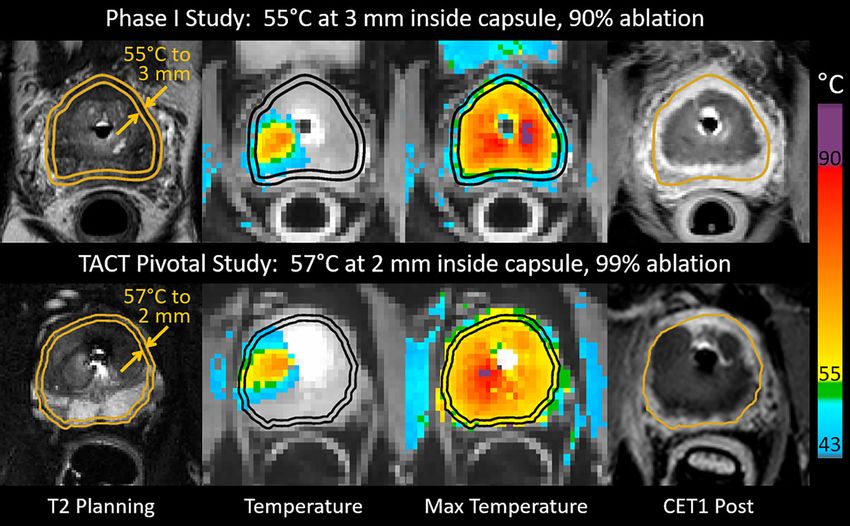
Frontiers in Oncology | www.frontiersin.org 2 October 2021 | Volume 11 | Article 782546Hatiboglu et al. TULSA: Treatment Success Using Two Protocols FIGURE 1 | Intraoperative images from patients who underwent MRI-guided transurethral ultrasound ablation (TULSA) using conservative treatment parameters in the Phase I study (top), and intensified parameters designed to achieve whole-gland ablation in the pivotal study (bottom). Transverse images at one location in the midgland for each patient depict interoperative treatment planning on T2-weighted images, real-time MRI temperature images used to control treatment, maximum temperature projections used to assess ablation coverage during treatment, and post-treatment contrast-enhanced T1-weighted images (CET1) demonstrating greater ablation extent in the example from the pivotal study (bottom). according to the Prostate Imaging Reporting and Data System patients treated in both studies and with completed follow-up. A p (PI-RADS) (9). The first follow-up was after 2 weeks to remove value 3 + 3) at 12 months, for the pooled cohort of 4.5). Follow-up biopsies at 12 months detected clinically significant Frontiers in Oncology | www.frontiersin.org 3 October 2021 | Volume 11 | Article 782546
Hatiboglu et al.                                                                                                               TULSA: Treatment Success Using Two Protocols

TABLE 1 | Clinical baseline and pathological characteristics between the Phase I and pivotal trial patients treated at our institution.

                                                                                     Phase 1 N = 14                             Pivotal N = 15                            P-value

Age, median (IQR)                                                                    71.0 (69.2-73.0)                           67.0 (64.9-71.9)                            0.10
Prostate Volume, median (IQR)                                                        41.0 (33.8-65.7)                           44.5 (33.4-54.5)                            0.76
PSA, median (IQR)                                                                     6.6 (4.0-8.1)                              6.6 (4.5-7.3)                              0.79
Gleason Score                                                                                                                                                               0.01
   Gleason 3 + 3                                                                            14                                         9
   Gleason 3 + 4                                                                             0                                         6
IIEF, median (IQR)                                                                    11.5 (3.8-26.5)                           22.5 (5.0-28.8)                             0.21
IPSS, median (IQR)                                                                    8.5 (5.0-15.5)                            10.0 (5.5-14.8)                             0.74
IPSS quality of life, median (IQR)                                                     2.5 (1.8-4.0)                             3.0 (2.0-3.8)                              0.91
Number of pads, median (IQR)                                                              0 (0-0)                                   0 (0-0)                                 1.00
Testosterone, median (IQR)                                                             4.2 (3.7-5.4)                             3.8 (2.3-5.5)                              0.36
Mode of initial biopsy                                                                                                                                                      0.74
   12 core TRUS                                                                             11                                         11
   MRI fusion/systematic biopsy                                                             3                                          4
Number of obtained biopsy cores                                                      25.5 (16.0-28.0)                           27.0 (16.0-30.0)                            0.67
median (IQR)
Number of positive biopsy cores                                                        3.0 (2.0-4.3)                              4.0 (1.0-6.0)                             0.95
median (IQR)

Groups were compared by chi-square tests for categorical variables and Mann-Whitney-U tests for continuous variables. A p value < 0.05 was considered statistically significant.
Statistical significant values are displayed in bold.

TABLE 2 | Oncological follow-up: PSA values at baseline and follow-up, PSA nadir, time to PSA nadir and results of 12 months prostate biopsy for phase 1 and pivotal
trial patients.

                                                                                         Phase 1 N = 14                           Pivotal N = 15                          P-value

PSA, median (IQR)
Baseline                                                                                     6.6 (4.0-8.1)                         6.6 (4.5-7.3)                            0.79
  1 month                                                                                    0.9 (0.5-2.0)                         0.8 (0.4-1.5)                            0.78
  3 months                                                                                   0.9 (0.3-1.7)                         0.5 (0.2-1.1)                            0.13
  6 months                                                                                   0.7 (0.4-1.3)                         0.5 (0.3-1.4)                            0.60
  12 months                                                                                  0.9 (0.6-1.7)                         1.0 (0.7-1.5)                            0.82
  24 months                                                                                  0.9 (0.4-2.5)                         0.9 (0.4-2.2)                            0.86
PSA Nadir at 12 months                                                                       0.7 (0.2-0.8)                         0.5 (0.2-1.2)                            0.42
Time to PSA nadir                                                                            3.0 (1.0-6.0)                         3.0 (3.0-4.5)                            0.45
Any Recurrence at 12 months biopsy (%)                                                       5/13 (38.5%)                          5/14 (35.7%)                             0.88
Gleason Score (recurrence)                                                                                                                                                  0.10
  Gleason 3 + 3                                                                                   2                                      4
  Gleason 3 + 4                                                                                   0                                      1
  Gleason 4 + 3                                                                                   3                                      0
  Clinically significant                                                                       4 (28.6%)                              2 (14.3%)                               0.2

Groups were compared by Mann-Whitney-U test. A p value < 0.05 was considered statistically significant.

cancer (high volume Gleason=3+3 or any Gleason >3+3)                                          patients from both studies (Table 3). Hereby, patient age
in four patients (29%) of the Phase I study and two patients                                  (OR:0.71; p=0.03), PSA at 12 months (OR:4.01; p=0.04), PSA
(14%) in the pivotal trial. Clinically insignificant findings (small                            nadir (OR:16.52; p=0.02), mode of initial biopsy (OR:7.5; p=0.04)
volume Gleason 3 + 3) were detected in one additional patient in                              and number of biopsy cores sampled at initial biopsy (OR:0.87;
Phase I, and three additional patients in the pivotal trial.                                  p=0.03) had statistically significant impact on oncological outcome.
   Table 3 compares all patients with cancer recurrence to those                              Intensifying treatment parameters had no statistically significant
with negative 12-month biopsy. At time of diagnosis, men who                                  impact on cancer recurrence. A separate univariate evaluation
eventually had recurrence were significantly younger (p=0.02), had                             focusing on patients with clinically significant tumor recurrence
lower cancer risk classification (p=0.03), had fewer biopsy cores                              revealed PSA at 12 months (OR: 4.3; p=0.04) and PSA nadir (OR:
sampled at initial diagnosis (p=0.02), and were less likely to have                           13.2; p=0.03) as predictive factors.
had a MRI fusion/systematic initial biopsy (p=0.03). During follow-                               Patients with clinically significant tumor recurrence were
up, men who had recurrence on 12-month biopsy had higher PSA at                               referred for salvage treatment. In the Phase I cohort, two
nadir (p=0.01), higher PSA values at 3 to 12-month visits, and had                            patients underwent salvage radiation therapy and two patients
higher PIRADS scores at 12-month multiparametric MRI (p=0.01).                                salvage prostatectomy. In the pivotal trial, the two patients with
   Univariate analysis of predictive factors for any cancer                                   clinically significant tumor recurrence both underwent salvage
recurrence at 12-month biopsy was performed including all                                     prostatectomy. Patients with clinically insignificant tumor

Frontiers in Oncology | www.frontiersin.org                                              4                                         October 2021 | Volume 11 | Article 782546
Hatiboglu et al. TULSA: Treatment Success Using Two Protocols
TABLE 3 | Comparison of patients with and without cancer recurrence.
No recurrence N = 17 Recurrence N = 10 P-value
Age, median (IQR) 71.7 (68.8-73.9) 68.2 (54.8-70.2) 0.016
Prostate Volume, median (IQR) 44.0 (35.4-51.9) 43.1 (31.8-58.0) 0.980
Initial PSA, median (IQR) 6.7 (5.0-7.8) 6.6 (4.7-7.9) 0.880
PSA 3 months, median (IQR) 0.5 (0.2-1.3) 1.2 (0.7-2.7) 0.031
PSA 6 months, median (IQR) 0.5 (0.3-0.9) 1.2 (0.8-1.7) 0.017
PSA 12 months, median (IQR) 0.8 (0.5-1.2) 1.4 (1.0-2.5) 0.007
PSA nadir 12 months 0.5 (0.2-0.6) 1.0 (0.6-1.3) 0.010
Median (IQR)
Gleason Score 0.097
Gleason 3 + 3 12 10
Gleason 3 + 4 4 0
Risk classification 0.033
Low risk 11 10
Intermediate risk 6 0
Initial Biopsy 0.029
TRUS 2 5
MRI fusion/systematic 15 5
Initial MRI 0.184
Yes 17 9
No 0 1
IIEF, median (IQR) 11.0 (3.5-28.0) 22.5 (10.5-29.0) 0.172
IPSS, median (IQR) 8.0 (4.5-14.5) 10.0 (7.5-14.3) 0.465
Quality of life, median (IQR) 3.0 (2.0-4.0) 2.5 (1.8-4.0) 0.938
Testosterone, median (IQR) 4.0 (2.7-6.0) 4.8 (4.0-5.3) 0.547
Percentage Non-Perfused Volume (%) 54.0 (47.5-61.0) 53.5 (37.3-57.3) 0.407
No of biopsy cores (initial biopsy) 28.0 (20.5-30.5) 17.5 (12.3-26.5) 0.023
No of pos. cores (initial biopsy) 3.0 (1.5-5.0) 3.5 (1.8-4.5) 0.839
PIRADS Score (initial biopsy) 4 (3-4) 4 (4-5) 0.337
No of biopsy cores Follow-Up 12.0 (12.0-15.0) 12.5 (12.0-14.8) 0.847
Any Lesion follow-up MRI 0.883
Yes 8 5
no 9 5
PIRADS score at 12 months 3 (3-4) 4 (4-5) 0.008
Treatment protocol 0.883
Phase 1 Study 8 5
Pivotal Trial 9 5
Groups were compared by Mann-Whitney-U test for continuous variables and chi-square for categorial variables. A p value < 0.05 was considered statistically significant.
Statistical significant values are displayed in bold.
recurrence are under surveillance with regular PSA controls and month, four pivotal study patients reported the use of pads related
scheduled re-biopsies. One patient of our Phase I cohort to urine leakage, but by the 6-month visit all men in both cohorts
discontinued follow-up to pursue active surveillance before the were pad-free and remained so at 2 years.
one-year visit, and four patients in our pivotal cohort withdrew Between baseline and 24 months, only one patient from each
from the study to pursue active surveillance before the two- study lost the ability to achieve erections sufficient for penetration
year visit. (IIEF question 2 ≥ 2). IIEF-15 erectile function domain scores
showed recovery by 3 months with wide variability in both groups.
Functional Outcomes and Safety Perioperative adverse events to 6 months have been reported
Functional outcomes for patients in the Phase I and pivotal studies previously (5). No late adverse events related to the procedure
are listed in Table 4. Catheterization durations were longer in were noted beyond 6 months.
pivotal study versus Phase I patients: median 20.0 (IQR 10-42)
days vs median 14.5 (IQR 13-25) days, respectively (p=0.18).
After an initial worsening of urinary symptoms at 1 month in
both groups, IPSS recovery was seen in both groups at 3 and 6 DISCUSSION
months. At 12 to 24 months, the IPSS score returned to baseline in
both cohorts, with improvement beyond baseline noted in pivotal We compare two different treatment protocols for TULSA with
patients (median 56% improvement from baseline to 24 months). regards to functional and oncologic outcomes at 2 years for the
IPSS quality of life was improved compared to baseline starting at subgroup of men treated at our institution. By this means, we also
3 months in Phase I and 6 months in the pivotal study, with most assess the impact of intensified treatment parameters and identify
men from both trials reporting that they were “pleased” or prognostic factors for treatment success. Whole-gland prostate
“delighted” with their urinary condition at 24 months. At one ablation delivered using MRI-guided TULSA led to lower rates of
Frontiers in Oncology | www.frontiersin.org 5 October 2021 | Volume 11 | Article 782546Hatiboglu et al. TULSA: Treatment Success Using Two Protocols
TABLE 4 | Functional follow-up: Comparison of catheter indwelling time, IPSS, quality of life and pad usage for evaluation of micturition and IIEF questionnaire data for
erectile function at baseline and during follow-up for phase 1 and pivotal trial patients.
Phase 1 N = 14 Pivotal N = 15 P-value
Catheter indwelling time, median (IQR) 14.5 (13.0-25.0) 20.0 (10.3-42.0) 0.18
IPSS, median (IQR)
Baseline 8.5 (5.0-15.5) 10.0 (5.5-14.8) 0.74
1 month 15.5 (11.0-21.0) 14.5 (11.5-18.5) 0.84
3 months 5.0 (3.0-10.0) 7.5 (3.5-10.0) 0.38
6 months 5.0 (3.0-7.0) 7.0 (2.0-9.0) 0.50
12 months 6.0 (3.5-6.5) 4.5 (1.8-10.5) 0.77
24 months 8.5 (6.0-9.0) 3.0 (2.5-9.5) 0.18
Quality of life, median (IQR)
Baseline 1.5 (0.8-3.0) 1.0 (1.0-3.0) 0.86
1 month 3.0 (1.0-3.0) 4.0 (2.0-6.0) 0.12
3 months 1.0 (0.0-1.0) 2.0 (1.0-3.0) 0.01
6 months 0.5 (0.0-1.0) 1.0 (0.0-2.0) 0.06
12 months 1.0 (0.0-1.0) 1.0 (0.0-2.3) 0.50
24 months 0.5 (0.0-1.0) 0.0 (0.0-1.5) 0.78
Number of pads (IQR; range)
Baseline 0 (0.0; 0.0) 0 (0.0; 0.0) 1.00
1 month 0 (0.0; 0.0) 0 (0.0; 0.2) 0.04
3 months 0 (0.0; 0.0) 0 (0.0; 0.1) 0.33
6 months 0 (0.0; 0.0) 0 (0.0; 0.0) 1.00
12 months 0 (0.0; 0.0) 0 (0.0; 0.0) 1.00
24 months 0 (0.0; 0.0) 0 (0.0; 0.0) 1.00
IIEF, median (IQR)
Baseline 11.5 (3.8-26.5) 25.0 (8.0-29.0) 0.20
1 month 3.0 (1.0-9.3) 4.5 (1.8-9.3) 0.58
3 months 11.5 (4.8-16.5) 14.0 (1.0-29.0) 0.54
6 months 11.0 (5.3-20.0) 14.0 (2.0-25.0) 0.98
12 months 19.0 (8.0-25.0) 14.5 (8.8-25.0) 1.00
24 months 17.5 (3.8-24.3) 7.0 (1.5-25.5) 0.86
IIEF Q2 erection sufficient for penetration
Baseline 2.5 (0.8-5.0) 4.0 (0.0-5.0) 0.69
1 month 0.0 (0.0-1.5) 0.0 (0.0-1.8) 0.57
3 months 2.0 (0.0-3.3) 2.5 (0.0-4.8) 0.37
6 months 2.5 (1.0-4.3) 2.5 (0.0-4.8) 0.95
12 months 4.0 (2.5-4.0) 3.5 (1.8-5.0) 0.94
24 months 3.0 (0.3-4.8) 1.0 (0.0-4.5) 0.58
Groups were compared by Mann-Whitney-U test. A p value < 0.05 was considered statistically significant.
Statistical significant values are displayed in bold.
clinically significant tumor recurrence compared with wide safety patients reported pad use due to urgency and mild incontinence
margins. The intensified parameters had no impact on clinical at 1 month, but recovered to pad-free continence by six months, as
safety and minimal impact on functional outcomes at 24 months. did all of our patients in the Phase I study. IPSS and IPSS quality of
In the Phase I trial, safety and treatment precision were the life scores showed an initial increase in both subgroups, followed
primary study outcomes, not oncological efficacy. A wide safety by an improvement to better than baseline levels in the pivotal
margin of 3 mm inside the prostate capsule was intentionally study. Bladder outlet obstruction as a typical side effect of other
spared regardless of cancer location, leaving a rim of viable thermoablative treatments (11, 12) did not occur in any of
prostate tissue (10). In the subsequent pivotal study designed to our patients.
assess treatment efficacy, treatment parameters were intensified The incidental, ameliorating effect of TULSA treatment on
to remove the safety margin and increase treatment temperature lower urinary tract symptoms (LUTS) has recently been described
and exposure time (by reducing minimal rotation speed). This for a subgroup of the overall population of Phase I patients who
reduced safety margins to the prostatic capsule and increased entered the study with cancer and concomitant LUTS (13).
ablation coverage from 90% to 99% of the gland (5). For the However, a comparison between the Phase I and TACT cohorts
subgroups of men treated at our center, the rate of clinically is described here for the first time. While micturition symptoms
significant disease on the 12-month biopsy was reduced from reported through IPSS scores recovered to baseline by 2 years in
29% to 14% by intensifying treatment parameters, similar to the the Phase I subgroup, improvements relative to baseline were seen
reduction from 31% to 15% seen in the full cohorts (2, 4). in the pivotal study subgroup at the 24-month visit. We attribute
Despite more aggressive treatment plans, there were no the enhanced urinary symptom relief to the higher treatment
material differences in urinary function between the two temperatures and exposure times applied in the pivotal study, in
subgroups from 6 to 24 months. Four of our pivotal study line with previous in-vitro studies demonstrating that increased
Frontiers in Oncology | www.frontiersin.org 6 October 2021 | Volume 11 | Article 782546Hatiboglu et al. TULSA: Treatment Success Using Two Protocols
thermal dose improved ablation effect for hyperplastic prostate predominantly intermediate risk cancers in the pivotal trial must
tissue (14, 15). be taken into consideration while only men with low-risk cancer
Baseline erection firmness sufficient for penetration (IIEF were treated at our center in Phase I. Furthermore, two-year
question 2 ≥2) was maintained for all but one patient from each follow-up was lacking for 6 of 15 men in the pivotal study: four
subgroup. However, IIEF-15 erectile function domain scores who declined additional follow-up having low PSA, and two who
trended towards improved recovery in the Phase I subgroup. The had undergone salvage treatment. Although clinical safety and
reduced ablation safety margins of the pivotal study were expected functional outcome can be evaluated, the data on oncological
to result in increased heating to within 1mm of the neurovascular outcomes need further evaluation with a longer follow-up.
bundles adjacent to the prostatic capsule, compared to the wide
3mm subcapsular margin applied in the Phase I study which may
have resulted in better preservation of erectile function. While the CONCLUSION
differences were not statistically significant in this small sample size,
they suggest that the intensified parameters per the pivotal trial In patients at one institution involved in two different multicenter
whole-gland ablation protocol could be considered non-nerve- studies, intensified treatment parameters for whole-gland prostate
sparing, whereas a safety margin of 3 mm appears to be suitable ablation using MRI-guided TULSA led to lower rates of clinically
for regions of the gland where nerve-sparing is intended and significant tumor recurrence while having no impact on clinical
oncologically acceptable. safety or 24-month functional outcomes. PSA course and especially
In our evaluation, the PSA course and especially PSA nadir PSA nadir, as well as baseline use of MRI fusion biopsy technique
were significant predictors for tumor recurrence. As previously predicted histopathological evidence of residual disease. Close
demonstrated, PSA nadir is a prognostic indicator for disease-free follow-up through PSA monitoring with 12-month MRI and
survival in patients undergoing HIFU treatment (16, 17) or biopsy is recommended for early detection of disease recurrence.
radiotherapy (18). Definition of a threshold for biochemical
recurrence post-TULSA awaits long-term outcomes and
evaluation of larger cohorts. In these cohorts with ablation of at DATA AVAILABILITY STATEMENT
least 90% of the prostate, 75% of those with nadir ≤ 0.6 ng/ml were
disease-free, while 75% with nadir above 0.6 ng/ml had The raw data supporting the conclusions of this article will be
histological recurrence. Of four men with histological recurrence made available by the authors, without undue reservation.
after nadir ≤ 0.6 ng/ml, two with only one core of residual Gleason
Grade 1 disease had PSA remain within nadir + 0.5 ng/ml
thereafter. The other two each had 3/12 positive cores and ETHICS STATEMENT
presence of residual Gleason Grade 2 disease, and had PSA
The studies involving human participants were reviewed and
increase to nadir + 0.7 and nadir + 1.2 ng/ml by one year,
approved by Ethikkommission Heidelberg, Universität
eventually exceeding nadir + 2.0 ng/ml. Routine follow-up
Heidelberg. The patients/participants provided their written
biopsy at 12 months is therefore recommended to ensure timely
informed consent to participate in this study.
detection of residual disease. For patients with tumor recurrence,
standard salvage treatments like radical prostatectomy (19) or
radiotherapy (20) are viable options.
Another factor that influenced tumor recurrence at 12
AUTHOR CONTRIBUTIONS
months was the use of stereotactic or MRI-guided biopsy at GH, project development, data collection, data analysis, manuscript
baseline. The accuracy of MRI guided stereotactic biopsy has writing and editing. VP, data collection and data analysis. DB, data
been described before and showed significantly better diagnostic collection, data analysis, manuscript writing and editing. MB and
accuracy (21, 22). The combination of MRI fusion biopsy with RS, project development, data analysis, manuscript writing and
systematic biopsy has shown even better accuracy in detecting editing. FD and JR, data analysis, manuscript writing and editing.
clinically significant prostate cancer (23) or the index tumor (24) SP, project development, data collection, and data analysis. JM, data
than 12-core systematic biopsy alone. In the Phase I trial, more of collection and supervision. HS, project development, supervision,
our patients had a previous MRI-guided or stereotactic biopsy and critical revision of manuscript. JN-D, data analysis, manuscript
than in other centers (3), while the pivotal trial mandated writing and editing, and critical revision of manuscript. All authors
preoperative multiparametric prostate MRI for all patients. We contributed to the article and approved the submitted version.
assume that our center’s lower recurrence rate in Phase I
compared to other centers is related to the initial mode of
biopsy. The increased diagnostic certainty accessible with MRI- ACKNOWLEDGMENTS
guided or stereotactic biopsy is essential for appropriate patient
counseling and decision-making, and was correlated with Profound Medical Inc. helped design and conduct the study,
decreased tumor recurrence at 12 months. manage and analyze the data, and prepare and approve
Limitations of this evaluation include the short follow-up of the manuscript.
24 months and the small cohorts. In addition, the treatment of
Frontiers in Oncology | www.frontiersin.org 7 October 2021 | Volume 11 | Article 782546Hatiboglu et al. TULSA: Treatment Success Using Two Protocols
REFERENCES 15. Madersbacher S, Susani M, Marberger M. Thermal Ablation of BPH With
Transrectal High-Intensity Focused Ultrasound. Prog Clin Biol Res (1994)
1. Mottet N, van den Bergh RCN, Briers E, Van den Broeck T, Cumberbatch 386:473–8.
MG, De Santis M, et al. EAU-EANM-ESTRO-ESUR-SIOG Guidelines on 16. Ganzer R, Robertson CN, Ward JF, Brown SC, Conti GN, Murat FJ, et al.
Prostate Cancer-2020 Update. Part 1: Screening, Diagnosis, and Local Correlation of Prostate-Specific Antigen Nadir and Biochemical Failure After
Treatment With Curative Intent. Eur Urol (2021) 79(2):243–62. High-Intensity Focused Ultrasound of Localized Prostate Cancer Based on the
doi: 10.1016/j.eururo.2020.09.042 Stuttgart Failure Criteria - Analysis From the @-Registry. BJU Int (2011) 108
2. Chopra R, Burtnyk M, Haider MA, Bronskill MJ. Method for MRI-Guided (8 Pt 2):E196–201. doi: 10.1111/j.1464-410X.2011.10091.x
Conformal Thermal Therapy of Prostate With Planar Transurethral 17. Chen PY, Chiang PH, Liu YY, Chuang YC, Cheng YT. Primary Whole-Gland
Ultrasound Heating Applicators. Phys Med Biol (2005) 50(21):4957–75. Ablation for Localized Prostate Cancer With High-Intensity Focused
doi: 10.1088/0031-9155/50/21/001 Ultrasound: The Important Predictors of Biochemical Recurrence. Int J
3. Chin JL, Billia M, Relle J, Roethke MC, Popeneciu IV, Kuru TH, et al. Magnetic Urol (2018) 25(6):615–20. doi: 10.1111/iju.13581
Resonance Imaging-Guided Transurethral Ultrasound Ablation of Prostate 18. Buyyounouski MK. Radiotherapy: PSA Nadir Predicts Long-Term Mortality.
Tissue in Patients With Localized Prostate Cancer: A Prospective Phase 1 Nat Rev Clin Oncol (2010) 7(4):188–90. doi: 10.1038/nrclinonc.2010.33
Clinical Trial. Eur Urol (2016) 70(3):447–55. doi: 10.1016/j.eururo.2015.12.029 19. Nair SM, Stern N, Dewar M, Siddiqui K, Smith E, Gomez JA, et al. Salvage
4. Klotz L, Pavlovich CP, Chin J, Hatiboglu G, Koch M, Penson D, et al. MRI- Open Radical Prostatectomy for Recurrent Prostate Cancer Following MRI-
Guided Transurethral Ultrasound Ablation of Prostate Cancer. J Urol (2020) Guided Transurethral Ultrasound Ablation (TULSA) of the Prostate:
205(3):769–79. doi: 10.1097/JU.0000000000001362 Feasibility and Efficacy. Scand J Urol (2020) 54(3):215–9. doi: 10.1080/
5. Hatiboglu G, Popeneciu V, Bonekamp D, Burtnyk M, Staruch R, Pahernik S, 21681805.2020.1752795
et al. Magnetic Resonance Imaging-Guided Transurethral Ultrasound 20. Nair SM, Hatiboglu G, Relle J, Hetou K, Hafron J, Harle C, et al. Magnetic
Ablation of Prostate Tissue in Patients With Localized Prostate Cancer: Resonance Imaging-Guided Transurethral Ultrasound Ablation in Patients
Single-Center Evaluation of 6-Month Treatment Safety and Functional With Localised Prostate Cancer: 3-Year Outcomes of a Prospective Phase I
Outcomes of Intensified Treatment Parameters. World J Urol (2020) 38 Study. BJU Int (2021) 127(5):544–52. doi: 10.1111/bju.15268
(2):343–50. doi: 10.1007/s00345-019-02784-w 21. Siddiqui MM, Rais-Bahrami S, Truong H, Stamatakis L, Vourganti S, Nix J,
6. Burtnyk M, Hill T, Cadieux-Pitre H, Welch I. Magnetic Resonance Image et al. Magnetic Resonance Imaging/Ultrasound-Fusion Biopsy Significantly
Guided Transurethral Ultrasound Prostate Ablation: A Preclinical Safety and Upgrades Prostate Cancer Versus Systematic 12-Core Transrectal Ultrasound
Feasibility Study With 28-Day Followup. J Urol (2015) 193(5):1669–75. Biopsy. Eur Urol (2013) 64(5):713–9. doi: 10.1016/j.eururo.2013.05.059
doi: 10.1016/j.juro.2014.11.089 22. Hadaschik BA, Kuru TH, Tulea C, Rieker P, Popeneciu IV, Simpfendorfer T,
7. Chopra R, Burtnyk M, N'Djin WA, Bronskill M. MRI-Controlled et al. A Novel Stereotactic Prostate Biopsy System Integrating Pre-
Transurethral Ultrasound Therapy for Localised Prostate Cancer. Int J Interventional Magnetic Resonance Imaging and Live Ultrasound Fusion.
Hyperthermia (2010) 26(8):804–21. doi: 10.3109/02656736.2010.503670 J Urol (2011) 186(6):2214–20. doi: 10.1016/j.juro.2011.07.102
8. Partanen A, Yerram NK, Trivedi H, Dreher MR, Oila J, Hoang AN, et al. 23. Siddiqui MM, Rais-Bahrami S, Turkbey B, George AK, Rothwax J, Shakir N,
Magnetic Resonance Imaging (MRI)-Guided Transurethral Ultrasound et al. Comparison of MR/ultrasound Fusion-Guided Biopsy With Ultrasound-
Therapy of the Prostate: A Preclinical Study With Radiological and Guided Biopsy for the Diagnosis of Prostate Cancer. JAMA (2015) 313
Pathological Correlation Using Customised MRI-Based Moulds. BJU Int (4):390–7. doi: 10.1001/jama.2014.17942
(2013) 112(4):508–16. doi: 10.1111/bju.12126 24. Radtke JP, Schwab C, Wolf MB, Freitag MT, Alt CD, Kesch C, et al.
9. Weinreb JC, Barentsz JO, Choyke PL, Cornud F, Haider MA, Macura KJ, et al. Multiparametric Magnetic Resonance Imaging (MRI) and MRI-Transrectal
PI-RADS Prostate Imaging - Reporting and Data System: 2015, Version 2. Ultrasound Fusion Biopsy for Index Tumor Detection: Correlation With
Eur Urol (2016) 69(1):16–40. doi: 10.1016/j.eururo.2015.08.052 Radical Prostatectomy Specimen. Eur Urol (2016) 70(5):846–53. doi: 10.1016/
10. Bonekamp D, Wolf MB, Roethke MC, Pahernik S, Hadaschik BA, Hatiboglu j.eururo.2015.12.052
G, et al. Twelve-Month Prostate Volume Reduction After MRI-Guided
Transurethral Ultrasound Ablation of the Prostate. Eur Radiol (2019) 29 Conflict of Interest: RS and MB are employees of Profound Medical and receive a
(1):299–308. doi: 10.1007/s00330-018-5584-y salary and stock options.
11. Crouzet S, Chapelon JY, Rouviere O, Mege-Lechevallier F, Colombel M,
Tonoli-Catez H, et al. Whole-Gland Ablation of Localized Prostate Cancer The remaining authors declare that the research was conducted in the absence of
With High-Intensity Focused Ultrasound: Oncologic Outcomes and any commercial or financial relationships that could be construed as a potential
Morbidity in 1002 Patients. Eur Urol (2014) 65(5):907–14. doi: 10.1016/ conflict of interest.
j.eururo.2013.04.039
12. Netsch C, Pfeiffer D, Gross AJ. Development of Bladder Outlet Obstruction Publisher’s Note: All claims expressed in this article are solely those of the authors
After a Single Treatment of Prostate Cancer With High-Intensity Focused and do not necessarily represent those of their affiliated organizations, or those of
Ultrasound: Experience With 226 Patients. J Endourol (2010) 24(9):1399–403. the publisher, the editors and the reviewers. Any product that may be evaluated in
doi: 10.1089/end.2009.0607 this article, or claim that may be made by its manufacturer, is not guaranteed or
13. Elterman D, Li W, Hatiboglu G, Relle J, Zorn KC, Bhojani N, et al. Relief of endorsed by the publisher.
Lower Urinary Tract Symptoms After MRI-Guided Transurethral Ultrasound
Ablation (TULSA) for Localized Prostate Cancer: Subgroup Analyses in Copyright © 2021 Hatiboglu, Popeneciu, Bonekamp, Burtnyk, Staruch, Distler,
Patients With Concurrent Cancer and Benign Prostatic Hyperplasia. Radtke, Motsch, Schlemmer, Pahernik and Nyarangi-Dix. This is an open-access
J Endourol (2021) 35(4):497–505. doi: 10.1089/end.2020.0511 article distributed under the terms of the Creative Commons Attribution License
14. Bhowmick P, Coad JE, Bhowmick S, Pryor JL, Larson T, de la Rosette J, et al. (CC BY). The use, distribution or reproduction in other forums is permitted, provided
In Vitro Assessment of the Efficacy of Thermal Therapy in Human Benign the original author(s) and the copyright owner(s) are credited and that the original
Prostatic Hyperplasia. Int J Hyperthermia (2004) 20(4):421–39. doi: 10.1080/ publication in this journal is cited, in accordance with accepted academic practice. No
02656730310001637343 use, distribution or reproduction is permitted which does not comply with these terms.
Frontiers in Oncology | www.frontiersin.org 8 October 2021 | Volume 11 | Article 782546You can also read