Malaria abrogates O'nyong-nyong virus pathologies by restricting virus infection in nonimmune cells - Life Science Alliance
←
→
Page content transcription
If your browser does not render page correctly, please read the page content below
Published Online: 17 January, 2022 | Supp Info: http://doi.org/10.26508/lsa.202101272
Downloaded from life-science-alliance.org on 12 February, 2022

Research Article

Malaria abrogates O’nyong–nyong virus pathologies by
restricting virus infection in nonimmune cells
Anthony Torres-Ruesta1,2, Teck-Hui Teo1, Yi-Hao Chan1, Siti Naqiah Amrun1 , Nicholas Kim-Wah Yeo1 ,
Cheryl Yi-Pin Lee1, Samantha Yee-Teng Nguee1 , Matthew Zirui Tay1, Francois Nosten3,4 , Siew-Wai Fong1,
Fok-Moon Lum1 , Guillaume Carissimo1 , Laurent Renia1,5,6,7 , Lisa FP Ng1,2,8,9

O’nyongnyong virus (ONNV) is a re-emerging alphavirus previ-                        such as polyarthralgia and myalgia may persist in a small pro-
ously known to be transmitted by main malaria vectors, thus                         portion of the cases (3). ONNV was first isolated in 1959 in Gulu,
suggesting the possibility of coinfections with arboviruses in co-                  Uganda (4), during an outbreak that lasted 3 yr (1959–1962) and
endemic areas. However, the pathological outcomes of such in-                       involved more than two million cases (5). After this outbreak, ONNV
fections remain unknown. Using murine coinfection models, we                        caused two other major epidemics: one in south-central Uganda in
demonstrated that a preexisting blood-stage Plasmodium in-                          1996 (3, 6) and another in Liberia and Ivory Coast in 2003 involving
fection suppresses ONNV-induced pathologies. We further showed                      thousands of cases (7). More recently, epidemiological surveys have
that suppression of viremia and virus dissemination are dependent                   reported high seroprevalence of ONNV in Coastal Kenya (8) and
on Plasmodium-induced IFNγ and are associated with reduced                          Uganda (9), suggesting an underestimated burden of ONNV in-
infection of CD452 cells at the site of virus inoculation. We further               fections in sub-Saharan Africa.
proved that treatment with IFNγ or plasma samples from Plas-                           The re-emergence and expansion of alphavirus infections in the
modium vivax–infected patients containing IFNγ are able to restrict                 tropics during the last decade introduce a new risk of coinfections
ONNV infection in human fibroblast, synoviocyte, skeletal muscle,                    with other highly prevalent endemic mosquito-borne diseases such
and endothelial cell lines. Mechanistically, the role of IFNγ in                    as malaria. Limited serological studies in malaria-endemic countries
restricting ONNV infection was confirmed in in vitro infection                       in Africa have reported evidence of coinfections between Plasmo-
assays through the generation of an IFNγ receptor 1 α chain                         dium parasites and alphaviruses. Specifically, anti-CHIKV antibodies
(IFNγR1)–deficient cell line.                                                        were detected in a Nigerian cohort of Plasmodium falciparum-in-
                                                                                    fected patients (10). In another study in Tanzania (11), a considerable
DOI 10.26508/lsa.202101272 | Received 23 October 2021 | Revised 4 January
                                                                                    proportion of febrile malaria patients were seropositive for CHIKV,
2022 | Accepted 4 January 2022 | Published online 17 January 2022
                                                                                    suggesting pre-exposure or active CHIKV infection. However, these
                                                                                    reports should be interpreted cautiously because antibodies against
                                                                                    CHIKV and ONNV are highly cross-reactive (9).
Introduction                                                                           ONNV and Plasmodium parasites share common anopheline
                                                                                    vectors such as Anopheles gambiae and Anopheles funestus (1),
O’nyongnyong virus (ONNV) is an enveloped, positive-sense, single-                  thus increasing the likelihood of co-transmission. However, reports
stranded RNA virus that belongs to the Alphavirus genus of the                      on ONNV and Plasmodium coinfections in humans are lacking
Togaviridae family (1). It is closely related to other arthritogenic                despite the increasing rates of ONNV transmission and the over-
alphaviruses from the Semliki Forest antigen complex, such as                       whelming presence of malaria in Sub-Saharan Africa. This, together
chikungunya virus (CHIKV), Ross River virus (RRV), and Mayaro virus                 with the highly inflammatory signature of both infections (12, 13, 14,
(MAYV) (2). ONNV pathology in humans is characterized by fever,                     15, 16), the detrimental role of T cell–mediated immunity in the
maculopapular skin rash, myalgia, incapacitating polyarthralgia                     pathologies induced by both infections (17, 18, 19, 20, 21), and the
and to a lesser extent lymphadenopathy (1). The disease is gen-                     immunosuppressive nature of malaria (22, 23, 24, 25), strongly
erally self-limiting and resolves within some days, but symptoms                    suggest that immune modulation could happen upon coinfection.

1
 A*STAR Infectious Diseases Labs (A*STAR ID Labs), Agency for Science, Technology and Research (A*STAR), Singapore, Singapore 2Department of Biochemistry, Yong Loo
Lin School of Medicine, National University of Singapore, Singapore, Singapore 3Shoklo Malaria Research Unit, Mahidol-Oxford Tropical Medicine Research Unit (MORU),
Faculty of Tropical Medicine, Mahidol University, Mae Sot, Thailand 4Nuffield Department of Medicine, Centre for Tropical Medicine and Global Health, University of
Oxford, Oxford, UK 5Singapore Immunology Network, Agency for Science, Technology and Research (A*STAR), Singapore, Singapore 6Lee Kong Chian School of Medicine,
Nanyang Technological University, Singapore, Singapore 7School of Biological Sciences, Nanyang Technological University, Singapore, Singapore 8National Institute of
Health Research, Health Protection Research Unit in Emerging and Zoonotic Infections, University of Liverpool, Liverpool, UK 9Institute of Infection, Veterinary and
Ecological Sciences, University of Liverpool, Liverpool, UK

Correspondence: lisa_ng@idlabs.a-star.edu.sg; renia_laurent@idlabs.a-star.edu.sg

© 2022 Torres-Ruesta et al.                                                             https://doi.org/10.26508/lsa.202101272   vol 5 | no 4 | e202101272   1 of 15
In this study, we describe the interactions between ONNV and           setting, no effect on ONNV-induced joint swelling (Figs 1H and S1C)
rodent Plasmodium parasites in a mammalian host. Using mouse              or viremia (Fig 1I) was observed, indicating that neither PbA nor
models of coinfections, we demonstrated that preexisting murine           Py17x infection could alter the course of a preexisting ONNV
malaria restricts ONNV-associated pathologies and this protective         infection.
effect is driven mainly by Plasmodium-induced IFNγ by limiting               The effects of ONNV infection on the dynamics of malarial growth
ONNV infection in the CD45-cell compartment. In vitro experiments         and survival were also assessed. The inoculation of ONNV 4 d post
using human cell lines and plasma from Plasmodium vivax–                  Py17x or PbA infection did not alter parasitemia levels (Fig S2A and
infected patients confirmed the antiviral role of IFNγ in restricting      B) or PbA-induced mortality (Fig S2B). Concurrent coinfection with
ONNV infection. Our findings have potential implications in arbo-          nonlethal Py17x and ONNV resulted in increased parasitemia levels
virus and malaria control programs in endemic regions where               (Fig S2C). Py17x parasitemia resolution was delayed as coinfected
Plasmodium parasites and arboviruses co-circulate.                        animals took 26–28 d to clear blood-stage parasites compared with
                                                                          20–22 d in controls. On the other hand, simultaneous inoculation
                                                                          with PbA and ONNV did not affect the development of parasitemia
                                                                          or ECM mortality in coinfected mice (Fig S2D). Finally, the infection
Results                                                                   with Plasmodium parasites 4 d post ONNV inoculation resulted in
                                                                          aggravated Py17x and PbA parasitemia (Fig S2E and F) but did not
Murine malaria suppresses ONNV-induced joint swelling                     impact ECM mortality (Fig S2F).
and viremia
                                                                          Early stages of ONNV replication in footpad tissues are impaired
An immunocompetent mouse model was previously established                 in animals preinfected by Plasmodium parasites
to recapitulate ONNV-induced joint pathologies (inflammation,
edema, muscle necrosis, synovitis, and tenosynovitis) and acute           As shown in Fig 1C, a preexisting Plasmodium infection, either by
viremia (26). Using this model, we first assessed whether a pre-           PbA or Py17x, significantly reduced viremia in coinfected animals. To
existing acute blood-stage Plasmodium infection could alter the           assess any possible differences in the kinetics of virus replication at
development of ONNV pathologies. To do so, 3-wk-old C57BL/6J              the site of inoculation (right hind limb footpad), viral dissemination
mice were inoculated with 1E6-infected red blood cells (iRBCs) from       in vivo was assessed using a luciferase-tagged ONNV clone that
either Plasmodium berghei ANKA clone 231cl1 (PbA), which induces          mimics wild type ONNV infection in mice. Bioluminescence mea-
lethal neuropathology known as experimental cerebral malaria              surements from infected footpads correlate with viral burden in
(ECM), or the nonlethal self-resolving strain Plasmodium yoelii           these tissues (26). ONNV-infected and coinfected animals were
17XNL clone 1.1 (Py17x). When patent parasitemia was detected at          monitored during the first 24 hours postvirus inoculation (hpi) given
4 d post iRBC injection, 1E6 ONNV PFU were injected subcutaneously        that ONNV dissemination peaks at 12 hpi (31). Significant differences
in the right paw. Viremia and joint swelling were measured for 12         in whole body and footpad radiance were detected as early as 1 hpi
and 14 days postinfection, respectively (dpi) (Fig 1A). The patent        in mice preinfected with PbA or Py17x and were maintained at 3, 6,
blood-stage Py17x infection protected coinfected animals from the         12, and 24 hpi (Fig 2A–C and Video 1).
development of ONNV-induced joint swelling (Figs 1B and S1A) and             The absence of ONNV bioluminescence signals into adjacent
significantly reduced viremia levels (Fig 1C). Similarly, a preexisting    tissues such as the tail (Fig 2C) in the coinfected mice prompted us
blood-stage PbA infection was able to abolish virus-induced               to further assess ONNV dissemination into other major mouse
footpad swelling (Fig 1B). Of note, viremia in animals preinfected        tissues. Appendages (hind limb footpads and tail), internal organs
with PbA was undetectable during the entire follow-up suggesting a        (spleen, liver, and pLN) and muscle tissue (gastrocnemius and
stronger protective effect by PbA than by Py17x against ONNV (Fig         quadriceps) of ONNV-infected and coinfected animals were har-
1C).                                                                      vested at 1 dpi and viral RNA was quantified. Lower viral loads were
   To assess whether the suppression of viremia and ONNV-                 observed in most of the tissues assessed, with major differences
induced joint swelling by murine malaria was dependent on the             occurring in organs distant from the site of inoculation such as liver
timing of Plasmodium inoculation, two additional coinfection              and spleen where viral burden in coinfected mice were on average
conditions were explored: concurrent coinfection and postviral            ≈10,000-fold and ≈1,000-fold lower, respectively (Fig 2D).
coinfection. In concurrent coinfection, mice were coinfected with
1E6 ONNV PFU and 1E6 PbA or Py17x iRBC at the same time (Fig 1D).         Preexisting Plasmodium infection renders CD45+ and CD452
Concurrent coinfection did not affect the development of viremia in       footpad cells less susceptible to ONNV infection
coinfected animals (Fig 1F) but had strain-specific effects on in-
flammation. Mice coinfected with Py17x did not display significant          Our data strongly suggested that a preexisting blood-stage Plas-
reduction in joint swelling, whereas mice coinfected with PbA             modium infection renders footpad cells less susceptible to ONNV.
displayed a significant reduction in joint swelling from 5 dpi on-         To identify the cellular subsets involved in the suppression of ONNV
wards with a major suppression at the peak of swelling at 6 dpi and       infection, we defined the differences in ONNV infection of non-
an earlier resolution of the pathology at 10 dpi compared with            immune (CD45−) and immune (CD45+) cells in footpads of coinfected
ONNV-infected controls (Figs 1E and S1B). Finally, we assessed            mice at 12 hpi (peak of footpad viral load in Fig 2B). For this purpose,
postviral Plasmodium infection, where mice were infected with 1E6         a ZsGreen-tagged ONNV infectious clone detectable by flow
PbA or Py17x iRBC 4 d after 1E6 ONNV PFU injection (Fig 1G). In this      cytometry under the FITC channel was used.

Plasmodium-induced IFNg restricts ONNV infection   Torres-Ruesta et al.    https://doi.org/10.26508/lsa.202101272   vol 5 | no 4 | e202101272   2 of 15
Figure 1. Preexisting murine malaria protects mice
                                                                                                  from ONNV-induced pathologies.
                                                                                                  (A) Previral Plasmodium infection: mice were infected
                                                                                                  with PbA or Py17x 4 d prior ONNV inoculation according
                                                                                                  to the schematic in (A). (B, C) Joint swelling and (C)
                                                                                                  viremia measurements in ONNV, Py17x(-4dpi)+ONNV,
                                                                                                  and PbA(-4dpi)+ONNV groups. (D) Concurrent
                                                                                                  coinfection: animals were simultaneously infected
                                                                                                  with ONNV and PbA or Py17x on the same day according
                                                                                                  to the schematic in (D). (E, F) Joint swelling and (F)
                                                                                                  viremia measurements in ONNV, Py17x+ONNV, and
                                                                                                  PbA+ONNV groups. (G) Postviral Plasmodium infection:
                                                                                                  mice were infected with ONNV 4 d prior PbA or Py17x
                                                                                                  inoculation according to the schematic in (G).
                                                                                                  (H, I) Joint swelling and (I) viremia measurements in ONNV,
                                                                                                  ONNV(-4dpi)+Py17x, and ONNV(-4dpi)+PbA groups. Data are
                                                                                                  presented as mean ± SD of at least five animals per
                                                                                                  experimental group and are representative of two
                                                                                                  independent experiments. Differences between ONNV
                                                                                                  controls and coinfected mice with PbA (++P < 0.01,
                                                                                                  +++P < 0.01) or Py17x (*P < 0.05, **P < 0.01, ***P < 0.001)
                                                                                                  were calculated using two-tailed Kruskal–Wallis and
                                                                                                  post hoc Dunn’s tests. When PbA-infected animals
                                                                                                  succumbed to ECM, differences between ONNV singly
                                                                                                  infected controls and coinfected mice with Py17x were
                                                                                                  computed using two-tailed Mann–Whitney U test
                                                                                                  instead. “†” represents one mouse that succumbed of
                                                                                                  PbA-induced ECM on the respective day. Horizontal
                                                                                                  dashed lines in (C), (F) and (I) represent the qRT-PCR
                                                                                                  detection limit. Vertical dashed line in (H) represents
                                                                                                  the day on which Plasmodium parasites were
                                                                                                  inoculated.

   To understand the individual contribution of CD45+ and CD45−           observations highlight a major role of CD45− cells as an early target
compartments to the total ONNV-infected cells at 12 hpi, the in-          for ONNV replication.
fectivity profile of footpads was analyzed in ONNV-infected mice. It          The impact of coinfections on the infectivity rates of CD45+ and
was observed that ~80% of the ONNV-infected footpad cells at              CD45− cells was then assessed. Animals preinfected with either PbA
12 hpi were part of the CD45− nonimmune compartment (Fig S3A).            or Py17x displayed a marked reduction in the percentage and total
When the analysis was extended to median fluorescence intensity            counts of CD45+ZsGreen+ and CD45−ZsGreen+ cells compared with
values, CD45− cells appeared to harbour on average a higher               ONNV controls (Fig S3C and D). Given the importance of nonimmune
number of ONNV-ZsGreen copies than CD45+ cells (Fig S3B). These           cells as alphavirus targets during the early stages of infection

Plasmodium-induced IFNg restricts ONNV infection   Torres-Ruesta et al.    https://doi.org/10.26508/lsa.202101272   vol 5 | no 4 | e202101272        3 of 15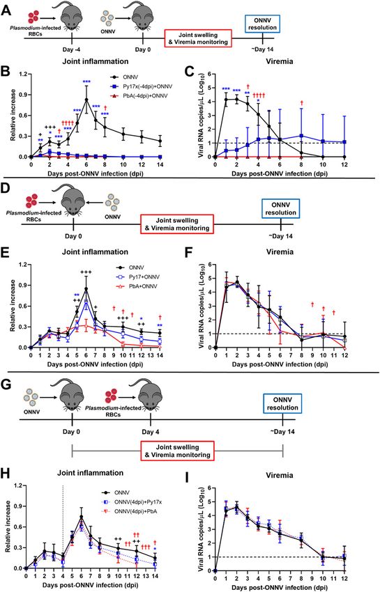
Figure 2. Early stages of ONNV replication and dissemination are supressed by preexisting Plasmodium infection.
Mice were infected with Py17x or PbA and 4 d postinfection were inoculated with a firefly luciferase-tagged ONNV clone in the right hind limb footpad. Before data
acquisition, mice were injected with 100 μl of D-Luciferin (5 mg/ml) subcutaneously. (A, B) Whole body radiance and (B) footpad radiance of ONNV, Py17x(-4dpi)+ONNV,
and PbA(-4dpi)+ONNV groups at 1, 3, 6, 12, and 24 hpi. (C) Representative pseudo-coloured images of bioluminescence readings depicting reduction of tissue viral load and
restriction of viral dissemination in coinfected animals at 12 hpi (yellow-doted boxes). (D) Tissue viral load in mouse appendages, internal organs and muscle detected
by qRT-PCR at 24 hpi. Data are presented as mean ± SD of at least five animals per experimental group. Differences between ONNV controls and coinfected mice with PbA
or Py17x were calculated using two-tailed Kruskal–Wallis and post hoc Dunn’s tests (*P < 0.05 **P < 0.01, ***P < 0.001).

(27, 28, 29), the differences in infectivity within the CD45− compartment              included in these sets of experiments because of the high mortality
were further characterized using Uniform Manifold Approximation                        rate induced by ECM. Interestingly, suppression of ONNV infection
and Projection (UMAP) on the flow cytometry data. Manually gated                        in coinfected animals was observed across all the different time
CD45− populations based on the expression of signature surface                         points assessed, suggesting a sustained suppression of viral load
markers (30, 31, 32, 33, 34) were overlapped in the UMAP plot and                      and not a delay in viral load appearance (Fig 3C).
allowed the identification of four major cell lineages: endo-
thelial cells (CD45−CD29+CD31+), myoblasts (CD45−CD31−Sca-1+), fibro-                   Murine malaria up-regulates proinflammatory immune mediators
blasts (CD45−CD9+CD29+CD31−Sca-1−), and mesenchymal stem cells                         in footpad tissues
(CD45−CD9−CD29−CD31−Sca-1−) (Fig 3A). UMAP plots displaying the dis-
tribution of ZsGreen+ events in ONNV-infected and coinfected mice are                  Acute Plasmodium infections are known to induce strong pro-
also shown in Fig 3A.                                                                  inflammatory responses characterized by the production and re-
    As observed from the UMAP plots, ONNV infection was globally                       lease of cytokines/chemokines and other mediators into the
suppressed in endothelial cells, myoblasts, and fibroblasts from                        bloodstream to control parasite burden (15, 16). Thus, we hy-
coinfected animals at 12 hpi (Fig 3A). A similar trend was observed                    pothesized that some of these soluble factors could be responsible
when ZsGreen+ cells were quantified revealing a reduced number of                       for the decreased susceptibility to ONNV infection observed in
infected endothelial cells, myoblasts, and fibroblasts (Fig 3B). To                     footpad tissues.
explore whether the reduced infectivity in CD45− cells was a                              36 different immune mediators were assessed in sera (Fig 4A)
transient or maintained effect beyond 12 hpi, footpads from                            and footpad tissue lysates (Fig 4B) of mice singly infected with
coinfected mice with Py17x were harvested at 48 and 72 hpi and                         lethal PbA or self-resolving Py17X at 4 d postparasite infection (time
analyzed by flow cytometry. Coinfected mice with PbA were not                           of ONNV inoculation in the coinfection model). Interestingly, results

Plasmodium-induced IFNg restricts ONNV infection      Torres-Ruesta et al.              https://doi.org/10.26508/lsa.202101272    vol 5 | no 4 | e202101272      4 of 15
Figure 3. Prior Plasmodium infection restricts ONNV replication in CD452 footpad cells.
(A) UMAP analysis of 105,000 live CD45− footpad cells from naive, ONNV, PbA(-4dpi)+ONNV, and Py17x(-4dpi)+ONNV groups at 12 hpi. The UMAP plot was generated by
concatenation of samples containing 5,000 randomly selected live CD45− cells from each sample. ONNV, PbA(-4dpi)+ONNV, and Py17x(-4dpi)+ONNV UMAP plots show the
global distribution of ZsGreen+ events (ONNV-infected cells). Colored dashed boxes highlight myoblasts (M), fibroblasts (F), and endothelial cells (E) and median ONNV
infection rates per population. (B) Total counts of ONNV-ZsGreen+ cells in endothelial cells, myoblasts, fibroblasts and mesenchymal stem cells (MSCs) in ONNV,
PbA(-4dpi)+ONNV, and Py17x(-4dpi)+ONNV at 12 hpi. (C) Percentages of infected CD45− cells in ONNV and Py17x(-4DPI)+ONNV groups at 12, 48, and 72 hpi. Data are

Plasmodium-induced IFNg restricts ONNV infection     Torres-Ruesta et al.             https://doi.org/10.26508/lsa.202101272   vol 5 | no 4 | e202101272      5 of 15
showed that PbA- and Py17x-infected mice generated distinct pro- cells, fibroblasts, and mesenchymal stem cells (Fig 5C and D). To further
inflammatory cytokine profiles. Using principal component analysis validate these findings, in vivo IFNγ neutralization was performed using
on these cytokine profiles, a distinct separation was revealed anti-mouse IFNγ antibodies. Coinfected animals treated with anti-
between samples from mock (green), PbA-infected (red), and Py17x- mouse IFNγ displayed comparable ONNV tissue viral loads at 3, 6, 12,
infected mice (green) (Fig 4C). and 24 hpi (Fig 5E and Video 3) than isotype-control treated mice,
Levels of pro-inflammatory IFNγ were increased by ~292- and suggesting a major role of IFNγ in the antiviral effects exerted by
~28-folds in sera from PbA and Py17x-infected mice, respectively preexisting Plasmodium infections.
(Fig 4D), in line with previous reports (35, 36, 37). Up-regulation of Blood-stage Plasmodium infections in humans and mice are
chemokines such as CXCL10 (IFNγ-induced protein 10: IP-10), CXCL1, known to induce the production of type I IFN responses (39, 40, 41).
CCL2, CCL3, CCL5, and CCL7 was also observed in the sera (Fig 4D). Type I IFN is a major regulator of susceptibility to alphavirus in-
Locally at the footpad, changes in immune mediator profiles were fection (13, 42, 43) and evidence has suggested crosstalk mecha-
characterized by increased chemokine production, particularly nisms with IFNγ signaling (44, 45, 46). To evaluate any possible
IFNγ-induced CXCL10, which was found to be elevated by ~20- and contribution of type I IFN responses in the reduced susceptibility to
~11-folds in tissue lysates of PbA and Py17x-infected animals, re- ONNV upon coinfection, the effect of preexisting murine malaria on
spectively (Fig 4D). Other up-regulated chemokines shared between ONNV replication was assessed in IFNaR−/− mice (deficient of IFN-
PbA and Py17x-infected mice were CCL2, CCL3, CCL4, CCL5, CCL7, and α/β receptor). Viremia measurements at 12, 24, and 48 hpi in
CCL11. In addition, IL-18 (IFNγ inducing factor: IGIF) and IFNγ were coinfected IFNaR−/− mice (Fig S6) revealed that murine malaria was
found to be slightly elevated in footpad tissues upon PbA and Py17x still able to restrict ONNV infection, ruling out the involvement of
infection (Fig 4D). type I IFN responses in the antiviral effects exerted by Plasmodium-
Differentially regulated cytokines/chemokines in the footpads induced IFNγ.
were subjected to STRING analysis (performed with a high confi-
dence threshold of 0.9), revealing key interactions between eight of IFNγ inhibits ONNV infection in human fibroblast, synoviocyte,
these immune mediators (Fig S4). The predicted interactions are skeletal muscle, and endothelial cell lines
linked to cellular responses to IFNγ, host-negative regulation of
viral transcription and leukocyte recruitment and activation. Col- The antiviral role of IFNγ in nonimmune cell lineages upon ONNV
lectively, these data suggest that Plasmodium infection (either by infection remains poorly defined. To extrapolate our findings in the
PbA or Py17x) triggers a systemic immune response characterized by human context, the antiviral effect of IFNγ was assessed in human
the up-regulation of pro-inflammatory immune mediators not only cell lines representing skin fibroblasts (BJ), synoviocytes (SW982),
in the blood but also locally in footpad tissues. Importantly, the endothelial cells (HPMECs), and skeletal muscle cells (RD) in an in
increased levels of immune mediators linked to IFNγ signaling, a vitro infection system. Interestingly, each cell type displayed dif-
known antiviral cytokine (38), in footpad lysates of Plasmodium- ferent susceptibility to ONNV infection. Particularly, skin fibroblast
infected mice suggested a possible involvement of this cytokine in and synoviocyte cell lines are highly susceptible to ONNV, whereas
the restriction of ONNV infection. endothelial cells and skeletal muscle cells are poorly infected.
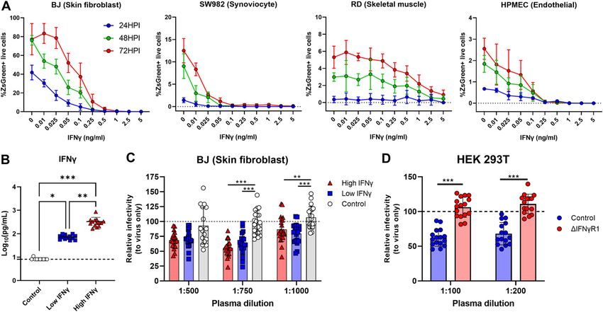
Nevertheless, regardless of cell type, IFNγ pretreatment success-
Plasmodium-induced IFNγ mediates the suppression of ONNV fully reduced ONNV infection in BJ, SW982, HPMEC, and RD cells at
replication and dissemination in coinfected animals 24, 48, and 72 hpi in a dose-dependent manner (Fig 6A).
Finally, we characterized the ability of plasma samples from
To assess the potential role of IFNγ in suppressing ONNV pathol- acute P. vivax–infected patients to render human cell lines less
ogies, IFNγ-deficient animals were infected with nonlethal Py17x. susceptible to ONNV infection. Plasma IFNγ levels were quantified,
4 d postinfection, luciferase-tagged ONNV was inoculated subcu- and samples were categorized and pooled into low (n = 13, median
taneously in the right footpad. Lack of IFNγ in coinfected animals IFNγ concentration = 69.53 pg/ml) and high IFNγ producers (n = 14,
abolished the antiviral effects exerted by Py17x infection in mice. median IFNγ concentration = 293.675 pg/ml). 10 plasma samples
Bioluminescence readings at 3, 6, 12, and 24 hpi were comparable from healthy individuals were pooled and included in the exper-
with those from IFNγ-deficient animals singly infected with ONNV iments as controls (IFNγ concentration under the quantification
(Fig 5A and Video 2). Similarly, ONNV viremia in coinfected IFNγ- limit) (Fig 6B). Incubation of skin fibroblasts (BJ) with plasma from
deficient mice was also restored (Fig 5B) despite a suppression of either low or high IFNγ producers reduced ONNV infection com-
joint swelling at 6 dpi (Fig S5). This suggests that coinfection might pared with those treated with healthy control plasma (Fig 6C). To
modulate other immune responses leading to joint swelling sup- prove that IFNγ present in the plasma samples from acute P.
pression in the absence of IFNγ. vivax–infected patients was responsible for the antiviral effects
Because nonimmune cells express IFNγ receptors (38) and can exerted in vitro, we generated a HEK293T cell line with impaired IFNγ
respond to IFNγ signaling, the infection profile of these subsets was signaling (Fig S7A and B) by knocking down the expression of the
assessed in coinfected mice deficient in IFNγ. Likewise, ONNV in- IFNγ receptor 1 α chain (IFNγR1). Upon treatment with plasma from
fectivity profiles of CD45− cells were restored in myoblasts, endothelial malaria patients, HEK293T cells with intact IFNγR1 expression
presented as mean ± SD of at least five animals per experimental group. Differences between ONNV controls and coinfected mice with PbA or Py17x were calculated
using two-tailed Kruskal–Wallis and post hoc Dunn’s tests (**P < 0.01, ***P < 0.001).
Plasmodium-induced IFNg restricts ONNV infection Torres-Ruesta et al. https://doi.org/10.26508/lsa.202101272 vol 5 | no 4 | e202101272 6 of 15Figure 4. Footpads of Plasmodium-infected mice display a pro-inflammatory milieu.
(A, B) Heat map plots showing the detected cytokines/chemokines in (A) serum and (B) footpad lysates of mock-infected (green), PbA-infected (red), and Py17x-infected
(blue) mice at 4 dpi. Analyte concentrations (pg/ml + 1) were logarithmically transformed (Log10) and Z-scores were calculated for representation purposes. Principal
component analysis (PCA) and heat map plots were constructed using ClustVis. (C) PCA using differentially expressed analytes in footpad lysates and sera of mock, PbA-
infected (4 dpi), and Py17x-infected (4 dpi) groups. PCA plot shows that PC1 (responsible for 35.7% of the variation) and PC2 (responsible for 12.7% of the variation)
segregate the populations in three clusters: mock (green), PbA-infected (red), and Py17x-infected (blue). Colored ellipses were calculated with 95% confidence levels.
(D) Radar plots showing median fold changes of differentially expressed cytokines/chemokines in serum and footpad lysates of PbA-infected (4 dpi) and Py17x-infected
(4 dpi) groups relative to mock animals. Each cytokine/chemokine is grouped according to its immunological function (green: pro-inflammatory, yellow: anti-
inflammatory) or homing receptors (purple) as indicated. Shared chemokine receptors are shown in dashed lines. Data correspond six animals per experimental group.
Differences between naı̈ve, PbA, or Py17x-infected mice calculated using two-tailed Kruskal–Wallis and post hoc Dunn’s tests.
displayed lower ONNV infection compared with untreated controls. development of ONNV pathologies by restricting viral infection at
This antiviral effect was lost in cells with impaired IFNγR1 expression the site of inoculation and dissemination to distant organs. We
(ΔIFNγR1) (Fig 6D), in agreement with an IFNγ-specific effect. demonstrated that Plasmodium-induced IFNγ is the main cytokine
driving the antiviral effects observed.
IFNγ is a pleiotropic cytokine known for its ability to regulate immune
Discussion responses by promoting macrophage activation, enhancing antigen
presentation, modulating helper T cell development and mediating viral
O’nyongnyong virus and Plasmodium parasites share common and bacterial immunity, among others (47). In ONNV-infected animals, it
anopheline vectors and co-circulate in sub-Saharan Africa with risk was observed that nearly 80% of the total virus-infected cells at 12 hpi
of human coinfection. This is the first study investigating the (Fig S3A) belonged to the CD45− compartment corroborating previous
pathological outcomes of coinfection by Plasmodium parasites and observations from other closely related alphaviruses, whereby non-
alphavirus ONNV in a mammalian host. Here, we showed that a immune cells support the early stages of viral replication (13, 27, 28, 29,
preexisting murine Plasmodium infection is able to suppress the 48). In contrast, animals harboring a Plasmodium infection displayed
Plasmodium-induced IFNg restricts ONNV infection Torres-Ruesta et al. https://doi.org/10.26508/lsa.202101272 vol 5 | no 4 | e202101272 7 of 15Figure 5. Plasmodium-induced IFNγ mediates the suppression of ONNV replication and dissemination in coinfected animals. (A, B) In vivo luminescence readings of (A) whole body and footpad radiance at 3, 6, 12, and 24 hpi and (B) viremia at 24 hpi of ONNV and Py17x(-4dpi)+ONNV wild-type (WT), or IFNγ-deficient (IFNγ −/−) animals. (C) UMAP analysis of 160,000 live CD45− footpad cells from WT or IFNγ −/− ONNV, PbA(-4dpi)+ONNV, and Py17x(-4dpi)+ONNV groups at 12 hpi. The UMAP plot was generated by concatenation of samples containing 5,000 randomly selected live CD45− cells from each sample. ONNV, PbA(-4dpi)+ONNV, and Py17x(-4dpi)+ONNV UMAP plots show the global distribution of ZsGreen+ events (ONNV-infected cells). Colored dashed boxes highlight ONNV infection in myoblasts, M; fibroblasts, F and endothelial cells, E. (D) Frequency of CD45-ZsGreen+ footpad cells of WT or IFNγ −/− ONNV, PbA(-4dpi)+ONNV, and Py17x(-4dpi)+ONNV groups at 12 hpi. Myo, myoblasts; Fibro, fibroblasts; ECs, endothelial cells and MSCs, mesenchymal stem cells. (E) Footpad radiance at 1, 3, 6, 12, and 24 hpi of ONNV, PbA(-4dpi)+ONNV, and Py17x(-4dpi)+ONNV groups in animals treated with mouse anti-IFNγ or isotype control. Data are presented as mean ± SD of at least five animals per experimental group. (A, B) Two-tailed Mann–Whitney U test was used to compute differences between ONNV and Py17x(-4dpi)+ONNV groups in (A) and (B) (*P < 0.05 **P < 0.01). Differences Plasmodium-induced IFNg restricts ONNV infection Torres-Ruesta et al. https://doi.org/10.26508/lsa.202101272 vol 5 | no 4 | e202101272 8 of 15
reduced numbers of ONNV-infected myoblasts, fibroblasts and endo-                  infection in human cell lines in a dose dependent manner. Similarly, we
thelial cells and this protective effect was reverted in IFNγ-deficient            also observed that stimulation of skin fibroblasts with plasma from
mice or upon in vivo IFNγ neutralization. We therefore hypothesize                acute P. vivax–infected patients containing IFNγ-reduced cell suscep-
that production of IFNγ in response to acute blood-stage Plasmo-                  tibility to ONNV infection. Of note, the biological effects of IFNγ are
dium infection could stimulate cells from the CD45− compartment                   mediated conventionally through the activation of the JAK/STAT pathway
activating antiviral processes (Fig S4) which in turn restrict a sub-             (55). We postulate that the attenuation of ONNV infection by Plasmo-
sequent ONNV infection. In line with this, the antiviral effects of IFNγ          dium-induced IFN-γ observed in mouse models could be translated to a
have been observed in other alphaviruses such as Sindbis virus                    real coinfection scenario in endemic populations given that the JAK/
(SINV). In vitro studies suggested that IFNγ affects SINV replication in          STAT signaling pathway between humans and mice is highly conserved
mature neurons by interfering with the synthesis of genomic and                   (56, 57). This suggests that similar downstream effector proteins could be
sub-genomic viral RNA (49) and that this effect is dependent on JAK/              involved in the IFNγ-mediated restriction of alphavirus infection.
STAT signaling (50). The contribution of Plasmodium-induced type I                   A recent study (58) showed that Plasmodium infection protected
IFN (40, 41) to the reduced susceptibility to ONNV infection was also             mice from Ebola virus (EBOV)–induced mortality via up-regulation of
assessed in IFNaR−/− mice. Considerable viremia differences were                  IFNγ, supporting field reports where coinfected patients by EBOV and P.
observed between ONNV-infected wild type and IFNaR−/− controls                    falciparum displayed increased survival rates (59). Conversely, two other
(~4–5 Log10 at 48 hpi) highlighting the importance of IFN-α/β sig-                murine coinfection models with respiratory viral pathogens such as
naling in the control of ONNV infection as observed in other                      murine pneumonia virus (PVM) and murine gammaherpesvirus 68
alphavirus animal models (42, 43, 51, 52). Nonetheless, type I IFN-               (MHV68) using nonlethal Plasmodium chabaudi and P. yoelii 17XNL have
induced upon Plasmodium infection seems to be negligible for the                  reported detrimental outcomes for the host such as increased viral
establishment of protective effects by murine malaria as coinfected               loads in the lungs (60) and mortality due to severe anemia (61). These
IFNaR−/− mice still displayed reduced ONNV infection to a compa-                  observations were linked to altered type I IFN production (60) and
rable level than coinfected wild-type mice (Fig S6).                              antiviral humoral responses (61) upon coinfection. Thus, the protective
   It is important to note that in our experiments, mice only ex-                 or detrimental effects of murine malaria on viral pathogens are likely
perienced suppression of ONNV viremia and virus dissemination                     associated to the modulation of distinct immune responses governing
after 4 d post-Plasmodium inoculation (Figs 1B and 2A–D) and not                  the control of different viral infections. Conversely, other protozoan
upon concurrent or sequential (postviral) coinfection. These ob-                  parasites highly prevalent in the tropics and known to induce the up-
servations strongly suggested that the timing of parasite inocu-                  regulation of IFNγ in response to infection such as Leishmania spp. (62),
lation and induction of IFNγ are critical for the protective effects to           Toxoplasma spp. (63), or Trypanosoma spp. (64) could potentially display
happen. Interestingly, although the main suppression of ONNV                      similar protective effects in coinfection settings. In line with this, mice
infection occurred in joint footpad cells, we observed lower con-                 infected with Trypanosoma brucei were shown to be resistant to cu-
centrations of IFNγ in joint footpad tissues compared with serum                  taneous leishmaniasis through the induction of IFNγ generating a
samples at 4 d post-Plasmodium inoculation. Thus, it is likely that               hostile pro-inflammatory environment impairing L. major colonization
IFNγ levels in joint footpads could have increased at an earlier time             of the skin (65).
point. In support of this, we observed high concentrations of IFNγ-                  Results in Fig 1B and E suggest the impairment of ONNV-induced
induced immune mediators in joint footpad tissues, particularly                   joint pathologies upon Plasmodium infection. Interestingly, con-
CXCL10 (53) and CCL7, known to be produced by fibroblasts and                      current coinfection with PbA parasites did not affect viremia levels
mononuclear cells upon IFNγ stimulation (54). The development of                  but significantly reduced the major peak of footpad swelling at 6
T-cell responses, major IFNγ-producing subsets during malaria                     dpi. This can be the result of two different mechanisms. First, the
(ref), could also influence the outcome of a Plasmodium–ONNV                       development of viremia in concurrently coinfected mice can be
coinfection in murine models. Early in a blood-stage infection, a                 attributed to the absence of Plasmodium-induced IFNγ in the early
large number of IFNγ-secreting Th1 cells are produced, whereas                    stages of ONNV infection in the footpads. It has been shown that the
Th2-like responses govern during the chronic phase of infection                   earliest IFNγ production during blood-stage murine malaria only
(17). Because ONNV inoculation occurs in the early stages of murine               occurs after 24 h postparasite injection (35). During this period,
malaria (4 dpi), it is likely that the antiviral effects of IFNγ are              footpad cells in concurrently coinfected mice are still susceptible to
associated to the establishment of Th1 immunity against the                       ONNV infection which results in viremia levels similar to control
parasite. It can be speculated that the degree of virus suppression               mice (Fig 1F). On the other hand, the suppression of joint swelling
might differ if ONNV is inoculated during the chronic stage of the                upon coinfection could be linked to the dysregulation of virus-
murine malaria, particularly when Th1 responses are weaning.                      specific CD4 T-cell responses, main drivers of joint inflammation at
   We explored the relevance of our findings in the context of ONNV                6 dpi (26) by malaria. It has been reported that murine Plasmodium
human infection by treating four human cell lines from different                  infections impair the development of CD4 T-cell responses against
nonimmune lineages (fibroblast, synoviocyte, endothelial, and                      heterologous antigens (66, 67). Thus, PbA infections could alter the
skeletal muscle cells) with IFNγ before ONNV infection. Mechanis-                 establishment of virus-specific CD4 T-cell immunity resulting in
tically, our results demonstrated that IFNγ is able of restricting ONNV           decreased footpad swelling.

between ONNV, PbA(-4dpi)+ONNV, and Py17x(-4dpi)+ONNV groups were calculated using two-tailed Kruskal–Wallis and post hoc Dunn’s tests (*P < 0.05 **P < 0.01,
***P < 0.001).

Plasmodium-induced IFNg restricts ONNV infection   Torres-Ruesta et al.            https://doi.org/10.26508/lsa.202101272   vol 5 | no 4 | e202101272   9 of 15
Figure 6. In vitro stimulation with human IFNγ or plasma from malaria patients reduces susceptibly to ONNV infection.
For (A), cells were treated with recombinant human IFNγ for 24 h before ONNV infection at MOI 10. (A) ONNV infection rates in BJ, SW982, HPMEC, and RD at 24, 48, and 72
hpi. (B) IFNγ levels in plasma of healthy controls (HC, n = 10), low (n = 13), and high (n = 14) IFNγ responders. For (C), BJ cells were treated with pooled plasma dilutions from
P. vivax–infected patients or healthy controls for 12 h before ONNV infection at MOI 1. For (D), control or ΔIFNγR1 HEK293T cells were treated with pooled plasma (1:100 or
1:200) from P. vivax-infected patients for 12 h before ONNV infection at MOI 1. Differences between three groups were calculated using two-tailed Kruskal–Wallis and
post hoc Dunn’s tests, differences between two groups were calculated using two-tailed Mann–Whitney U test (*P < 0.05 **P < 0.01, ***P < 0.001). Data are presented as
mean ± SD values and representative of two independent experiments.

   The great success of malaria control programs during the last                            specific pathogen-free conditions at the Biological Resource Centre
decade has been accompanied of by a sharp rise in the number of                             of the Agency for Science, Technology, and Research, Singapore
arbovirus infections worldwide (68). Our data highlight a possible                          (A*STAR).
causative link: that this phenomenon could be due in part to the
loss of protective effects exerted by Plasmodium infections on                              Viruses
alphavirus-induced pathologies that had hitherto masked the real
burden of ONNV and other arbovirus infections. This may occur via                           The IMTSSA/5163 ONNV isolate used in this study was obtained from
two mechanisms: first, higher arbovirus case reporting because of                            an acute patient in Chad in 2004 (kindly provided by Marc Gran-
more severe symptoms of arbovirus infections in the absence of                              dadam from the Unité de Virologie Tropicale, IMTSSA) (69). Full-
malaria; second, and worse, increased arbovirus cases might reflect                          length infectious cDNA clones of the IMTSSA/5163 isolate were used
increased arbovirus transmissibility and/or fitness because of                               to generate ONNV variants expressing the firefly luciferase gene
increased viral titers in the absence of malaria. Our data in this                          (ONNV-Fluc) and ZsGreen protein (ONNV-ZsGreen) (70, 71). Viruses
report will be of value in the fight against Plasmodium and ONNV                             were propagated in Aedes albopictus C6/36 cell line (ATCC CRL-
infections in areas where both pathogens co-circulate, particularly                         1660) and purified by sucrose-gradient ultracentrifugation. Viral
highlighting the need for screening and clinical studies of un-                             stock titers were determined by standard plaque assay using Vero
derlying alphavirus infections in malaria intervention programs.                            E6 cells (ATCC CCL-81).

                                                                                            Parasites
Materials and Methods
                                                                                            P. yoelii 17XNL clone 1.1 (referred as Py17x) was used to induce self-
Mice                                                                                        resolving infections in mice (72). Lethal infections were induced by
                                                                                            inoculation of P. berghei ANKA (PbA) (231cl1) expressing luciferase
3- to 4-wk-old gender-matched wild-type (JAX #000664), IFNγ −/− (JAX                        and GFP under the control of the ef1-α promoter (73). iRBCs were
#002287), and IFNaR1−/− (JAX #028288) mice in C57BL/6J background                           obtained by in vivo serial passage in C57BL/6J mice and were stored
were used in this study. Animals were bred and maintained under                             in Alsever’s solution in liquid nitrogen.

Plasmodium-induced IFNg restricts ONNV infection         Torres-Ruesta et al.               https://doi.org/10.26508/lsa.202101272     vol 5 | no 4 | e202101272         10 of 15
Human cell lines Eppendorf tubes and mixed with 230 μl of chloroform and incu-
bated at RT for 2 min. Samples were centrifuged at 12,000g for
BJ (ATCC CRL-2522), SW-982 (ATCC HTB-93), and RD (ATCC CCL-136) 10 min at 4°C and recovered supernatants were transferred into
cells were grown in DMEM medium with 10% heat-inactivated FBS. clean Eppendorf Tubes and mixed with 70% ethanol (1:1 volume).
HPMEC (#3000; ScienCell) were grown in supplemented EC medium RNA was purified using the RNeasy Mini Kit (QIAGEN) according to
(#1001; ScienCell). Cells were grown at 37°C, relative humidity of the manufacturer’s protocol. Viral RNA copies were quantified by
95%, and 5% CO2. qRT-PCR as described above.
Generation of ΔIFNγR1 HEK293T cell line In vivo virus tissue dissemination assay
ΔIFNγR1 HEK293T cell line was generated by phosphorylating and To quantify tissue viral load and virus dissemination in vivo, mice
annealing primers (59-CACCGACATGAACCCTATCGTATAT-39) and (59- were inoculated with a firefly luciferase-tagged ONNV infectious
AAACATATACGATA GGGTTCATGTC-39) (NG_007394.1) using T4 Poly- clone (ONNV-Fluc) and virus dissemination was tracked using the
nucleotide Kinase (NEB M0201S) in provided buffer supplemented IVIS Spectrum In Vivo Imaging System (Perkin-Elmer) (26). Animals
with 1 mM ATP (A2383; Sigma-Aldrich) at 37°C for 30 min followed by were kept anesthetized during the experiment using an oxygen flow
5 min at 95°C and ramped down to 25°C. Annealed primers were rate of 1 liter/minute with 2% isoflurane. Full-body shaved mice
then ligated using Instant Sticky-end Ligase Master Mix (M0370; were subcutaneously injected with 100 μl of D-luciferin potassium
NEB) in pSB-CRIPSR (kindly gifted by Dr Gao and Dr Hu (74)) pre- salt (Caliper Life sciences) diluted in PBS (5 mg/ml). Whole body
viously linearized using Esp3I (R0734S; NEB) in CutSmart Buffer and footpad bioluminescence readings were independently taken 7
(NEB) and purified using Nucleospin gel and PCR clean up kit min post–D-luciferin injection with a field of view (FOV) of 21.7 cm
(740609.50; Macherey-Nagel). HEK293T cells were co-transfected (ventral position) and 13.1 cm (dorsal position) for whole body (FOV-
with pCMV(CAT)T7-SB100 (a gift from Zsuzsanna Izsvak, plasmid # D) and footpad (FOV-C) measurements, respectively. Two pictures
34879; Addgene; RRID: Addgene_34879 (75)) and pSB-CRISPR (at 1:1 were taken per FOV with exposure times set to “AUTO” and 60 s.
ratio) using Lipofectamine 2000 (11668019; Thermo Fisher Scientific) Regions of interest were drawn using the software Living Image 3.0
in Opti-MEM medium (31985070; Gibco) following the manufac- and total flux values (photons/second) were calculated. Readings
turer’s recommendations. 3 d post co-transfection, cells were of naı̈ve mice injected with D-luciferin were used for background
passaged and cultured in complete media containing 1 μg/ml subtraction.
puromycin (P8833; Sigma-Aldrich). When control cells fully suc-
cumbed to puromycin selection, co-transfected cells were cultured Plasmodium infection and disease monitoring
in complete media and IFNγR1 expression was assessed by flow
cytometry. Mice were infected with Py17x or PbA by i.p. injection of 106 iRBC in
Alsever’s buffer. Parasitemia was monitored by flow cytometry
ONNV infection and disease monitoring as previously described (76) using a staining mix containing anti-
mouse APC-tagged CD45 antibodies, 8 μM dihydroethidium (Sigma-
Mice were infected with ONNV by subcutaneous inoculation of 106 Aldrich), and 5 μg/ml Hoechst 33342 (Sigma-Aldrich). Successful
PFU in 30 μl of PBS in the ventral side of the right hind footpad. infections were confirmed 4 d postinoculation.
Viremia was monitored daily from 1 to 6 dpi and thereafter on every
alternate day until 12 dpi. Briefly, 10 μl of blood collected from the Isolation of footpad cells
tail of each mouse was mixed in 120 μl of PBS supplemented with
10 μl of citrate-phosphate-dextrose solution (Sigma-Aldrich). RNA Homogenous cell suspensions were obtained for immune profiling
isolation was performed using the QIAamp Viral RNA kit (QIAGEN) of footpads of infected and naı̈ve animals. Mice were culled by
following the manufacturer’s instructions with a final elution vol- cervical dislocation and right paws were harvested and immedi-
ume of 60 μl. 1 μl of purified RNA was quantified by qRT-PCR using ately placed in 4 ml of digestion medium containing Collagenase IV
QuantiTect Probe RT-PCR (QIAGEN) as previously described (26). (20 μg/ml; Sigma-Aldrich), Dispase I (2 U/ml; Invitrogen), and DNase
Joint swelling was measured for 2 wk post-ONNV inoculation as a I (50 μg/ml; Roche Applied Science) mixed in RPMI medium com-
function of height × width relative to measurements preinfection plemented with 10% FBS. Using forceps, footpads were deskinned
(relative increase) (26). and deboned to maximize digestion. Processed samples were
placed on a shaker and incubated at 37°C at 100 rpm (Biosan PSU-
Tissue viral load determination 10i) for 3 h. After digestion, tissues were passed through a 40-μm cell
strainer (Fisherbrand). Any remaining tissue trapped in the strainer
Ketamine xylazine–anesthetized mice (150 mg/kg of ketamine, 10 was grinded using the top of a 1 ml syringe plunger to maximize cell
mg/kg of xylazine) were intracardially perfused with PBS and or- recovery. 1× Flow Cytometry Mouse Lysis Buffer (R&D Systems) was
gans were collected in tubes containing zirconia beads (TOMY used to lyse contaminating RBCs. Samples were resuspended in
Digital Biology) and 1 ml TRIzol (Invitrogen) and stored at −80°C. To 1 ml of complete RPMI, overlaid to 35% vol/vol Percoll (Sigma-
isolate RNA, tissues were thawed on ice and homogenized using the Aldrich)/RPMI mixture, and centrifuged at 2,400 rpm for 20 min at
Bead Ruptor Elite (OMNI International) at a speed of 6 m/s (3 cycles 4°C. Footpad cell pellets were washed and resuspended in ap-
of lysis × 30 s). Tissue lysates were then transferred to 1.5-ml propriate volumes for counting using haemocytometers.
Plasmodium-induced IFNg restricts ONNV infection Torres-Ruesta et al. https://doi.org/10.26508/lsa.202101272 vol 5 | no 4 | e202101272 11 of 15Profiling of immune and nonimmune cells by flow cytometry In vitro IFNγ treatment and ONNV infection
Cell suspensions were stained for viability using LIVE/DEAD Aqua Recombinant human IFNγ (PHC4033; Gibco) diluted in supple-
dye (Life Technologies). Cells were washed and resuspended in mented DMEM at various concentrations was used to treat skin
50 μl of blocking buffer containing TruStain FcX PLUS (anti-mouse fibroblasts (BJ), synoviocytes (SW-982), skeletal muscle cells (RD),
CD16/32, clone S17011E) antibody diluted in PBS and incubated in and endothelial cells (HPMEC) for 24 h before virus infection. Cells
the dark for 15 min on ice. Conjugated anti-mouse antibodies CD45 were washed with PBS and then infected with ONNV-ZsGreen virus
(30-F11), CD9 (eBioKMC8), CD29 (HMβ1-1), CD31 (390), and Integrin α 7 at MOI of 10. Cells were harvested at 24-, 48-, and 72-h postinfection
(334908), Sca-1 (D7) were used to stain cell surface markers for (hpi) and percentage of infection was quantified by flow cytometry.
30 min on ice. Finally, cells were fixed with 50 μl of eBioscience IC
Fixation Buffer (Thermo Fisher Scientific) for 5 min and acquired Data and statistical analyses
using a 5-laser LSR II flow cytometer (BD Biosciences) with BD
FACSDiva software. Data were analyzed with FlowJo v10.6.2 (Becton, Statistical analyses were performed using GraphPad Prism 8.4.3
Dickinson and Company). (GraphPad Software). Data are presented as mean ± SD unless
otherwise specified. Nonparametric Mann–Whitney U statistical test
Dimensionality reduction analysis of flow cytometry data was used to compute differences between two groups. Differences
between three groups were calculated using two-tailed Kruskal–
Live CD45− singlets events were pregated and then randomly down- Wallis and post hoc Dunn’s tests. Values obtained for viremia,
sampled to a fixed number (n = 5,000) for each sample using FlowJo parasitemia, in vivo imaging, and cytokine/chemokines were log-
v10.7 (Becton, Dickinson and Company). Down-sampled files were transformed for representation purposes. P-values < 0.05 were
concatenated and analyzed using UMAP for Dimension Reduction considered statistically significant.
plug-in v3.1 using default parameters (number of nearest neigh-
bours = 15, minimum distance = 0.1). Study approval
Animal experiments were approved by the Institutional Animal Care
In vivo IFNγ neutralization
and Use Committee (IACUC #211635) of A*STAR in accordance with
the guidelines of the Agri-Food and Veterinary Authority (AVA) and
Anti-mouse IFNγ (0.5 mg per mouse, clone XMG1.2, Bio X Cell) was i.p.
the National Advisory Committee for Laboratory Animal Research of
injected at 0-, 2-, and 4 d postparasite inoculation. Control groups
Singapore (NACLAR). Plasma samples from febrile P. vivax–infected
were given rat IgG1 Isotype control (0.5 mg, clone TNP6A7, Bio X Cell)
patients from Mae Sot, Thailand, were collected and tested in
at similar time points as treatment groups.
accordance with protocols approved by the University of Oxford
Tropical Research Ethics Committee (OXTREC 17-11) and the Ethics
Cytokine/chemokine quantification by multiplexed bead-based Committee of the Faculty of Tropical Medicine at Mahidol University
immunoassays (MUTM 2008-215). Written informed consent was received before
participation.
Cytokine and chemokine concentrations (pg/ml) were quantified in
footpad and serum samples. For footpad samples, animals were
anesthetized with ketamine-xylazine and intracardially perfused Supplementary Information
with PBS. The right paw was cut at the ankle and placed in a
gentleMACS M tube (Miltenyi) filled with 1.5 ml of RIPA buffer (50 mM Supplementary Information is available at https://doi.org/10.26508/lsa.
Tris–HCl, pH 7.4, 1% NP-40, 0.25% sodium deoxycholate, 150 mM 202101272.
NaCl, and 1 mM EDTA) complemented with 1× cOmplete Protease
Inhibitor Cocktail (Roche). Samples were lysed in a Xiril Dispomix
Tissue Homogenizer, centrifuged and the supernatants transferred Acknowledgements
into clean 2-ml microcentrifuge tubes for sonication in a Branson
Ultrasonics Sonifier S-450 (70% intensity × 15 s). For serum samples, The authors would like to thank Dr Carla Claser for critical discussion and
blood was collected from the retro-orbicular sinus using a glass valuable suggestions on the study. We also thank the SIgN Flow Cytometry
Pasteur pipette and allowed to clot for 30 min at room temperature. Core and SIgN Mouse Core for assistance with cytometry analyses and
Clotted blood was centrifuged at 14,000 rpm for serum isolation. support in animal breeding, respectively. We thank Wilson How from the SIgN
Immunomonitoring platform for his support in the multiplexed bead-based
Footpad lysates and serum samples were analyzed using the Cy-
immunoassays. We also thank Professor Andres Merits from the University of
tokine & Chemokine 36-Plex Mouse ProcartaPlex Panel 1A (Thermo Tartu for providing the tagged ONNV infectious clones used in this study. The
Fisher Scientific) according to the manufacturer’s protocol. Human study was supported by a core research grant provided to A*STAR Infectious
plasma samples were analyzed using the Cytokine/Chemokine/ Diseases Labs and Singapore Immunology Network by the Biomedical Re-
Growth Factor 45-plex Human ProcartaPlex Panel 1 (Thermo Fisher search Council (BMRC) from the Agency for Science, Technology and Re-
search (A*STAR). A Torres-Ruesta is supported by the A*STAR Singapore
Scientific). Data were acquired with Luminex FLEXMAP 3D instru- International Graduate Award (SINGA) scholarship. Flow cytometry platform
ment (Millipore) using xPONENT 4.0 software and analyzed with Bio- is supported by the Health and Biomedical Sciences (HBMS) Open Fund
Plex Manager 6.1.1 (Bio-Rad Laboratories). Shared Infrastructure Support Grant under the Immunomonitoring Service
Plasmodium-induced IFNg restricts ONNV infection Torres-Ruesta et al. https://doi.org/10.26508/lsa.202101272 vol 5 | no 4 | e202101272 12 of 15Platform project (NRF2017_SISFP09). The funders had no role in the study Anopheles mosquitoes. Trans R Soc Trop Med Hyg 59: 300–306.
design, data collection and analysis, decision to publish, or preparation of doi:10.1016/0035-9203(65)90012-x
the manuscript. 5. Haddow AJ, Davies CW, Walker AJ (1960) O’nyong-nyong fever: An
epidemic virus disease in East Africa 1. Introduction. Trans R Soc Trop
Med Hyg 54: 517–522. doi:10.1016/0035-9203(60)90025-0
Author Contributions
6. Rwaguma EB, Lutwama JJ, Sempala SD, Kiwanuka N, Kamugisha J, Okware
S, Bagambisa G, Lanciotti R, Roehrig JT, Gubler DJ (1997) Emergence of
A Torres-Ruesta: data curation, formal analysis, investigation, vi-
epidemic O’nyong-nyong fever in southwestern Uganda, after an
sualization, methodology, and writing—original draft, review, and absence of 35 years. Emerg Infect Dis 3: 77. doi:10.3201/eid0301.970112
editing.
7. Posey DL, O’Rourke T, Roehrig JT, Lanciotti RS, Weinberg M, Maloney S
T-H Teo: data curation, formal analysis, investigation, methodology, (2005) O’Nyong-nyong fever in West Africa. Am J Trop Med Hyg 73: 32.
and writing—review and editing. doi:10.4269/ajtmh.2005.73.1.0730032
Y-H Chan: formal analysis, investigation, methodology, and wri- 8. LaBeaud AD, Banda T, Brichard J, Muchiri EM, Mungai PL, Mutuku FM,
ting—review and editing. Borland E, Gildengorin G, Pfeil S, Teng CY, et al (2015) High rates of
SN Amrun: formal analysis, investigation, methodology, and wri- O’nyong nyong and Chikungunya virus transmission in coastal Kenya.
ting—review and editing. PLoS Negl Trop Dis 9: e0003436. doi:10.1371/journal.pntd.0003436
NK-W Yeo: formal analysis, investigation, methodology, and wri- 9. Clements TL, Rossi CA, Irish AK, Kibuuka H, Eller LA, Robb ML, Kataaha P,
ting—review and editing. Michael NL, Hensley LE, Schoepp RJ (2019) Chikungunya and O’nyong-
nyong viruses in Uganda: Implications for diagnostics. Open Forum
CY-P Lee: formal analysis, investigation, methodology, and wri-
Infect Dis 6: ofz001. doi:10.1093/ofid/ofz001
ting—review and editing.
SY-T Nguee: formal analysis, investigation, methodology, and wri- 10. Baba M, Logue CH, Oderinde B, Abdulmaleek H, Williams J, Lewis J, Laws
TR, Hewson R, Marcello A, D’Agaro P (2013) Evidence of arbovirus co-
ting—review and editing. infection in suspected febrile malaria and typhoid patients in Nigeria.
MZ Tay: formal analysis, investigation, methodology, and wri- J Infect Dev Ctries 7: 51–59. doi:10.3855/jidc.2411
ting—review and editing.
11. Kinimi E, Patrick BN, Misinzo G (2018) Serological evidence of
F Nosten: formal analysis, investigation, methodology, and wri- chikungunya and malaria co-infection among febrile patients seeking
ting—review and editing. health care in district, Tanzania. Tanzania J Health Res 20: 1–8.
S-W Fong: formal analysis, investigation, methodology, and wri- doi:10.4314/thrb.v20i4.1
ting—review and editing. 12. Mostafavi H, Abeyratne E, Zaid A, Taylor A (2019) Arthritogenic
F-M Lum: formal analysis, investigation, methodology, and wri- alphavirus-induced immunopathology and targeting host inflammation
ting—review and editing. as A therapeutic strategy for alphaviral disease. Viruses 11: 290.
doi:10.3390/v11030290
G Carissimo: formal analysis, investigation, methodology, and
writing—review and editing. 13. Assunção-Miranda I, Cruz-Oliveira C, Da Poian AT (2013) Molecular
mechanisms involved in the pathogenesis of -induced arthritis. Biomed
L Renia: conceptualization, supervision, and writing—original draft,
Res Int 2013: 973516. doi:10.1155/2013/973516
review, and editing.
14. Septembre-Malaterre A, Bedoui Y, Giry C, Gasque P, Guiraud P,
LFP Ng: conceptualization, resources, supervision, funding acqui-
Sélambarom J (2021) Quercetin can reduce viral RNA level of O’nyong-
sition, project administration, and writing—original draft, review, nyong virus and resulting innate immune cytokine responses in
and editing. cultured human synovial fibroblasts. Sci Rep 11: 6369. doi:10.1038/
s41598-021-85840-z
Conflict of Interest Statement 15. Clark IA (2007) The advent of the cytokine storm. Immunol Cell Biol 85:
271–273. doi:10.1038/sj.icb.7100062
The authors declare that they have no conflict of interest. 16. Clark IA, Alleva LM, Budd AC, Cowden WB (2008) Understanding the role
of inflammatory cytokines in malaria and related diseases. Trav Med
Infect Dis 6: 67–81. doi:10.1016/j.tmaid.2007.07.002
17. Kurup SP, Butler NS, Harty JT (2019) T cell-mediated immunity to malaria.
References Nat Rev Immunol 19: 457–471. doi:10.1038/s41577-019-0158-z
18. Rénia L, Howland SW, Claser C, Charlotte Gruner A, Suwanarusk R, Hui
1. Rezza G, Chen R, Weaver SC (2017) O’nyong-nyong fever: A neglected Teo T, Russell B, Ng LF (2012) Cerebral malaria: Mysteries at the blood-
mosquito-borne viral disease. Pathog Glob Health 111: 271–275. brain barrier. Virulence 3: 193–201. doi:10.4161/viru.19013
doi:10.1080/20477724.2017.1355431 19. Howland SW, Claser C, Poh CM, Gun SY, Rénia L (2015) Pathogenic CD8+ T
2. Powers AM, Brault AC, Shirako Y, Strauss EG, Kang W, Strauss JH, Weaver cells in experimental cerebral malaria. Semin Immunopathol 37: 221–231.
SC (2001) Evolutionary relationships and systematics of the doi:10.1007/s00281-015-0476-6
alphaviruses. J Virol 75: 10118–10131. doi:10.1128/JVI.75.21.10118-10131.2001 20. Rénia L, Potter SM, Mauduit M, Rosa DS, Kayibanda M, Deschemin JC,
3. Sanders EJ, Rwaguma EB, Kawamata J, Kiwanuka N, Lutwama JJ, Snounou G, Grüner AC (2006) Pathogenic T cells in cerebral malaria. Int J
Ssengooba FP, Lamunu M, Najjemba R, Were WA, Bagambisa G, et al Parasitol 36: 547–554. doi:10.1016/j.ijpara.2006.02.007
(1999) O’nyong-nyong fever in south-central Uganda, 1996-1997: 21. Claser C, Nguee SYT, Balachander A, Howland SW, Becht E, Gunasegaran
Description of the epidemic and results of a household-based B, Hartimath SV, Lee AWQ, Ho JTT, Ong CB, et al (2019) Author Correction:
seroprevalence survey. J Infect Dis 180: 1436–1443. doi:10.1086/315073 Lung endothelial cell antigen cross-presentation to CD8+T cells drives
4. Williams MC, Woodall JP, Corbet PS, Gillett JD (1965) O’nyong-Nyong malaria-associated lung injury. Nat Commun 10: 5066. doi:10.1038/
fever: An epidemic virus disease in East Africa. 8. Virus isolations from s41467-019-13025-4
Plasmodium-induced IFNg restricts ONNV infection Torres-Ruesta et al. https://doi.org/10.26508/lsa.202101272 vol 5 | no 4 | e202101272 13 of 15You can also read