Low-level laser therapy on skeletal muscle inflammation: evaluation of irradiation parameters
←
→
Page content transcription
If your browser does not render page correctly, please read the page content below
Low-level laser therapy on skeletal
                 muscle inflammation: evaluation of
                 irradiation parameters

                 Matías Mantineo
                 João P. Pinheiro
                 António M. Morgado

Downloaded From: https://www.spiedigitallibrary.org/journals/Journal-of-Biomedical-Optics on 15 Nov 2021
Terms of Use: https://www.spiedigitallibrary.org/terms-of-use
Journal of Biomedical Optics 19(9), 098002 (September 2014)

                Low-level laser therapy on skeletal muscle
                inflammation: evaluation of irradiation parameters
                Matías Mantineo,a,b João P. Pinheiro,c and António M. Morgadoa,b,*
                a
                  University of Coimbra, Instrumentation Center, Department of Physics, Coimbra 3004-516, Portugal
                b
                  IBILI-Institute for Biomedical Imaging and Life Sciences, Azinhaga de Santa Comba-Celas, Coimbra 3000-548, Portugal
                c
                  University of Coimbra, Faculty of Medicine, Azinhaga de Santa Comba-Celas, Coimbra 3000-548, Portugal

                              Abstract. We evaluated the effect of different irradiation parameters in low-level laser therapy (LLLT) for treating
                              inflammation induced in the gastrocnemius muscle of rats through cytokines concentration in systemic blood and
                              analysis of muscle tissue. We used continuous (830 and 980 nm) and pulsed illuminations (830 nm). Animals
                              were divided into five groups per wavelength (10, 20, 30, 40, and 50 mW), and a control group. LLLT was applied
                              during 5 days with a constant irradiation time and area. TNF-α, IL-1β, IL-2, and IL-6 cytokines were quantified by
                              ELISA. Inflammatory cells were counted using microscopy. Identical methodology was used with pulsed illumi-
                              nation. Average power (40 mW) and duty cycle were kept constant (80%) at five frequencies (5, 25, 50, 100, and
                              200 Hz). For continuous irradiation, treatment effects occurred for all doses, with a reduction of TNF-α, IL-1β, and
                              IL-6 cytokines and inflammatory cells. Continuous irradiation at 830 nm was more effective, a result explained by
                              the action spectrum of cytochrome c oxidase (CCO). Best results were obtained for 40 mW, with data suggesting
                              a biphasic dose response. Pulsed wave irradiation was only effective for higher frequencies, a result that might
                              be related to the rate constants of the CCO internal electron transfer process. © 2014 Society of Photo-Optical
                              Instrumentation Engineers (SPIE) [DOI: 10.1117/1.JBO.19.9.098002]

                              Keywords: low-level laser therapy; inflammation; skeletal muscle; blood cytokines; irradiation parameters evaluation.
                              Paper 140205PR received Mar. 31, 2014; revised manuscript received Aug. 5, 2014; accepted for publication Aug. 19, 2014; pub-
                              lished online Sep. 8, 2014.

                1    Introduction                                                                  response,15 a condition of oxidative unbalance can cause additional
                Low-level laser therapy (LLLT) is both an active research topic                    injury, amplifying the inflammatory response and expanding
                                                                                                   the injured area. LLLT promotes oxidative balance by increasing
                and an expanding clinical therapeutic technique. In addition to
                                                                                                   the levels of superoxide dismutase (SOD) enzyme. Higher expres-
                the need for more evidence on therapy results and adequate dose
                                                                                                   sion of SOD causes less expression of cyclooxygenases and a
                and beam parameters, it is still necessary to clarify the cellular
                                                                                                   lower release of prostaglandins. The interactions between SOD,
                mechanisms mediated by LLLT.1–4 It is currently thought that
                                                                                                   RNS and ROS, subsequent to LLLT, seem to balance the oxidants’
                the main mechanism of action results from photon absorption
                                                                                                   beneficial and harmful effects by reducing the oxidants concentra-
                by mitochondrial chromophores, namely cytochrome c oxidase
                                                                                                   tion without compromising cell proliferation.9,16
                (CCO), leading to increased adenosine triphosphate (ATP) pro-
                                                                                                       There is evidence that LLLT also acts on the muscle repair
                duction and reduction of oxidative stress and restored mitochon-
                                                                                                   phase, stimulating the activation of myogenic satellite cells and
                drial function, starting a cascade of effects promoting tissue                     promoting angiogenesis.17–19 The activation of satellite cells is
                repair and reducing inflammation.5                                                 regulated by growth factors such as transforming growth factor
                    There are several studies showing the therapeutic effect of                    ðTGFÞ-β, basic fibroblast growth factor (FGFb), and insulin-like
                LLLT on all stages of the skeletal muscle repair process after                     growth factor (IGF-1).20 TGF-β influences successful muscle
                lesion.6–10 In the acute inflammation phase, LLLT acts on mito-                    repair since excessive production of this growth factor leads to
                chondria, increasing electron transfer along the electron transport                a higher production of type I collagen and the formation of non-
                chain and proton transport across the inner mitochondrial mem-                     functional fibrotic tissue.20,21 Activated satellite cells express
                brane. The consequence is a substantial rise in ATP production                     more myogenic regulatory factors (MRFs). These MRFs induce
                that leads to the reactivation of DNA, RNA and proteins synthe-                    differentiation of myoblasts into myocytes to regenerate skeletal
                sis, increasing cell proliferation.11–13 During the inflammation                   muscle tissue.7,20–22 Angiogenesis, which is fundamental for
                phase, LLLT also plays an antioxidative role. The recruitment                      successful muscle regeneration, is regulated by vascular endo-
                of immune cells, triggered by inflammatory mediators, is a nec-                    thelial growth factor (VEGF). It was already shown that LLLT
                essary response for the phagocytosis of cellular debris. However,                  increases the expression of MRFs and VEGF expression while
                the higher number of neutrophils and macrophages in the injured                    concurrently reducing TGF-β and, consequently, collagen
                area results in a greater concentration of reactive oxygen and                     deposition and muscle fibrosis.23,24
                nitrogen species (ROS and RNS).14 As these oxidants modulate                           LLLT effects in irradiated biological tissues depend directly
                transcription factor NF-κB, which in turn regulates the expression                 on irradiation and treatment parameters such as wavelength,
                of cytokines and chemokines that control the inflammatory                          radiant power, irradiated area, and exposure time. From these
                                                                                                   parameters, it is possible to calculate radiant exposure or energy

                *Address all correspondence       to:   António   Miguel   Morgado,   E-mail:
                 miguelmorgado@uc.pt                                                               0091-3286/2014/$25.00 © 2014 SPIE

                Journal of Biomedical Optics                                               098002-1                                      September 2014   •   Vol. 19(9)

Downloaded From: https://www.spiedigitallibrary.org/journals/Journal-of-Biomedical-Optics on 15 Nov 2021
Terms of Use: https://www.spiedigitallibrary.org/terms-of-use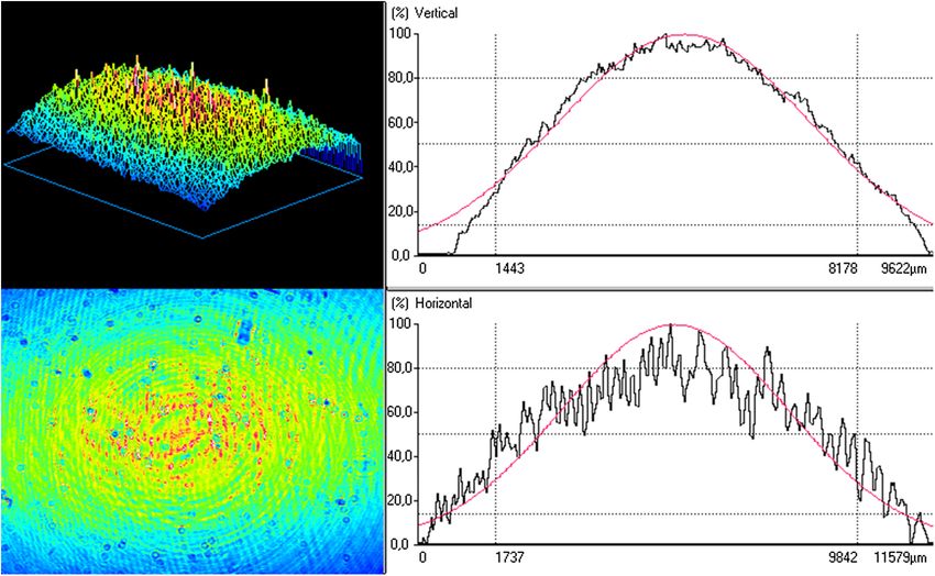
Mantineo, Pinheiro, and Morgado: Low-level laser therapy on skeletal muscle inflammation: evaluation. . .

                dose (energy per irradiated area), irradiance, and total radiant
                energy. The published studies concerning LLLT for muscle
                repair present a considerable variety of irradiation and treatment
                parameters and protocols. Laser wavelengths vary between
                632.8 and 904 nm. Therapeutic effects are reported25,26 for radi-
                ant exposures from 1 J∕cm2 to more than 300 J∕cm2 . Protocols
                show a large variation in the number of treatment days and of
                daily treatment sessions.
                    Another important feature concerns the modality of radiation
                delivery: continuous wave (CW) or pulsed wave (PW).The most
                used modality is CW irradiation, which has been used for the
                whole range of LLLT applications. There is some evidence
                that PW irradiation can be more effective than CW illumination
                with equivalent irradiation and dosimetry parameters, particu-
                larly for wound healing and poststroke management applica-
                tions.27 As far as we know, no study was published concerning
                PW irradiation for skeletal muscle repair.
                    Here, we report on the effect of different LLLT doses, using
                both CW (830 and 980 nm) and PW irradiations (830 nm), in
                the inflammation phase of skeletal muscle injury, induced by
                mechanical trauma in the gastrocnemius muscle of Wistar
                rats. LLLT treatment effect was evaluated measuring the con-
                centration of cytokines (TNF-α, IL-1β, IL-2, and IL-6) in sys-
                temic blood and through histological analysis of muscle tissue.
                Our goal was to evaluate treatment effectiveness with different
                irradiation parameters. First, we evaluated different energy
                doses for two distinct wavelengths with CW irradiation. Then,
                we compared CW and PW irradiations using the wavelength and                                Fig. 1 Drop-mass press device for inflammation induction.
                average power that produced the largest treatment effect on the
                first experiment. Although our experiments were not designed to
                validate any of the LLLT action mechanisms, we discuss our                         at 9:00 a.m. The average duration of the process was approxi-
                results in the framework of the currently most accepted                            mately 3 min per rat.
                LLLT mechanism, based on the role of CCO as a primary photo-
                acceptor for red-NIR radiation. Parts of this work were previ-
                ously presented at SPIE BiOS 2014.28                                               2.3       Laser Therapy
                                                                                                   Laser radiation was administered using a Thorlabs ITC4001
                2     Materials and Methods                                                        Laser Diode/TEC Controller, with a single HL8338 GaAlAs
                                                                                                   laser diode (830 nm) or L9805E2P5 GaAlAs laser diode
                2.1     Animals                                                                    (980 nm) (Thorlabs, Newton, New Jersey, USA). Laser diode
                We used 85 Rattus norvegiccus (Wistar strain) male adult rats,                     temperature was controlled.
                with an average body mass of 256.35  21.22 g and 8 weeks of                          The 830-nm wavelength was selected since it is a wavelength
                age. The rats were housed in collective standard cages (three                      located in the band of highest transmission through the combi-
                animals per cage), with a controlled room temperature of 21°C,                     nation of skin and muscle (770–850 nm),33 and is one of the two
                cage ventilation rates of about 75 air changes per hour and rel-                   most common wavelengths used in therapeutic light sources (the
                ative humidity of 70%. Bedding was replaced once a week.29,30                      other is 632.8 nm) and also is a peak of the CCO absorption
                All procedures were approved by the Commission of Ethics of                        spectrum, attributed to the relatively oxidized CuA chromo-
                the Faculty of Medicine of the University of Coimbra, which                        phores of CCO.34 It is important to remember that the absorption
                follows the Directive 2010/63/EU of the European Parliament                        spectrum of CCO is similar to the action spectra for biological
                and of the European Council.31                                                     responses to light, such as DNA and RNA synthesis, a fact
                                                                                                   that supports the role of CCO as a primary photoacceptor for
                                                                                                   red-NIR radiation in LLLT.
                2.2     Trauma Induction                                                              One of the reasons for selecting the 980-nm wavelength was
                A controlled and reproducible inflammation condition between                       the fact that it does not correspond to any CCO absorption band.
                different animals was created, using the model of muscle trauma                    At this wavelength, there is a large water absorption band, mak-
                proposed by Rizzi et al.32 Gastrocnemius muscle injury was                         ing 980-nm photons more likely to produce tissue heating than
                induced by single impact, with a press developed by Industrias                     photochemical effects. The fact that reported results for 980-nm-
                Mantineo, Mendoza, Argentina (Fig. 1), after shaving the ani-                      based LLLT are mixed also leads us to choose this wavelength.
                mal’s leg. Injury was done by a metal mass (0.300 kg) falling                      The literature presents conflicting results in wound healing,35,36
                through a guide from a 30-cm height. The impact energy was                         positive effects on neuropathic pain relief,37 and no effects on
                0.889 J. During the procedure, rats were anesthetized with                         traumatic brain injury.38 Published data on the effectiveness of
                a mixture of isoflurane (2.5%) and oxygen (97.5%) at a flow                        980 nm in the treatment of skeletal muscle inflammation are
                of 1.5 l∕ min. Inflammation was produced at day 0, starting                        very scarce, a fact that also prompted us to use this wavelength.

                Journal of Biomedical Optics                                              098002-2                                          September 2014   •   Vol. 19(9)

Downloaded From: https://www.spiedigitallibrary.org/journals/Journal-of-Biomedical-Optics on 15 Nov 2021
Terms of Use: https://www.spiedigitallibrary.org/terms-of-use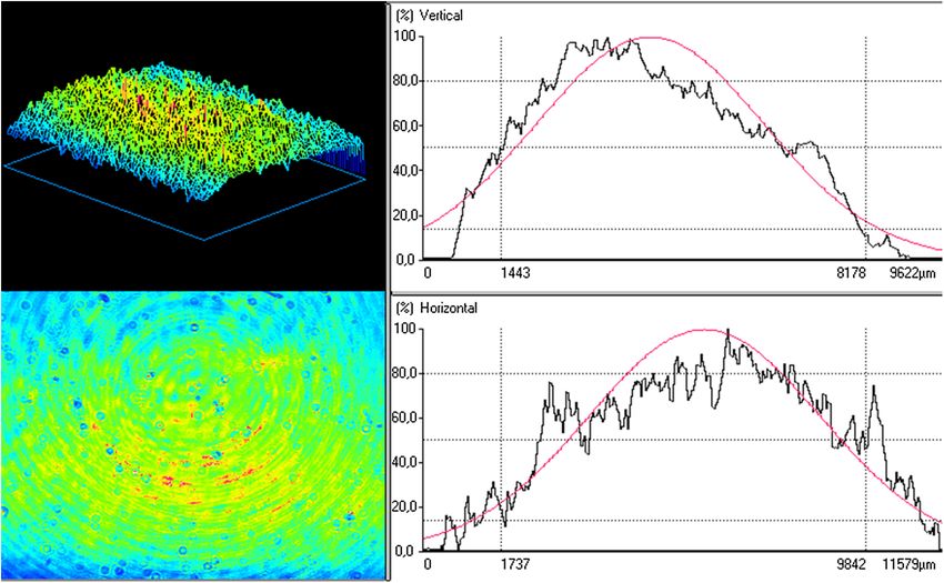
Mantineo, Pinheiro, and Morgado: Low-level laser therapy on skeletal muscle inflammation: evaluation. . .

                    The laser beam profile and shape were measured with a                          PW groups. Laser treatment was applied daily, under artificial
                BeamStar PCI-PAL 100345 beam profiler (Ophir, Jerusalem,                           light, at the same time of the day (9:00 a.m.) for 5 days (from
                Israel) at a plane corresponding to the animal leg. The 830-                       day 1 to day 5) directly to shaved skin by transcutaneous appli-
                nm beam presented an elliptical shape, with horizontal and ver-                    cation. Beam laser incidence was kept perpendicular to the irra-
                tical profiles whose correlation coefficients to Gaussian shape                    diation area. Only one spot was irradiated. During irradiation,
                were 78.5% and 90.7%, respectively. Profiles widths at 1∕e2                        animals were anesthetized with a mixture of isoflurane (2.5%)
                were 9.74  0.003 mm, for the horizontal profile, and                              and oxygen (97.5%) at a flow of 1.5 l∕ min. Control rats were
                8.31  0.001 mm, for the vertical profile, resulting in a beam                     also anesthetized to ensure standardization, but did not receive
                spot size at target of 0.80 cm2 . The 830-nm beam two-dimen-                       laser treatment.
                sional (2-D) and three-dimensional (3-D) shapes are shown in                          Regarding the used irradiation parameters, the total energy
                Fig. 2, along with its horizontal and vertical profiles.                           was selected according to the World Association for Laser
                    The 980-nm laser also presented an elliptical shape. The cor-                  Therapy (WALT) recommended treatment doses for LLLT,
                relation coefficients between the horizontal and vertical profiles                 which vary between 4 and 16 J. The radiant powers were
                and a Gaussian profile were 80.1% and 82.2%, respectively. The                     selected in order to obtain irradiances in the higher half of the
                measured profiles widths at 1∕e2 were 9.26  0.001 mm, for the                     common range of values used for stimulation and healing (5 to
                horizontal profile, and 7.42  0.001 mm, for the vertical profile,                 50 mW∕cm2 )39 and are similar to those reported for modulation
                resulting in a beam spot size at target of 0.69 cm2 . Figure 3                     of cytokines expression in skeletal muscle following acute
                presents the 2-D and 3-D shapes and horizontal and vertical pro-                   injury.10,32,40
                files for the 980-nm laser beam.                                                      For PW repetition rates, due to the lack of reference values
                    These measurements required the use of neutral density (ND)                    for the treatment of skeletal muscle inflammation, we choose to
                filters to prevent CCD saturation. Speckle is not evident in the                   use values similar to those reported for pain reduction, since
                                                                                                   pain relief seems to be obtained by the anti-inflammatory action
                actual treatment conditions. The large ring patterns visible in
                                                                                                   of LLLT.
                Figs. 2 and 3 probably result from interference effects due to
                                                                                                      It is important to stress that we performed the experiments
                reflections on the ND filters. The small circular patterns are
                                                                                                   sequentially. First, we did the CW treatments. The PW measure-
                most probably due to dust on the filters.
                                                                                                   ments were done afterward and using only the wavelength and
                    The animals were randomly divided into one control group
                                                                                                   average power that yielded the best results with CW irradiation.
                (n ¼ 10) and 15 treatment groups (n ¼ 5), defined by different
                                                                                                   This way we complied with the reduction principle on animal
                irradiation and treatment parameters. Ten groups concern CW                        experimentation, minimizing the number of animals used.
                irradiation (radiant powers between10 and 50 mW, at 10 mW
                steps, for both 830 and 980 nm), while the remaining five groups
                                                                                                   2.4     Blood Sampling
                underwent PW irradiation (Peak power: 50 mW, Average Power:
                40 mW, Duty cycle: 80%, frequencies: 5, 25, 50, 100, and                           Blood was collected on days 0 (5 h after inflammation induc-
                200 Hz).                                                                           tion), 3, and 6 (before animal sacrifice), always at 2:00 p.m.
                    LLLT was applied daily, perpendicular to the muscle’s sur-                     A blood volume of 1 ml was taken through the jugular vein.
                face. Exposure time was always constant and equal to 3 min.                        This is in accordance with the blood sample limit of 8 ml∕kg
                Average energy per application was 0 J (control group), 1.8,                       each 14 days.41,42 During blood collection, the animals were
                3.6, 5.4, 7.2, and 9.0 J, in the CW groups, and 7.2 J in the                       anesthetized in the conditions previously described.

                                       Fig. 2 Beam shape and profile for the 830-nm laser beam. The smooth line indicates a Gaussian profile.

                Journal of Biomedical Optics                                              098002-3                                      September 2014   •   Vol. 19(9)

Downloaded From: https://www.spiedigitallibrary.org/journals/Journal-of-Biomedical-Optics on 15 Nov 2021
Terms of Use: https://www.spiedigitallibrary.org/terms-of-use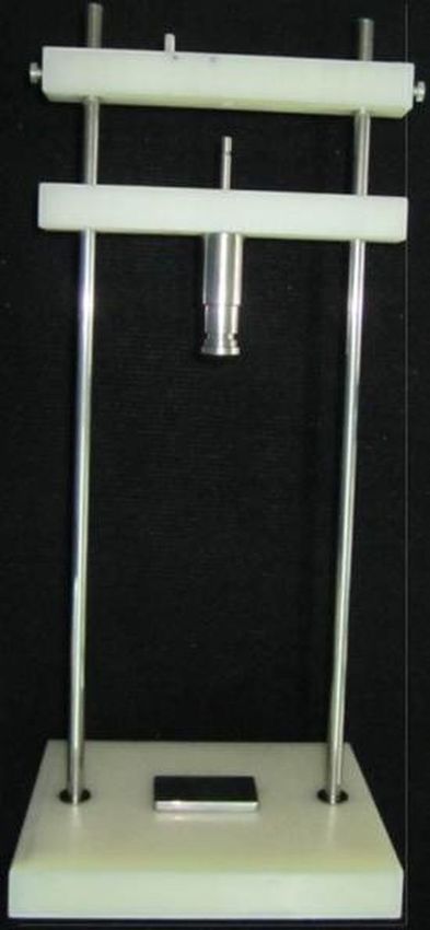
Mantineo, Pinheiro, and Morgado: Low-level laser therapy on skeletal muscle inflammation: evaluation. . .

                                       Fig. 3 Beam shape and profile for the 980-nm laser beam. The smooth line indicates a Gaussian profile.

                    All blood samples were placed in BD Vacutainer Plastic SST                     −80°C until analysis. The surgical procedure took less than
                II Advance tubes (BD, Franklin Lakes, New Jersey, USA) for                         15 min.
                subsequent centrifuging (15 min., 3500 rpm at 4°C). The serum                         5-μm thick cuts were made transversely to the muscular
                was removed and the samples were stored at −20°C. The                              fibers with a glass knife using a Leica Microsystems
                followed work schedule is summarized in Fig. 4.                                    CM3350S cryostat (Leica Microsystems, Wetzlar, Germany).
                                                                                                   After dewaxing and hydration, the samples were colored with
                2.5     ELISA Analysis                                                             hematoxylin-eosin and fixed with DPX mountant for micros-
                                                                                                   copy in order to observe the hematoma area and other visible
                ELISA serum analysis was done using Peprotech ELISA Kits                           changes.
                (PeproTech EC Ltd., London, United Kingdom) for quantifying                           The cross sections were observed with a Motic AE 31
                TNF-α, IL-1β, IL-2, and IL-6 cytokines. We used a BioTek                           inverted microscope (Motic Ltd. Hong-Kong, China), using
                Synergy HT microplate reader (BioTek Instruments, Inc.,                            10X, 20X, and 40X objectives. The muscles images were
                Winooski, Vermont, USA) at 405 nm, with a wavelength cor-                          captured using a high resolution camera Motic Moticam 2.
                rection set at 650 nm. The plate was monitored at 5-min intervals                  The most representative cuts were selected. Hematoma areas
                for 45 min.43 The samples’ concentrations were calculated by                       were identified by visual inspection.
                interpolation of the regression curve using the Gen 5 HT soft-                        The number of inflammatory cells was compared using the
                ware (BioTek Instruments, Inc., Winooski, Vermont, USA).                           images obtained with the 20X objective. Cells were counted
                    Comparisons between different cytokines concentrations and                     using an unbiased counting frame.45 Comparisons between dif-
                concentrations decrease were done using analysis of variance                       ferent rats were analyzed using ANOVA procedure, with post
                (ANOVA) with post hoc between-group comparisons by the                             hoc between-group comparisons through the Tukey test, with
                Tukey test.44 A significance level of 0.05 was considered in                       a significance level of 0.05. For each animal, 10 images were
                all cases.                                                                         used for inflammatory cell counting.

                2.6     Muscle Sample Preparation and Examination
                                                                                                   2.7     Monte Carlo Simulation of Light Transport in
                Rats were killed on day 6 for histological analysis of muscle.                             Tissue
                The animals were anesthetized before blood sampling and cer-
                vical dislocation.                                                                 The expected dose in muscle tissue was evaluated through
                   The gastrocnemius muscle was rapidly removed from the                           computer Monte Carlo (MC) simulation of light transport in
                injured leg, snap frozen in cryopreservation resin, and stored at                  a heterogeneous medium. MC simulations were done with the

                                                                           Fig. 4 Experimental work schedule.

                Journal of Biomedical Optics                                              098002-4                                   September 2014   •   Vol. 19(9)

Downloaded From: https://www.spiedigitallibrary.org/journals/Journal-of-Biomedical-Optics on 15 Nov 2021
Terms of Use: https://www.spiedigitallibrary.org/terms-of-use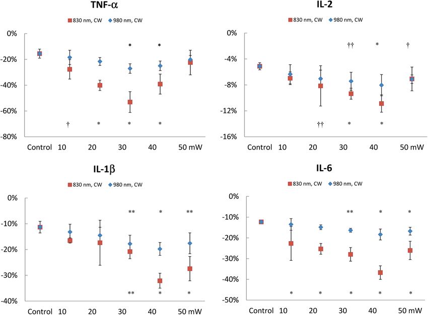
Mantineo, Pinheiro, and Morgado: Low-level laser therapy on skeletal muscle inflammation: evaluation. . .

                                        Table 1 Optical parameters for the tissue model used in Monte Carlo simulation of light transport.

                                                    μa (cm−1 )                                        μs (cm−1 )                                     g

                Layer                    830 nm                   980 nm                   830 nm                  980 nm              830 nm                 980 nm

                Skin                      0.17                     0.35                     74.49                  72.05                 0.82                   0.84

                Muscle                    1.15                     1.15                     91.82                  89.79                 0.88                   0.89

                mcxyz.c code developed and made available by Jacques et al.46                       decrease was significantly higher for almost all treated groups,
                We used a two-layer tissue model: skin (thickness: 2.1 mm) and                      the exception being the 50-mW group. This higher TNF-α
                muscle. The laser beam was modeled as a Gaussian beam with a                        decrease could be already observed at day 3 for the 20, 30,
                diameter of 8 mm at the 1∕e2 contour. The optical parameters                        and 40 mW groups. The highest variation between days 0
                were obtained from published research work47,48 and are sum-                        and 6 was found in the 30-mW group and was statistically
                marized in Table 1.                                                                 higher than those observed in the other treatment groups.
                                                                                                    The lower effectiveness of the 930-nm irradiation is revealed
                3      Results                                                                      by the lower number of treatment groups that achieved a sta-
                                                                                                    tistically higher relative TNF-α decrease than that measured
                3.1     Cytokines Measurement Through ELISA:
                                                                                                    for the control group. At day 6, LLLT was only effective for
                        Continuous-Wave Irradiation
                                                                                                    the 30 and 40-mW groups, with no statistically significant dif-
                Figure 5 shows the decrease in cytokines concentration at day 6,                    ference between them. When comparing between treated groups
                expressed as percentage of the concentration at day 0, for CW                       at 830 and 980 nm with the same radiant power and energy dose,
                irradiation at 830 and 980 nm. For each group, we tested                            we find that TNF-α concentration decrease is higher at 830 nm
                whether or not the measured cytokines relative concentration                        for the 20, 30, and 40 mW groups.
                decrease was different from those measured for the control                              IL-1β measurements at day 6 show higher relative concen-
                group. Significantly different concentration decreases are iden-                    tration decreases for the 30, 40, and 50 mW groups when com-
                tified. As Fig. 5 shows, the treatment effect was higher for irra-                  pared with controls for both irradiation wavelengths. This could
                diation at 830 nm. For this wavelength, the TNF-α concentration                     be already clearly observed at day 3 for the 40 and 50 mW

                                       Fig. 5 Cytokine concentration decrease for CW irradiation at 830 and 980 nm, at day 6. Values
                                       are expressed as percentage of the concentration at day 0. Error bars indicate standard deviations
                                       (SD ). Significant difference in relative cytokines concentration decrease for comparisons with control
                                       group: * p < 0.001; ** p < 0.008; † p < 0.05; †† p < 0.015.

                Journal of Biomedical Optics                                              098002-5                                      September 2014   •   Vol. 19(9)

Downloaded From: https://www.spiedigitallibrary.org/journals/Journal-of-Biomedical-Optics on 15 Nov 2021
Terms of Use: https://www.spiedigitallibrary.org/terms-of-use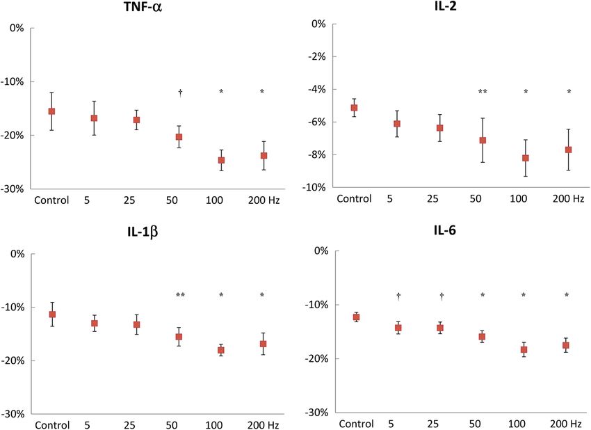
Mantineo, Pinheiro, and Morgado: Low-level laser therapy on skeletal muscle inflammation: evaluation. . .

                groups at 830 nm (p < 0.001), and for the 40-mW group at                           The highest concentration variation between days 0 and 6
                980 nm (p < 0.001). The highest variation between days 0                           occurred again at 40 mW for both wavelengths. For LLLT
                and 6 occurred in the 40-mW group for both wavelengths.                            with 830 nm, the IL-6 decrease at day 6 in the 40-mW
                Animals treated with the 830-nm laser show a statistically                         group is significantly higher than those obtained for the other
                higher IL-1β concentration decrease in the 40 mW groups                            treatment groups. This is also true at day 3, except when com-
                than those measured in the 10, 20, and 30 mW groups                                pared to the animals irradiated at 50 mW. For animals treated
                (p < 0.002). The concentration decrease was lower for animals                      with the 980-nm laser, differences between the 40 mW and
                treated at 980 nm. Comparison between groups treated at 830                        the other treatment groups were only found when compared
                and 980 nm with equal radiant power yielded a higher decrease                      to the 10 and 20 mW at day 6. The comparison between equiv-
                at 830 nm for the 40 (p ¼ 0.002) and 50 mW (p ¼ 0.016)                             alent LLLT groups at 830 and 980 nm produced significant
                groups.                                                                            differences for all compared groups, being highly significant
                    IL-2 measurements at day 6 show higher concentration                           (p < 0.001) for the 30 and 40 mW groups, For the 40 mW
                decreases for the 20, 30, and 40 mW groups at 830 nm, and for                      groups, this highly significant difference appears at day 3.
                the 30, 40, and 50 mW groups at 980 nm. A significant decrease
                at day 3 was observed only in the animals treated with the 830-
                                                                                                   3.2     Cytokines Measurement Through ELISA:
                nm laser at 40 mW (p ¼ 0.002, when compared with controls).
                                                                                                           Pulsed-Wave Irradiation
                Once again, the highest variation between days 0 and 6 occurred
                in the 40-mW group. Although this took place for both wave-                        Figure 6 shows the decrease in cytokines concentration at day 6,
                lengths, we only found differences when comparing the 10 and                       expressed as percentage of the concentration value at day 0 for
                the 50 mW groups and just for the animals treated with the 830-                    830-nm PW irradiation with constant average and peak powers
                nm laser. At 980 nm, no differences were found between treat-                      at different frequencies. Concentration decreases significantly
                ment groups at day 6. The comparison between LLLT groups                           different than those observed for the control group are identified.
                with the same radiant power at 830 and 980 nm yielded no                           As Fig. 6 shows, the treatment effect was higher for irradiation at
                differences.                                                                       frequencies higher than 50 Hz. The TNF-α concentration
                    The results at day 6 for IL-6 show higher concentration                        decrease was significantly higher for the 50, 100, and 200 Hz
                decreases for all treatment groups (p < 0.001) at 830 nm and                       groups. This higher TNF-α decrease could be already observed
                for the 30, 40, and 50 mW groups at 980 nm. At day 3, it was                       at day 3, mainly for the 100 Hz group (p ¼ 0.001) but also for
                already possible to observe significant decreases in the IL-6                      the 200 Hz group (p ¼ 0.010). The highest variation from days
                concentration in the 30, 40, and 50 mW groups treated with the                     0 to 6 was observed in the 100-Hz group. However, it was not
                830-nm laser. No significant IL-6 concentration decreases were                     statistically different from those observed for the 50 and 200 Hz
                observed at day 3 in the animals treated with the 980-nm laser.                    groups.

                                       Fig. 6 Cytokine concentration decrease for PW irradiation at 830 nm, at day 6. Values are expressed as
                                       percentage of the concentration at day 0. Error bars indicate standard deviations (SD ). Significant
                                       difference in relative cytokines concentration decrease for comparisons with control group: * p < 0.001;
                                       ** p < 0.008; † p < 0.05.

                Journal of Biomedical Optics                                              098002-6                                       September 2014   •   Vol. 19(9)

Downloaded From: https://www.spiedigitallibrary.org/journals/Journal-of-Biomedical-Optics on 15 Nov 2021
Terms of Use: https://www.spiedigitallibrary.org/terms-of-use
Mantineo, Pinheiro, and Morgado: Low-level laser therapy on skeletal muscle inflammation: evaluation. . .
Fig. 7 Comparison between cytokine concentration decrease for CW and PW irradiations at 830 nm at
day 6. Values are expressed as percentage of the concentration at day 0. Error bars indicate standard
deviations (SD ).
The IL-1β and IL-2 measurements show a similar behavior to muscle of control and CW treated animals. Figure 8 shows the
the TNF-α measurements. Significant differences were found at representative images of gastrocnemius muscle cuts. For each
day 6 for the 50, 100, and 200 Hz groups when compared to animal, the counting value is the average of measurements
controls. However, at day 3, differences were only found in done in 10 slides. The values presented are the average for all
IL-2 measurements for 100 and 200 Hz. The highest variation animals in a given group. Significant differences (p < 0.001)
between days 0 and 6 was measured for the 100 Hz but, again, were found between the control and all treatment groups for
with no statistically differences from the variations observed in both irradiation wavelengths. The lowest counts of inflamma-
the 50 and 200 Hz groups. tory cells were obtained at 40 mW. For the 830-nm laser,
The IL-6 cytokine concentration decrease at day 6 is signifi- there are differences between the cell counting at 40 mW and
cantly higher for all treatment groups when compared with the at 10, 20, and 30 mW. With the 980-nm laser, the differences
control group. This was already true at day 3 for the higher exist only when compared to the 10 and 20 mW groups.
frequencies groups (50, 100, and 200 Hz). The highest variation The images from animals irradiated with the 830-nm laser
between days 0 and 6 occurred again for the 100-Hz group. This present less inflammatory cells when compared with the muscle
time, the concentration decrease observed for 100 Hz is sta- cuts from animals treated at 980 nm. However, as Table 2 shows,
tistically different from the one measured for 50 Hz, but not the differences are not statistically significant for the level of
from the value obtained in the 200-Hz group. confidence used.
Figure 7 compares the decrease in cytokines concentration at
day 6 between PW irradiation at 50, 100, and 200 Hz and CW Table 2 Inflammatory cell counting in images of the gastrocnemius
irradiation at 40 mW. All values are for treatment at 830 nm. It is muscle of control and CW treated animals. Values are average SD.
easily seen that the cytokines relative concentration decrease
is larger for CW irradiation. The differences are statistically sig-
Group 830-nm laser 980-nm laser p (830 versus 980 nm)
nificant, with all but one p value smaller than 0.001 (p ¼ 0.005
for IL-2, CW versus 100 Hz). At day 3, there are already sta- Control 17.76 0.79 17.76 0.79
tistically significant differences between the PW group and the
CW irradiated animals for the variation of TNF-α, IL-1β, and 10 mW 14.30 1.59 14.88 1.59 0.97
IL-6 cytokines. For IL-2, no differences were found at day 3
20 mW 13.24 1.66 14.06 1.42 0.79
between the treatment groups.
30 mW 12.78 1.84 13.08 1.54 1.00
3.3 Inflammatory Cells Counting 40 mW 10.84 1.57 12.29 1.96 0.09
Table 2 shows the results from inflammatory cell counting on 50 mW 12.08 2.06 13.02 1.39 0.63
microscopy images of 5-μm thick cuts from the gastrocnemius
Journal of Biomedical Optics 098002-7 September 2014 • Vol. 19(9)
Downloaded From: https://www.spiedigitallibrary.org/journals/Journal-of-Biomedical-Optics on 15 Nov 2021
Terms of Use: https://www.spiedigitallibrary.org/terms-of-useMantineo, Pinheiro, and Morgado: Low-level laser therapy on skeletal muscle inflammation: evaluation. . .
Fig. 8 Images from gastrocnemius muscle cuts. Control rat: (a) 20×; (b) 40×; rat from 40-mW group:
(c) 20×; (d) 40×. In the control rat without treatment, it is possible to observe an infiltration of inflammatory
cells. The treated rat shows an improved condition, although still presents inflammatory cells.
Table 3 presents the inflammatory cell count for PW irradi- irradiation profiles between 830 and 980 nm. Therefore,
ation and CW 40 mW at 830 nm. Significant differences were differences of treatment effects between those wavelengths
found between the control group and each of the CW, 50, 100, are not due to irradiance differences in the target area.
and 200 Hz groups (p < 0.001). The number of inflammatory
cells for the CW is also significantly lower than for every PW 4 Discussions and Conclusions
irradiation group (p < 0.001). For PW irradiation, no significant Our objective was to evaluate the effect of different LLLT irra-
differences can be found among the 50, 100, and 200 Hz groups. diation parameters, namely radiant power, wavelength, and con-
tinuous versus pulsed illumination, on the inflammation phase
3.4 Simulation of Light Transport in Tissue of skeletal muscle injury. A quantitative evaluation of LLLT
effects was achieved by measuring the concentration of inflam-
Figure 9 shows the irradiance (W∕cm2 ∕W delivered) distribu- matory cytokines (TNF-α, IL 1β, IL-2, and IL-6) in the systemic
tion in the tissue model for irradiation at 830 and 980 nm. It also blood. Tumor necrosis factor ðTNFÞ-α and interleukin (IL)-1 are
includes the normalized irradiance as a function of tissue depth. two key cytokines, produced in response to trauma, that promote
The results show that there are no differences in the depth inflammatory responses, including the recruitment of immune
cells to the injured area. IL-6 is also a proinflammatory cytokine
Table 3 Inflammatory cell counting in images of the gastrocnemius that is responsible, with TNF-α and IL-1, for increasing the liver
muscle of control and 40 mW (average power), 830-nm treated synthesis of most acute-phase proteins. IL-2 has both pro- and
animals (CW and PW). Values are average SD. anti-inflammatory roles. It is a potent inducer of T-cell prolif-
eration, but also has regulatory roles, namely in the development
and function of regulatory T cells. Thus, IL-2 contributes both to
Group Cell counting
the induction and the end of acute inflammatory responses.
Control 17.76 0.79 Cytokines have been used to quantify LLLT effects in treat-
ing inflammation. Piva et al.25 reviewed the effect of LLLT on
CW 10.84 1.57 the initial stages of tissue repair, reporting several studies where
LLLT decreases the expression of TNF-α, IL-1β, and IL-6. In
5 Hz 16.62 1.61 what concerns skeletal muscle injury, one of the reviewed stud-
25 Hz 16.56 1.92 ies shows that TNF-α, IL-1β, and IL-6 mRNA expression is
decreased when using LLLT to treat inflammation of the sub-
50 Hz 15.64 1.74 plantar muscle of a rat paw.49 LLLT was also able to reduce
the TNF-α and IL-1β concentrations in rat tibialis anterior
100 Hz 14.66 1.25 muscle after cryolesion.9,10 Although in most LLLT studies,
200 Hz 15.18 1.14
cytokines concentration is measured in a muscle sample homog-
enate, we choose to measure the cytokines concentration in
Journal of Biomedical Optics 098002-8 September 2014 • Vol. 19(9)
Downloaded From: https://www.spiedigitallibrary.org/journals/Journal-of-Biomedical-Optics on 15 Nov 2021
Terms of Use: https://www.spiedigitallibrary.org/terms-of-useMantineo, Pinheiro, and Morgado: Low-level laser therapy on skeletal muscle inflammation: evaluation. . .
Fig. 9 Irradiance (W∕cm2 ∕W delivered) distribution in the tissue, for irradiation at 830 nm (a) and 980 nm
(b) and normalized irradiance profile as a function of tissue depth (c). In (a) and (b), the dashed top line
identifies the air-skin interface. The dashed bottom line corresponds to the skin–muscle interface. In
(c) the dashed line corresponds to the skin–muscle interface.
systemic blood serum. This allows sampling during treatment to lower muscle irradiance for that laser wavelength. Our MC
without sacrificing the animals. Moreover, this quantifica- simulations showed that the normalized irradiance at the muscle
tion method can be applied to human studies. Zhevago and is equal for 830 and 980 nm. In fact, although skin absorption is
Samoilova50 have previously shown in humans, that transcuta- higher for 980-nm, scattering in the skin is higher at 830 nm.
neous irradiation with visible and infrared light modulates cyto- The combination of the two processes seems to result in very
kines concentration on peripheral systemic blood, namely by similar profiles for the dependence of irradiance with tissue
decreasing the concentration of TNF-α and IL-6. depth.
Our results show treatment effects, particularly for irradiation The lower treatment effect at 980 nm seems to result from
with the 830-nm laser. At day 6, the concentration of all mea- specific absorption properties of the chromophores mediating
sured proinflammatory cytokines in the 30 and 40 mW groups LLLT effects. The probable photo acceptor in mammalian
was significantly lower than for the control group. IL-6 concen- cells for visible and near-infrared (NIR) light is CCO, the ter-
tration was reduced for all treatment groups and TNF-α for all minal electron acceptor of the mitochondrial electron transport
but the 50-mW group. The number of inflammatory cells in chain in eukaryotic cells.34 It is known that the action spectrum
muscle tissue samples was also significantly lower in all treat- of CCO has a peak at 825 nm, and is thought to be due to the
ment groups when compared to the control animals. relatively oxidized CuA chromophores.34 Specific extinction
The best results were obtained with a radiant power of spectrum of oxidized and reduced CCO from bovine heart tissue
40 mW at 830 nm. This was the only group of animals where shows larger extinction coefficients at 830 nm when compared
the concentration of all measured cytokines was already signifi- with values measured at 980 nm (1.7 times higher for oxidized
cantly lower at day 3 when compared to the control group. The CCO and 1.2 times higher for reduced CCO).54 This difference
lowest counts of inflammatory cells were also obtained in the may justify the larger treatment effect observed at 830 nm.
40-mW group. As Fig. 1 clearly shows, the treatment effects The irradiance values on the central region of the irradiated
decrease both for radiant powers below and above 40 mW. tissue volume raise the issue of whether thermal effects play a
This behavior may suggest a biphasic dose response.39,51 We role on the experiments. In fact, we planned our experiments
varied the delivered energy dose (J∕cm2 ) per application by considering a priori that thermal effects were not significant.
adjusting the laser power while keeping the irradiation time con- This was based on measurements in humans reported by
stant at 3 min. For the used radiant powers and the measured Joensen et al.,55 using a 810-nm laser with an output power
beam areas at skin (0.80 cm2 for 830 nm; 0.69 cm2 for of 200 mW, spot size of 0.0314 cm2 and power density of
980 nm), this amounts to a dose range between 2.25 and 6.37 W∕cm2 , values that produce local irradiances much higher
13.0 J∕cm2 , with the peak effect at 9 − 10 J∕cm2 . There are than those we used. The measurements showed small thermal
some published studies reporting biphasic responses for compa- effects in light skin (a condition closer to our experiments with
rable energy doses. In one study with macrophage cell lines irra- albino rats), with temperature increases ranging from 0.38°C to
diated at 820 nm, Bolton et al.52 observed cell proliferation from 1.58°C, for 9 J of delivered energy.
2.4 to 9.6 J∕cm2 , finding a maximum at 7.2 J∕cm2 . An animal The MC simulations of light propagation allow us to do a
study53 on mouse pleurisy induced by carrageenan treated with a simple evaluation of possible thermal effects by calculating the
650-nm laser at three dose values (3, 7.5, and 15 J∕cm2 ), found average irradiance in skin and muscle and the temperature
the largest inflammatory cell migration reduction at 7.5 J∕cm2 . increase in these tissues. Simulation data show that thermal
A final conclusion for the biphasic response behavior requires effects are only relevant in the central region of the beam,
additional measurements for doses greater than 13.0 J∕cm2 to taken as the region of the beam profile where intensity is higher
verify if the LLLT effect still decreases for those doses. than 80% of the peak intensity. The temperature increase in the
LLLT treatment was less effective at 980 nm. The light trans- muscle is not significant. The calculated value, without consid-
port in a two-layer tissue model was simulated to assess if ering the effects of thermal diffusion or blood convection, was
the lower effect observed with irradiation at 980 nm was due close to 1°C. The temperature increase is larger in the skin since
Journal of Biomedical Optics 098002-9 September 2014 • Vol. 19(9)
Downloaded From: https://www.spiedigitallibrary.org/journals/Journal-of-Biomedical-Optics on 15 Nov 2021
Terms of Use: https://www.spiedigitallibrary.org/terms-of-useMantineo, Pinheiro, and Morgado: Low-level laser therapy on skeletal muscle inflammation: evaluation. . .
it absorbs more light, and this is more pronounced for 980-nm several repetition rates (6, 18, 36, 100, and 600 Hz) through
irradiation. This further suggests that thermal effects are not melanin filters. The authors found that cell proliferation was
responsible for the observed treatment effects, which are increased in the groups treated with PW irradiation, with maxi-
more for pronounced for 830 nm. mal effects at 100 Hz, suggesting that penetration of PW light
For the animals treated with PW irradiation, cytokines reduc- through tissues with high melanin content depends on pulse fre-
tion was only significant for the higher frequencies (50, 100, and quency. However, this effect does not play a role in our experi-
200 Hz), although cytokines concentration decrease was much ments since we used albino Wistar rats. Multiple nitric oxide
lower than the one obtained for CW irradiation with the same photodissociation events from a protein binding site are another
radiant power. The reduction in inflammatory cells was also sig- mechanism proposed for explaining PW effects in LLLT.27
nificantly lower with PW irradiation than the one measured for Although this mechanism may play a significant role in irradi-
CW illumination. These results suggest that pulsed irradiation is ation with pulsed light in the red region, NIR wavelengths are
less effective than CW irradiation with the same average power absorbed by a part of the CCO not involved in NO binding,57
in the reduction of the inflammatory phase of skeletal muscle suggesting that photodissociation is not responsible for the pos-
injury. itive effects of PW for NIR irradiation.
There is published evidence that pulsed irradiation produces The observed larger effects of PW irradiation for 50, 100, and
different effects than CW irradiation. Hashmi et al. recently 200 Hz frequencies suggest the existence in this frequency range
reviewed the effects of pulsing in LLLT,27 reporting nine studies of some fundamental frequency in involved biological systems
comparing CW and PW irradiations, none of them on muscle or some process with a time scale of milliseconds. The most
inflammation. Of those, seven found beneficial effects from obvious time constant is the thermal relaxation time of blood
pulsed irradiation with only one study finding a higher treatment vessels, which again raises the question of the involvement of
effect with CW irradiation, although by a minimal margin. thermal effects.
Biological reasons are usually proposed for the increased effi- Considering the thermal relaxation time of an infinite cylin-
ciency of PW irradiation, namely modulation of ion channels der, we find that time constants between 5 and 20 ms can be
kinetics in the milliseconds time range or promotion of multiple associated with thermal relaxation of vessels with diameters
nitric oxide photodissociation events from a protein binding site.27 between 100 and 200 μm. These diameters are larger than
In our experiments, the duty cycle was 80%, resulting in a those found in dermis capillaries.58 We simulated the thermal
peak power 20% higher than the CW radiant power. Therefore, behavior of blood vessels for vessel diameters between 50
during pulse exposure, the irradiance at muscle tissue is 20% and 220 μm and considering the frequencies used in our PW
higher than during CW irradiation, although the radiant expo- measurements. For that purpose, we used the values of average
sure is kept equal. If we examine the existence of irradiance irradiance for skin and muscle obtained through our MC sim-
effects on LLLT, which are clearly suggested by the observed ulations and computed the thermal behavior following a meth-
biphasic dose responses that imply a lack of compliance to odology identical to that used by Stuart Nelson et al.59 For
the Bunsen–Roscoe rule of reciprocity,51 a direct comparison vessels located within the muscle tissue, the temperature
between CW and PW irradiations with the same average increase is always negligible (lower than 0.03°C). Significant
power may be partially hampered by such effects. The higher temperature effects may occur for skin blood vessels with diam-
irradiance stimulus occurring with PW irradiation may inhibit eters larger than 70 μm. However, these vessel diameters are
to some degree the LLLT action, resulting in a treatment effect rarely found in normal dermis. It is also important to note
lower than that observed for CW irradiation at 40 mW. This is that temperature effects are more significant for low frequencies,
supported by the results obtained with CW irradiation at 50 mW. which were the frequencies that resulted in lower treatment
It is relevant to note that our data yield mixed results when effects. For these reasons, it seems very unlikely that the fre-
we compare CW irradiation at 50 mW and PW irradiation at the quency dependence of the measurements is due to the thermal
higher frequencies. PW irradiation was significantly more effec- relaxation of blood vessels.
tive than CW irradiation for reducing TNF-α concentration, as Currently, the more accepted cellular level mechanism for
effective as CW irradiation for decreasing IL-2 concentration, LLLT is the absorption radiation by components of the cellular
and less effective than CW irradiation concerning IL-1β and respiratory chain. Therefore, we looked at this chain for proc-
IL-6. CW irradiation at 50 mW also resulted in a lower count esses with time constants in the range of milliseconds. Starting
of inflammatory cells than PW irradiation. A new set of PW from fully oxidized CCO, the electronic transfer rate from cyto-
measurements using a peak power of 40 mW could be useful chrome a to cytochrome a3 occurs in the millisecond time range,
to properly address the impact of irradiance effects. even with large reductant concentrations.60 Simulations done by
None of the published studies comparing CW and PW irra- Brunori et al.60 resulted in forward and reverse rate constants for
diations deal with skeletal muscle inflammation. Our data sug- the electronic transfer from cytochrome a to cytochrome a3
gest that CW irradiation is more effective in the treatment of equal to 25 and 125 s−1 , respectively. Karu34 suggests that irra-
the inflammation phase of skeletal muscle injury than PW irra- diation intensifies the cytochrome a to cytochrome a3 electron
diation with the same radiant exposure. However, irradiance transfer stage, since this is the rate-limiting step in the whole
effects may hinder this conclusion. Therefore, further investiga- electron transfer within the CCO, making more electrons avail-
tions are required. able for the reduction of dioxygen. Taking its rate constants, it is
PW irradiation only produced treatment effects for higher possible to suggest that PW irradiation with frequencies compa-
frequencies (50, 100, and 200 Hz). Once again, we could not rable to those rates will be more effective in intensifying the
find any study comparing pulse repetition rates in the treatment cytochrome a to cytochrome a3 electron transfer when com-
of muscle inflammation. An in vitro study,56 designed to evalu- pared with irradiation at lower frequencies.
ate if pulsed light can overcome the filtering effects of melanin In conclusion, we were able to quantify the effect of LLLT on
exposed human HEP-2 cells to 670-nm CW or PW light at the treatment of inflammation induced in the gastrocnemius
Journal of Biomedical Optics 098002-10 September 2014 • Vol. 19(9)
Downloaded From: https://www.spiedigitallibrary.org/journals/Journal-of-Biomedical-Optics on 15 Nov 2021
Terms of Use: https://www.spiedigitallibrary.org/terms-of-useMantineo, Pinheiro, and Morgado: Low-level laser therapy on skeletal muscle inflammation: evaluation. . .
muscle of Wistar rats by measuring the concentration of proin- 8. R. Albertini et al., “Effects of different protocol doses of low power
flammatory cytokines in the systemic blood, a method that gallium-aluminum-arsenate (Ga-Al-As) laser radiation (650 nm) on
carrageenan induced rat paw ooedema,” J. Photochem. Photobiol. B
allows following the treatment effect without sacrificing ani-
74(2–3), 101–107 (2004).
mals. The results showed that CW irradiation at 830 nm pro- 9. L. Assis et al., “Low-level laser therapy (808 nm) reduces inflammatory
duced the largest treatment effects, a result in accordance with response and oxidative stress in rat tibialis anterior muscle after cryo-
the action spectrum of CCO. Best results were obtained with an lesion,” Lasers Surg. Med. 44(9), 726–735 (2012).
irradiation power of 40 mW, with the data suggesting a biphasic 10. R. A. Mesquita-Ferrari et al., “Effects of low-level laser therapy on
dose response. This suggestion requires further confirmation expression of TNF-alpha and TGF-beta in skeletal muscle during the
repair process,” Lasers Med. Sci. 26(3), 335–340 (2011).
through experiments using higher radiant powers. PW irradia-
11. X. Gao and D. Xing, “Molecular mechanisms of cell proliferation
tion at 830 nm and 40-mW average power was only effective for induced by low power laser irradiation,” J. Biomed. Sci. 16(1), 4 (2009).
the tested frequencies equal to or higher than 50 Hz. This result 12. W. P. Hu et al., “Helium-neon laser irradiation stimulates cell prolifer-
might be related to the rate constants of the CCO internal elec- ation through photostimulatory effects in mitochondria,” J. Invest.
tron transfer stage between cytochrome a and cytochrome a3 . Dermatol. 127(8), 2048–2057 (2007).
LLLT have been used since 1960s to improve the healing of 13. P. C. Silveira et al., “Evaluation of mitochondrial respiratory chain activ-
different soft-tissue pathologies and to reduce the perception of ity in muscle healing by low-level laser therapy,” J. Photochem.
Photobiol. B 95(2), 89–92 (2009).
both nociceptive and neuropathic pain. Histological studies
14. H. Toumi and T. Best, “The inflammatory response: friend or enemy for
report increased microvascularization and a positive influence muscle injury?,” Br. J. Sports Med. 37, 284–286 (2003).
on fibroblast proliferation, collagen synthesis, and tissue regen- 15. R. van den Berg et al., “Transcription factor NF-kappaB as a potential
eration. In rehabilitation medicine, LLLT was introduced as biomarker for oxidative stress,” Br. J. Nutr. 86(Suppl 1), S121–S127
a noninvasive and safe treatment, but its efficacy is still contro- (2001).
versial because several clinical trials have reported its ineffec- 16. W. Lim et al., “The anti-inflammatory mechanism of 635 nm light-emit-
ting-diode irradiation compared with existing COX inhibitors,” Lasers
tiveness to treat pain and inflammation in musculoskeletal
Surg. Med. 39(7), 614–621 (2007).
disorders. 17. D. M. Iyomasa et al., “Ultrastructural analysis of the low level laser
Researchers and clinicians should consistently report the therapy effects on the lesioned anterior tibial muscle in the gerbil,”
characteristics of the device, the irradiation parameters, and Micron 40(4), 413–418 (2009).
the treatment procedures. If we are able to quantify the effect 18. A. C. Amaral, N. A. Parizotto, and T. F. Salvini, “Dose-dependency of
of LLLT on the relief of pain and inflammation using a rigorous low-energy HeNe laser effect in regeneration of skeletal muscle in
methodology, we can choose the best therapeutic window, mice,” Lasers Med. Sci. 16(1), 44–51 (2001).
19. J. Nakano et al., “Low-level laser irradiation promotes the recovery of
increasing the efficiency and credibility of this physical agent. atrophied gastrocnemius skeletal muscle in rats,” Exp. Physiol. 94(9),
This work is intended to be a contribution toward this goal. Its 1005–1015 (2009).
potential clinical impact result lies in the methodology used to 20. T. A. Butterfield, T. M. Best, and M. A. Merrick, “The dual roles of
quantify inflammation relief and on the identification of the best neutrophils and macrophages in inflammation: a critical balance
irradiation and treatment parameters for achieving that relief. between tissue damage and repair,” J. Athl. Train. 41(4), 457–465 (2006).
21. Y. Li et al., “Transforming growth factor-beta1 induces the differentia-
tion of myogenic cells into fibrotic cells in injured skeletal muscle: a key
event in muscle fibrogenesis,” Am. J. Pathol. 164(3), 1007–1019
Acknowledgments
(2004).
This study was supported in part by the Fundação para a Ciência 22. M. D. Cressoni et al., “The effects of a 785-nm AlGaInP laser on the
e a Tecnologia (FCT) under program COMPETE FCOMP/01- regeneration of rat anterior tibialis muscle after surgically-induced
0124-FEDER-022709 and by Erasmus Mundus EADIC injury,” Photomed. Laser Surg. 26(5), 461–466 (2008).
Scholarships. Thanks to “Industrias Mantineo,” Mendoza, 23. L. Assis et al., “Low-level laser therapy (808 nm) contributes to muscle
regeneration and prevents fibrosis in rat tibialis anterior muscle after
Argentina, for building the inflammation induction equipment. cryolesion,” Lasers Med. Sci. 28(3), 947–955 (2013).
We also thank the help of Mr. António Correia, MSc with the 24. T. O. de Souza et al., “Phototherapy with low-level laser affects the
Monte Carlo simulations. remodeling of types I and III collagen in skeletal muscle repair,”
Lasers Med. Sci. 26(6), 803–14 (2011).
25. J. A. Piva et al., “Effect of low-level laser therapy on the initial stages of
References tissue repair: basic principles,” An. Bras. Dermatol. 86(5), 947–954
(2011).
1. J. D. Carroll, “Photomedicine and LLLT literature watch,” Photomed.
26. L. Ramos et al., “Infrared (810 nm) low-level laser therapy in experi-
Laser Surg. 27(4), 689–90 (2009).
mental model of strain-induced skeletal muscle injury in rats: effects on
2. C. Antipa et al., “Our clinical experience in low-energy laser medical
functional outcomes,” Photochem. Photobiol. 88(1), 154–160 (2012).
treatments,” Opt. Eng. 35(5), 1367–1371 (1996).
27. J. T. Hashmi et al., “Effect of pulsing in low-level light therapy,” Lasers
3. R. A. B. Lopes-Martins et al., “Low level laser therapy [LLLT] in
Surg. Med. 42(6), 450–466 (2010).
inflammatory and rheumatic diseases: a review of therapeutic mecha-
28. M. Mantineo, J. P. Pinheiro, and A. M. Morgado, “Evaluation of low
nisms,” Curr. Rheumatol. Rev. 3(2), 147–154 (2007).
level laser therapy irradiation parameters on rat muscle inflammation
4. K. Nomura, M. Yamaguchi, and Y. Abiko, “Inhibition of interleukin-1
through systemic blood cytokines,” Proc. SPIE 8932, 89320M (2014).
beta production and gene expression in human gingival fibroblasts
by low-energy laser irradiation,” Lasers Med. Sci. 16(3), 218–223 29. W. O. P. Heine, Environmental Management in Laboratory Animal
(2001). Units: Basic Technology and Hygiene Methods and Practice,
5. H. Chung et al., “The nuts and bolts of low-level laser (light) therapy,” PABST Science Publishers, Berlin (1998).
Ann. Biomed. Eng. 40(2), 516–533 (2012). 30. M. F. W. Festing et al., The Design of Animal Experiments: Reducing
6. A. C. M. Rennó et al., “Comparative effects of low-intensity pulsed the Use of Animals in Research Through Better Experimental Design,
ultrasound and low-level laser therapy on injured skeletal muscle,” Laboratory Animals Ltd., Ed., pp. 79–81, Royal Society of Medicine
Photomed. Laser Surg. 29(1), 5–10 (2011). Press Ltd., London (2011).
7. G. Shefer et al., “Low-energy laser irradiation promotes the survival and 31. European Commission, “Directive 2010/63/EU of the European
cell cycle entry of skeletal muscle satellite cells,” J. Cell Sci. 115(Pt 7), Parliament and of the Council of 22 September 2010 on the protection
1461–1469 (2002). of animals used for scientific purposes,” http://eur-lex.europa.eu/
Journal of Biomedical Optics 098002-11 September 2014 • Vol. 19(9)
Downloaded From: https://www.spiedigitallibrary.org/journals/Journal-of-Biomedical-Optics on 15 Nov 2021
Terms of Use: https://www.spiedigitallibrary.org/terms-of-useMantineo, Pinheiro, and Morgado: Low-level laser therapy on skeletal muscle inflammation: evaluation. . .
LexUriServ/LexUriServ.do?uri=OJ:L:2010:276:0033:0079:EN:PDF 50. N. A. Zhevago and K. A. Samoilova, “Pro- and anti-inflammatory cyto-
(2010). kine content in human peripheral blood after its transcutaneous (in vivo)
32. C. F. Rizzi et al., “Effects of low-level laser therapy (LLLT) on the and direct (in vitro) irradiation with polychromatic visible and infrared
nuclear factor (NF)-kappaB signaling pathway in traumatized muscle,” light,” Photomed. Laser Surg. 24(2), 129–139 (2006).
Lasers Surg. Med. 38(7), 704–713 (2006). 51. Y.-Y. Huang et al., “Biphasic dose response in low level light therapy,”
33. K. R. Byrnes et al., “Light promotes regeneration and functional recov- Dose-Response 7(4), 358–383 (2009).
ery and alters the immune response after spinal cord injury,” Lasers 52. P. Bolton, S. Young, and M. Dyson, “Macrophage responsiveness to
Surg. Med. 36(3),171–185 (2005). light therapy: a dose response study,” Laser Ther. 2(3), 101–106 (1990).
34. T. I. Karu, “Multiple roles of cytochrome c oxidase in mammalian cells 53. R. A. Lopes-Martins et al., “Spontaneous effects of low-level laser
under action of red and IR-A radiation,” IUBMB Life, 62(8), 607–610 therapy (650 nm) in acute inflammatory mouse pleurisy induced by
(2010). carrageenan,” Photomed. Laser Surg. 23(4), 377–381 (2005).
35. A. Gupta, T. Dai, and M. R. Hamblin, “Effect of red and near-infrared 54. P. R. Rich, A. J. Moody, and W. J. Ingledew, “Detection of a near infra-
wavelengths on low-level laser (light) therapy-induced healing of red absorption band of ferrohaem a3 in cytochrome c oxidase,” FEBS
partial-thickness dermal abrasion in mice,” Lasers Med. Sci. 29(1), Lett. 305(3), 171–173 (1992).
257–265 (2014). 55. J. Joensen et al., “The thermal effects of therapeutic lasers with 810 and
36. F. A. H. Al-Watban, X. Y. Zhang, and B. L. Andres “Low-level laser 904 nm wavelengths on human skin,” Photomed. Laser Surg. 29(3),
therapy enhances wound healing in diabetic rats: a comparison of differ- 145–153 (2011).
ent lasers,” Photomed. Laser Surg. 25(2), 72–77 (2007). 56. P. Brondon, I. Stadler, and R. J. Lanzafame, “Pulsing influences photo-
37. M. Masoumipoor et al., “Effects of 660- and 980-nm low-level laser radiation outcomes in cell culture,” Lasers Surg. Med. 41(3), 222–226
therapy on neuropathic pain relief following chronic constriction injury (2009).
in rat sciatic nerve,” Lasers Med. Sci. (2014) [Epub ahead of print]. 57. N. Lane, “Cell biology: power games,” Nature 443(7114), 901–903
38. W. Qiuhe et al., “Low level laser therapy for traumatic brain injury,” (2006).
Proc. SPIE 7552, 755206 (2010). 58. R. Archid et al., “Confocal laser-scanning microscopy of capillaries in
39. Y.-Y. Huang et al., “Biphasic dose response in low level light therapy— normal and psoriatic skin,” J. Biomed. Opt. 17(10), 101511. (2012).
an update,” Dose-Response 9(4), 602–618 (2011). 59. J. S. Nelson et al., “Laser pulse duration must match the estimated ther-
40. K. P. Fernandes et al., “Effect of photobiomodulation on expression mal relaxation time for successful photothermolysis of blood vessels,”
of IL-1beta in skeletal muscle following acute injury,” Lasers Med. Lasers Med. Sci. 10(1), 9–12 (1995).
Sci. 28(3), 1043–1046 (2013). 60. M. Brunori et al., “Internal electron transfer in Cu-heme oxidases:
41. S. Parasuraman, R. Raveendran, and R. Kesavan, “Blood sample thermodynamic or kinetic control?” J. Biol. Chem. 272(32), 19870–
collection in small laboratory animals,” J. Pharmacol. Pharmacother. 19874 (1997).
1(2), 87–93 (2010).
42. M. F. Toft et al., “The impact of different blood sampling methods on
Matías Mantineo is a PhD student in biomedical engineering at
laboratory rats under different types of anaesthesia,” Lab. Anim. 40(3), the University of Coimbra, Portugal. He received his MD degree in
261–274 (2006).. bioengineering from the University of Mendoza, Argentina, in 2009.
43. PeproTech Inc., “General Sandwich ELISA protocol,” https://www His current research interests include Laser–tissue interactions, clini-
.peprotech.com/Lists/PTProtocol/Attachments/19/Sandwich%20ELISA cal laser applications, photodynamic therapy, low-level laser therapy,
%20-%20web.pdf (2012). and medical technology. He is a member of SPIE.
44. J. H. Zar, Biostatistical Analysis, Prentice-Hall International Inc.,
Englewood Cliff, New Jersey (1984). João P. Pinheiro is professor of medicine in the Faculty of Medicine
45. H. J. Gundersen et al., “Some new, simple and efficient stereological of the University of Coimbra, specialist in physical and rehabilita-
methods and their use in pathological research and diagnosis,” tion medicine and sports medicine and Head of the Physical and
APMIS 96(1–6), 379–394 (1988). Rehabilitation Medicine Department in University Hospital of Coimbra.
46. S. Jacques, T. Li, and S. Prahl, “mcxyz.c, a 3D Monte Carlo simulation He is a member of the European Academy of Physical and
of heterogeneous tissues,” http://omlc.ogi.edu/software/mc/mcxyz/ Rehabilitation Medicine.
index.html (2013).
47. W. F. Cheong, S. A. Prahl, and A. J. Welch, “A review of the optical- António M. Morgado received his PhD degree in physics and is
properties of biological issues,” IEEE J. Quantum Electron. 26(12), assistant professor in the Department of Physics of the University
2166–2185 (1990). of Coimbra. He is a researcher in IBILI–Institute for Biomedical
Imaging and Life Sciences where in works mainly on the development
48. L. Oliveira et al., “The optical properties of rat abdominal wall muscle,”
of optoelectronic instrumentation and techniques for ocular imaging,
Presented at Workshop on Internet Biophotonics VI, Report 2, Saratov
namely corneal metabolic imaging using fluorescence lifetime micros-
Fall Meeting 2013, Saratov, Russia, http://sfm.eventry.org/report/713 copy, corneal confocal microscopy, and retinal OCT imaging. His
(25–28 September 2013). research interests also include low-level laser therapy and laser
49. R. Albertini et al., “Cytokine mRNA expression is decreased in the sub- safety. He is a member of SPIE.
plantar muscle of rat paw subjected to carrageenan-induced inflamma-
tion after low-level laser therapy,” Photomed. Laser Surg. 26(1), 19–24
(2008).
Journal of Biomedical Optics 098002-12 September 2014 • Vol. 19(9)
Downloaded From: https://www.spiedigitallibrary.org/journals/Journal-of-Biomedical-Optics on 15 Nov 2021
Terms of Use: https://www.spiedigitallibrary.org/terms-of-useYou can also read