LncRNA H19 regulates smooth muscle cell functions and participates in the development of aortic dissection through sponging miR-193b-3p
←
→
Page content transcription
If your browser does not render page correctly, please read the page content below
Bioscience Reports (2021) 41 BSR20202298
https://doi.org/10.1042/BSR20202298

Research Article

LncRNA H19 regulates smooth muscle cell functions
and participates in the development of aortic
dissection through sponging miR-193b-3p
Mingming Ren1 , Tao Wang1 , Xiaolong Wei2 , Yizeng Wang1 , Chun Ouyang1 , Yilian Xie3 , Xiaoqiang Ye1 and
  Zhen Han1

                                                                                                                                                                                           Downloaded from http://portlandpress.com/bioscirep/article-pdf/41/1/BSR20202298/902631/bsr-2020-2298.pdf by guest on 09 October 2021
1 Department of Cardiovascular Surgery, Peking University Shenzhen Hospital, 1120 Lianhua Road, Shenzhen City, Guangdong Province, PR China; 2 Department of Vascular
Surgery, Shanghai Changhai Hospital, Shanghai, PR China; 3 Department of Surgery, School of Medicine, Shantou University, Shantou, PR China
Correspondence: Zhen Han (hanzhen0431@sohu.com)

                                           Background: Multiple studies showed that long-chain noncoding RNA H19 (LncRNA H19)
                                           is high-expressed in human and mouse abdominal aortic aneurysms (AAAs). We specu-
                                           lated that it plays an important role in arterial disease, and therefore studied the role and
                                           mechanism of H19 in aortic dissection (AD).
                                           Methods: The expressions of related genes in human aortic smooth muscle cells
                                           (HASMCs) induced by platelet-derived growth factor BB (PDGF-BB) or in the aortic tis-
                                           sue of AD patients/mice were identified by Western blot and quantitative real-time poly-
                                           merase chain reaction. The targeting relationship between H19 and miR-193b-3p was pre-
                                           dicted and verified by bioinformatics analysis, dual luciferase assay, RNA pull-down as-
                                           say, RNA immunoprecipitation (RIP), and Pearson correlation coefficient. The H19 and
                                           miR-193b-3p effects on the biological functions of tissues and cells were examined
                                           by MTT (3-(4,5-dimethyl-2-thiazolyl)-2,5-diphenyl-2-H-tetrazolium bromide, thiazolyl blue
                                           tetrazolium bromide) assay, wound-healing assay, and Hematoxylin–Eosin (HE) staining.
                                           Results: LncRNA H19 was abnormally high-expressed in thoracic aorta tissues of AD
                                           patients, and it could competitively bind to and inhibit miR-193b-3p. In the PDGF-BB
                                           group, the expressions of H19, matrix metallopeptidase (MMP) 2 (MMP-2) and MMP-9
                                           were up-regulated and the expressions of miR-193b-3p, α-SMA, and SM22α were
                                           down-regulated; moreover, the proliferation and migration rate of HASMCs were increased.
                                           However, H19 silencing reversed the regulation of PDGF-BB on HASMCs. More interest-
                                           ingly, miR-193b-3p inhibitor could partially reverse the effect of H19 silencing. In addition,
                                           the above results were verified by animal experiments, showing that shH19 and up-regulated
                                           miR-193b-3p could significantly reduce the thoracic aorta pathological damage in AD mice.
                                           Conclusion: LncRNA H19 regulated smooth muscle cell function by sponging miR-193b-3p
                                           and it participated in the development of AD.

Received: 14 July 2020
                                         Introduction
Revised: 03 December 2020                Aortic rupture/dissection (AD) is caused by an intima-media tear in the aorta under the impact of high
Accepted: 23 December 2020               velocity and pressure blood flow, forming a false or true lumen [1]. AD has a high mortality rate and a
                                         poor prognosis, and is a clinically urgent problem to be solved [2]. Thoracic AD (TAD) is a type of AD,
Accepted Manuscript online:
06 January 2021                          according to pathological morphology [2]. Recent studies found that TAD is a comprehensive patholog-
Version of Record published:             ical change process caused by pathological changes involving multiple blood vessel constituents, such as
22 January 2021                          human aortic smooth muscle cells (HASMCs) and extracellular matrix (ECM) [3,4].

© 2021 The Author(s). This is an open access article published by Portland Press Limited on behalf of the Biochemical Society and distributed under the Creative Commons Attribution   1
License 4.0 (CC BY).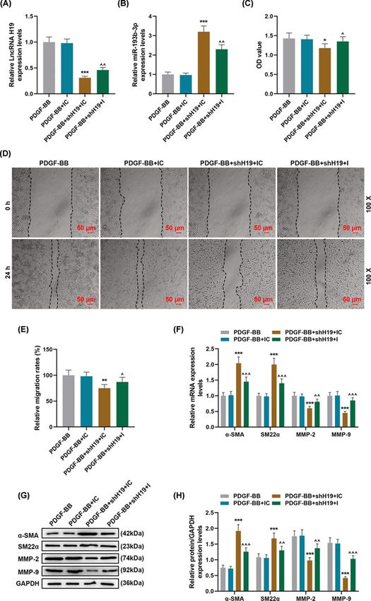
Bioscience Reports (2021) 41 BSR20202298
                                                                                                                                         https://doi.org/10.1042/BSR20202298

              In the pathogenesis of TAD, vascular smooth muscle cells (VSMCs) play an important role in the process of aortic
           wall contraction and synthesis in the presence of the stimulation of various cells to promote vascular remodeling [5].
           With the phenotypic change, VSMCs transform from contraction (differentiation) phenotype to synthesis (dediffer-
           entiation) phenotype [6,7]. Dedifferentiated VSMCs showed higher viability in terms of proliferation, migration and
           synthesis, and at the same time, the expressions of differentiation markers α-SMA and SM22α were down-regulated
           [6]. In addition, ECM is the main component that forms the morphology of the aortic blood vessel wall, and in the
           vascular wall tissues of TAD patients, ECM shows obvious abnormalities [8]. Although a large number of studies
           demonstrated that some factors such as matrix metalloproteinases (MMPs) directly participate in the degradation of
           the ECM of aorta [9], but the interaction of these factors and their upstream regulatory factors are still not clear.
              Abnormal expressions or functions of long-chain noncoding RNAs (lncRNAs) are closely related to many dis-
           eases such as cardiovascular diseases, cancers, and neurodegenerative diseases [10–12]. Study found that lncRNA
           H19 (H19) may play a mediating role between c-Myc and downstream gene expressions in colon cancer [13], and is

                                                                                                                                                                                           Downloaded from http://portlandpress.com/bioscirep/article-pdf/41/1/BSR20202298/902631/bsr-2020-2298.pdf by guest on 09 October 2021
           up-regulated in liver cancer [14], bladder cancer [15] and breast cancer [16], suggesting that H19 may be related to
           the occurrence of cancer. Moreover, H19 also plays an important role in the network structure of multiple gene ex-
           pressions in the human body [17]. For example, Wang et al. found that BRG1 interacts with lncRNA HIF1α-AS1, and
           that both may play an important role in the pathogenesis of TAD by regulating MMP-2/-9 expression level, apoptosis
           of VSMCs and phenotype conversion [18]. But this may not be the only mechanism for the development of TAD. The
           expression of H19 is significantly higher in the abdominal aorta samples of mouse abdominal aortic aneurysm (AAA)
           model than in normal aortic tissues [19,20]. Such a finding indicated that H19 may have a certain correlation with
           the AD process. LncRNAs, as an miRNA host transcript, regulate mRNA stability, and participate in intracellular life
           processes [21].
              However, it is unclear whether the specific molecular mechanism of H19 in AD was consistent with the previ-
           ously reported pathway. Therefore, the present study combined previous research to discuss the role and molecular
           mechanism of H19 in AD through clinical experiments, cell experiments, animal experiments, and other aspects.

           Materials and methods
           Tissues, cells, and infection
           Thoracic aortic tissue of 25 patients with AD were collected during the operation, and 15 normal aortic samples were
           collected from age-sex matched patients undergoing valve replacement (August 2019 to December 2019).
              HASMCs (PCS-100-012) were purchased from ATCC (U.S.A.). HASMCs were divided into the following four
           groups for research: Control, platelet-derived growth factor BB (PDGF-BB), PDGF-BB + shNC, and PDGF-BB +
           shH19. In the latter three groups, PDGF-BB (20 ng/ml) was used to treat HASMCs for 12 h, based on previous re-
           ports [22,23]. The cells were cultured in a 37◦ C and 5% CO2 incubator in DMEM/F-12 medium (11320033, Gibco,
           U.S.A.). The shNC lentiviral and shH19 lentiviral (pLKO.1, Target Sequence: CCCGTCCCTTCTGAATTTAAT) were
           constructed and packaged by Geneseed Biotech Co., Ltd. (CA). The cells were plated in 24-well plates at a density
           of 1 × 10⁵ /well. When the degree of cell fusion reached 70–80%, the fresh culture medium containing 10 μg/ml
           polybrene was used to replace the original medium, and the cells were added with appropriate amount of virus sus-
           pension (shNC or shH19) and incubated at 37◦ C. To verify the effect of miR-193b-3p on the shH19 in ameliorating
           PDGF-BB-induced HASMCs, we divided HASMCs into the following groups: PDGF-BB, PDGF-BB + miR-193b-3p
           inhibitor control (IC), PDGF-BB+shH19 + IC, PDGF-BB+shH19+inhibitor (I). IC (miR2N0000001-1-5) and I
           (miR-193b-3p) were purchased from Ribobio (China).

           AD animal model construction
           As previously described [24], Apolipoprotein E-deficient male mice (6–8 weeks old, Shanghai SLAC Company, CA)
           were used to construct AD animal models. All animals were randomly divided into the following four groups (n=12):
           Sham, AD, AD + shNC, AD + shH19. The mice were housed in specific SPF animal houses (12-h light/12-h dark)
           in Peking University Shenzhen Hospital. During 28 days of model construction, the mice were anesthetized with 3%
           isoflurane (Y0000858, Sigma–Aldrich, Germany). A subcutaneous osmotic micropump containing Ang II solution
           was implanted into the back of the mice in prone position through a small incision and closed with sutures (the flow
           rate was 1000 ng/kg/min). The mice in the AD + shNC and AD + shH19 groups received tail vein injection of lentivirus
           (1 × 10¹¹ pfu/mouse) carrying shNC and shH19, followed by subcutaneous osmotic micropump implantation. Sham
           mice received an equal volume of saline injection. AD + agomiRNA Control and AD + agomiR-193b-3p groups
           mice (n=12) were injected with 10 mg/kg agomiR-193b-3p or agomiRNA Control via tail vein injection once a week
           (for 3 weeks), and other operations were the same as above. At the end of the model construction, after anesthesia

2   © 2021 The Author(s). This is an open access article published by Portland Press Limited on behalf of the Biochemical Society and distributed under the Creative Commons Attribution
    License 4.0 (CC BY).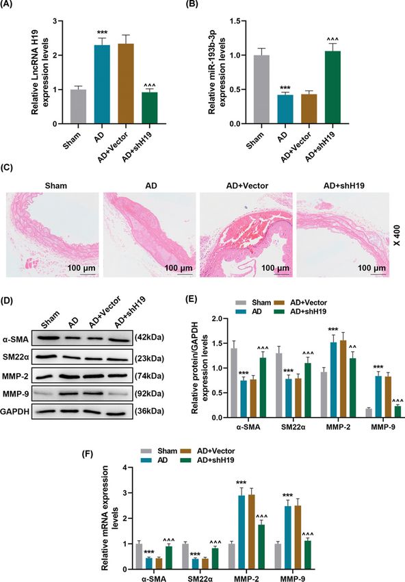
Bioscience Reports (2021) 41 BSR20202298
https://doi.org/10.1042/BSR20202298

Table 1 All primer in the present study
  ID                                                             Forward sequence (5 –3 )                                      Reverse sequence (5 –3 )

  H19                                                            ATGAAAGGTGAGGGGCTTCC                                            CCTTCCAGAGCCGATTCCTG
  GAPDH                                                          GGAGCGAGATCCCTCCAAAAT                                           GGCTGTTGTCATACTTCTCATGG
  H19-m                                                          GAACAGAAGCATTCTAGGCTGG                                          TTCTAAGTGAATTACGGTGGGTG
  GAPDH-M                                                        TGGCCTTCCGTGTTCCTAC                                             GAGTTGCTGTTGAAGTCGCA
  U6                                                             CTCGCTTCGGCAGCACA                                               AACGCTTCACGAATTTGCGT
  miR-193b-3p                                                    AAAGTCCCGCTGTCGTATCC                                            GTATCCAGTGCGTGTCGTGG
  α-SMA                                                          AAAAGACAGCTACGTGGGTGA                                           GCCATGTTCTATCGGGTACTTC
  α-SMA-M                                                        CCCAGACATCAGGGAGTAATGG                                          TCTATCGGATACTTCAGCGTCA
  SM22α                                                          GAAACCCACCCTCTCAGTCAG                                           TTGGCCATGTCTGGGGAAAG
  SM22α-M                                                        CCAACAAGGGTCCATCCTACG                                           ATCTGGGCGGCCTACATCA

                                                                                                                                                                                           Downloaded from http://portlandpress.com/bioscirep/article-pdf/41/1/BSR20202298/902631/bsr-2020-2298.pdf by guest on 09 October 2021
  MMP2                                                           GCATCCAGACTTCCTCAGGC                                            CCATTAGCGCCTCCATCGTAG
  MMP2-M                                                         ACCTGAACACTTTCTATGGCTG                                          CTTCCGCATGGTCTCGATG
  MMP9                                                           CGACGTCTTCCAGTACCGAG                                            TTGTATCCGGCAAACTGGCT
  MMP9-M                                                         GGACCCGAAGCGGACATTG                                             CGTCGTCGAAATGGGCATCT

(Pentobarbital Sodium, 85 mg/kg, 57-33-0, Sigma, U.S.A., intraperitoneal injection), the mice were killed by cervical
dislocation, the thoracic aorta was excised, and histochemical staining and expression analysis were performed.

Real-time quantitative PCR
Total RNA was extracted using TRIzol reagent (15596018, Invitrogen, U.S.A.). Cytoplasmic & Nuclear RNA Purifi-
cation Kit (NGB-21000, NorgenBiotek, Canada) was performed to extract nuclear and cytoplasmic RNA. To obtain
the required cDNA, PrimeScript RT Master Reagent (RR047A, Takara, CA) was used here. The quantitative reverse
transcription real-time polymerase chain reaction (qRT-PCR) was detected by qRT-PCR instrument (QuantStudio 5,
ABI, U.S.A.) and Real-time fluorescence quantitative PCR TB Green kit (RR820A). The qRT-PCR conditions were as
follows (40 cycles): pre-denaturation at 95◦ C for 10 min, denaturation at 95◦ C for 15 s, annealing at 60◦ C for 1 min.
The U6 and GAPDH were served as controls. The results of the experiment were quantitatively analyzed using the
2−Ct method [25]. The primers were displayed in Table 1.

Bioinformatics analysis
The GEO database (https://www.ncbi.nlm.nih.gov/geo/) (GSE92427) was used to analyze the expression of re-
lated miRNA in the aortic dissection of AD patients and healthy controls. Identification of differentially expressed
(DE)-miRNA was performed by Limmar package (Fold Change = −2.16, P=0.01597). starBase v2.0 software was
used to predict the potential targeted binding sites of H19 and miR-193b-3p.

Dual-luciferase reporter assay
Wild-type H19 sequence fragments (5 -UGGGGCCUGAGGCCAGU-3 ) and mutant-type H19 sequence fragments
(5 -UUAGGUCACGCUAUCUA-3 ) were inserted into the pmirGLO vector (E1330, Promega, CA, U.S.A.) to form
H19-WT and H19-MUT plasmids, respectively. HEK293T cells (CRL-11268, ATCC, U.S.A.) were simultaneously
transfected with miR-193b-3p mimic and H19-WT or H19-MUT. After 48 h of plasmids transfection of each group,
the medium was discarded and the dual-luciferase reaction intensity was measured in strict accordance with the kit
instructions (FR201-01, TransGen Biotech, CA).

RNA pull-down assay
According to the instructions of the RNA pull-down assay kit (11685597910, Pierce, Rockford, U.S.A.), the biotiny-
lated RNA and the structure buffer were mixed at a ratio of 1:500. Afterward, the magnetic beads (95◦ C, 2 min; ice
bath, 3 min) were resuspended. The magnetic bead–RNA complex was washed three times with 500 μl of washing
solution, and added with cell lysate (10 μl). Subsequently, RNA pull-down washing solution (500 μl) was added, and
cell lysate (10 μl) was added. After measuring the protein concentration using the BCA method, Western blot analysis
was performed to determine protein expressions.

© 2021 The Author(s). This is an open access article published by Portland Press Limited on behalf of the Biochemical Society and distributed under the Creative Commons Attribution   3
License 4.0 (CC BY).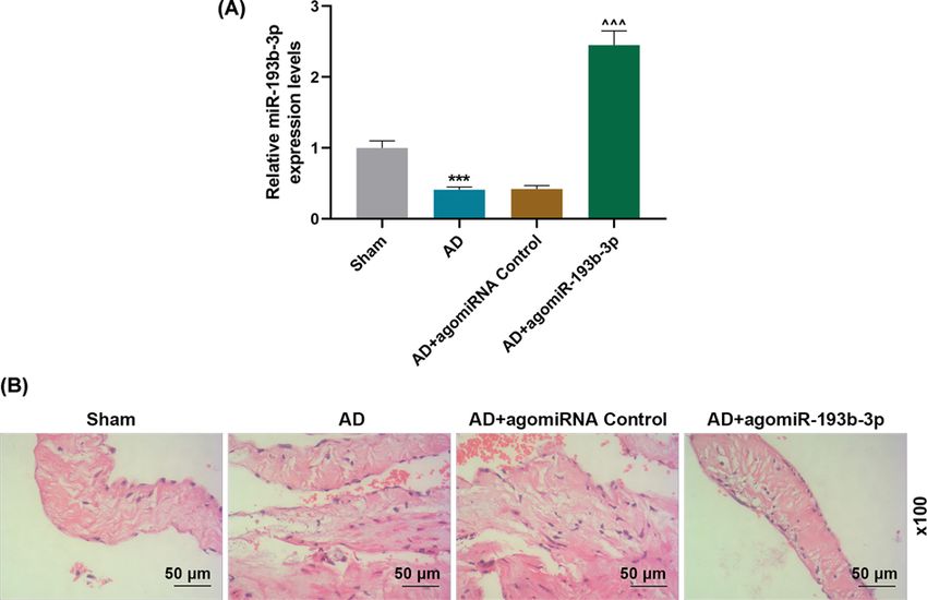
Bioscience Reports (2021) 41 BSR20202298
                                                                                                                                         https://doi.org/10.1042/BSR20202298

           RNA immunoprecipitation
           The binding of H19 to argonaute 2 (AGO2) proteins was performed according to the instructions of the RNA im-
           munoprecipitation (RIP) kit (RIP-12RXN, Merck Millipore, U.S.A.). Briefly, cell extracts were incubated with anti-
           bodies and magnetic beads at 4◦ C overnight. The magnetic bead antibody complex was resuspended in RIP-wash
           buffer (900 μl). To collect the magnetic bead binding protein complex, the sample was placed on a magnetic base.
           After separation by proteinase K, RNA was extracted from the sample and part of the cell extract sample, and then
           detected by PCR. RIP antibodies were AGO2 (ab32381, 1:50, Abcam, U.S.A.) and immunoglobulin G (IgG) (1: 100,
           ab109489, Abcam, U.S.A.).

           Detection of cell viability
           The trypsin EDTA solution (0.25%, 25200-056, GIBCO, U.S.A.) was added to each group of HASMCs to

                                                                                                                                                                                           Downloaded from http://portlandpress.com/bioscirep/article-pdf/41/1/BSR20202298/902631/bsr-2020-2298.pdf by guest on 09 October 2021
           prepare a cell suspension (1 × 10⁴ /ml). The cell suspension (100 μl/well) was added to a 96-well cul-
           ture plate, followed by routine incubation for 24, 48, and 72 h, respectively. Ten microliters of MTT
           (3-(4,5-dimethyl-2-thiazolyl)-2,5-diphenyl-2-H-tetrazolium bromide, thiazolyl blue tetrazolium bromide) reagent
           (CT02, Sigma–Aldrich, Germany) was added to the culture well to culture the cells for 4 h. After removing the MTT
           supernatant, 100 μl/well DMSO (ST038, Beyotime, U.S.A.) was added to the culture wells and shaken at low speed
           for 10 min. A microplate reader (1681130, Bio-Rad, U.S.A.) was used to measure the absorbance at 570 nm.

           Wound-healing assay
           The concentration of HASMCs suspension was adjusted to 1 × 10⁵ cells/ml. The cell suspension of each group was
           routinely incubated for 24 h to achieve a monolayer cell fusion degree of 70–80%. A sterile pipette tip was used to draw
           a ‘1’ on the cell membrane. The culture medium was gently washed twice to remove the detached cells. Subsequently,
           fresh medium was added to each well and cultivation continued for 24 h. Microscope (BZ-8100, Keyence, Japan) and
           Image-Pro Plus 4.1 analysis software (Media Controlnetics Company, U.S.A.) were used for observation and image
           analysis.

           Western blotting assay
           The expression of α-SMA, SM22α, MMP-2, MMP-9 in the HASMCs and aortic tissues were detected by western blot
           [26]. The RIPA method (P0013, Beyotime, CA) was used to extract the total protein from each group of HASMCs
           and aortic tissues, and the BCA method was used to determine the protein content. The protein was separated by
           SDS/PAGE and then transferred to the PVDF membrane (Immobilon-P Transfer Membrane, EMD Millipore Cor-
           poration, MA). The membrane was sealed in a box with 5% skimmed milk at room temperature for 2 h. Then the
           primary antibodies (α-SMA (ab5694, 42 kDa, 1 μg/ml, Abcam, U.K.), SM22α (ab14106, 23 kDa, 1 μg/ml), MMP-2
           (ab92536, 74 kDa, 1/1000), MMP-9 (ab38898, 92 kDa, 1/1000), and GAPDH (ab181602, 36 kDa, 1/10000)) were
           added for overnight incubation in a refrigerator at 4◦ C. After washing three times with TBST, the corresponding
           secondary antibodies such as anti-Mouse IgG (1:5000, ab205719) or anti-Rabbit IgG (1:5000, ab205718) were added
           and incubated for 1 h. The chemiluminescence kit (SL1350: 100 ml, Coolaber, China) was performed for exposure.
           The results were displayed by relative expression of protein (gray value of target protein/gray value of GAPDH), and
           analyzed with ImageJ analysis software (version 5.0, Bio-Rad, U.S.A.).

           Hematoxylin–Eosin staining
           Mouse thoracic aorta tissues were fixed with 4% paraformaldehyde (P0099, Beyotime, CA) and embedded into paraf-
           fin. The sections were routinely deparaffinized, and the staining experiment was performed according to the instruc-
           tions of the Hematoxylin–Eosin (HE) kit (C0105, Beyotime, CA). After the slices were dehydrated, made transparent,
           and sealed, the histopathological changes of the thoracic aorta in each group were observed and photographed under
           the CKX53 microscope (Olympus, Japan).

           Statistical analysis
           The correlation between H19 and miR-193b-3p expression was analyzed by Pearson correlation coefficient. Each
           experiment was repeated three times independently. The data were expressed as mean +
                                                                                              − SD, followed by analysis with
           SPSS v21.0 software. Comparisons between two groups were performed by t test. Multiple groups were expressed by
           one-way ANOVA for comparison. P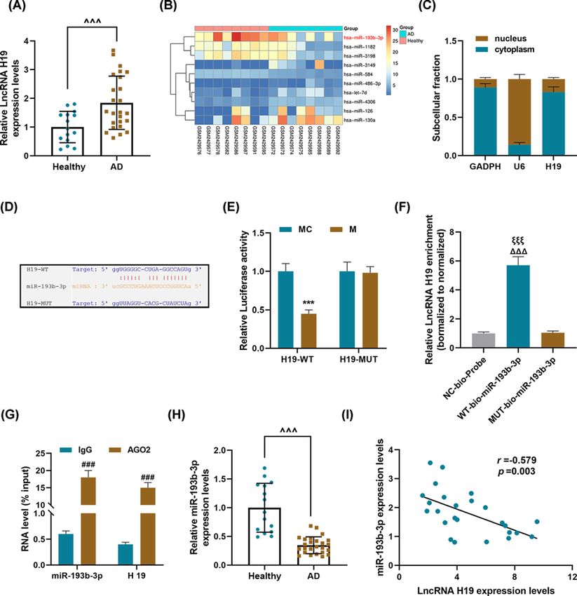
Bioscience Reports (2021) 41 BSR20202298
https://doi.org/10.1042/BSR20202298

Results
LncRNA H19 was abnormally high-expressed in thoracic aorta tissues of
AD patients, and could bind and inhibit miR-193b-3p
We determined the expression of H19 in the aorta samples of AD patients and healthy controls, and found that the
expression of H19 in the AD group was significantly higher than that in the healthy group (P
Bioscience Reports (2021) 41 BSR20202298
                                                                                                                                         https://doi.org/10.1042/BSR20202298

                                                                                                                                                                                           Downloaded from http://portlandpress.com/bioscirep/article-pdf/41/1/BSR20202298/902631/bsr-2020-2298.pdf by guest on 09 October 2021
           Figure 1. LncRNA H19 was abnormally high-expressed in thoracic aorta tissues of AD patients, and could competitively
           bind and inhibit miR-193b-3p
           (A) qRT-PCR was used to detect the expression of lncRNA H19 in the aorta of AD patients (n=25) and healthy aorta
           samples (n=15). GAPDH served as a control. Each sample was set to three replicate wells. (B) The GEO database
           (https://www.ncbi.nlm.nih.gov/geo/) was used to analyze related miRNA expression in AD. (C) The location of H19 in the HASMCs.
           GAPDH and U6 were used as internal reference genes. (D) The targeted binding site of lncRNA H19 and miR-193b-3p was predicted
           using starBase. (E) Dual-luciferase assay, (F) RNA pull-down assay, and (G) RIP experiments were used to verify the targeted binding
           relationship between lncRNA H19 and miR-193b-3p. (H) The expression level of miR-193b-3p in the sample tissues was detected
           by qRT-PCR. U6 serves as an internal reference gene. Each sample was set to six replicate wells. (I) The correlation between the
           expressions of lncRNA H19 and miR-193b-3p was analyzed using Pearson’s correlation coefficient. Each experiment was repeated
           three times independently at least, and the results were expressed as the means +    ˆˆ
                                                                                           − SD. P
Bioscience Reports (2021) 41 BSR20202298
https://doi.org/10.1042/BSR20202298
Downloaded from http://portlandpress.com/bioscirep/article-pdf/41/1/BSR20202298/902631/bsr-2020-2298.pdf by guest on 09 October 2021
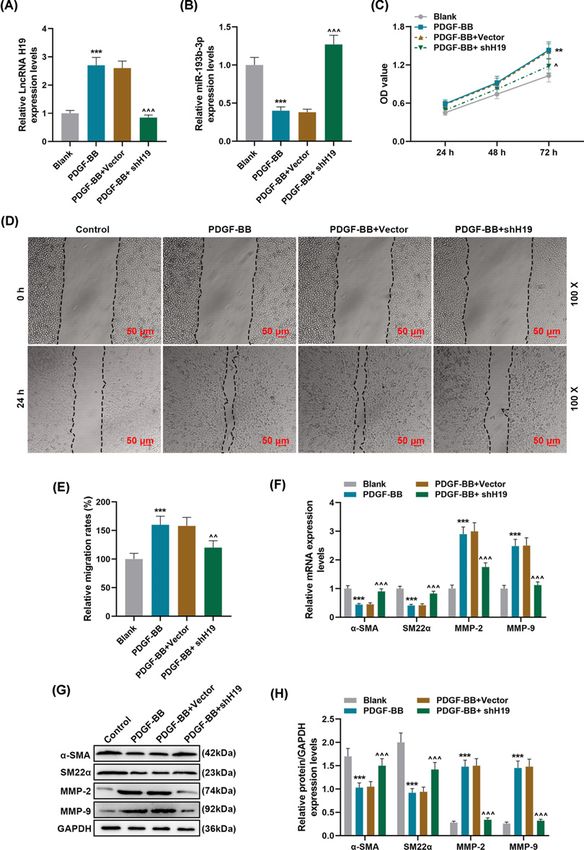
Figure 2. Effects of silencing lncRNA H19 on PDGF-BB-induced phenotypic differentiation and migration of HASMCs
(A,B) The expression levels of lncRNA H19 and miR-193b-3p in Blank, PDGF-BB, PDGF-BB + Vector, PDGF-BB + shH19 groups
were detected by qRT-PCR. GAPDH and U6 were used as internal reference genes. (C) After 24-, 48, and 72-h incubation of cells
in each group, cell viability was detected by MTT method. (D,E) The cell migration of each group was detected by wound-healing
assay. (F–H) qRT-PCR and Western blot were used to detect the levels of phenotypic differentiation markers α-SMA and SM22α,
and the expressions of MMP-2 and MMP-9. GAPDH served as a control. Each experiment was repeated three times independently,
and the results were the means + ˆˆ ˆˆ
− SD. **PBioscience Reports (2021) 41 BSR20202298
https://doi.org/10.1042/BSR20202298
Downloaded from http://portlandpress.com/bioscirep/article-pdf/41/1/BSR20202298/902631/bsr-2020-2298.pdf by guest on 09 October 2021
Figure 3. The effect of silencing miR-193b-3p on shH19 inhibiting PDGF-BB-induced dedifferentiation and migration of
HASMCs
(A,B) The expressions of lncRNA H19 and miR-193b-3p in PDGF-BB, PDGF-BB + IC, PDGF-BB + shH19 + IC, PDGF-BB + shH19
+ I groups were detected by qRT-PCR. GAPDH and U6 were used as internal reference genes. (C) After 72 h of incubation, the
cell viability was detected by MTT method. (D,E) The cell migration of each group was detected by wound-healing assay. (F–H)
qRT-PCR and Western blot were used to detect the levels of phenotypic differentiation markers α-SMA and SM22α, as well as
the expressions of MMP-2 and MMP-9. GAPDH served as a control. Each experiment was repeated three times independently,
and the results were expressed by the means +− SD. I: miR-193b-3p inhibitor. *PBioscience Reports (2021) 41 BSR20202298
https://doi.org/10.1042/BSR20202298
Downloaded from http://portlandpress.com/bioscirep/article-pdf/41/1/BSR20202298/902631/bsr-2020-2298.pdf by guest on 09 October 2021
Figure 4. Effect of lncRNA H19 silencing on AD animal models
(A,B) The expression levels of lncRNA H19 and miR-193b-3p in Sham, AD, AD + Vector, AD + shH19 groups were detected by
qRT-PCR (n=6). GAPDH and U6 were used as internal reference genes. (C) The histopathological changes of each group were
evaluated by HE staining (n=6). (D–F) qRT-PCR and Western blot were used to detect the levels of phenotypic differentiation markers
α-SMA and SM22α and the expression of MMP-2 and MMP-9 in each group. GAPDH served as a control. Each experiment was
repeated three times independently at least, and the results were the means +
− SD. qRT-PCR: quantitative reverse transcription real
time polymerase chain reaction. ***PBioscience Reports (2021) 41 BSR20202298
https://doi.org/10.1042/BSR20202298
Downloaded from http://portlandpress.com/bioscirep/article-pdf/41/1/BSR20202298/902631/bsr-2020-2298.pdf by guest on 09 October 2021
Figure 5. Effect of miR-193b-3p agomir on AD animal models
(A) The expression levels of miR-193b-3p in the Sham, AD, AD + agomiRNA Control, and AD + agomiR-193b-3p groups were
detected by qRT-PCR (n=6). U6 were used as internal reference genes. (B) The histopathological changes in each group were
evaluated by HE staining (n=6). ***PBioscience Reports (2021) 41 BSR20202298
https://doi.org/10.1042/BSR20202298
atherosclerosis and increased risk of coronary heart disease [28]. A large number of studies confirmed that there
is a mutual regulation relationship between lncRNAs and miRNAs that exert their physiological and pathological
functions in various diseases through different regulatory forms [21,29,30]. It has been reported that lncRNA H19
promotes hepatocellular carcinoma metastasis and invasion by triggering and activating the miR-193b/MAPK1 axis
[31]. Li et al. analyzed the development and progression of non-coding RNA in thoracic and AAA diseases, and
pointed out the lack of study on the role of lncRNA in AAA [32]. The role of H19 in AAA has been previously men-
tioned. For example, H19 plays a pathogenic role in the formation of AAA through let-7a/IL-6 inflammation pathway
[20]. In view of the complexity of the molecular regulatory network in diseases, this may not be the only regulatory
pathway of H19. Our research associated H19 with miR-193b-3p for the first time. The current results showed that
H19 competitively bound to and inhibited miR-193b-3p during AD development, suggesting that H19 acts as an ad-
sorbent sponge for miR-193b-3p. Mechanistically, H19 can capture the target sequence of miR-193b-3p and isolate
miR-193b-3p from its target mRNA, thereby regulating the function of related physiological pathology in AD.
Downloaded from http://portlandpress.com/bioscirep/article-pdf/41/1/BSR20202298/902631/bsr-2020-2298.pdf by guest on 09 October 2021
PDGF-BB co-expression has been found to regulate VEGF-induced abnormal angiogenesis by regulating VEGF-R2
signaling and endothelial cell proliferation [22,23]. Our study also applied PDGF-BB co-expression to induce prolif-
eration and migration and phenotypic dedifferentiation of HASMCs, and found that silencing H19 can reverse the
effect of PDGF-BB on HASMCs. The current research results suggest that H19 was abnormally expressed in various
malignant tumors and had proto-carcinogenic activity [33,34]. However, H19 has also been found to have cancer
suppressing activity, which can inhibit the malignant proliferation, invasion, and metastasis of tumors, and the for-
mation of tumor neovascularization [35,36]. The above studies indicate that the function of H19 may be different
in different diseases. According to the literature, H19-derived miR-675 directly targets PTEN to stimulate VSMC
proliferation, similarly, VSMC apoptosis is induced by H19 via HIF1α [37]. The present study found that silencing
H19 through miR-193b-3p can inhibit PDGF-BB-induced cell proliferation and migration, and promote HASMCs
phenotypic differentiation.
The main pathological change of AD is the tearing of the middle layer of blood vessels, and VSMCs are the main
cellular components of the middle layer of the aorta [3,4]. Animal experiments showed that AD mice had obvious
vascular media degeneration, muscle fiber assembly disorder, VSMCs became large and round. These changes are
consistent with the pathological changes of AD. More importantly, H19 silencing and miR-193b-3p agomir can reduce
the pathological damage of thoracic aorta in AD mice. α-SMA and SM22α are specific marker proteins of aortic
smooth muscle cells, and are also commonly used to identify the contractile phenotype of VSMCs [23,38]. MMPs can
degrade many components in ECM and basement membrane, and can destroy the connective tissues of the arterial
wall [23]. Among them, MMP-2 and MMP-9 are the most closely related to occurrence and development with AD
[39]. Increased viability of MMP-2 can cause arteries to dilate, and increased vitality of MMP-9 can cause rupture
of aortic aneurysms [40]. Consistent with previous studies [18], in this study, both in vivo and in vitro experiments
proved that H19 silencing further promoted up-regulation of α-SMA and SM22α, and down-regulated MMP-2 and
MMP-9 expressions, suggesting that H19 may participate in the pathogenesis of AD by regulating the expressions of
MMP-2/-9 and the proliferation, migration, and phenotype transformation of HASMCs.
In summary, our research indicated that H19, as an miR-193b-3p sponge, regulates smooth muscle cell function
and participates in AD vascular remodeling. Importantly, for the first time, we discovered that H19/miR-193b-3p is
a new pathogenic pathway in AD. This provides a direction for further research on the role of H19 in AD and new
theoretical support for the pathological mechanism of AD.
Data Availability
The analyzed datasets generated during the study are available from the corresponding author on reasonable request.
Competing Interests
The authors declare that there are no competing interests associated with the manuscript.
Funding
This work was supported by the Shenzhen Scientific Innovation Committee [grant number JCYJ20170412150405310].
Author Contribution
Substantial contributions to conception and design: M.m.R. Data acquisition, data analysis and interpretation: T.W., X.l.W., Y.z.W.,
C.O., Y.l.X., X.q.Y., Z.H. Drafting the article or critically revising it for important intellectual content: M.m.R. Final approval of the
version to be published: all authors. Agreement to be accountable for all aspects of the work in ensuring that questions related to
the accuracy or integrity of the work are appropriately investigated and resolved: all authors.
© 2021 The Author(s). This is an open access article published by Portland Press Limited on behalf of the Biochemical Society and distributed under the Creative Commons 11
Attribution License 4.0 (CC BY).Bioscience Reports (2021) 41 BSR20202298
https://doi.org/10.1042/BSR20202298
Ethics Approval
All the animal experiments were performed following Guide for the Care and Use of Laboratory Animals, and had obtained the
approval of the Institutional Animal Care and Use Committees of Peking University Shenzhen Hospital (DX201907031). Clinical
experiments were approved by the Ethics Committee of Peking University Shenzhen Hospital (XXW201906008). Patients and their
families signed and agreed to donate aortic tissue samples.
Abbreviations
AAA, abdominal aortic aneurysm; AD, aortic dissection; AGO2, argonaute 2; α-SMA, alpha-smooth muscle actin; DMEM/F12,
Dulbecco’s Modified Eagle Medium /nutrient mixture F12; ECM, extracellular matrix; GAPDH, glyceraldehyde-3 phosphate de-
hydrogenase; GEO, Gene Expression Omnibu Altered long noncoding RNA expression profiles in the myocardium of rats with
ischemic heart failure.; HASMC, human aortic smooth muscle cell; HE, Hematoxylin–Eosin; HIF1α-AS1, Hypoxiainducible
factor1α antisense RNA 1; IC, inhibitor control; IgG, immunoglobulin G; lncRNA, long-chain noncoding RNA; MMP, matrix met-
Downloaded from http://portlandpress.com/bioscirep/article-pdf/41/1/BSR20202298/902631/bsr-2020-2298.pdf by guest on 09 October 2021
allopeptidase/metalloproteinase; MTT, 3-(4,5-dimethyl-2-thiazolyl)-2,5-diphenyl-2-H-tetrazolium bromide, thiazolyl blue tetra-
zolium bromide; PDGF-BB, platelet-derived growth factor BB; qRT-PCR, quantitative reverse transcription real-time polymerase
chain reaction; RIP , RNA immunoprecipitation; SM22α, Smooth muscle22 alpha; SPF, Specific pathogen free; TAD , thoracic
AD; VEGF, vascular endothelial growth factor; VSMC, vascular smooth muscle cell.
References
1 Silaschi, M., Byrne, J. and Wendler, O. (2017) Aortic dissection: medical, interventional and surgical management. Heart 103, 78–87,
https://doi.org/10.1136/heartjnl-2015-308284
2 Larson, H. (2017) Aortic dissection. Radiol. Technol. 89, 193–195
3 Milewicz, D.M., Trybus, K.M., Guo, D.C., Sweeney, H.L., Regalado, E., Kamm, K. et al. (2017) Altered smooth muscle cell force generation as a driver of
thoracic aortic aneurysms and dissections. Arterioscler. Thromb. Vasc. Biol. 37, 26–34, https://doi.org/10.1161/ATVBAHA.116.303229
4 Wang, L., Zhang, J., Fu, W., Guo, D., Jiang, J. and Wang, Y. (2012) Association of smooth muscle cell phenotypes with extracellular matrix disorders in
thoracic aortic dissection. J. Vasc. Surg. 56, 1698–1709, 709e1, https://doi.org/10.1016/j.jvs.2012.05.084
5 Wei, X., Sun, Y., Wu, Y., Zhu, J., Gao, B., Yan, H. et al. (2017) Downregulation of Talin-1 expression associates with increased proliferation and migration
of vascular smooth muscle cells in aortic dissection. BMC Cardiovasc. Disord. 17, 162, https://doi.org/10.1186/s12872-017-0588-0
6 An, Z., Liu, Y., Song, Z.G., Tang, H., Yuan, Y. and Xu, Z.Y. (2017) Mechanisms of aortic dissection smooth muscle cell phenotype switch. J. Thorac.
Cardiovasc. Surg. 154, 1511e6–1521e6, https://doi.org/10.1016/j.jtcvs.2017.05.066
7 An, Z., Qiao, F., Lu, Q., Ma, Y., Liu, Y., Lu, F. et al. (2017) Interleukin-6 downregulated vascular smooth muscle cell contractile proteins via
ATG4B-mediated autophagy in thoracic aortic dissection. Heart Vessels 32, 1523–1535, https://doi.org/10.1007/s00380-017-1054-8
8 Jia, L.X., Zhang, W.M., Zhang, H.J., Li, T.T., Wang, Y.L., Qin, Y.W. et al. (2015) Mechanical stretch-induced endoplasmic reticulum stress, apoptosis and
inflammation contribute to thoracic aortic aneurysm and dissection. J. Pathol. 236, 373–383, https://doi.org/10.1002/path.4534
9 Zhang, X., Wu, D., Choi, J.C., Minard, C.G., Hou, X., Coselli, J.S. et al. (2014) Matrix metalloproteinase levels in chronic thoracic aortic dissection. J.
Surg. Res. 189, 348–358, https://doi.org/10.1016/j.jss.2014.03.027
10 Uchida, S. and Dimmeler, S. (2015) Long noncoding RNAs in cardiovascular diseases. Circ. Res. 116, 737–750,
https://doi.org/10.1161/CIRCRESAHA.116.302521
11 Sanchez Calle, A., Kawamura, Y., Yamamoto, Y., Takeshita, F. and Ochiya, T. (2018) Emerging roles of long non-coding RNA in cancer. Cancer Sci. 109,
2093–2100, https://doi.org/10.1111/cas.13642
12 Riva, P., Ratti, A. and Venturin, M. (2016) The long non-coding RNAs in neurodegenerative diseases: novel mechanisms of pathogenesis. Curr.
Alzheimer Res. 13, 1219–1231, https://doi.org/10.2174/1567205013666160622112234
13 Barsyte-Lovejoy, D., Lau, S.K., Boutros, P.C., Khosravi, F., Jurisica, I., Andrulis, I.L. et al. (2006) The c-Myc oncogene directly induces the H19
noncoding RNA by allele-specific binding to potentiate tumorigenesis. Cancer Res. 66, 5330–5337, https://doi.org/10.1158/0008-5472.CAN-06-0037
14 Yoshimura, H., Matsuda, Y., Yamamoto, M., Michishita, M., Takahashi, K., Sasaki, N. et al. (2018) Reduced expression of the H19 long non-coding RNA
inhibits pancreatic cancer metastasis. Lab. Invest. 98, 814–824, https://doi.org/10.1038/s41374-018-0048-1
15 Luo, M., Li, Z., Wang, W., Zeng, Y., Liu, Z. and Qiu, J. (2013) Long non-coding RNA H19 increases bladder cancer metastasis by associating with EZH2
and inhibiting E-cadherin expression. Cancer Lett. 333, 213–221, https://doi.org/10.1016/j.canlet.2013.01.033
16 Collette, J., Le Bourhis, X. and Adriaenssens, E. (2017) Regulation of human breast cancer by the long non-coding RNA H19. Int. J. Mol. Sci. 18, 2319,
https://doi.org/10.3390/ijms18112319
17 Lorenzen, J.M. and Thum, T. (2016) Long noncoding RNAs in kidney and cardiovascular diseases. Nat. Rev. Nephrol. 12, 360–373,
https://doi.org/10.1038/nrneph.2016.51
18 Wang, S., Zhang, X., Yuan, Y., Tan, M., Zhang, L., Xue, X. et al. (2015) BRG1 expression is increased in thoracic aortic aneurysms and regulates
proliferation and apoptosis of vascular smooth muscle cells through the long non-coding RNA HIF1A-AS1 in vitro. Eur. J. Cardiothorac. Surg. 47,
439–446, https://doi.org/10.1093/ejcts/ezu215
19 Li, D.Y., Busch, A., Jin, H., Chernogubova, E., Pelisek, J., Karlsson, J. et al. (2018) H19 induces abdominal aortic aneurysm development and
progression. Circulation 138, 1551–1568, https://doi.org/10.1161/CIRCULATIONAHA.117.032184
20 Sun, Y., Zhong, L., He, X., Wang, S., Lai, Y., Wu, W. et al. (2019) LncRNA H19 promotes vascular inflammation and abdominal aortic aneurysm formation
by functioning as a competing endogenous RNA. J. Mol. Cell Cardiol. 131, 66–81, https://doi.org/10.1016/j.yjmcc.2019.04.004
12 © 2021 The Author(s). This is an open access article published by Portland Press Limited on behalf of the Biochemical Society and distributed under the Creative Commons
Attribution License 4.0 (CC BY).Bioscience Reports (2021) 41 BSR20202298
https://doi.org/10.1042/BSR20202298
21 Paraskevopoulou, M.D. and Hatzigeorgiou, A.G. (2016) Analyzing miRNA-lncRNA interactions. Methods Mol. Biol. 1402, 271–286,
https://doi.org/10.1007/978-1-4939-3378-5˙21
22 Gianni-Barrera, R., Butschkau, A., Uccelli, A., Certelli, A., Valente, P., Bartolomeo, M. et al. (2018) PDGF-BB regulates splitting angiogenesis in skeletal
muscle by limiting VEGF-induced endothelial proliferation. Angiogenesis 21, 883–900, https://doi.org/10.1007/s10456-018-9634-5
23 Wang, Y., Dong, C.Q., Peng, G.Y., Huang, H.Y., Yu, Y.S., Ji, Z.C. et al. (2019) MicroRNA-134-5p regulates media degeneration through inhibiting VSMC
phenotypic switch and migration in thoracic aortic dissection. Mol. Ther. Nucleic Acids 16, 284–294, https://doi.org/10.1016/j.omtn.2019.02.021
24 Zhang, Z., Zou, G., Chen, X., Lu, W., Liu, J., Zhai, S. et al. (2019) Knockdown of lncRNA PVT1 inhibits vascular smooth muscle cell apoptosis and
extracellular matrix disruption in a murine abdominal aortic aneurysm model. Mol. Cells 42, 218–227
25 Singh, C. and Roy-Chowdhuri, S. (2016) Quantitative real-time PCR: recent advances. Methods Mol. Biol. 1392, 161–176,
https://doi.org/10.1007/978-1-4939-3360-0˙15
26 Kurien, B.T. and Scofield, R.H. (2015) Western blotting: an introduction. Methods Mol. Biol. 1312, 17–30,
https://doi.org/10.1007/978-1-4939-2694-7˙5
27 Gao, W., Wang, Z.M., Zhu, M., Lian, X.Q., Zhao, H., Zhao, D. et al. (2015) Altered long noncoding RNA expression profiles in the myocardium of rats with
Downloaded from http://portlandpress.com/bioscirep/article-pdf/41/1/BSR20202298/902631/bsr-2020-2298.pdf by guest on 09 October 2021
ischemic heart failure. J. Cardiovasc. Med. (Hagerstown) 16, 473–479, https://doi.org/10.2459/JCM.0b013e32836499cd
28 Gao, W., Zhu, M., Wang, H., Zhao, S., Zhao, D., Yang, Y. et al. (2015) Association of polymorphisms in long non-coding RNA H19 with coronary artery
disease risk in a Chinese population. Mutat. Res. 772, 15–22, https://doi.org/10.1016/j.mrfmmm.2014.12.009
29 He, J.H., Han, Z.P., Zou, M.X., Wang, L., Lv, Y.B., Zhou, J.B. et al. (2018) Analyzing the LncRNA, miRNA, and mRNA regulatory network in prostate
cancer with bioinformatics software. J. Comput. Biol. 25, 146–157, https://doi.org/10.1089/cmb.2016.0093
30 Ye, Y., Li, S.L. and Wang, S.Y. (2018) Construction and analysis of mRNA, miRNA, lncRNA, and TF regulatory networks reveal the key genes associated
with prostate cancer. PLoS ONE 13, e0198055, https://doi.org/10.1371/journal.pone.0198055
31 Ye, Y., Guo, J., Xiao, P., Ning, J., Zhang, R., Liu, P. et al. (2020) Macrophages-induced long noncoding RNA H19 up-regulation triggers and activates the
miR-193b/MAPK1 axis and promotes cell aggressiveness in hepatocellular carcinoma. Cancer Lett. 469, 310–322,
https://doi.org/10.1016/j.canlet.2019.11.001
32 Li, Y. and Maegdefessel, L. (2017) Non-coding RNA contribution to thoracic and abdominal aortic aneurysm disease development and progression.
Front. Physiol. 8, 429, https://doi.org/10.3389/fphys.2017.00429
33 Gao, S., Zhao, Z.Y., Wu, R., Zhang, Y. and Zhang, Z.Y. (2018) Prognostic value of long noncoding RNAs in gastric cancer: a meta-analysis. Onco Targets
Ther. 11, 4877–4891, https://doi.org/10.2147/OTT.S169823
34 Xu, J., Xia, Y., Zhang, H., Guo, H., Feng, K. and Zhang, C. (2018) Overexpression of long non-coding RNA H19 promotes invasion and autophagy via the
PI3K/AKT/mTOR pathways in trophoblast cells. Biomed. Pharmacother. 101, 691–697, https://doi.org/10.1016/j.biopha.2018.02.134
35 Raveh, E., Matouk, I.J., Gilon, M. and Hochberg, A. (2015) The H19 Long non-coding RNA in cancer initiation, progression and metastasis - a proposed
unifying theory. Mol. Cancer 14, 184, https://doi.org/10.1186/s12943-015-0458-2
36 Juan, V., Crain, C. and Wilson, C. (2000) Evidence for evolutionarily conserved secondary structure in the H19 tumor suppressor RNA. Nucleic Acids
Res. 28, 1221–1227, https://doi.org/10.1093/nar/28.5.1221
37 Wu, Z.Y., Trenner, M., Boon, R.A., Spin, J.M. and Maegdefessel, L. (2020) Long noncoding RNAs in key cellular processes involved in aortic aneurysms.
Atherosclerosis 292, 112–118, https://doi.org/10.1016/j.atherosclerosis.2019.11.013
38 Zhong, L., He, X., Si, X., Wang, H., Li, B., Hu, Y. et al. (2019) SM22alpha (smooth muscle 22alpha) prevents aortic aneurysm formation by inhibiting
smooth muscle cell phenotypic switching through suppressing reactive oxygen species/NF-kappaB (nuclear factor-kappaB). Arterioscler. Thromb. Vasc.
Biol. 39, e10–e25, https://doi.org/10.1161/ATVBAHA.118.311917
39 Maguire, E.M., Pearce, S.W.A., Xiao, R., Oo, A.Y. and Xiao, Q. (2019) Matrix metalloproteinase in abdominal aortic aneurysm and aortic dissection.
Pharmaceuticals (Basel) 12, 118, https://doi.org/10.3390/ph12030118
40 Petersen, E., Wagberg, F. and Angquist, K.A. (2002) Proteolysis of the abdominal aortic aneurysm wall and the association with rupture. Eur. J. Vasc.
Endovasc. Surg. 23, 153–157, https://doi.org/10.1053/ejvs.2001.1572
© 2021 The Author(s). This is an open access article published by Portland Press Limited on behalf of the Biochemical Society and distributed under the Creative Commons Attribution 13
License 4.0 (CC BY).You can also read