Kidney disease in the setting of HIV infection: conclusions from a Kidney Disease: Improving Global Outcomes (KDIGO) Controversies Conference
←
→
Page content transcription
If your browser does not render page correctly, please read the page content below
www.kidney-international.org                                                                     KDIGO executive conclusions

Kidney disease in the setting of HIV infection:
conclusions from a Kidney Disease:                                                                                                           OPEN
Improving Global Outcomes (KDIGO)
Controversies Conference
Charles R. Swanepoel1, Mohamed G. Atta2, Vivette D. D’Agati3, Michelle M. Estrella4, Agnes B. Fogo5,
Saraladevi Naicker6, Frank A. Post7, Nicola Wearne1, Cheryl A. Winkler8, Michael Cheung9,
David C. Wheeler10, Wolfgang C. Winkelmayer11 and Christina M. Wyatt12; for Conference Participants13
1
  Division of Nephrology and Hypertension, University of Cape Town, Cape Town, South Africa; 2Department of Medicine, Johns Hopkins
University School of Medicine, Baltimore, Maryland, USA; 3Department of Pathology & Cell Biology, Columbia University Medical Center,
New York, New York, USA; 4Department of Medicine, San Francisco VA Medical Center and University of California, San Francisco,
California, USA; 5Department of Pathology, Microbiology and Immunology, Vanderbilt University, Nashville, Tennessee, USA;
6
  Department of Internal Medicine, Faculty of Health Sciences, University of the Witwatersrand, Johannesburg, South Africa; 7King’s
College Hospital NHS Foundation Trust, London, UK; 8Basic Research Laboratory, Center for Cancer Research, National Cancer Institute,
National Institutes of Health and Leidos Biomedical Research, Frederick National Laboratory, Frederick, Maryland, USA; 9KDIGO, Brussels,
Belgium; 10University College London, London, UK; 11Selzman Institute for Kidney Health, Section of Nephrology, Department of Medicine,
Baylor College of Medicine, Houston, Texas, USA; and 12Division of Nephrology, Department of Medicine, Icahn School of Medicine at
Mount Sinai, New York, New York, USA

HIV-positive individuals are at increased risk for kidney                   KEYWORDS: antiretroviral therapy; APOL1; CKD progression; HIV; immune
disease, including HIV-associated nephropathy,                              complex kidney disease; podocytopathy; renal pathology
                                                                            Copyright ª 2017, International Society of Nephrology. Published by
noncollapsing focal segmental glomerulosclerosis,
                                                                            Elsevier Inc. This is an open access article under the CC BY-NC-ND license
immune-complex kidney disease, and comorbid kidney                          (http://creativecommons.org/licenses/by-nc-nd/4.0/).
disease, as well as kidney injury resulting from prolonged
exposure to antiretroviral therapy or from opportunistic

                                                                            W
infections. Clinical guidelines for kidney disease prevention                          orldwide, an estimated 37 million people are living
and treatment in HIV-positive individuals are largely                                  with HIV infection, and more than 2 million new
extrapolated from studies in the general population, and                               infections are diagnosed annually.1 HIV-positive
do not fully incorporate existing knowledge of the unique                   individuals are at increased risk for both acute and chronic
HIV-related pathways and genetic factors that contribute to                 kidney disease (CKD). The classic kidney disease of HIV
the risk of kidney disease in this population. We convened                  infection, HIV-associated nephropathy (HIVAN), has become
an international panel of experts in nephrology, renal                      less common with widespread use of antiretroviral therapy
pathology, and infectious diseases to define the pathology                   (ART); however, there has been a simultaneous increase in the
of kidney disease in the setting of HIV infection; describe                 prevalence of other kidney diseases. HIV-positive individuals
the role of genetics in the natural history, diagnosis, and                 are also exposed to lifelong ART, with the potential to cause
treatment of kidney disease in HIV-positive individuals;                    or exacerbate kidney injury. Newer guidelines recommending
characterize the renal risk-benefit of antiretroviral therapy                earlier initiation of ART may further reduce the incidence of
for HIV treatment and prevention; and define best practices                  HIVAN, but the overall risk-benefit for kidney health is
for the prevention and management of kidney disease in                      unknown.
HIV-positive individuals.                                                       Clinical guidelines for CKD prevention and treatment in
Kidney International (2018) 93, 545–559; https://doi.org/10.1016/           HIV-positive individuals are extrapolated from studies in the
j.kint.2017.11.007                                                          general population,2 and current therapies do not target
                                                                            unique HIV-related pathways and genetic factors that
                                                                            contribute to CKD progression. In March 2017, Kidney
                                                                            Disease: Improving Global Outcomes (KDIGO) convened a
Correspondence: Charles R. Swanepoel, Division of Nephrology and
Hypertension, University of Cape Town, Cape Town, South Africa. E-mail:
                                                                            multidisciplinary, international panel of clinical and scientific
charles.swanepoel@uct.ac.za; or Christina M. Wyatt, Mount Sinai School of   experts to identify and discuss key issues relevant to the
Medicine, Nephrology, Box 1243, One Gustave L. Levy Place, New York, New    optimal diagnosis and management of kidney disease in HIV-
York 10029, USA. E-mail: christina.wyatt@mssm.edu                           positive individuals. The primary goals were to define the
13
    See Appendix for list of other conference participants.                 pathology of kidney disease in the setting of HIV infection;
Received 15 August 2017; revised 23 October 2017; accepted 8                describe the role of genetics in the natural history, diagnosis,
November 2017; published online 2 February 2018                             and treatment of kidney disease in HIV-positive individuals;

Kidney International (2018) 93, 545–559                                                                                                           545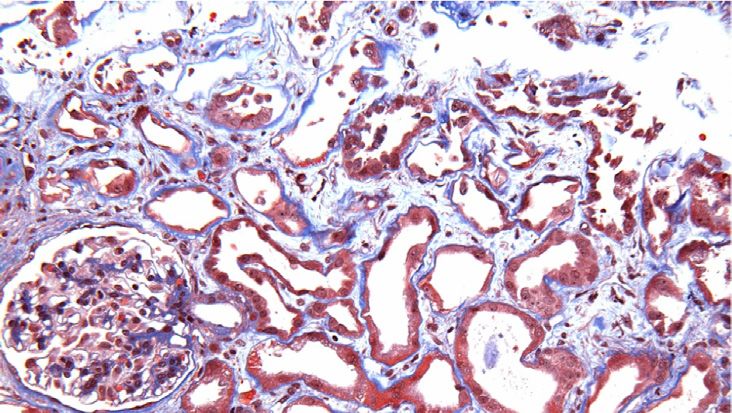
KDIGO executive conclusions                                      CR Swanepoel et al.: Kidney disease and HIV: a KDIGO conference report

characterize the renal risk-benefit of ART; and define best         Table 1 | Pathologic classification of HIV-related kidney
practices to delay the progression of kidney disease and to       diseases
treat end-stage kidney disease (ESKD) in HIV-positive             I. Glomerular-dominanta
individuals.                                                      a. Podocytopathies (all characterized by extensive foot process
                                                                      effacement)b
Renal pathology in the setting of HIV infection                           i. Classic HIVAN
                                                                         ii. FSGS (NOS) in the setting of HIV
The spectrum of renal pathology in HIV-positive individuals            iii. Minimal change disease in the setting of HIV
is diverse, including lesions directly related to intrarenal           iv. Diffuse mesangial hypercellularity in the setting of HIV
HIV gene expression and lesions related to comorbidities,                v. Other podocytopathy in the setting of HIV
drug effects, immune dysregulation, and co-infections.3           b. Immune complex-mediated glomerular diseasea
                                                                          i. IgA nephropathy in the setting of HIV
Kidney biopsy is required to distinguish between these le-               ii. Lupus-like glomerulonephritis in the setting of HIV
sions. A useful approach to classification is based on the               iii. Lupus nephritis in the setting of HIV
major tissue compartment affected (Table 1). A brief                   iv. Membranous nephropathy in the setting of HIV
                                                                              Indicate whether HBV positive, HCV positive, PLA2R positive
description of each histologic lesion is provided below, and
                                                                                (should not preclude workup for other secondary causes)
more comprehensive descriptions are available in the Sup-                v. Membranoproliferative pattern glomerulonephritis in the setting of
plementary Appendix.                                                         HIV
    Glomerular-dominant     diseases:    podocytopathy. Glo-                  Indicate whether HCV positive (should not preclude workup for
                                                                                other secondary causes)
merular-dominant diseases include 2 main subcategories:                vi. Endocapillary proliferative and exudative glomerulonephritis in the
podocytopathies and immune complex–mediated.                                 setting of HIV
    Four major subtypes of podocytopathy are seen in the                      Post-streptococcal, staphylococcal-associated, other

setting of HIV: classic HIVAN, focal segmental glomerulo-              vii. Fibrillary or immunotactoid glomerulonephritis in the setting of HIV
                                                                      viii. Other immune complex disease in the setting of HIV
sclerosis (FSGS) not otherwise specified (NOS), and rarer          II. Tubulointerstitial-dominanta
cases of minimal change disease and diffuse mesangial             a. Tubulointerstitial injury in the setting of classic HIVAN
hypercellularity.4 All exhibit extensive podocyte foot process            i. Hyaline droplet tubulopathy
effacement and proteinuria, with absent or minimal im-                   ii. Tubular microcysts
                                                                       iii. Tubulointerstitial inflammation
mune complex deposition. There is a well-established              b. Acute tubular injury or acute tubular necrosis
causal relationship between HIVAN and HIV infection,                      i. Ischemic
mediated by direct HIV infection of renal epithelial cells,              ii. Toxic (associated with ART vs. other)
intrarenal viral gene expression, and dysregulation of host       c. Drug-induced tubulointerstitial nephritis (other than ART)
                                                                          i. Antibiotics
genes governing cell differentiation and cell cycle.5 The role           ii. Proton pump inhibitors
of genetic susceptibility in the pathogenesis of HIVAN and             iii. NSAIDs
other podocytopathies is discussed in detail in the next               iv. Other
                                                                  d. Direct renal parenchymal infection by pathogens (bacterial, viral,
section.
                                                                      fungal, protozoal, etc.)
    We recommend distinguishing classic HIVAN from FSGS           e. Immunologic dysfunction-related tubulointerstitial inflammation
(NOS) in the setting of HIV infection. Direct causality of HIV            i. Diffuse infiltrative lymphocytosis syndrome (DILS)
can only be established with reasonable certainty in classic             ii. Immune reconstitution inflammatory syndrome (IRIS)
                                                                   f. Other tubulointerstitial inflammation in the setting of HIV
HIVAN and congenital cases of podocytopathy in infants born       III. Vascular-dominanta
to HIV-positive mothers. We recommend that the biopsy             a. Thrombotic microangiopathy in the setting of HIV
report should indicate the degree of certainty that the           b. Arteriosclerosis
pathology is causally related to HIV infection as high,           IV. Other, in the setting of HIV infection
                                                                  a. Diabetic nephropathy
moderate, or low.                                                 b. Age-related nephrosclerosis
    Classic HIVAN. Classic HIVAN is defined as collapsing
                                                                  ART, antiretroviral therapy; HBV, hepatitis B virus; HCV, hepatitis C virus; FSGS, focal
glomerulopathy and attendant tubulointerstitial disease,          segmental glomerulosclerosis; HIVAN, HIV-associated nephropathy; NOS, not
including tubular microcyst formation, interstitial inflam-        otherwise specified; NSAID, nonsteroidal anti-inflammatory drug; PLA2R, M-type
                                                                  phospholipase A2 receptor.
mation, and tubular injury (Figure 1).6,7 Glomerular              a
                                                                   Indicates likelihood of HIV causality.
“collapse” is defined as at least 1 glomerulus with collapse of    b
                                                                    Indicates association with APOL1 risk allele genotype.
glomerular basement membranes accompanied by hypertro-
phy and hyperplasia of the overlying glomerular epithelial
cells. These hyperplastic cells may fill the urinary space,        droplets may stain for albumin and Ig. In late stages, the
forming pseudocrescents.8,9                                       sclerotic tuft is retracted into a tight solid sphere, capped
    By electron microscopy, diffuse podocyte foot process         by a monolayer of cobblestone epithelium; this has been
effacement and endothelial tubuloreticular inclusions             described as resembling a “fetal glomerulus.”10 Phenotypic
(interferon footprints) are classic features.6,7 By immuno-       studies suggest that the glomerular epithelial cell monolayer
fluorescence, there may be staining for IgM, C3, and C1q in        is composed of parietal epithelial cells.8 In some
collapsed segments and mesangial areas.7 Protein resorption       cases, sequential biopsy and postmortem studies have shown

546                                                                                                             Kidney International (2018) 93, 545–559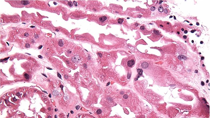
CR Swanepoel et al.: Kidney disease and HIV: a KDIGO conference report                         KDIGO executive conclusions

   a                                             b                                              c

       100 μm                                      100 μm
                                                        m                                           200 μm
                                                                                                         m

  d                                e                                                       f

       10 μm
           m                           1 μm
                                          m                                                    100 μm
                                                                                                    m

Figure 1 | Classic HIV-associated nephropathy (HIVAN) and focal segmental glomerulosclerosis (FSGS) not otherwise specified (NOS) in
the setting of HIV. (a,b) Classic HIVAN shows typical global collapse of the glomerular tuft with loss of luminal patency and hypertrophy
and hyperplasia of the overlying glomerular epithelial cells, some of which contain intracytoplasmic protein resorption droplets (a, Jones
methenamine silver x400; b, Masson trichrome, x400). (c) The tubulointerstitium shows focal tubular microcysts (arrow) containing glassy casts,
associated with tubular atrophy, interstitial fibrosis, and inflammation (Masson trichrome, x200). (d) There is marked foot process effacement
overlying the collapsed capillaries associated with glomerular epithelial cell hyperplasia forming a pseudocrescent. Some glomerular epithelial
cells contain numerous intracytoplasmic protein resorption droplets. No immune-type electron dense deposits are seen (electron micrograph
x4000). (e) Glomerular endothelial cells may contain intracytoplasmic tubuloreticular inclusions (arrows). Foot processes are effaced (electron
micrograph, x40,000). (f) FSGS (NOS) in the setting of HIV shows discrete segmental scars with segmental adhesions to Bowman’s capsule.
No collapsing features or glomerular epithelial cell hyperplasia are identified (H&E, x400). To optimize viewing of this image, please see the
online version of this article at www.kidney-international.org.

an evolution from collapsing glomerulopathy to FSGS                      and biopsy findings may be difficult to distinguish from
(NOS).7                                                                  arterionephrosclerosis of hypertension, aging, and APOL1-
   Tubulointerstitial disease is an invariable component of              associated nephropathy. Such cases typically lack prominent
HIVAN and often appears out of proportion to the glomer-                 tubulointerstitial disease, and the degree of podocyte efface-
ular disease,6,7 causing kidney enlargement and hyperechoic              ment is generally less severe than in HIVAN (Figure 1). These
appearance by ultrasound. Tubular microcysts are dilated                 differences have been hypothesized to reflect attenuation of
tubules (at least 3-fold larger than normal) containing glassy           the renal phenotype by ART.9
proteinaceous casts and lined by simplified epithelium.                      Podocytopathy in perinatal HIV infection. In addition to
Tubular microcysts are easily distinguished from tubular                 classic HIVAN, children with perinatal HIV infection can
thyroidization based on their larger diameter, irregular size,           present with minimal change disease or diffuse mesangial
and the absence of tubular atrophy or colloid-type casts.11              hypercellularity with numerous endothelial tubuloreticular
The microcysts may involve all tubular segments, and                     inclusions and marked foot process effacement.17 Tubular
intracellular viral transcript expression has been demon-                microcysts and interstitial inflammation are often lacking.
strated.12 Prominent interstitial inflammation7 and tubular               Such cases are rare in the ART era.
degenerative and regenerative changes may also occur.13
                                                                            Glomerular-dominant diseases: immune complex kidney
Interstitial edema in the acute phase is followed by fibrosis
                                                                         disease in the setting of HIV. Numerous forms of immune
and tubular atrophy.
                                                                         complex-mediated glomerular disease have been reported in
    FSGS (NOS) in the setting of HIV. In ART-treated patients,           HIV-positive individuals.18 We recommend that the
noncollapsing FSGS (NOS) is more commonly encountered                    commonly used term “HIV immune complex kidney disease”
at biopsy.9,14–16 Causality is presumed when no other etiology           (HIVICK) be replaced with a specific description of the
for FSGS can be identified. Viral load is often undetectable,             pattern of immune complex disease “in the setting of HIV.”

Kidney International (2018) 93, 545–559                                                                                                    547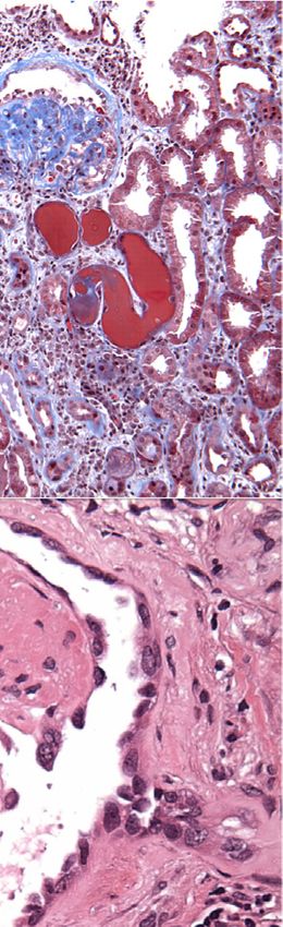
KDIGO executive conclusions                                             CR Swanepoel et al.: Kidney disease and HIV: a KDIGO conference report

The rationale for this approach is the heterogeneous spectrum            hepatitis B virus co-infection or anti-PLA2R autoantibodies)
of disease and the lack of certainty of HIV causality in most            and membranoproliferative glomerulonephritis (hepatitis C
cases. Early studies that eluted glomerular immune deposits              virus co-infection).24–26
and demonstrated immune complexes containing HIV antigen                    Tubulointerstitial disease in the setting of HIV. As described
and specific anti-HIV antibody were performed on a small                  above, classic HIVAN is a pan-nephropathy with an impor-
number of well-characterized cases in the research setting and           tant tubulointerstitial component;6,7 in biopsies with under-
are not practicable in routine pathology laboratories.19,20 Re-          sampled glomeruli, the characteristic glomerular lesions may
flex diagnosis as HIVICK may preclude workup for other                    not be demonstrable. Acute tubular necrosis may occur in
secondary, treatable causes.                                             association with sepsis, volume depletion, and other ischemic
   A unique lupus-like nephritis with full-house immune                  or toxic insults.4 The commonly used antiretroviral agent
staining but negative serologies and no clinical signs of sys-           tenofovir disoproxil fumarate can cause proximal tubulopathy
temic lupus erythematosus has been reported in HIV-positive              with characteristic dysmorphic mitochrondria (Figure 2).27
individuals;21 true lupus nephritis also occurs.22 It remains            Tubulointerstitial nephritis can occur secondary to
unclear whether IgA nephropathy in the setting of HIV is                 antibiotics, proton pump inhibitors, nonsteroidal anti-
coincidental and related to undergalactosylated IgA1 or due to           inflammatory drugs, protease inhibitors, and other medica-
deposition of IgA directed to viral antigen, as demonstrated             tions, as well as in response to mycobacterial infection.28–30
in a well-characterized case.19 An unusual ultrastructural               Direct infection of the renal parenchyma by other patho-
appearance of subepithelial deposits, or “ball in cup” lesion,           gens can also occur.7
has been described in reports from South Africa,10,23 but is                Two rare but distinct forms of tubulointerstitial injury
rarely observed in other settings. Other secondary causes                relate to immunologic dysfunction in the setting of HIV
should be sought in cases of membranous nephropathy (i.e.,               infection. Diffuse infiltrative lymphocytosis syndrome is a

   a                                                  c                                                e

       200 μm                                            200 μm
                                                              m                                             5 μm
                                                                                                               m

   b                                                 d                                                  f

       100 μm
            m                                            100 μm
                                                              m                                             3.3 μm
                                                                                                                 m

Figure 2 | Tenofovir nephrotoxicity. (a) Acute tenofovir nephrotoxicity is characterized by irregular proximal tubular profiles with atypical,
irregular lining epithelial cells and mild interstitial edema (hematoxylin and eosin, original magnification 200). (b) The atypical proximal
tubular cells display loss of brush border, marked irregularity of tubular epithelial height and shape, focal shedding of cytoplasmic fragments,
and enlarged atypical nuclei with prominent nucleoli (hematoxylin and eosin, original magnification 400). (c) Chronic nephrotoxicity displays
increased separation of the irregular proximal tubules by interstitial fibrosis and mild inflammation with focal tubular atrophy (Masson
trichrome, original magnification 200). The tubules show focal loss and flattening of lining epithelium leaving some desquamated tubular
basement membranes, as well as prominent epithelial simplification and irregularity with atypical nuclei. (d) There is intervening interstitial
fibrosis and mild inflammation, without tubulitis (Masson trichrome, original magnification 400). (e) The characteristic features are focal giant
mitochondria with few residual peripheral cristae (arrow) within the proximal tubular epithelial cells, as well as cytoplasmic swelling with
disruption of brush border, (electron micrograph, original magnification 8000). (f) In some cases, the dysmorphic mitochrondria exhibit
irregular size and shape with bizarre patterning of their cristae (electron micrograph, original magnification 12,000). To optimize viewing of
this image, please see the online version of this article at www.kidney-international.org.

548                                                                                                             Kidney International (2018) 93, 545–559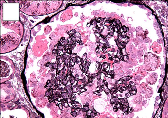
CR Swanepoel et al.: Kidney disease and HIV: a KDIGO conference report                              KDIGO executive conclusions

hyperimmune reaction against HIV that involves the kidneys                  Table 2 | Prevalence of APOL1 high-risk genotypes and asso-
in approximately 10% of cases.31–33 Immune reconstitution                   ciation with kidney disease in HIV-positive African Americans
inflammatory syndrome is an inflammatory disorder associ-                     and Black South Africans
ated with paradoxical unmasking or worsening of preexisting                                                   Population      Odds ratio
infectious processes after ART initiation,34 rarely involving               Histology       Population         controls Cases (95% CI) Reference
the kidney. Both conditions are characterized by prominent                  HIVAN     African American            13%       72%      29 (14, 68)        49

CD8 T-cell infiltrates.                                                                      (n ¼ 54)
    Vascular-dominant           diseases   in   the   setting   of   HIV.   HIVAN     African American            13%       62%           –             54

                                                                                            (n ¼ 60)
Thrombotic microangiopathy was reported in the early years                  HIVAN       South Africa              3%        79% 89 (18, 912)            51

of the AIDS epidemic, but is rare in the ART era.35 A role for                              (n ¼ 38)
direct endothelial dysregulation by HIV has been proposed.36                HIVþ FSGS African American            13%       63%           –             57

   Other pathologies in the setting of HIV. As patients with                                (n ¼ 35)
                                                                                                                                                        51
                                                                            HIVþ FSGS South Africa                3%         8%    2.1 (0.03, 44)
HIV infection age, comorbid kidney diseases such as diabetic                                (n ¼ 22)
nephropathy and arterionephrosclerosis are increasingly                     HIVþ ICD African American             13%        3%           –             57

common. When secondary FSGS develops in these contexts,                                     (n ¼ 31)
                                                                                                                                                        51
the potential overlap with HIV-related podocytopathy can be                 HIVþ ICD    South Africa              3%        25% 5.6 (0.4, 86)
                                                                                            (n ¼ 12)
diagnostically challenging. Molecular approaches demon-
                                                                            APOL1, apolipoprotein L1; CI, confidence interval; FSGS, focal segmental glomer-
strating renal epithelial cell infection by HIV have been used              ulosclerosis; HIVAN, HIV-associated nephropathy; ICD, immune complex kidney
in the research setting for decades, but have not been incor-               disease.
porated into routine diagnostic practice.37 In addition to
these established approaches, several novel and emerging
techniques could be incorporated into research and diag-                    diagnosis of HIVAN (Table 2). The estimated lifetime risk
nostic renal pathology to better characterize the causal rela-              associated with carrying 2 APOL1 risk alleles is 4% for FSGS
tionship between HIV and specific histologic lesions and to                  in the absence of HIV infection, and as high as 50% for
further delineate the host pathways involved. The conference                HIVAN (Supplementary Table S2).53 Despite the strong as-
attendees identified several particularly relevant techniques                sociation, w20% to 30% of African Americans with HIVAN
(Supplementary Table S1).38,39                                              have 0 or 1 APOL1 risk allele, suggesting that other genetic,
                                                                            viral, or environmental factors contribute to HIVAN.54
Genetics/genomics of kidney disease in the setting of HIV
infection
Classic HIVAN occurs predominantly in individuals of                        Table 3 | Features of APOL1-mediated kidney disease in the
African ancestry, with 18- to 50-fold increased prevalence.40               setting of HIV
Two studies involving mapping by admixture linkage
disequilibrium published in 2008 identified a region on                         MYH9 variants are not independently associated with HIVAN or FSGS
                                                                                (NOS)45,52,152
chromosome 22 strongly associated with idiopathic FSGS                         APOL1 kidney disease manifests as HIVAN or FSGS (NOS) with or
and HIVAN in African Americans;41,42 however, fine-                              without microcystic tubular dilatation49,56,57
mapping revealed no coding variants to explain the associ-                     S342G and N388Y389/– confer risk of kidney disease; therefore geno-
ation of intronic single-nucleotide polymorphisms in the                        typing only the APOL1 G1 rs73885319 missense and G2 rs71785313
                                                                                indel (i.e., insertion-deletion mutations) variants are sufficient to
candidate gene MYH9 with kidney disease.43,44 Subsequently,                     determine risk of CKD49
using data from the 1000 Genomes Project, Genovese et al.                      HIVAN is associated with low CD4þ cell counts, and often improves
identified 2 missense variants (G1 allele) and a 6 bp deletion                   with effective ART56
                                                                               HIV-associated FSGS is associated with higher CD4þ cell counts and
(G2 allele) in the adjacent APOL1 gene that were recessively
                                                                                occurs in patients undergoing ART56
associated with FSGS and nondiabetic ESKD.45 APOL1 en-                         APOL1 high-risk genotypes are associated with progression to ESKD in
codes apolipoprotein L1, which confers innate immunity                          HIV-positive patients with non-HIVAN kidney diseases57
against most strains of Trypanosoma brucei;46,47 G2 variants                   Histological features of HIVAN in patients carrying 2 copies of APOL1
                                                                                risk variants are similar to those carrying 0 or 1 copy54
extend immunity to T.b. rhodesiense and G1 associates with
                                                                               HIV-positive children with CKD and high-risk genotypes have lower
asymptomatic carriage of T.b. gambiense, the causes of acute                    eGFR and experience more rapid progression58,153
and chronic African human trypanosomiasis, respec-                             Multiple mechanisms have been proposed for APOL1-mediated podo-
tively.45,48 Coding variants in APOL1 are present only on                       cyte injury, but they converge in perturbations of endosomal trafficking,
                                                                                increased membrane permeability, and cytotoxicity61,63–65
African-ancestry haplotypes.49,50                                              APOL1, a component of the innate immune system, is up-regulated by
   APOL1 was strongly associated with FSGS (odds ratio                          interferons61,62
[OR] 17) and HIVAN (OR 29) in African Americans and with                       High levels of APOL1 may be a “second hit” and sufficient to cause
HIVAN in South Africans (OR 89).49,51 In contrast, HIV-                         kidney disease61,62
positive Ethiopians, who lack APOL1 risk variants, do not                   APOL1, apolipoprotein L1; ART, antiretroviral therapy; CKD, chronic kidney disease;
                                                                            eGFR, estimated glomerular filtration rate; ESKD, end-stage kidney disease; FSGS
develop HIVAN.52 Subsequent studies have confirmed the                       (NOS), focal segmental glomerulosclerosis, not otherwise specified; HIVAN,
strong association between the high-risk genotypes and the                  HIV-associated nephropathy; MYH9, myosin heavy chain 9 gene.

Kidney International (2018) 93, 545–559                                                                                                                      549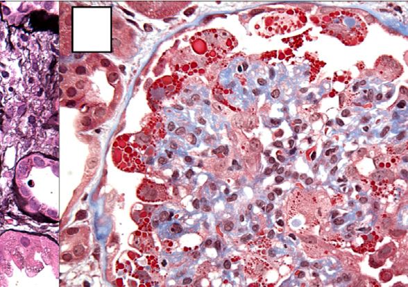
KDIGO executive conclusions                                        CR Swanepoel et al.: Kidney disease and HIV: a KDIGO conference report

Characteristics of APOL1-mediated kidney disease are sum-           loss of potassium, cell swelling, and cell lysis.66 Studies in
marized in Table 3.                                                 yeast, Drosophila, and human cells indicate that variant
    The distribution of the APOL1 coding variants varies            apolipoprotein L1 depolarizes cell membranes, which dis-
greatly among sub-Saharan African populations, with the             rupts intracellular processes including endosomal trafficking,
highest frequencies reported in Western Africa (>40% for            vesicle acidification, and mitochondrial function.63–65
G1) and much lower frequencies elsewhere in Africa                     In vivo, the expression of high-risk APOL1 variants
(Supplementary Figure S1).50,52,55 As a consequence of the          in transgenic mouse models has produced variable effects.
West African diaspora in the Americas and more recent               In a model with inducible APOL1 expression, high-risk
African emigrations, APOL1 variants are widely dispersed            variants disrupted endosomal trafficking and vesicle acidifi-
globally (e.g., 21% and 13% for G1 and G2, respectively, in         cation, similar to the effects observed in vitro. Affected
African Americans).45,50                                            animals developed podocyte death, proteinuria, and glomer-
    Prediction of histology. Given the strong genetic associa-      ulosclerosis.61 However, another transgenic mouse model
tion, investigators in the United States (US) evaluated whether     with constitutive expression of APOL1-G2 did not develop
APOL1 genotype could be used to predict HIVAN or FSGS               kidney disease.67
(NOS) histology in HIV-positive patients of African                    APOL1 is encoded in the genome of only a few primate
descent.56 Inclusion of the high-risk genotype did not              species, complicating the extrapolation of data from murine
significantly add to a predictive model including CD4þ cell          models. Mechanistic studies have also been limited by the use
count and HIV-RNA, suggesting that APOL1 genotype cannot            of overexpression assays. In vitro, the overexpression of wild-
replace kidney biopsy for definitive diagnosis of HIVAN.             type APOL1-G0 in cultured human renal epithelial cells also
    Carriage of APOL1 high-risk genotypes in HIV-positive           induces cell death, suggesting that the overexpression model
individuals is not associated with immune complex kidney            may not be biologically relevant.62,68,69
disease (Table 2). In a US series, high-risk genotypes were
present in only 3% of patients with biopsy-proven immune            Antiretroviral therapy (ART) nephrotoxicity
complex disease.57 Similarly, in a South African series, high-      HIV treatment guidelines recommend immediate initiation of
risk genotypes were present in 79% of HIVAN cases but in            ART in all HIV-positive individuals. Immuno-virological
only 25% of those with HIV and immune complex kidney                control is an important strategy to reduce the incidence
disease.51                                                          of acute kidney injury (AKI) and HIV-related kidney
    Renal survival and ESKD risk. In general population             diseases.70–73
studies, the high-risk APOL1 genotypes have been associated            The presence of CKD affects the choice and dosing of
with increased risk of CKD progression and with lower esti-         renally cleared antiretrovirals. Kidney function and CKD risk
mated glomerular filtration rate (eGFR).58 In children with          factors should be assessed prior to ART initiation (Figure 3).
perinatal HIV infection, those with a high-risk genotype had        CKD risk scores have been developed to guide clinicians,
3-fold increased odds of CKD and presented at a younger             although future studies are needed to determine their utility
median age compared with those with 0 or 1 risk allele.51 In        in diverse populations (Supplementary Table S3).74,75
HIV-positive adults with non-HIVAN kidney disease on                   The widely used antiretroviral agent tenofovir disoproxil
biopsy, carriage of 2 APOL1 risk alleles was associated with        fumarate (TDF) is generally safe and well tolerated, but has
more rapid progression and a 2-fold greater risk of ESKD.57         important potential for cumulative nephrotoxicity. Sub-
Carriage of 2 APOL1 risk alleles has been associated with           clinical proximal tubular dysfunction (low-level proteinuria
proteinuria in HIV-infected women and with accelerated              and excessive phosphaturia) is common, and approximately
decline in longitudinal kidney function in unsuppressed HIV-        1% to 2% of recipients develop treatment-limiting tubul-
infected men.59,60                                                  opathy.76 Risk factors for tubulopathy include aging, im-
    Mechanisms of APOL1-mediated disease. Two APOL1 risk            munodeficiency, diabetes, prolonged exposure, and
alleles are required to confer increased risk of kidney disease.    concomitant use of didanosine or ritonavir-boosted protease
However, the presence of high-risk genotypes in healthy             inhibitors.77 Severe tubulopathy may progress to eGFR
populations suggests that disease expression requires a “sec-       decline, osteomalacia, and pathological fractures. In large
ond hit,” such as infections (e.g., HIV or viral hepatitis),        observational studies, TDF has also been associated with
interferon, gene-gene interactions, illicit drug use, and other     decreased eGFR or creatinine clearance,78,79 as well as with
CKD risk factors.                                                   rapid eGFR decline and proteinuria.78,79 Co-administration
    The mechanism of APOL1-mediated kidney disease is               of TDF with ritonavir-boosted protease inhibitors increases
currently unknown. Evidence from in vitro experiments in            the risk.78,79 Although not well studied, the newer phar-
human cells and APOL1 transgenic mouse models suggests              macoenhancer cobicistat also increases tenofovir exposure
that interferon upregulates APOL1 expression, causing               and may increase the risk of toxicity. TDF discontinuation
podocyte injury.61,62 Intracellular apolipoprotein L1 in renal      and switches from TDF to the newer prodrug tenofovir
epithelium may cause apoptosis or autophagy by increasing           alafenamide (TAF) have been associated with improved
cellular and mitochondrial membrane permeability.63–65 In           kidney function, although the long-term safety of TAF is not
cell culture, G1 and G2 APOL1 variants induce intracellular         known.80–83

550                                                                                                      Kidney International (2018) 93, 545–559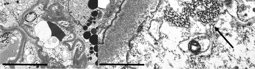
CR Swanepoel et al.: Kidney disease and HIV: a KDIGO conference report KDIGO executive conclusions
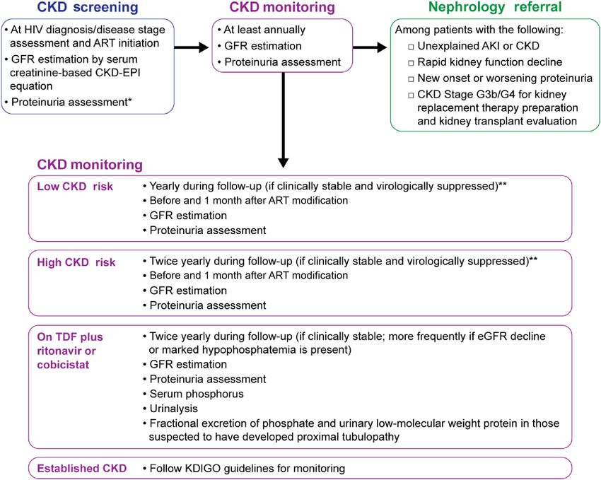
Perform CKD risk straƟficaƟon
Low risk High risk
eGFR >90 eGFR 3–5 ml/min per 1.73 m2 per dividuals, traditional CKD risk factors, particularly
year), or at high CKD risk. The threshold for avoiding or hypertension and diabetes, are of increasing concern world-
discontinuing these agents may be influenced by local wide.94–96 Hepatitis B virus (HBV) and hepatitis C virus (HCV)
circumstances. In resource-limited settings, TDF dose adap- co-infections are associated with a 2- to 3-fold increased risk of
tation may be an option. Dual therapy (i.e., boosted protease progressive CKD.97,98 Other co-infections such as tuberculosis
inhibitor plus lamivudine or raltegravir) has been proposed as and syphilis may also contribute to CKD risk.99–101 In addition,
a way to avoid concomitant use of boosted protease inhibitors severe AKI has been associated with a 3.8- to 20-fold increased
with TDF, thereby minimizing the nephrotoxic potential.91–93 risk of progression to ESKD.102
Pharmacological considerations. Several antiretrovirals CKD screening and monitoring. Studies to inform the
require dose adjustment in individuals with decreased eGFR optimal CKD screening and monitoring strategies among
(Supplementary Table S4). If continued use of TDF is HIV-positive individuals are lacking. Until such studies exist,
required when eGFR is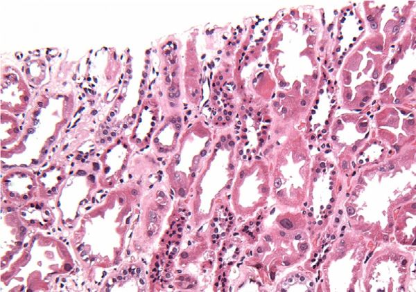
KDIGO executive conclusions                                           CR Swanepoel et al.: Kidney disease and HIV: a KDIGO conference report

                                                Exposures and
                                                  co-morbid
      Host geneƟc             Socio-                                HIV-related                                          Underlying CKD
                                                non-infecƟous                            Co-infecƟons
      suscepƟbility        demographics                               factors                                               eƟology
                                               condiƟons and
                                               their treatment

                                                    Diabetes                                                                    HIVAN
                                                                                         HepaƟƟs B or C
      APOL1 G1 and               Age                Obesity
                                                                                             virus
      G2 risk variants                                                HIV viremia
                                                                                                                              FSGS (NOS)
                                                 Hypertension
                                                                                                                           Immune complex
                            Race/Ethnicity       Cardiovascular                           Tuberculosis                        disease
                                                    disease                                                                    DiabeƟc
      Sickle cell trait?                                                                                                     nephropathy
                                                Recurrent/ Severe   CD4+ cell count
                                                  acute kidney
                            Illicit drug use          injury                                 Syphilis                    Arterionephrosclerosis

                                                  Malignancy                                                               TubulointersƟƟal
                                                                     ComposiƟon
         ABCC2/4                                                                                                               diseases
                            Socioeconomic                            and Ɵming of            ParasiƟc
         variants?
                                status            TradiƟonal/        ART iniƟaƟon           infecƟons
                                                herbal medicines                                                          ART nephrotoxicity

Figure 4 | Risk factors and underlying etiologies of CKD in HIV-positive individuals. APOL1, apolipoprotein L1; ABCC, ATP-binding cassette
transporter proteins; ART, antiretrovial therapy; CKD, chronic kidney disease; FSGS (NOS), focal segmental glomerulosclerosis, not otherwise
specified; GN, glomerulonephritis; HIVAN, HIV-associated nephropathy.

    Serum creatinine is the preferred biomarker for esti-                  In most HIV-positive individuals who are stable on ART,
mating GFR.2,103 Serum cystatin C may be considered in                  annual monitoring of kidney function appears appropriate. In
patients receiving medications that alter tubular creatinine            those with or at increased risk of CKD and those who receive
handling. Cystatin C may also better predict long-term                  TDF with ritonavir- or cobicistat-boosted protease inhibitors,
mortality,104,105 but is susceptible to bias in the setting of          more frequent monitoring is recommended, typically 2–4
inflammation. The serum creatinine-based CKD-EPI equa-                   times per year depending on risk factors.113 Kidney function
tion is generally preferred;2,103 however, none of the avail-           should also be carefully monitored during hospitalization,
able estimates have been validated in diverse populations or            particularly in individuals receiving TDF and concomitant
in the setting of drugs that alter creatinine secretion.106–108         nephrotoxic medications.
Use of the antiretrovirals dolutegravir or rilpivirine or the              If CKD is identified, patients should undergo work-up
pharmacoenhancers ritonavir or cobicistat may result in                 based on available resources and risk stratification, including
average reductions in calculated creatinine clearance of                consideration of potential medication toxicity; screening for
around 5 to 20 ml/min, which should be taken into account               hypertension, diabetes, and co-infections; and assessment of
when interpreting eGFR or creatinine clearance.109 Clini-               region-specific risk factors such as traditional medicines. HIV-
cians should also be aware that serum creatinine measure-               specific CKD risk scores may facilitate risk-stratification,74,75
ments may not be standardized in resource-limited regions               although these scores have not been validated in diverse
and that extrarenal factors may alter both serum creatinine             populations or in resource-limited settings (Supplementary
and cystatin C concentrations (Supplementary Table S5).110–             Table S3). Referral to a nephrologist should be considered in
112
     Rather than a single eGFR value, eGFR trajectories are             certain settings (Figure 5).103 When the cause of CKD is un-
useful for identifying individuals with progressive decline in          clear, CKD progression is rapid, or prognostication is needed,
kidney function.                                                        a kidney biopsy should be considered.
    Urinalysis should be performed in all HIV-positive                     CKD management. Evidence from observational studies
individuals to detect worsening or new onset of proteinuria             strongly supports the beneficial effect of early ART initiation
or hematuria. Where feasible, quantification of proteinuria              on the risk of classic HIVAN.114 The impact of ART on CKD
(urine albumin-to-creatinine or protein-to-creatinine ratio)            progression in patients with immune complex kidney diseases
should also be performed. In individuals receiving TDF,                 is more variable.71,72 Given the overwhelming benefit on
urinalysis may also detect glycosuria, and plasma phosphate             survival, ART is recommended for all HIV-positive
should be monitored if possible. Evaluation of cystatin C,              individuals.115 Evaluation of other treatment strategies for
low–molecular weight (“tubular”) proteinuria, or phosphate              kidney disease in the setting of HIV has been limited to small,
reabsorption is not indicated in individuals with stable kidney         single-center studies with short duration, and has focused
function and no indication of TDF toxicity.113                          largely on HIVAN (Supplementary Table S6). No rigorous

552                                                                                                         Kidney International (2018) 93, 545–559
CR Swanepoel et al.: Kidney disease and HIV: a KDIGO conference report                       KDIGO executive conclusions

Figure 5 | Recommendations for kidney disease screening and monitoring in HIV-positive adults. *Urinalysis should be performed in all
HIV-positive individuals to detect worsening or new onset of proteinuria or hematuria. Where feasible, quantification of proteinuria (spot urine
albumin-to-creatinine or protein-to-creatinine ratio) should also be performed. **More frequent monitoring is recommended in persons who
are clinically unstable, severely immunocompromised, or viremic. AKI, acute kidney injury; ART, antiretroviral therapy; CKD, chronic kidney
disease; CKD-EPI, CKD Epidemiology Collaboration; eGFR, estimated glomerular filtration rate; GFR, glomerular filtration rate; KDIGO, Kidney
Disease: Improving Global Outcomes; TDF, tenofovir disoproxil fumarate.

study has evaluated the efficacy of blood pressure control,               resources.123,124 Arteriovenous fistulas are the preferred
diabetes treatment, or angiotensin-converting-enzyme in-                 vascular access, as arteriovenous grafts and catheters are
hibitors and angiotensin receptor blockers in slowing CKD                associated with higher risk of infection and thrombosis.125
progression in HIV-positive individuals. However, extrapo-               There is no evidence supporting isolation of HIV-positive
lating from the strong evidence supporting the efficacy of                patients in HD units, except those with HBV co-infec-
these interventions in the general population is reasonable              tion.126 Dialyzer reuse by the same patient is practiced in
(Table 4).103,116,117 Treatment of HBV, HCV, and tuberculosis            resource-limited settings as a cost-saving alternative. Evidence
co-infections should be considered based on existing treat-              supporting the safety of dialyzer reuse by HIV-positive
ment guidelines.118–121                                                  individuals is limited,127,128 and precautions must be
    Kidney replacement therapy (KRT) in HIV-positive individu-           adhered to in order to avoid HIV transmission to other
als. With ART, survival of HIV-positive individuals                      patients and dialysis staff. HIV-positive PD patients may have
receiving KRT is comparable to their HIV-negative counter-               higher risk of PD catheter infections; however, PD catheter
parts.122 Therefore, HIV serostatus should not influence                  failure rates are similar in HIV-negative patients.129 PD
candidacy for KRT. Observational studies demonstrate similar             consumables must be discarded properly, as HIV persists in
outcomes between hemodialysis (HD) and peritoneal dialysis               PD materials and fluid.130,131
(PD) among ART-treated individuals, and modality selection                   Kidney transplantation in HIV-positive individuals. Kidney
depends     upon     patient  preference    and    regional              transplantation in HIV-positive recipients is associated with

Kidney International (2018) 93, 545–559                                                                                                    553
KDIGO executive conclusions                                                          CR Swanepoel et al.: Kidney disease and HIV: a KDIGO conference report

Table 4 | Recommendations for management of CKD risk                                  Table 5 | Selection criteria for potential HIV-positive kidney
factors in HIV-positive individuals                                                   transplant recipients
Risk factor                                Recommendations                            ➢ Meets standard criteria for kidney transplant recipients, plus the
                                                                                        following:
Hypertension
                                                                                      ➢ Effective HIV suppression for $6 months prior to transplantation
  Nonproteinuric             Target systolic blood pressure #140 mm Hg116
                                                                                           Undetectable plasma HIV-1 RNA
  Proteinuric                Target systolic blood pressure #130 mm Hg116
                                                                                           CD4þ cell count > 200 cells/mm
                                                                                                                              3
                             Preferred antihypertensive: ACE inhibitors or
                                                                                      ➢ No active opportunistic infections
                              angiotensin receptor blockers116
                                                                                      ➢ No history of:
Diabetes mellitus            Target hemoglobin A1c w7%*103
                                                                                           Progressive multifocal leukoencephalopathy
Hepatitis B virus            Treat per existing guidelines118,121
                                                                                           Primary central nervous system lymphoma
  co-infection               TAF may be used in patients with eGFR $ 30
                                                                                           Pulmonary aspergillosis
                              ml/min per 1.73 m2.154
                                                                                           Visceral Kaposi’s sarcoma
                             Where TAF is unavailable or in patients with
                                                                                           Coccidiomycosis
                              eGFR < 30 ml/min per 1.73 m2, dose-adjusted
                                                                                           Chronic intestinal cryptosporidiosis >1 month
                              TDF or entecavir may be considered.
                                                                                      ➢ Hepatology evaluation for patients co-infected with hepatitis B or
Hepatitis C virus            Treatment per existing guidelines120,155
                                                                                        hepatitis C virus
  co-infection               In patients with HCV genotypes 1 and 4 and CKD
                              G4-5, ribavirin-free grazoprevir/elbasvir156–158        Criteria adapted from Stock et al. and Muller E et al.132,137
                              or glecaprevir/pibrentasvir regimens may be
                              effective164,165
                             In patients with genotypes 2, 3, 5, and 6 and CKD
                                                                                      steady-state drug levels, integrase inhibitors and nucleoside
                              G4-5, the pan-genotypic glecaprevir/pibrentasvir        reverse transcriptase inhibitors are the preferred antiretroviral
                              regimen can be used164,165; sofosbuvir-based            agents, while protease inhibitors and the pharmacoenhancers
                              regimens can be used in patients with any               ritonavir and cobicistat are best avoided.145 Given the
                              genotype, but should be avoided or dose
                              adjusted in patients with eGFR < 30 ml/min per
                                                                                      complexity of issues, a multidisciplinary team comprising
                              1.73 m2.159–161 In addition, the combination of         experts in transplant nephrology, infectious disease, and
                              ledipasvir and sofosbuvir with TDF should               clinical pharmacology is imperative.
                              be avoided.                                                 Given the strong association between the APOL1 risk
ACE, angiotensin-converting enzyme; ARB, angiotensin receptor blocker; eGFR,          variants and HIVAN, HIV-positive recipients of African
estimated glomerular filtration rate; HCV, hepatitis C; TAF, tenofovir alafenamide;
TDF, tenofovir disoproxil fumarate.                                                   descent and those who receive an allograft from a donor of
*Hemoglobin A1c may underestimate glycemia in HIV-positive individuals.162,163        African descent should be monitored for recurrent HIVAN.146
                                                                                      The relative contribution of donor and recipient APOL1
                                                                                      risk status to the risk of HIVAN recurrence is the subject
excellent 1-year and 3-year recipient and allograft survival
                                                                                      of ongoing research. The APOL1 Long-term Kidney Trans-
rates, intermediate to those observed in the overall US kidney
                                                                                      plantation Outcomes (APOLLO) Research Network147 will
transplant population and in a higher risk subgroup of
                                                                                      investigate the influence of donor APOL1 risk variants on
recipients $65 years of age.132 Registry data also suggest good
                                                                                      long-term outcomes among recipients and African American
5- and 10-year outcomes, with an improvement in survival
                                                                                      donors, including those with HIV.
compared with patients who remain on the wait-list.133
                                                                                          Based on experience in South Africa, there is growing
Studies in other settings have confirmed the safety of kidney
                                                                                      evidence to support the safety of kidney transplantation from
transplantation in individuals with well-controlled
                                                                                      HIV-positive donors.148,149 The US HIV Organ Policy Equity
HIV.132,134–138 Eligible patients with advanced CKD and
                                                                                      (HOPE) Act allows the use of organs from HIV-positive
well-controlled HIV infection should be referred for kidney
                                                                                      donors in approved research programs.150,151 Questions
transplant evaluation (Table 5).
                                                                                      remain about the implications of super-infection in settings
   Immunosuppressant protocols for the general population
                                                                                      where ART resistance is common.
can be applied to HIV-positive individuals. In view of the
increased immunological risk, some centers prefer induction                           Children and adolescents with HIV
therapy with an interleukin-2 receptor antagonist, polyclonal                         As in adults, CKD screening and monitoring are recom-
antithymocyte globulin, or alemtuzumab.132,134,139 Tacroli-                           mended, and ART should be provided as per international
mus is the calcineurin inhibitor of choice for maintenance                            and regional guidelines (Supplementary Table S7).2,115
immunosuppression.132,140
   Existing guidelines for prophylaxis against opportunistic                          Conclusion
infections141,142 and management of hepatitis co-infection                            Despite improved survival with ART, HIV-positive individuals
should be followed.143,144 Outcomes for HCV–co-infected                               remain at increased risk for kidney disease. This report
recipients are poorer compared with recipients with HIV or                            summarizes recommendations for diagnosis, management,
HCV mono-infection, but are still superior to those of                                and prevention of kidney disease in this population, including
patients who remain on the wait-list. Clinicians should be                            a proposed histologic classification. In the absence of data
aware of significant drug-drug interactions among immuno-                              from randomized controlled trials, these recommendations
suppressive agents, ART, and antiviral medications for HCV                            reflect the expert opinion of conference attendees, incorpo-
co-infection. To minimize drug-drug interactions and achieve                          rating combined clinical experience and evidence from

554                                                                                                                                 Kidney International (2018) 93, 545–559
CR Swanepoel et al.: Kidney disease and HIV: a KDIGO conference report KDIGO executive conclusions
Table 6 | Controversies, knowledge gaps, and areas for future Table 6 j (Continued)
research
How well do creatinine-based eGFR estimates predict true GFR in
Renal pathology ART-treated individuals, especially those undergoing ART that interferes
What is the spectrum of renal pathology in the setting of HIV infection
with creatinine secretion and in sub-Saharan African populations?
What is the role of serum cystatin C, alone or in combination with
in the current era and in diverse patient populations?
How do pathologic features correlate with the duration of ART, HIV viral
creatinine, in evaluating kidney function in specific clinical contexts,
load, racial and geographic origin, and APOL1 risk allele genotype? such as the use of ART that interferes with creatinine secretion?
What is the clinical utility of novel urine biomarkers of kidney injury in
What is the relative contribution of de-differentiated podocytes versus
parietal epithelial cells to the glomerular epithelial cell hyperplasia seen assessing and monitoring kidney health?
Are clinical guidelines for diabetes, hypertension, and cardiovascular
in HIVAN?
What are the roles of specific HIV transcript expression in promoting
disease developed in the general populations effective in preventing
proliferation and possible transdifferentiation of podocytes and parietal CKD onset and progression in HIV-positive individuals?
Do ACE inhibitors and ARBs confer similar renoprotective effects among
epithelial cells, and in mediating the tubular phenotype of cell cycle
arrest and microcyst formation? HIV-positive individuals with CKD as in the general population?
What is the impact of tuberculosis co-infection and its treatment on the
What is the pattern of HIV viral transcript expression in specific renal cell
types and tissue compartments in FSGS (NOS) and other non-HIVAN risks of CKD development and progression among HIV-positive
lesions in the setting of HIV? individuals?
What is the role of adjunctive therapy with corticosteroids or immu-
Is FSGS (NOS) in the setting of HIV representative of attenuated or
partially treated HIVAN? nosuppressive therapy in patients with HIVAN or other kidney disease
How can immune complex disease that is causally related to HIV
that may be causally related to HIV infection?
What is the role of HIV infection in immune complex kidney disease,
infection be distinguished from coincident disease?
Can HIV infection of renal dendritic cells, infiltrating monocyte and/or
and what is the optimal therapy for specific immune complex diseases
macrophages, or intrinsic renal epithelial cells produce a viral reservoir in this setting?
Has the epidemiology of acute kidney injury changed in the era of
that is capable of reactivation?
What is the composition of the inflammatory infiltrates in HIV-related
modern ART, and what is the impact on CKD risk in the setting of HIV?
What is the optimal antiviral therapy for HBV or HCV co-infection with
tubulointerstitial disease?
Genetics and genomics regard to efficacy and safety in HIV-positive individuals?
Does treatment of HBV or HCV co-infection impact CKD prognosis?
What is the prevalence of APOL1 risk alleles among ethnic and tribal
How does the peritonitis risk among ART-treated HIV-positive patients
populations in sub-Saharan Africa, particularly in central and
southeastern Africa? undergoing peritoneal dialysis compare with that of their
What is the prevalence of APOL1 risk alleles in African admixed pop-
HIV-negative counterparts?
Are existing treatment guidelines for catheter-related infections
ulations as a consequence of the African diaspora in Central and South
America and in the Caribbean? developed in HIV-negative populations effective among HIV-positive
What other genes or viral or environmental factors cause HIVAN in 30%
patients with ESKD?
What are the optimal strategies for anemia and mineral-bone disease
of individuals with 0 or 1 APOL1 risk allele? Why is HIVAN not observed
more frequently in other populations lacking APOL1 risk alleles? management in the HIV-positive population with CKD or ESKD?
Why do APOL1 gain-of-function variants show recessive inheritance?
Kidney transplantation
Among HIV-positive patients being considered for kidney
Is a single copy of APOL1 G1 or G2 sufficient to cause HIVAN in a setting
of HIV infection? transplantation, what is the optimal timing of HBV or HCV treatment
What are the genetic and environmental factors that affect penetrance
relative to kidney transplantation? This is particularly important based
of APOL1, and does APOL1 penetrance differ by ethnicity or ancestry? on the worse post-transplant outcomes among recipients with HIV-HCV
What is the role of APOL1 in children with HIV infection?
co-infection.
What is the optimal induction therapy for highly sensitized HIV-positive
What are the mechanisms by which APOL1 precipitates kidney disease?
Do these mechanisms differ in the setting of HIV infection? transplant recipients?
What are the optimal ART and immunosuppressive regimens for HIV-
Is APOL1 an initiator of HIVAN or a progression factor?
What are the public health implications of APOL1 testing in
positive kidney transplant recipients?
What is the optimal strategy for selecting and matching potential HIV-
resource-limited settings?
Antiretroviral therapy and nephrotoxicity positive organ donors and recipients?
What are the long-term implications of HIV-to-HIV kidney
What is the clinical significance of TDF-induced subclinical renal tubular
dysfunction, and what is the value of monitoring for low–molecular transplantation on patient and allograft outcomes and HIV disease
weight proteinuria and reduced phosphate reabsorption in patients course?
undergoing TDF? ACE, angiotensin-converting enzyme; APOL1, apolipoprotein L1; ARB, angiotensin
What is the rate of TDF nephrotoxicity in individuals without access to receptor blocker; ART, antiretroviral treatment; CKD, chronic kidney disease; eGFR,
regular kidney function monitoring, including HIV-negative individuals estimated glomerular filtration rate; ESKD, end-stage kidney disease; FSGS, focal
taking TDF to prevent HIV infection? segmental glomerulosclerosis; GFR, glomerular filtration rate; HBV, hepatitis B virus;
HCV, hepatitis C C; HIVAN, HIV-associated nephropathy; NOS, not otherwise speci-
What is the long-term renal safety of TAF in individuals with a history of
fied; TDF, tenofovir disoproxil fumarate; US, United States.
TDF-associated nephrotoxicity, CKD, or relevant comorbidities?
What is the long-term safety of TAF in children, particularly with respect
to bone health? observational studies and laboratory research. A second major
Would epidemiologic studies linking ritonavir-boosted protease
inhibitors to decreased eGFR yield similar results with cystatin C-based outcome of this conference was the identification of knowl-
eGFR estimates? edge gaps and areas for future research (Table 6), with the
Management of CKD and ESKD long-term goal of improving the diagnosis and management
What are the optimal strategies for assessing and monitoring kidney
of kidney disease in HIV-positive individuals.
health among ART-treated adults and children in resource-rich and
resource-limited settings?
Are existing CKD risk scores developed in HIV-positive US and European
DISCLOSURE
populations valid in other populations? The conference was sponsored by KDIGO and jointly held with
African Association of Nephrology (AFRAN).
Kidney International (2018) 93, 545–559 555KDIGO executive conclusions CR Swanepoel et al.: Kidney disease and HIV: a KDIGO conference report
CRS declared owning stock equity from Aspen. MGA declared 5. Ross MJ. Advances in the pathogenesis of HIV-associated kidney
having received research support from National Institute on Drug diseases. Kidney Int. 2014;86:266–274.
Abuse and National Institutes of Health (NIH)/National Institute of 6. Cohen AH, Nast CC. HIV-associated nephropathy. A unique combined
Diabetes and Digestive and Kidney Diseases (NIDDK). VDD glomerular, tubular, and interstitial lesion. Mod Pathol. 1988;1:87–97.
7. D’Agati V, Suh JI, Carbone L, et al. Pathology of HIV-associated
declared having received research support from NIH/NIDDK.
nephropathy: a detailed morphologic and comparative study. Kidney
MME declared having received research support from NIH/NIDDK. Int. 1989;35:1358–1370.
FAP declared having received consultancy fees from Gilead Sciences, 8. Dijkman HB, Weening JJ, Smeets B, et al. Proliferating cells in HIV and
Merck Sharp & Dohme, and ViiV Healthcare; speaker honoraria from pamidronate-associated collapsing focal segmental glomerulosclerosis
Astellas, Gilead Sciences, and Janssen; and research support from are parietal epithelial cells. Kidney Int. 2006;70:338–344.
Gilead Sciences and ViiV Healthcare. NW declared having received 9. Wyatt CM, Klotman PE, D’Agati VD. HIV-associated nephropathy: clinical
consultancy fees from Adcock Ingram and research support from presentation, pathology, and epidemiology in the era of antiretroviral
Medical Research Council of South Africa. DCW declared having therapy. Semin Nephrol. 2008;28:513–522.
received consultancy fees from Akebia, Amgen, Bio Nano Consulting, 10. Wearne N, Swanepoel CR, Boulle A, et al. The spectrum of renal
histologies seen in HIV with outcomes, prognostic indicators and
Boehringer Ingelheim, Bristol Myers Squibb, GlaxoSmithKline,
clinical correlations. Nephrol Dial Transplant. 2012;27:4109–4118.
Janssen, Otsuka, UCB Celltech, and Vifor Fresenius Medical Care Renal
11. Nadasdy T, Laszik Z, Blick KE, et al. Tubular atrophy in the end-stage kidney:
Pharma; speaker honoraria from Amgen, Fresenius Medical Care, a lectin and immunohistochemical study. Hum Pathol. 1994;25:22–28.
Janssen, Vifor Fresenius Medical Care Renal Pharma, and ZS Pharma; 12. Ross MJ, Bruggeman LA, Wilson PD, et al. Microcyst formation and HIV-
and research support from Australian National Health & Medical 1 gene expression occur in multiple nephron segments in
Research Council, British Heart Foundation, Healthcare Quality HIV-associated nephropathy. J Am Soc Nephrol. 2001;12:2645–2651.
Improvement Partnership, Kidney Research UK, and National Institute 13. Rosenstiel PE, Gruosso T, Letourneau AM, et al. HIV-1 Vpr inhibits
for Health Research. WCW declared having received consultancy fees cytokinesis in human proximal tubule cells. Kidney Int. 2008;74:1049–1058.
from Akebia, AMAG Pharmaceuticals, Amgen, AstraZeneca, Bayer, 14. Berliner AR, Fine DM, Lucas GM, et al. Observations on a cohort of
Daiichi Sankyo, Medtronic, Relypsa, and Vifor Fresenius Medical Care HIV-infected patients undergoing native renal biopsy. Am J Nephrol.
2008;28:478–486.
Renal Pharma. All the other authors declared no competing interests.
15. Lescure FX, Flateau C, Pacanowski J, et al. HIV-associated kidney
glomerular diseases: changes with time and HAART. Nephrol Dial
Transplant. 2012;27:2349–2355.
ACKNOWLEDGMENTS 16. Mallipattu SK, Salem F, Wyatt CM. The changing epidemiology of HIV-
We gratefully acknowledge AFRAN for accommodating the KDIGO related chronic kidney disease in the era of antiretroviral therapy.
conference on the final day of the AFRAN congress. Kidney Int. 2014;86:259–265.
17. Strauss J, Abitbol C, Zilleruelo G, et al. Renal disease in children with the
acquired immunodeficiency syndrome. N Engl J Med. 1989;321:
SUPPLEMENTARY MATERIAL 625–630.
18. Nobakht E, Cohen SD, Rosenberg AZ, et al. HIV-associated immune
Appendix S1. Pathologic classification of kidney disease in the
complex kidney disease. Nat Rev Nephrol. 2016;12:291–300.
setting of HIV: detailed description. 19. Kimmel PL, Phillips TM, Ferreira-Centeno A, et al. Brief report: idiotypic
Table S1. Established and emerging approaches for future research IgA nephropathy in patients with human immunodeficiency virus
and diagnostic testing in renal pathology. infection. N Engl J Med. 1992;327:702–706.
Table S2. Lifetime risk of HIVAN or FSGS (NOS) in the setting of HIV 20. Kimmel PL, Phillips TM, Ferreira-Centeno A, et al. HIV-associated
by number of APOL1 risk alleles. immune-mediated renal disease. Kidney Int. 1993;44:1327–1340.
Table S3. Risk scores for development of chronic kidney disease in 21. Haas M, Kaul S, Eustace JA. HIV-associated immune complex
glomerulonephritis with “lupus-like” features: a clinicopathologic study
patients with HIV. of 14 cases. Kidney Int. 2005;67:1381–1390.
Table S4. Antiretroviral dose adjustments in chronic kidney disease 22. Chang BG, Markowitz GS, Seshan SV, et al. Renal manifestations of
according to creatinine clearance (CrCl). concurrent systemic lupus erythematosus and HIV infection. Am J
Table S5. Factors that affect serum creatinine and cystatin C levels in Kidney Dis. 1999;33:441–449.
the setting of HIV. 23. Gerntholtz TE, Goetsch SJ, Katz I. HIV-related nephropathy: a South
Table S6. Treatment strategies for specific kidney diseases in the African perspective. Kidney Int. 2006;69:1885–1891.
24. Cheng JT, Anderson HL Jr, Markowitz GS, et al. Hepatitis C virus-
setting of HIV.
associated glomerular disease in patients with human
Table S7. Recommendations for HIV-positive children and immunodeficiency virus coinfection. J Am Soc Nephrol. 1999;10:
adolescents. 1566–1574.
Figure S1. APOL1 frequencies in geographic regions and among 25. Mohan S, Herlitz LC, Tan J, et al. The changing pattern of glomerular
ethnic groups in Africa. disease in HIV and hepatitis C co-infected patients in the era of HAART.
Supplementary References. Clin Nephrol. 2013;79:285–291.
26. Stokes MB, Chawla H, Brody RI, et al. Immune complex
Supplementary material is linked to the online version of the paper at
glomerulonephritis in patients coinfected with human
www.kidney-international.org. immunodeficiency virus and hepatitis C virus. Am J Kidney Dis. 1997;29:
514–525.
27. Herlitz LC, Mohan S, Stokes MB, et al. Tenofovir nephrotoxicity: acute
REFERENCES tubular necrosis with distinctive clinical, pathological, and
1. UNAIDS. AIDSinfo. Available at: http://aidsinfo.unaids.org/. Accessed mitochondrial abnormalities. Kidney Int. 2010;78:1171–1177.
July 19, 2017. 28. Fine DM, Perazella MA, Lucas GM, et al. Kidney biopsy in HIV: beyond
2. Lucas GM, Ross MJ, Stock PG, et al. Clinical practice guideline for the HIV-associated nephropathy. Am J Kidney Dis. 2008;51:504–514.
management of chronic kidney disease in patients infected with HIV: 29. Parkhie SM, Fine DM, Lucas GM, et al. Characteristics of patients with
2014 update by the HIV Medicine Association of the Infectious Diseases HIV and biopsy-proven acute interstitial nephritis. Clin J Am Soc Nephrol.
Society of America. Clin Infect Dis. 2014;59:e96–e138. 2010;5:798–804.
3. Rosenberg AZ, Naicker S, Winkler CA, et al. HIV-associated 30. Zaidan M, Lescure FX, Brocheriou I, et al. Tubulointerstitial
nephropathies: epidemiology, pathology, mechanisms and treatment. nephropathies in HIV-infected patients over the past 15 years: a clinico-
Nat Rev Nephrol. 2015;11:150–160. pathological study. Clin J Am Soc Nephrol. 2013;8:930–938.
4. D’Agati V, Appel GB. HIV infection and the kidney. J Am Soc Nephrol. 31. Fox C, Walker-Bone K. Evolving spectrum of HIV-associated rheumatic
1997;8:138–152. syndromes. Best Pract Res Clin Rheumatol. 2015;29:244–258.
556 Kidney International (2018) 93, 545–559You can also read