Intercellular transfer of exosomal wild type EGFR triggers osimertinib resistance in non-small cell lung cancer
←
→
Page content transcription
If your browser does not render page correctly, please read the page content below
Wu et al. Molecular Cancer      (2021) 20:17
https://doi.org/10.1186/s12943-021-01307-9

 RESEARCH                                                                                                                                           Open Access

Intercellular transfer of exosomal wild type
EGFR triggers osimertinib resistance in non-
small cell lung cancer
Shaocong Wu1, Min Luo1, Kenneth K. W. To2, Jianye Zhang3, Chaoyue Su1,3, Hong Zhang1, Sainan An1,
Fang Wang1, Da Chen1 and Liwu Fu1*

  Abstract
  Background: Epidermal growth factor receptor (EGFR)-mutated lung cancer constitutes a major subgroup of non-
  small cell lung cancer (NSCLC) and osimertinib is administrated as first-line treatment. However, most patients with
  osimertinib treatment eventually relapse within one year. The underlying mechanisms of osimertinib resistance
  remain largely unexplored.
  Methods: Exosomes isolation was performed by differential centrifugation. Co-culture assays were conducted to
  explore the alteration of drug sensitivity by cell viability and apoptosis assays. Immunofluorescence and flow
  cytometry were performed to visualize the formation or absorption of exosomes. Exosomes secretion was
  measured by Nanoparticle Tracking Analysis or ELISA. The xenograft tumor model in mice was established to
  evaluate the effect of exosomes on osimertinib sensitivity in vivo.
  Results: Intercellular transfer of exosomal wild type EGFR protein confers osimertinib resistance to EGFR-mutated
  sensitive cancer cells in vitro and in vivo. Co-culture of EGFR-mutated sensitive cells and EGFR-nonmutated resistant
  cells promoted osimertinib resistance phenotype in EGFR-mutated cancer cells, while depletion of exosomes from
  conditioned medium or blockade of exosomal EGFR by neutralizing antibody alleviated this phenotype.
  Mechanistically, osimertinib promoted the release of exosomes by upregulated a Rab GTPase (RAB17). Knockdown
  of RAB17 resulted in the decrease of exosomes secretion. Moreover, exosomes could be internalized by EGFR-
  mutated cancer cells via Clathrin-dependent endocytosis and then the encapsulated exosomal wild type EGFR
  protein activated downstream PI3K/AKT and MAPK signaling pathways and triggered osimertinib resistance.
  Conclusions: Intercellular transfer of exosomal wild type EGFR promotes osimertinib resistance in NSCLC, which
  may represent a novel resistant mechanism of osimertinib and provide a proof of concept for targeting exosomes
  to prevent and reverse the osimertinib resistance.
  Keywords: Wild type EGFR, Exosomes, Osimertinib, NSCLC, Acquired resistance

* Correspondence: fulw@mail.sysu.edu.cn
1
 State Key Laboratory of Oncology in South China, Collaborative Innovation
Center for Cancer Medicine; Guangdong Esophageal Cancer Institute; Sun
Yat-sen University Cancer Center, Guangzhou 510060, People’s Republic of
China
Full list of author information is available at the end of the article

                                        © The Author(s). 2021 Open Access This article is licensed under a Creative Commons Attribution 4.0 International License,
                                        which permits use, sharing, adaptation, distribution and reproduction in any medium or format, as long as you give
                                        appropriate credit to the original author(s) and the source, provide a link to the Creative Commons licence, and indicate if
                                        changes were made. The images or other third party material in this article are included in the article's Creative Commons
                                        licence, unless indicated otherwise in a credit line to the material. If material is not included in the article's Creative Commons
                                        licence and your intended use is not permitted by statutory regulation or exceeds the permitted use, you will need to obtain
                                        permission directly from the copyright holder. To view a copy of this licence, visit http://creativecommons.org/licenses/by/4.0/.
                                        The Creative Commons Public Domain Dedication waiver (http://creativecommons.org/publicdomain/zero/1.0/) applies to the
                                        data made available in this article, unless otherwise stated in a credit line to the data.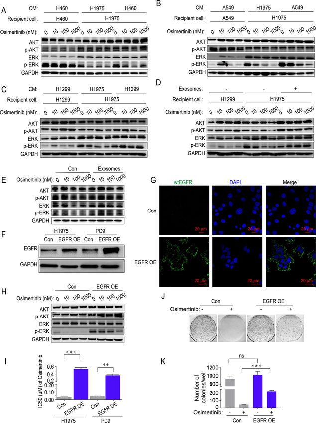
Wu et al. Molecular Cancer   (2021) 20:17                                                                     Page 2 of 17

Background                                                     ) [12–15]. Previous studies have showed that mutated
Lung cancer is a leading cause of cancer-related deaths        EGFR gene arise somatically during tumorigenesis. Only
worldwide. NSCLC is the major histological subtype of          a small sub-population of cancer cells carry the hetero-
lung cancer, which constitutes more than 85% of all lung       zygous activating EGFR mutations whereas others har-
cancer cases [1, 2]. While traditional cytotoxic chemother-    bor the wild type counterparts [16, 17]. Recently, the
apy remains an important treatment option for advanced         relationship between intratumor heterogeneity and re-
NSCLC, recent advancement in personalized medicine             sponsiveness to EGFR TKIs in NSCLC patients has been
allowed a subset of NSCLC patient harboring specific           extensively studied, especially at the gene level [16, 18–
oncogenic mutations to respond well to targeted therapy        20]. A higher percentage of cancer cells bearing the mu-
with minimal adverse reaction. In particular, molecular        tated EGFR gene within the tumor was highly correlated
targeted tyrosine kinase inhibitors (TKIs) specific to EGFR    with better responsiveness, longer progression-free sur-
have been extensively studied [3]. Importantly, the muta-      vival and overall survival in mutEGFR NSCLC patients.
tions located in the EGFR gene were more frequently            However, the specific mechanism underlying intratumor
found in mainland China adenocarcinoma NSCLC pa-               heterogeneity in EGFR mutation and osimertinib re-
tients (up to 50% ) [4]. NSCLC patients harboring these        sponsiveness remains unclear.
sensitizing EGFR mutations were exceptionally sensitive          Exosomes (40–150 nm) are small extracellular vesicles
to the reversible first-generation EGFR TKIs (gefitinib and    originating from multivesicular bodies (MVBs) produced
erlotinib) and the irreversible second-generation EGFR         inside the endosomal compartment of most eukaryotic
TKIs (afatinib and dacomitinib) [5, 6].                        cells. They are released into the extracellular milieu
   Despite the excellent initial clinical responsiveness and   upon the fusion of MVBs with plasma membranes. Exo-
disease control rates, these patients inevitably developed     somes act as mediators of cell-to-cell communication by
acquired resistance to EGFR TKIs within an average of          delivering molecular constituents of cells (including nu-
one year [7]. Among the various mechanisms leading to          cleic acids and proteins) as their cargoes, thereby alter-
EGFR TKIs resistance, the gatekeeper T790M point mu-           ing physiological state and biological functions of the
tation of EGFR close to the catalytic site is the most         recipient cells [21, 22]. Recently, exosomes have been
prevalent. EGFR T790M mutation is known to increase            shown to mediate the transfer of the drug resistance
the affinity of the receptor tyrosine kinase for adenosine     phenotype. Cancer cells would develop drug resistance
triphosphate (ATP) and also sterically hinder the binding      after the incorporation of exosomes from drug-resistant
of EGFR to TKIs, thereby leading to the failure of TKI         cancer cells. Among these investigations, the transfer of
treatment [8, 9]. Osimertinib is a novel mutant-selective      microRNAs and long non-coding RNAs (lncRNAs)
irreversible third-generation EGFR TKI, which exhibits         through exosomes is the most extensively studied [23–
minimal effect on wild type EGFR (wtEGFR) and there-           29]. In contrast, only a few recent studies reported about
fore less adverse effects, demonstrating potent antican-       the acquisition and spread of drug resistance by protein
cer activity in EGFR-mutated (mutEGFR) NSCLC                   cargoes delivered in exosomes [30–32].
patients harboring the T790M gatekeeper mutation [5].            In the clinic, the mutation status of EGFR gene in
After demonstrating robust objective response rate             NSCLC patients is an important determinant of therapy
(ORR) and prolonging progression-free survival (PFS) in        decision [33]. The recent findings about the influence of
the AURA study (NCT01802632), osimertinib has been             intratumor heterogeneity on the responsiveness of
clinically approved as first-line treatment of advanced        tumor to EGFR TKIs were intriguing [34]. This study
mutEGFR NSCLC patients regardless of T790M muta-               aims to investigate the possible intercellular transfer of
tion status [10, 11]. While the discovery of osimertinib       wtEGFR protein via exosomes to mutEGFR NSCLC cells
represents a breakthrough in the treatment of NSCLC,           and subsequently activates PI3K/AKT and MAPK signal-
all patients eventually relapsed and developed resistance      ing pathways and triggers cell resistance to osimertinib.
to the treatment. Importantly, there is no further effect-     Moreover, the effect of osimertinib on exosomes forma-
ive therapeutic option for these progressing patients          tion and secretion and the pathway of exosomes uptake
after failure of osimertinib.                                  were also investigated, which provides novel insights in
   Acquired resistance to osimertinib can be categorized       the field of osimertinib resistance induced by exosomes
into EGFR-dependent (such as EGFR C797S, G796D,                transfer.
G796S/R, L792F/Y/H, C797G, L718Q or L798I muta-
tion, exon19 deletion and wild type EGFR gene amplifi-         Materials and methods
cation, or EGFR T790M loss) and EGFR-independent               Chemicals and reagents
mechanisms (such as bypass pathway activation via              Osimertinib, gefitinib, afatinib, cisplatin and chlorpro-
MET or ERBB2, constitutive MAPK pathway activation             mazine (CPZ) were purchased from Selleck Chemicals
by mutated KRAS, MEK or Src-AKT pathway activation             (Houston, TX, USA). Osimertinib, gefitinib, afatinib and

Wu et al. Molecular Cancer   (2021) 20:17                                                                       Page 3 of 17

CPZ were prepared in dimethyl sulfoxide (DMSO) at a              at Beckman Coulter Avanti J30I at 4 °C). Exosomes were
concentration of 10 mM or 100 mM whereas cisplatin               sedimented, re-suspended and washed in PBS followed by
was dissolved in dimethyl formamide (DMF). All stock             another ultracentrifugation procedure and the pellet was
solutions were kept at − 20 °C. Stock solutions were di-         resuspended in 100 μL PBS.
luted to the appropriate concentrations with growth                For neutralizing assay, exosomes (20 μg) resolved in 1
medium right before administration. Cetuximab was                mL PBS were incubated with cetuximab (5 μg/mL) at
purchased from MedChemExpress (HY-P9905). PKH-67                 4 °C for 2 h and then performed ultracentrifugation at
and PKH-26 were purchased from Sigma-Aldrich                     150,000 g for 2 h to remove excess antibody before incu-
(MINI67, MINI26). Generally, for in vivo experiment,             bated with H1975 cells or PC9 cells for 6 h.
phospho-ERK1/2 (T204), ERK1/2, phospho-AKT1/2/3                    For sucrose density gradients, pellet collected from
(S473) and AKT were purchased from Cell Signaling                ultracentrifugation (80 μg) was re-suspended in PBS,
Technology (Danvers, MA, USA). For in vitro experi-              which was then overlaid with a linear sucrose gradient
ment, phospho-ERK1/2 (T204), ERK1/2, phospho-                    (10–70% w/v, pH 7.4) created by a gradient fractionator
AKT1/2/3 (S473) and AKT were purchased from Santa                (Biocomp YIQI 113) in a SW41 tube (Beckman Coulter).
Cruz Biotechnology (Dallas, TX, USA). CD63 and Alix              The gradients were subjected to ultracentrifuge (Beck-
antibodies were purchased from Santa Cruz Biotechnol-            man Coulter OptimaTM L-100 XP) at 100,000 g for 16 h
ogy (Dallas, TX, USA). EGFR and Caveolin-1 antibody              at 4 °C. Gradient fractions (1 mL) were collected from
were purchased from Cell Signaling Technology (Dan-              top to bottom and then the fractions were washed in
vers, MA, USA). TSG101 and Calnexin antibodies were              PBS followed by ultracentrifugation (Beckman Coulter
purchased from Affinity Biosciences (Cincinnati, OH,             Avanti J30I) in JA30.50 tube at 100,000 g at 4 °C for 2 h.
USA). RAB17 and GAPDH antibodies were purchased                  Pellets were directly lysed in RIPA buffer for further im-
from Proteintech Group (Rosemont, IL, USA). Clathrin             munoblot analysis.
heavy chain antibody were purchased from Beyotime
Biotechnology (Shanghai, CN). Polyclonal goat anti-              Exosomes characterization
mouse antibody and goat anti-rabbit antibodies were ob-          The exosome markers (TSG101, CD63, Alix) were used
tained from R&D systems (Minneapolis, MN, USA).                  as positive control whereas the endoplasmic reticulum
                                                                 protein Calnexin was used as negative control in West-
Cell culture                                                     ern blot analysis. Number and size distribution of exo-
H460, A549, H1299 (human NSCLC cell lines harboring              somes were analyzed by the Nanosight NS300 system
wtEGFR), H1975 (human NSCLC cell line harboring                  (Nanosight Technology, Malvern, UK) according to
L858R and T790M EGFR), PC9 (human NSCLC cell line                manufacturer’s instructions. Exosomes resuspended in
harboring Exon 19del EGFR) and K562 (human EGFR-                 50 μL PBS (pooled by 10 million cells for 48 h) were
null chronic myeloid leukemia cell line) were cultured in        negatively stained with 2% uranyl acetate solution and
DMEM medium (Gibco, USA) supplemented with 100                   imaged by JEM-1400 electron microscope (JEOL Ltd.,
U/mL penicillin, 100 U/mL streptomycin and 10% fetal             Japan), operated at 120 kV.
bovine serum (FBS) in a humidified incubator at 37 °C
with 5% CO2. To eliminate exosomes from FBS used in              Exosomes internalization analysis by flow cytometry
cell culture, diluted FBS (1,4) was centrifuged at 150,000       To calculate the internalization of exosomes, PC9 cells
g for 16 h at 4 °C in 70ti tube (Beckman Coulter Opti-           (1.5 × 10⁵/well) were seeded on 6-well plates. Exosomes
maTM L-100 XP).                                                  (5 μg/well) were stained with PKH-67 according to man-
                                                                 ufacturer’s instructions and then added onto the PC9
Exosomes isolation                                               cells under various condition. After incubation, cells
Cancer cells were plated at a density of 10 million cells        were washed with PBS twice and subjected to analysis
per T225 cm2 flask (Corning, USA) and cultured in                using flow cytometry.
DMEM contained 5% exosomes-depleted FBS for 48 h.
Cancer cells-derived conditioned medium was pooled and           Immunofluorescence
exosomes were isolated by differential centrifugation ac-        To visualize the internalization of exosomes, PC9 cells
cording to previous published protocol with minor modi-          (2 × 10⁵/mL) were seeded on 15 mm glass bottom cell
fications [31]. Briefly, after removing cells and debris by at   culture disk (Nest, Cat#801002). Exosomes (5 μg/mL)
300 g for 5 min and 2000 g for 25 min, the supernatant           were stained with PKH-26 according to manufacturer’s
was harvested and centrifuged (Beckman Coulter Avanti            instructions and then added onto the PC9 cells under
J30I) at 15,000 g for 30 min to remove large extracellular       various condition. After pre-incubation for 12 h, cells
vesicles. Finally, the supernatant was centrifuged at 100,       were washed with PBS twice and fixed with 4% parafor-
000 g for 90 min (all centrifugation steps were performed        maldehyde for 15 min at room temperature and stained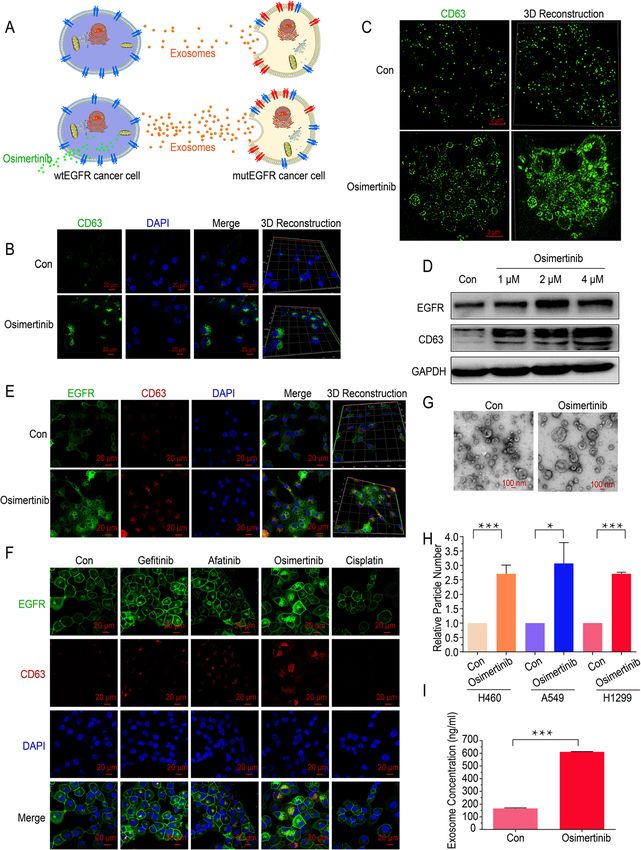
Wu et al. Molecular Cancer   (2021) 20:17                                                                       Page 4 of 17

with DAPI (5 μg/mL) for 15 min before imaging by                 analysis. The targeting sequences for specific genes were
ZEISS LSM880 confocal microscope.                                shown in Additional file: Table S1.
  To visualize the internalization of exosomal EGFR pro-
tein, H1975 cells (2 × 10⁵/mL) stably expressing RFP             Sanger sequencing of EGFR mutations
protein were seeded on 15 mm glass bottom cell culture           Genome DNAs of H460 cells, A549 cells, H1299 cells,
disk (Nest, Cat#801002). Exosomes (25 μg/mL) collected           H1975 cells and PC9 cells were extracted using DNA ex-
from H1299 cells expressing EGFR-GFP fusion protein              traction kit (TIANGEN, Cat#DP304–03) according to
were added onto the H1975 cells. After pre-incubation            the manufacturer’s instructions. The sequencing primers
for 6 h, cells were washed with PBS twice and fixed with         were shown in Additional file: Table S2.
4% paraformaldehyde for 15 min at room temperature
and stained with DAPI (5 μg/mL) for 15 min before im-            Western blot analysis
aging by ZEISS LSM880 or Nikon N-SIM confocal                    Expression of the proteins of interest was assessed by
microscope. For visualization of EGFR and CD63 protein           Western blot analysis, using the house-keeping protein
alteration, GFP or RFP-fusion protein were constructed           GAPDH for normalization. Briefly, cells were lysed in
accordingly for analysis. For visualization of the RAB17         RIPA buffer containing protease inhibitors and protein
and CD63 alteration after 36 h osimertinib treatment,            concentration was measured by BCA Protein Assay Kit
cells were washed with PBS twice and fixed with 4%               (Pierce Biotechnology, USA). Cell lysates were electropho-
paraformaldehyde for 15 min at 25 °C followed by mem-            resed in 10% SDS-PAGE gel and then transferred to a
brane permeabilization with 0.1% Triton X-100 for 10             polyvinylidene difluoride (PVDF) membrane (Millipore,
min. Then cells were blocked with 3% BSA for 45 min              USA). After blocking by 5% BSA, membranes were probed
and incubated at 4 °C with anti-CD63 mAb (1:250) and             with primary antibodies (1:1000) overnight at 4 °C and
anti-RAB17 antibody (1:250) diluent in 1% BSA for 1 h.           secondary antibody (1:5000) for 45 min at room
After incubating with secondary antibody conjugated              temperature. Protein bands were visualized by ECL im-
with AlexaFluor 488 or AlexaFluor 568 (1:1000) for 45            aging (Pierce ECL kit, Thermo Fisher Scientific, USA).
min and staining with DAPI, cells were subjected to im-
aging by ZEISS LSM880.                                           MTT assay
                                                                 Cells pre-treated with various conditions were harvested
                                                                 during logarithmic phase and seeded onto 96-well plates
Measurement of mean fluorescence intensity from
                                                                 at a density of 7000 cells/well in a final volume of
conditioned medium
                                                                 190 μL/well. After 12 h incubation, 10 μL of osimertinib,
To measure exosomes concentration, H1299 cells express-
                                                                 gefitinib, afatinib or cisplatin (indicated concentration)
ing CD63-GFP fusion protein was constructed and treated
                                                                 was added to 96-well plates. After 48 h treatment, 20 μL
with 4 μM osimertinib for 36 h in phenol-free DMEM
                                                                 MTT solution (5 mg/mL) was added to each well and
(Cat#21063029). After drug treatment, the supernatants
                                                                 incubated for 4 h at 37 °C. Then, the supernatant was
were pooled and centrifuged at 15,000 g for 30 min to re-
                                                                 discarded and MTT crystal was dissolved in 100 μL
move the dead cells, debris and large extracellular vesicles.
                                                                 DMSO. After incubating for 30 min, cell viability was
Fluorescence intensity was then measured using the Tecan
                                                                 measured by a microplate reader (Bio-Rad) at 570 nm
Spark fluorescence microplate reader.
                                                                 (using 630 nm as reference wavelength). The 50% inhibi-
                                                                 tory concentration (IC50) was determined using the Bliss
Construction and transfection of shRNA plasmid for               method. Experiments were performed at least three
RAB17, Caveolin-1, Clathrin and RAB27A knock-down                times.
Lentiviral shRNA vectors (pLKO.1 puro) targeting RAB17
were constructed according to standard procedure and             Apoptosis assay
the genetic sequence was verified in US National Center          Apoptosis assay was evaluated by Annexin V/PI apop-
for Biotechnology Information (NCBI). The shRNAs were            tosis detection kit (BD Biosciences; San Jose, CA) ac-
co-transfected with psPAX2 and pMD.2G into 293 T cells           cording to the manufacturer’s instructions. Briefly, after
at 70–80% confluence using Lipofectamine 2000 (Invitro-          treatment with 5 μM osimertinib for 36 h, cells were
gen) according to the manufacturer’s instructions. After         carefully harvested. After washing twice with PBS, cells
48 h, conditioned medium containing viral particles was          were stained with Annexin V and PI diluent for 15 min
pooled, filtered by 0.45 μm filter and cultured with H1299       and subjected to analysis using flow cytometry.
cells or PC9 cells for 24 h at 37 °C. At 48 h after lentiviral
infection, H1299 cells or PC9 cells were treated with puro-      Elisa
mycin (5 μg/mL). The changes in expression of the tar-           Briefly, H1299 cells were pre-treated with various con-
geted proteins were determined by Western blotting               centration of osimertinib for 36 h, after which the
Wu et al. Molecular Cancer   (2021) 20:17                                                                    Page 5 of 17

supernatant was pooled and centrifuged at 15,000 g for       Statistical analysis
30 min to remove dead cells, debris and large extracellu-    The data were showed as means ± SEM and analyzed by
lar vesicles. The expression levels of EGFR protein of the   one-way ANOVA or Student’s t-test analysis unless
supernatants were measured by Human EGFR ELISA               otherwise indicated. The statistical significance was de-
Kit (Solarbio, Cat#SEKH-0154) and the exosomes con-          termined to be p < 0.05. All statistical analyses were con-
centration was measured by Wako PS Capture™ exosome          ducted by SPSS 22.0 or GraphPad Prism 7.
ELISA Kit (WAKO, Cat# 297–79,201) according to the
manufacturers` instructions, respectively.                   Results
                                                             Intercellular transfer of exosomal wtEGFR protein confers
                                                             osimertinib resistance to sensitive mutEGFR cancer cells
Quantitative reverse transcription-PCR                       in vitro
Total cellular RNA was isolated by the Trizol reagent        Molecular intratumor heterogeneity is well recognized in
RNA extraction kit (Invitrogen, USA). Equal amount of        NSCLC and the percentage of EGFR-mutated cancer
RNA from different treatment groups was subjected to         cells has been reported to correlate well with the treat-
first-strain cDNA synthesis (EZBioscience, USA). After-      ment response to EGFR TKI [20]. To verify the effects
wards, real-time PCR was performed with Roche Light-         of NSCLC cells harboring wtEGFR on osimertinib-
Cycler 480. The expression of genes of interest in a         sensitive mutEGFR cancer cells proliferation and sur-
given sample was normalized to GAPDH. The 2-ΔΔCT             vival, NSCLC cells harboring wtEGFR (H460 cells, A549
method was applied to analyze the relative change in         cells, H1299 cells) and NSCLC cells with mutEGFR
gene expression. The primers were listed in Additional       (H1975 cells, PC9 cells) were employed. The EGFR mu-
file: Table S3.                                              tation status of four different NSCLC cells was con-
                                                             firmed by Sanger sequencing (Additional file: Fig. S1a)
Colony formation assay                                       and their sensitivity to osimertinib was evaluated. As ex-
Cancer cells (2000 cells per well) were plated onto 6 well   pected, H1975 cells and PC9 cells were much more sen-
plates and treated with 1 μM osimertinib for 24 h. Next,     sitive to osimertinib than other NSCLC cells bearing
culture medium was replaced with fresh DMEM con-             wtEGFR indicated by MTT assay (Additional file: Fig.
taining 10% FBS and cultured for another 8 days (ectopic     S1b).
wtEGFR-expressing assay) or 14 days (co-culture assay).         To examine the interaction between wtEGFR and
After that, the medium was removed and cells were            mutEGFR cancer cells, we established H1299 cells stably
washed with PBS twice, stained with 0.5% crystal violet,     expressing GFP and H1975 cells stably expressing RFP
washed with tap water and then subjected to imaging          to perform direct co-culture assay. As shown, the sur-
and data analysis.                                           vival of H1975 cells was dramatically increased upon osi-
                                                             mertinib treatment after cocultivation with H1299 cells
                                                             at a ratio of 1:1 (Fig. 1a and b). Furthermore, an in vitro
Establishment of in vivo xenograft tumor models              transwell cell culture assay was performed to exclude
In vivo experiments were conducted in accordance with        the influence of direct contact between the different cell
the guidelines for the use of laboratory animals of the      types on cell viability. Various cells (H460 cells, A549
Sun Yat-Sen University Institutional Animal Care and         cells, H1299 cells) were grown on the upper layer of a
Use Committee. H1975 cells (5 × 10⁶ cells) were sub-         cell culture insert whereas the other cells (H1975 cells)
cutaneously injected into the right flank of athymic nude    were grown on the surface of the bottom chamber, so
mice (BALB/c-nu/nu, female, 5 to 6 weeks old). When          that the two cell types were separated by a transwell
tumor volume reached 90 mm3, mice were randomized            membrane (pores 0.4 μm). Compared with H1975 cells
into three groups (seven in each group) and received dif-    alone, the IC50 value of osimertinib was significantly in-
ferent treatments: (a) Osimertinib (2–3 times/week, oral     creased in H1975 cells in the presence of upper layer
gavage, 5 mg/kg) plus exosomes (2–3 times/week, intra-       H460, A549 or H1299 cells (Fig. 1c). Moreover, H1975
tumor injection, 3 μg per mouse); (b) Osimertinib (2–3       cells were also incubated with the pooled conditioned
times/week, oral gavage, 5 mg/kg) plus PBS (2–3 times/       medium (CM) from NSCLC cells bearing wtEGFR (i.e.,
week, intratumorly); (c) Vehicle (5% DMSO+ 40%               H460 cells, A549 cells, H1299 cells), after which cell via-
PEG300 + 5% Tween80 + 50% ddH2O, 2–3 times/week,             bility (MTT and colony formation) and apoptosis assays
oral gavage) plus PBS (2–3 times/week, intratumorly).        were conducted following osimertinib treatment. Con-
Tumor size was measured with calipers every other day.       sistently, the presence of the CM from cancer cells bear-
Tumor volume was calculated by the formula:                  ing wtEGFR remarkably reduced the anti-proliferation
(length×width2/2). The mice were euthanized on day 29        and apoptotic effect induced by osimertinib in H1975
and the xenografts were excised and weighed.                 cells (Fig. 1d, Additional file: Fig. S1c-S1f).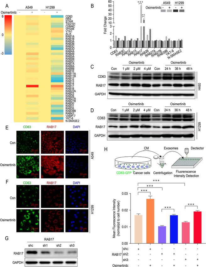
Wu et al. Molecular Cancer      (2021) 20:17                                                                                             Page 6 of 17

 Fig. 1 Intercellular transfer of exosomal wtEGFR protein confers osimertinib resistance to mutEGFR cancer cells in vitro. a-b Representative images
 of H1299 cells (GFP) alone, H1975 cells (RFP) alone or H1975 cells (RFP) co-cultured with H1299 cells (GFP) at a ratio of 1:1 treated by osimertinib
 (5 μM) for 72 h. Scale bar: 100 μm. c MTT assay of H1975 cells grown on the lower chamber indirectly pre-cultured with H460 cells, A549 cells and
 H1299 cells or control H1975 cells grown inside the transwell inserts (pores 0.4 μm) for 36 h. d MTT assay of H1975 cells pre-cultured with CM of
 H460 cells, A549 cells and H1299 cells or control H1975 cells for 36 h. e MTT assay of H1975 cells and PC9 cells pre-cultured with or without
 exosomes derived from H1299 cells for 6 h. f MTT assay of H1975 cells pre-cultured with CM or CM dExo derived from H1299 cells for 36 h. g-h
 Flow cytometric Annexin V/PI Apoptosis assay of H1975 cells pre-cultured with various concentration of exosomes for 6 h following osimertinib
 treatment (5 μM) for another 36 h. i MTT assay of H1975 cells pre-cultured with indicated exosomes (con, IgG or cetuximab pre-treated) derived
 from H1299 cells for 6 h. j-k Flow cytometric Annexin V/PI Apoptosis assay of H1975 cells pre-cultured with CM of K562 cells for 36 h following
 osimertinib treatment (5 μM) for another 36 h. All data are presented as means ± SEM. * P < 0.05, ** P < 0.01, *** P < 0.001

  To elucidate the mechanism by which wtEGFR-                                H1975 cells and PC9 cells after pre-treatment with the
bearing NSCLC cells endowed sensitive mutEGFR                                exosomes derived from H1299 cells (Fig. 1e). Import-
H1975 cells with osimertinib resistance, we isolated exo-                    antly, the osimertinib sensitiveness was rescued when
somes from CM of H1299 cells and then performed                              exosomes were depleted from CM (CM dExo) by ultra-
MTT and apoptosis assay to validate. As expected, the                        centrifugation or inhibited by knockdown of RAB27A
IC50 value of osimertinib were increased significantly in                    (Fig. 1f, Additional file: Fig. S1g-S1k), indicating the
Wu et al. Molecular Cancer   (2021) 20:17                                                                     Page 7 of 17

essential role of exosomes for the induction of osimerti-    were incubated with the CM of H1299 cells depleted of
nib resistance. Moreover, it was noteworthy that apop-       exosomes (Fig. 2e and f). Moreover, it was noteworthy
tosis rates of H1975 cells following osimertinib             that PKH-67-labeled H1299-derived exosomes could be
treatment were reduced dramatically in a concentration-      quickly internalized by PC9 cells within 12 h (Additional
dependent manner after pre-treatment with various con-       file: Fig. S2b and S2c). Furthermore, in order to visualize
centration of exosomes derived from H1299 cells (Fig.        the delivery of EGFR protein from H1299 cells to H1975
1g and h).                                                   cells, an EGFR-GFP fusion protein lentivirus plasmid
  Furthermore, to ascertain the functional role of           was engineered and transfected to H1299 cells. After
wtEGFR protein in the observed osimertinib resistance,       RFP-tagged H1975 cells were incubated with these
we applied cetuximab (5 μg/mL) as neutralized antibody       EGFR-GFP labeled H1299 cells-derived exosomes
to block exosomal wtEGFR and found that the osimerti-        (25 μg/mL), green fluorescence from EGFR-GFP was in-
nib resistance phenotype induced by exosomes derived         tegrated rapidly into the H1975 cells and could be de-
from H1299 cells was dramatically alleviated (Fig. 1i,       tected on the cell membrane by confocal microscopy
Additional file: Fig. S1l). Moreover, the EGFR-null K562     (Fig. 2g, Additional file: Fig. S2d,).
cells were employed to further validate (Additional file:       As known, there were two major endocytic pathways, a
Fig. S1m and S1n). As shown, pre-treatment with CM of        pathway associated with Caveolin-1 and another related to
K562 cells did not alter the apoptosis rates of H1975        Clathrin, facilitating receptor internalization [31]. To fur-
cells induced by osimertinib (Fig. 1j and k), implying the   ther investigate by which pathway cancer cells absorb exo-
essential role of EGFR in the observed osimertinib resist-   somes, we knocked down Caveolin-1 and Clathrin in PC9
ance. Additionally, it was worthy to note when cisplatin     cells using three shRNAs, respectively (Fig. 2h and i). As
was replacing osimertinib, there was ineligible difference   shown, the obvious decrease of Caveolin-1 did not de-
between the responsiveness of H1975 cells to cisplatin in    crease the internalization of exosomes in mutEGFR cancer
the presence or absence of supernatant from wtEGFR           cells, while knockdown of Clathrin contributed to a sig-
bearing NSCLC cells (Additional file: Fig. S1o). Collect-    nificant decrease of PKH-67-labeled cancer cells indicated
ively, intercellular transfer of exosomal wtEGFR protein     by flow cytometry (Fig. 2j and k). Moreover, chlorpromaz-
was responsible for the induction of osimertinib resist-     ine (CPZ), a pharmacological inhibitor of Clathrin, could
ance in sensitive mutEGFR cancer cells.                      remarkably decrease the internalization of exosomes in a
                                                             concentration-dependent manner (Fig. 2l and m, Add-
Exosomal wtEGFR protein can be internalized by sensitive     itional file: Fig. S2e).
mutEGFR cancer cells via Clathrin-mediated pathway              In conclusion, these results confirm that wtEGFR pro-
To further confirm whether exosomal wtEGFR protein           tein can be incorporated into exosomes and internalized
can be internalized by sensitive mutEGFR cancer cells,       by mutEGFR NSCLC cancer cells via Clathrin-mediated
exosomes were isolated from CM of H1299 cells by dif-        pathway.
ferential ultracentrifugation and characterized by West-
ern blot analysis (positive marker: CD63, TSG101, Alix;      Intercellular transfer of exosomal wtEGFR protein
negative marker: Calnexin; as well as transmembrane          activates PI3K/AKT and MAPK pathways in the presence
protein of EGFR) (Fig. 2a), Nanoparticle Tracking Ana-       of osimertinib in mutEGFR cancer cells
lysis (NTA) (Fig. 2b) and Transmission Electron Micro-       To ascertain whether the intercellular transfer of exoso-
scope (TEM) (Fig. 2c). Simultaneously, indicated by          mal wtEGFR protein activates EGFR downstream signal-
Western blot, EGFR protein was presented in exosomes         ing pathway in mutEGFR cancer cells under osimertinib
of H460 cells, A549 cells and H1299 cells (Additional        treatment, the status of the PI3K/AKT and MAPK path-
file: Fig. S2a). To further clarify whether EGFR protein     ways was evaluated after incubation with the CM from
was contained in exosomes, sucrose density gradient          wtEGFR-bearing NSCLC cells. As expected, higher level
centrifugation was performed and found that EGFR was         of phosphorylation ERK (p-ERK) were observed after
presented simultaneously with the exosome marker             H1975 cells pre-incubated with CM of H460 cells, A549
CD63 (Fig. 2d), suggesting that EGFR was preferentially      cells or H1299 cells, while the level of p-AKT was slight
encapsulated into exosomes.                                  elevated because of the limited inhibitory of osimertinib
   To investigate whether exosomal wtEGFR can be de-         on p-AKT in control H1975 cells (Fig. 3A-3C). Add-
livered to the osimertinib-sensitive cancer cells, H1975     itionally, the p-AKT and p-ERK were totally not blocked
cells were cultured with the CM of H1299 cells or            by osimertinib even up to 1000 nM in mutEGFR NSCLC
H1299 cells-derived exosomes and found that the EGFR         cells pre-incubated with H1299 cells-derived exosomes
protein was elevated without a significant change in         whereas there were moderate inhibition on p-AKT and
EGFR mRNA expression. However, EGFR protein and              dramatic decrease of p-ERK in control mutEGFR NSCL
mRNA level were not notably changed when H1975 cells         C cells (Fig. 3 d and e).
Wu et al. Molecular Cancer (2021) 20:17 Page 8 of 17 Fig. 2 (See legend on next page.)
Wu et al. Molecular Cancer (2021) 20:17 Page 9 of 17
(See figure on previous page.)
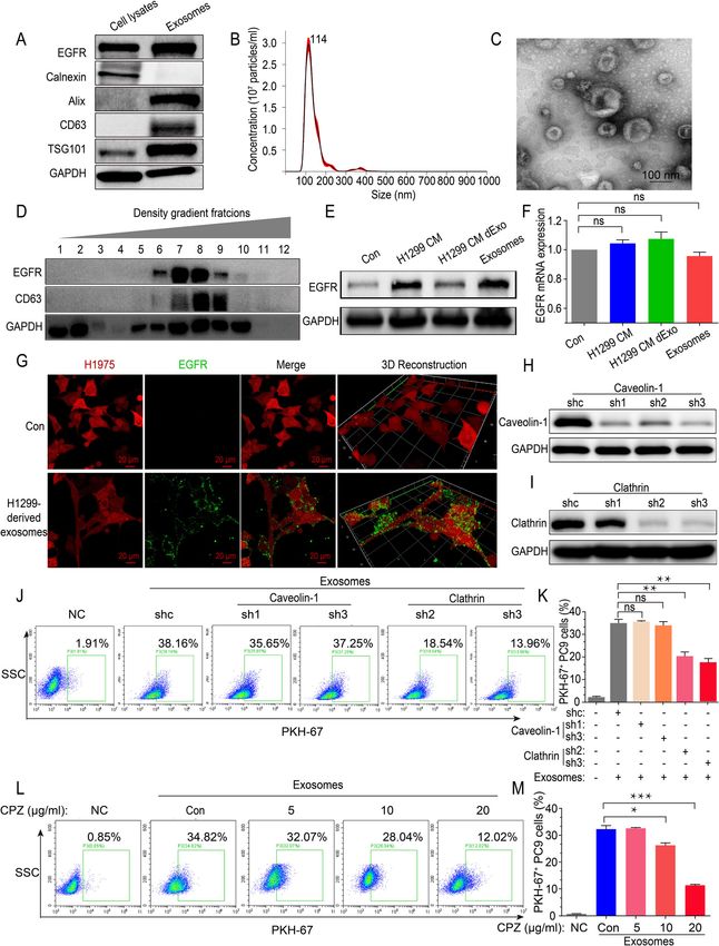
Fig. 2 Exosomal wtEGFR protein can be internalized by sensitive mutEGFR cancer cells via Clathrin-mediated pathway. a Western blot analysis of
indicated exosomal markers (positive markers: CD63, TSG101, Alix; negative marker: Calnexin) in H1299 whole cell lysates and H1299 cells-derived
exosomes. b Nanoparticle Tracking Analysis of H1299 cells-derived exosomes. c Transmission Electron Microscope imaging of H1299 cells-derived
exosomes. Scale bar: 100 nm. d Western blot analysis of colocalization of exosomal marker CD63 and transmembrane protein EGFR in various
extracellular vesicle fractions as separated by sucrose density gradient centrifugation. e Western blot analysis of EGFR protein level of H1975 cells after
pre-incubation with CM of H1299 cells, CM dExo of H1299 cells and H1299 cells-derived exosomes. f Real-time quantitative analysis of EGFR mRNA
level of H1975 cells after pre-incubation with CM of H1299 cells, CM dExo of H1299 cells and H1299 cells-derived exosomes. g Representative images
of internalization of H1299 cells-derived exosomes containing GFP-tagged wtEGFR in H1975 cells (RFP). Scale bar: 20 μm. (H) Western blot analysis of
Caveolin-1 protein expression to verify the efficacy of Caveolin-1 silencing by shRNAs. i Western blot analysis of Clathrin protein expression to verify
the efficacy of Clathrin silencing by shRNAs. j-k Flow cytometric exosomes absorption assay of control PC9 cells or Caveolin-1- and Clathrin-silenced
counterparts treated with PKH-67-labeled exosomes (5 μg/mL) for 12 h. l-m Flow cytometric exosomes absorption assay of PC9 cells pre-cultured with
various concentration of CPZ for 2 h following PKH-67-labeled exosomes (5 μg/mL) incubation for another 12 h. All data are presented as means ±
SEM. * P < 0.05, ** P < 0.01, *** P < 0.001
More importantly, to further verify the effect of file: Fig. S3a). In addition, it was noteworthy that we per-
wtEGFR protein on mutEGFR cancer cells, we ectopi- formed confocal imaging and found that EGFR expres-
cally overexpressed wtEGFR (Flag-tag) in mutEGFR sion was also increased and colocalized with exosomes
NSCLC cells and found that it was mainly localized in in presence of osimertinib (Fig. 4e, Additional file: Fig.
the plasma membrane confirmed by confocal micros- S3b). Likewise, to explore whether other anticancer
copy (Fig. 3 f and g). Moreover, in H1975 cells ectopi- drugs such as gefitinib, afatinib, and cisplatin also pro-
cally overexpressing wtEGFR, the p-ERK was totally not mote the formation of exosomes in wtEGFR-expressing
inhibited by osimertinib and the level of p-AKT was NSCLC cells, confocal imaging was conducted and the
much higher when compared with control H1975 cells results showed they did not significantly alter the forma-
(Fig. 3h). Further analysis revealed that ectopic overex- tion of exosomes in H1299 cells (Fig. 4f).
pression of wtEGFR in H1975 cells and PC9 cells signifi- Furthermore, to elucidate the effect of osimertinib on
cantly increased cell survival and colony formation the secretion of exosomes, TEM was conducted and
ability following osimertinib treatment (Fig. 3I-3K). indicated no alteration in the morphology of secreted
Taken together, these results suggest that the exosomal exosomes from H1299 cells after osimertinib treatment
wtEGFR might play an active role in activating PI3K/ (Fig. 4g). In addition, we pooled cell culture supernatant
AKT and MAPK pathways in the presence of osimerti- of wtEGFR-expressing NSCLC cells and detected the
nib in mutEGFR cancer cells. number of small particles (presumably exosomes) by
NTA. As expected, osimertinib dramatically promoted
Osimertinib promotes the formation and secretion of the secretion of exosomes (Fig. 4h). To further verify
exosomes in wtEGFR-expressing NSCLC cells this, exosomes derived from the CM of H1299 cells were
The data above demonstrates that exosomes from captured in Tim-4 immobilized plates and CD63 protein
wtEGFR- expressing NSCLC cells result in osimertinib level was detected by ELISA as exosomes concentration.
resistance in mutEGFR NSCLC cells. To further illus- Consistently, there was a significant increase of secreted
trate the mechanisms in detail, the effect of osimertinib exosomes after osimertinib treatment (Fig. 4i). Hence,
on the formation and secretion of exosomes in NSCLC we speculate that osimertinib promotes the formation
cells was also investigated (Fig. 4a). We first investigated and secretion of exosomes in wtEGFR-expressing NSCL
the effect of osimertinib on the formation of exosomes C cells.
in wtEGFR-expressing NSCLC cells. As shown, the clas-
sical exosome marker CD63 was remarkably increased Osimertinib promotes the release of exosomes by
in H1299 cells after treated with osimertinib (4 μM) (Fig. upregulating RAB17
4b). Furthermore, the location and quantified level of To investigate the molecular mechanism that enhanced
exosomes were assessed by Structured Illumination Mi- release of exosomes induced by osimertinib, quantitative
croscopy (N-SIM) and found that the intracellular for- real-time PCR was performed in A549 cells and H1299
mation of exosomes dramatically increased in the cells to confirm the alterations of gene expression which
presence of osimertinib compared with that in the ab- were critical for endosome formation or exosomes secre-
sence of osimertinib in H1299 cells (Fig. 4c). Consist- tion, including those encoding ESCRT proteins, Rab
ently, an increasing of CD63 protein level in wtEGFR- GTPase family and other related molecules (Fig. 5a). As
expressing NSCLC cells was observed in a shown, only RAB17 was identified as a key regulator in
concentration-dependent manner following osimertinib both A549 cells and H1299 cells exposed to osimertinib
treatment indicated by Western blot (Fig. 4d, Additional (Fig. 5b). The treatment of osimertinib led to a dramaticWu et al. Molecular Cancer (2021) 20:17 Page 10 of 17 Fig. 3 (See legend on next page.)
Wu et al. Molecular Cancer      (2021) 20:17                                                                                           Page 11 of 17

 (See figure on previous page.)
 Fig. 3 Intercellular transfer of exosomal wtEGFR protein activates PI3K/AKT and MAPK pathways in mutEGFR cancer cells. a Western blot analysis
 of indicated proteins in H1975 cells pre-treated with CM of H460 cells or control H1975 cells for 36 h and various concentration of osimertinib for
 another 6 h. b Western blot analysis of indicated proteins in H1975 cells pre-treated with CM of A549 cells or control H1975 cells for 36 h and
 various concentration of osimertinib for another 6 h. c Western blot analysis of indicated proteins in H1975 cells pre-treated with CM of H1299
 cells or control H1975 cells for 36 h and various concentration of osimertinib for another 6 h. d Western blot analysis of indicated proteins in
 H1975 cells pre-treated with H1299 cells-derived exosomes or PBS for 6 h and various concentration of osimertinib for another 6 h. e Western
 blot analysis of indicated proteins in PC9 cells pre-treated with H1299 cells-derived exosomes or PBS for 6 h and various concentration of
 osimertinib for another 6 h. f Western blot analysis of EGFR protein in H1975 cells and PC9 cells with or without ectopic overexpression of
 wtEGFR protein. g Representative images of membrane-anchored wtEGFR (Flag-tag) in H1975 cells. Labels: blue, nucleus; green, wtEGFR (Flag-
 tag). Scale bar: 20 μm. h Western blot analysis of indicated proteins in H1975 cells treated with various concentration of osimertinib with or
 without ectopic overexpression of wtEGFR protein. i MTT assay of H1975 cells and PC9 cells with or without ectopic overexpression of wtEGFR
 protein. j-k Colony formation assay of H1975 cells with or without ectopic overexpression of wtEGFR protein after treated by osimertinib (1 μM)
 for 24 h. All data are presented as means ± SEM. * P < 0.05, ** P < 0.01, *** P < 0.001

increase of RAB17 or CD63 protein level in a concentra-                      followed by osimertinib (5 mg/kg) treatment at one day
tion- and time-dependent manner (Fig. 5c and d, Add-                         after exosomes injection. While osimertinib remarkably
itional file: Fig. S3c). Moreover, it was worthy to note                     retarded tumor growth as expected, it is noteworthy that
that an increase of RAB17 and CD63 protein level was                         all treatment groups including the osimertinib+exo-
simultaneously detected in A549 cells and H1299 cells                        somes group did not cause appreciably animal body
by confocal microscopy (Fig. 5e and f).                                      weight loss (Fig. 6b). At the end point of measurement
   To further confirm the critical role of RAB17 on the                      (day 16), the osimertinib+exosomes group showed a lar-
release of exosomes, RAB17 expression was silenced in                        ger tumor size and heavier tumor weight than the osi-
H1299 cells using three different shRNAs and the effi-                       mertinib+PBS group (Fig. 6c and d). Additionally,
cient knock-down of RAB17 protein was verified by                            intratumor administration of H1299 cells-derived exo-
Western blot (Fig. 5g). In order to measure exosomes                         somes into the xenograft tumor significantly inhibited
concentration more efficiently, a new fluorescence-based                     osimertinib-mediated reduction in tumor volume (Fig.
detection method was employed. Briefly, we established                       6e and f). Importantly, the activation status of the PI3K/
H1299 cells expressing CD63-GFP fusion protein                               AKT and MAPK pathways were also evaluated by West-
(H1299 CD63-GFP+ cells) with or without RAB17 silen-                         ern blot. Consistent with the in vitro data, osimertinib
cing. The CM from these CD63-GFP+ cells was collected                        alone effectively inhibited the p-AKT and p-ERK and the
and the GFP fluorescence intensity was measured after                        co-administration of osimertinib and H1299 cells-
centrifugation. Using this fluorescence-based assay, we                      derived exosomes led to a considerable elevation of p-
found that osimertinib significantly promoted the release                    AKT and p-ERK (Fig. 6g). Collectively, these results
of exosomes (Fig. 5g), which was consistent with previ-                      demonstrate that exosomes derived from wtEGFR-
ous results of NTA and ELISA (Fig. 4h and i). Import-                        expressing H1299 cells induce osimertinib resistance in
antly, when RAB17 expression was knocked down in                             mutEGFR H1975 cells by activating PI3K/AKT and
H1299 cells by shRNAs, exosomes secreted by H1299                            MAPK pathways in vivo.
CD63-GFP+ cells were significantly reduced but this was
rescued by osimertinib (Fig. 5h). Collectively, RAB17 is                     Discussion
essential for the promotion of exosome release by                            Osimertinib is administrated as first-line treatment in
osimertinib.                                                                 advanced NSCLC patients harboring mutEGFR [10, 11].
                                                                             While the discovery of osimertinib represents a break-
Intercellular transfer of exosomal wtEGFR confers                            through in the treatment of NSCLC, almost all patients
osimertinib resistance to mutEGFR H1975 xenograft                            eventually relapsed and no further effective therapeutic
in vivo                                                                      options for the progressing patients. Mechanisms leading
To investigate the role of exosomes in osimertinib resist-                   to osimertinib resistance are multifactorial and most in-
ance in vivo, xenograft tumor model of mutEGFR                               vestigations focused on genetic alteration of EGFR (in
H1975 cells was established in nude mice by subcutane-                       particular, C797S), and MET and HER2 amplification
ously injection of the cells into the right flank of the ex-                 [12, 13]. Recent researches showed that the status of
perimental animals. Different treatment interventions                        intratumor heterogeneity was also responsible for osi-
were carried out shown in scheme after palpable tumors                       mertinib resistance. However, the specific mechanism
have developed in the mice (Fig. 6a). H1299 cells-                           remains unclear. In this study, we found osimertinib sig-
derived exosomes (3 μg) were injected into the H1975                         nificantly promoted the formation and secretion of exo-
xenograft tumors 2–3 times per week, which was                               somes in wtEGFR-expressing NSCLC cells. These
Wu et al. Molecular Cancer (2021) 20:17 Page 12 of 17 Fig. 4 Osimertinib promotes the formation and secretion of exosomes in wtEGFR-expressing NSCLC cells. a Graphical scheme showing our working model of the promotion of exosomes release by osimertinib. b Representative images of cellular expression and localization of GFP-tagged CD63 in H1299 cells with or without osimertinib treatment (4 μM) for 12 h. DAPI is used for nucleus staining. Scale bar: 20 μm. c N-SIM imaging of cellular localization of GFP-tagged CD63 in H1299 cells with or without osimertinib treatment (4 μM) for 12 h. Scale bar: 2 μm. d Western blot analysis of indicated proteins level in H1299 cells treated by various concentration of osimertinib for 36 h. e Representative images of cellular expression and localization of GFP-tagged EGFR and RFP-tagged CD63 in H1299 cells with or without osimertinib treatment (4 μM) for 12 h. DAPI is used for nucleus staining. Scale bar: 20 μm. f Representative images of cellular expression and localization of GFP-tagged EGFR and RFP-tagged CD63 in H1299 cells treated by gefitinib (4 μM), afatinib (4 μM), osimertinib (4 μM) or cisplatin (50 μM) for 12 h. DAPI is used for nucleus staining. Scale bar: 20 μm. g Transmission Electron Microscope imaging of exosomes derived from H1299 cells with or without osimertinib treatment for 36 h. Scale bar: 100 nm. h Nanoparticle Tracking Analysis of exosomes in CM of H460 cells, A549 cells and H1299 cells after osimertinib treatment (4 μM) for 36 h. i Exosomes concentration in CM of H1299 cells after osimertinib treatment (4 μM) for 36 h measured by ELISA. All data are presented as means ± SEM. * P < 0.05, ** P < 0.01, *** P < 0.001 exosomes were enriched in wtEGFR protein and could promoted the release of exosomes by upregulating be delivered to mutEGFR NSCLC cells, resulting in acti- RAB17 and then these exosomes could be internalized vation of PI3K/AKT and MAPK pathways under the via Clathrin-mediated pathway. Figure 6h depicts a stress of osimertinib. Mechanistically, osimertinib working model by which wtEGFR-incorporated
Wu et al. Molecular Cancer      (2021) 20:17                                                                                           Page 13 of 17

 Fig. 5 Osimertinib promotes the release of exosomes by upregulating RAB17. a Real-time quantitative PCR Screening for genes expression
 alteration encoding ESCRT or Rab GTPase family molecules in A549 cells and H1299 cells after osimertinib treatment (4 μM) for 12 h. Fold change
 in log2 transformation is shown as a color scheme. b Further validation of the changes in selected mRNAs in A549 cells and H1299 cells after
 osimertinib treatment (4 μM) by real-time quantitative PCR. c Western blot analysis of Rab17 and CD63 protein expression in H460 cells treated
 with various concentration of osimertinib for 36 h or treated with osimertinib (4 μM) for indicated time point. d Western blot analysis of Rab17
 and CD63 protein expression in H1299 cells treated with various concentration of osimertinib for 36 h or treated with osimertinib (4 μM) for
 indicated time point. e Representative images of cellular expression and localization of RAB17 and CD63 protein in A549 cells after osimertinib
 treatment (4 μM) for 36 h. DAPI is used for nucleus staining. Scale bar: 20 μm. f Representative images of cellular expression and localization of
 RAB17 and CD63 protein in H1299 cells after osimertinib treatment (4 μM) for 36 h. DAPI is used for nucleus staining. Scale bar: 20 μm. g Western
 blot analysis of RAB17 protein expression to verify the efficacy of RAB17 silencing by shRNAs. h Mean Fluorescence Intensity of GFP (indicating
 CD63 expression) in CM of H1299 cells or RAB17-silenced counterparts after treated by osimertinib (4 μM). All data are presented as means ±
 SEM. * P < 0.05, ** P < 0.01, *** P < 0.001

exosomes play a novel role in mediating osimertinib                         noteworthy that just a part of cancer cells carries hetero-
resistance.                                                                 zygous activating mutations, whereas others harbor wild
  It is well recognized that tumors are inherently hetero-                  type counterparts. In 2011, Zhou et al. investigated the
geneous and their plastic phenotypes allow them to re-                      correlation between EGFR mutation abundance and
sist treatment [35, 36]. In an individual patient, it is                    clinical benefit from gefitinib treatment and found that
Wu et al. Molecular Cancer     (2021) 20:17   Page 14 of 17

 Fig. 6 (See legend on next page.)
Wu et al. Molecular Cancer       (2021) 20:17                                                                                             Page 15 of 17

 (See figure on previous page.)
 Fig. 6 Intercellular transfer of exosomal wtEGFR confers osimertinib resistance to mutEGFR H1975 xenograft in vivo. a Schematic diagram depicting
 the in vivo experimental procedure. b Line graph showing the change in body weight of mice during experiment (n = 7). c Comparison of the tumor
 volume of three indicated treatment groups at end point (n = 7). d Comparison of the tumor weight of three indicated treatment groups at end point
 (n = 7). e Tumor growth curve at the indicated days with different treatments in BALB/c-nu/nu mice (n = 7). f Macroscopic view of tumor harvested at
 termination of three indicated treatment groups (n = 7). g Western blot analysis of indicated proteins level in xenograft tumors from three indicated
 treatment groups. h Working model showing the induction of osimertinib resistance by intercellular delivery of wtEGFR protein in exosomes from
 osimertinib-insensitive wtEGFR-expressing NSCLC cells to osimertinib-sensitive mutEGFR NSCLC cells. All data are presented as means ± SEM. * P < 0.05,
 ** P < 0.01, *** P < 0.001

median progression-free survival was significantly longer                        Exosomes are novel mediators of cell-to-cell commu-
in patients with high abundances of EGFR mutations, in-                       nication to modulate cell signaling and biological func-
dicating the relative EGFR mutation abundance could                           tion in recipient cells [22]. Recently, tumor-derived
potentially predict clinal benefit from EGFR-TKI treat-                       exosomes have been shown to mediate the transfer of
ment. Moreover, a study conducted by Guo et al. ana-                          drug resistance phenotype. Most researches in this area
lyzed 120 single tumor cells and confirmed the                                focused on the intercellular cross-talk by transferring
intratumor heterogeneity of EGFR-activating mutations                         miRNA or lncRNA from donor to recipient cells [23–
in lung adenocarcinoma on the single-cell level, which                        29]. However, both miRNAs and lncRNAs can simultan-
might closely relate to EGFR-TKIs response in lung                            eously affect multiple biological targets, which makes it
adenocarcinoma patients [16, 18–20]. Overall, the detec-                      difficult to predict their ultimate biological function and
tion of EGFR mutation abundance is highly recom-                              limits their clinical application. In NSCLC patients, the
mended in the clinic and could potentially predict                            EGFR tyrosine kinase play a central role on tumorigen-
EGFR-TKI response in NSCLC patients harboring                                 esis and the sensitivity to EGFR-targeted therapy [42].
mutEGFR.                                                                      Reported by several studies, EGFR protein could be in-
  Furthermore, NSCLC tumors bearing EGFR mutations                            corporated into exosomes and delivered to the recipient
are believed to express wtEGFR which could limit the                          cells to promote metastasis or enhance antiviral immun-
clinical responsiveness and lead to shorter PFS upon osi-                     ity [43–45]. In our study, we confirmed that exosomes
mertinib treatment. This hypothesis is partly supported                       carrying wtEGFR protein could be secreted from NSCL
by the findings that some NSCLC patients harboring                            C cells harboring wtEGFR and incorporated into the cy-
EGFR mutation are refractory to osimertinib but they                          tomembrane of mutEGFR H1975 cells within several
are responsive to erlotinib or afatinib [37, 38]. It has also                 hours, thus inducing osimertinib resistance. Moreover,
been reported that NSCLC patients who developed drug                          the depletion of exosomes from CM of H1299 cells, as
resistance during osimertinib targeted therapy could be                       well as the employment of neutralized body and CM of
partially overcome by afatinib [39, 40]. Therefore, we hy-                    EGFR-null K562 cells, could not induce osimertinib re-
pothesized that selective pressure from osimertinib treat-                    sistance in H1975 cells, at least in part. Given that exo-
ment allows tumor cells bearing wtEGFR to                                     somes are known to be stable in various biological
predominate and the cessation of osimertinib treatment                        fluids, it is possible that the expression level of exosomal
may lead to the resurgence of mutEGFR. Consistently, a                        wtEGFR protein detected in plasma sample from NSCL
recent clinical trial conducted by Ichihara et al. demon-                     C patients could be utilized to predict their responsive-
strated that re-administration of osimertinib to NSCLC                        ness to osimertinib treatment. Indeed, normal cells and
previously showed acquired resistance could lead to                           cancer cells would both secrete exosomes that contain-
tumor shrinkage once again with objective response and                        ing EGFR (data not shown). The differences of these
disease control rates of 33 and 73%, respectively [41].                       exosomal EGFR remain largely unexplored and it is a
Taken together, it is reasonable that the status of intra-                    challenging work to define the origin of them. Further
tumor heterogeneity might partially determine the effect                      investigation about the clinical translation of our find-
of osimertinib on mutEGFR NSCLC patients. Our study                           ings is warranted.
revealed that osimertinib-resistant wtEGFR-expressing                            Inspired by previous report that chemotherapeutic
NSCLC cells could secrete exosomal wtEGFR protein                             drugs could promote the secretion of extracellular vesicles
and deliver to osimertinib-sensitive cancer cells to acti-                    carrying the multidrug resistance transporter ABCB1 from
vate the MAPK pathway, thus inducing osimertinib re-                          multidrug resistant cancer cells and transfer to neighbor-
sistance. To this end, the co-administration of                               ing sensitive cells [31], the effect of osimertinib on exo-
osimertinib and first- or second- generation EGFR TKIs                        somes biogenesis was also investigated in wtEGFR-
might represent a useful strategy to overcome osimerti-                       expressing NSCLC cells in our study. As expected, we
nib resistance.                                                               found that exosomes formation and secretion were
Wu et al. Molecular Cancer          (2021) 20:17                                                                                                     Page 16 of 17

dramatically increased in wtEGFR-expressing NSCLC cells                           Funding
following osimertinib (4 μM) treatment. Furthermore,                              National Science & Technology Major Project “Key New Drug Creation and
                                                                                  Manufacturing Program”, China (No: 2018ZX09711002), the National Natural
screening aiming at molecules critical for endosome for-                          Science Foundation of China (No: 81673463).
mation or exosomes secretion identified a member of the
Rab GTPase family, RAB17, as the major contributor to                             Availability of data and materials
the observed induction of exosomes formation and secre-                           The datasets generated and/or analysed during the current study are
                                                                                  available on the Research Data Deposit public platform (www.researchdata.
tion by osimertinib. Unlike other Rab GTPases, RAB17 is                           org.cn). The approved RDD number is RDDB2021001047.
expressed most abundantly in epithelial cells. It has been
extensively studied in the delivery of hepatic transcytotic                       Ethics approval and consent to participate
vesicles [46]. Recently, RAB17 has been identified as a                           Animal study was performed with the permission of the institutional
                                                                                  committee of Sun Yat-sen University Cancer Center, in compliance with pro-
tumor suppressor gene and low expression of RAB17 pro-                            tocols approved by the Guangdong Provincial Animal Care and Use Commit-
motes the tumorigenesis via activation of the ERK path-                           tee and experimental guidelines of the Animal Experimentation Ethics
way in hepatocellular carcinoma [47]. Honestly, our study                         Committee of Sun Yat-sen University Cancer Center.
is the first to report the regulation of formation and secre-
                                                                                  Consent for publication
tion of exosomes by RAB17 under exposure of osimerti-                             Not applicable.
nib. Further studies are warranted to elucidate the
mechanisms leading to the upregulation of RAB17 by osi-                           Competing interests
mertinib and the subsequent promotion of exosome                                  The authors declare that they have no competing interests.

secretion.                                                                        Author details
                                                                                  1
                                                                                   State Key Laboratory of Oncology in South China, Collaborative Innovation
                                                                                  Center for Cancer Medicine; Guangdong Esophageal Cancer Institute; Sun
Conclusions                                                                       Yat-sen University Cancer Center, Guangzhou 510060, People’s Republic of
In summary, our finding demonstrates that intercellular                           China. 2School of Pharmacy, Faculty of Medicine, The Chinese University of
transfer of wtEGFR protein promotes osimertinib resist-                           Hong Kong, Room 801N, Area 39, Lo Kwee-Seong Integrated Biomedical
                                                                                  Sciences Building, Shatin, New Territories, Hong Kong, SAR, China. 3School of
ance by activating PI3K/AKT and MAPK signaling path-                              Pharmaceutical Sciences, Guangzhou Medical University, Guangzhou 511436,
ways both in vitro and in vivo. Moreover, osimertinib                             China.
promotes the formation and secretion of exosomes by up-
                                                                                  Received: 25 September 2020 Accepted: 2 January 2021
regulating RAB17, which represents novel molecular tar-
get for possible circumvention of osimertinib resistance.
                                                                                  References
                                                                                  1. Andrews Wright NM, Goss GD. Third-generation epidermal growth factor
Supplementary Information                                                             receptor tyrosine kinase inhibitors for the treatment of non-small cell lung
The online version contains supplementary material available at https://doi.          cancer. Transl Lung Cancer Res. 2019;8(Suppl 3):S247–64.
org/10.1186/s12943-021-01307-9.                                                   2. Siegel RL, Miller KD, Jemal A. Cancer statistics, 2019. CA Cancer J Clin. 2019;
                                                                                      69(1):7–34.
 Additional file 1: Figure S1. NSCLC cells harboring wtEGFR confer                3. Hirsch FR, Scagliotti GV, Mulshine JL, Kwon R, Curran WJ, Wu Y-L, Paz-Ares L.
 osimertinib resistance to sensitive mutEGFR cancer cells in vitro. Figure            Lung cancer: current therapies and new targeted treatments. Lancet. 2017;
 S2. Exosomal wtEGFR protein can be uptake by mutEGFR NSCLC cells via                 389(10066):299–311.
 Clathrin. Figure S3. Osimertinib promotes the release of exosomes via            4. Shi Y, Au JS, Thongprasert S, Srinivasan S, Tsai CM, Khoa MT, Heeroma K,
 RAB17. Table S1. shRNAs for RAB17, Caveolin-1, Clathrin and RAB27A.                  Itoh Y, Cornelio G, Yang PC. A prospective, molecular epidemiology study of
 Table S2. Sequencing primers for EGFR mutation. Table S3. qPCR                       EGFR mutations in Asian patients with advanced non-small-cell lung cancer
 primers for screening and validation                                                 of adenocarcinoma histology (PIONEER). J Thoracic Oncol. 2014;9(2):154–62.
                                                                                  5. Piotrowska Z, Sequist LV. Treatment of EGFR-mutant lung cancers after
                                                                                      progression in patients receiving first-line EGFR tyrosine kinase inhibitors : a
Abbreviations                                                                         review. JAMA Oncol. 2016;2(7):948–54.
CPZ: Chlorpromazine; EGFR: Epidermal growth factor receptor; wtEGFR: Wild         6. Wu S, Fu L. Tyrosine kinase inhibitors enhanced the efficacy of conventional
type EGFR; mutEGFR: EGFR-mutated; MVBs: Multivesicular bodies; N-                     chemotherapeutic agent in multidrug resistant cancer cells. Mol Cancer.
SIM: Structured Illumination Microscopy; NSCLC: Non-small cell lung cancer;           2018;17(1):25.
NTA: Nanoparticle Tracking Analysis; TEM: Transmission Electron Microscope;       7. Yu HA, Arcila ME, Rekhtman N, Sima CS, Zakowski MF, Pao W, Kris MG, Miller
TKI: Tyrosine kinase inhibitors                                                       VA, Ladanyi M, Riely GJ. Analysis of tumor specimens at the time of
                                                                                      acquired resistance to EGFR-TKI therapy in 155 patients with EGFR-mutant
                                                                                      lung cancers. Clin Cancer Res. 2013;19(8):2240–7.
Acknowledgements                                                                  8. Yun CH, Mengwasser KE, Toms AV, Woo MS, Greulich H, Wong KK,
This work was supported by grants from the National Science & Technology              Meyerson M, Eck MJ. The T790M mutation in EGFR kinase causes drug
Major Project “Key New Drug Creation and Manufacturing Program”, China                resistance by increasing the affinity for ATP. Proc Natl Acad Sci U S A. 2008;
(No: 2018ZX09711002), the National Natural Science Foundation of China                105(6):2070–5.
(No: 81673463).                                                                   9. Recondo G, Facchinetti F, Olaussen KA, Besse B, Friboulet L. Making the first
                                                                                      move in EGFR-driven or ALK-driven NSCLC: first-generation or next-
Authors’ contributions                                                                generation TKI? Nat Rev Clin Oncol. 2018;15(11):694–708.
Conception and design: SW, LF; Conducting experiments: SW, ML, CS, HZ,            10. Ramalingam SS, Yang JCH, Lee CK, Kurata T, Kim D-W, John T, Nogami N,
DC, SA, FW; Acquisition of data: SW, ML, JZ, TKK, HZ; Analysis of data: SW, JZ,       Ohe Y, Mann H, Rukazenkov Y, et al. Osimertinib as first-line treatment of
FW; Writing the manuscript: SW, TKK, LF. All authors had read and approved            EGFR mutation–positive advanced non–small-cell lung Cancer. J Clin Oncol.
the final manuscript.                                                                 2017;36(9):841–9.
You can also read