Inhibition of discoidin domain receptors by imatinib prevented pancreatic fibrosis demonstrated in experimental chronic pancreatitis model
←
→
Page content transcription
If your browser does not render page correctly, please read the page content below
www.nature.com/scientificreports
OPEN Inhibition of discoidin domain
receptors by imatinib prevented
pancreatic fibrosis demonstrated
in experimental chronic
pancreatitis model
Sapana Bansod2, Mohd Aslam Saifi1 & Chandraiah Godugu1*
Discoidin domain receptors (DDR1 and DDR2) are the collagen receptors of the family tyrosine
kinases, which play significant role in the diseases like inflammation, fibrosis and cancer. Chronic
pancreatitis (CP) is a fibro-inflammatory disease in which recurrent pancreatic inflammation leads
to pancreatic fibrosis. In the present study, we have investigated the role of DDR1 and DDR2 in CP.
The induced expression of DDR1 and DDR2 was observed in primary pancreatic stellate cells (PSCs)
and cerulein-induced CP. Subsequently, the protective effects of DDR1/DDR2 inhibitor, imatinib
(IMT) were investigated. Pharmacological intervention with IMT effectively downregulated DDR1
and DDR2 expression. Further, IMT treatment reduced pancreatic injury, inflammation, extracellular
matrix deposition and PSCs activation along with inhibition of TGF-β1/Smad signaling pathway. Taken
together, these results suggest that inhibition of DDR1 and DDR2 controls pancreatic inflammation
and fibrosis, which could represent an attractive and promising therapeutic strategy for the treatment
of CP.
Abbreviations
CP Chronic pancreatitis
CTGF Connective tissue growth factor
DDRs Discoidin domain receptors
ECM Extracellular matrix
IMT Imatinib
NF-κB Nuclear factor kappa B
PSCs Pancreatic stellate cells
PDGF Platelet-derived growth factor
α-SMA α-Smooth muscle actin
TGF-β Transforming growth factor-β
Chronic pancreatitis (CP) is a fibro-inflammatory disease of the pancreas, characterized by recurrent pancreatic
inflammation and fibrosis leading to irreversible impairment of exocrine and endocrine functions of pancreas1.
CP is a major risk factor for the progression of diabetes and pancreatic cancer in the developed c ountries2.
Although there has been an extensive research in CP during last few years, but the exact pathogenesis and spe-
cific molecular mechanism of CP needs to be elucidated. The well-established theory behind CP development
is premature activation of pancreatic digestive enzymes leading to autodigestion of pancreatic tissue and the
recruitment of the excess inflammatory cells that results in pancreatic inflammation3. Pancreatic fibrosis is a
complex pathological event in which wherein dysregulation of production and degradation of extracellular matrix
(ECM) is observed in the pancreas. Persistent inflammation causes the activation of pancreatic fibroblasts, known
as pancreatic stellate cells (PSCs) which promote development of pancreatic fibrosis. Upon activation, PSCs
undergo morphological changes and initiate pancreatic fibro-inflammation through the secretion of a number of
1
Department of Regulatory Toxicology, National Institute of Pharmaceutical Education and Research (NIPER),
Balanagar, Hyderabad, Telangana 500037, India. 2Division of Oncology, Department of Internal Medicine,
Washington University School of Medicine, St Louis, MO, USA. *email: chandra.niperhyd@gov.in
Scientific Reports | (2021) 11:12894 | https://doi.org/10.1038/s41598-021-92461-z 1
Vol.:(0123456789)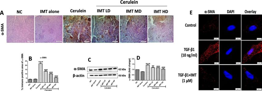
www.nature.com/scientificreports/
inflammatory mediators and excessive synthesis of ECM proteins such as collagen I, III and fibronectin4. Among
these mediators, transforming growth factor (TGF)-β1 is the potent profibrotic cytokine which regulates PSCs
activation and pancreatic fibrogenesis through its downstream Smad signaling p athway5. Currently, CP treatment
is only limited to the supportive care and there is no clinically available therapeutic agent to treat CP and associ-
ated fibrosis progression. Thus, there is a pressing need to identify novel therapeutic agents for the treatment of
CP on high priority, which could be helpful to reduce costs of long-term therapy and improve the quality of life.
Discoidin domain receptors (DDR1 and DDR2) are new class of tyrosine kinase receptors, which get activated
in response to collagens and are involved in the early embryonic d evelopment6. DDR1 is majorly expressed in
epithelial cells and activated by both fibrillar and nonfibrillar collagens (type I to V, VIII, and XI), while DDR2
is expressed in fibroblasts and activated by fibrillar collagens (type I and III). This binding causes phosphoryla-
tion and dimerization of the tyrosine kinase receptors which then leads to the activation of different signaling
pathways involved in inflammation and fi brosis7. Accumulating evidences have reported that upregulated expres-
sion of DDR1 and DDR2 are observed in the chronic injury, inflammation and fibrotic conditions8. Multiple
studies reported that inhibition of DDR1 reduced deposition of collagen I and IV in alcoholic liver fibrosis, lung
fibrosis, cirrhotic liver and chronic n ephropathies9–11. On the other hand, DDR2 is a key regulator of epithelial
to mesenchymal transition (EMT) program, which is involved in several pathological changes such as fibrosis
and tumor p rogression12. Although, the DDRs are involved in inflammatory and fibrotic diseases but their role
in CP and associated fibrosis is not explored fully.
Imatinib mesylate (IMT) is a standard Food and Drug Administration (FDA)-approved drug for the treatment
of patients with chronic myelocytic leukemia. IMT is the only tyrosine kinase inhibitor which potentially inhibits
both the profibrotic cytokines, TGF-β and PDGF which are involved in regulation of organ fibrosis process.
Emerging studies have shown that IMT has the potent tyrosine kinase DDR1 and DDR2 inhibitory activity13.
IMT is the type-2 DDRs inhibitor which inhibits tyrosine kinase domain by leveraging the ATP binding site as
well as the allosteric site, which is freely accessible in the inactive conformation14. However, the effect of IMT
on CP has not been studied yet and the underlying molecular mechanism remains to be explored. Therefore, in
the present study, we investigated the role of DDR1 and DDR2 in progression of CP and associated fibrosis. In
addition, we investigated the protective effects of pharmacological inhibitor of DDR1 and DDR2, IMT against
CP and pancreatic fibrosis. Our results showed that inhibition of DDR1 and DDR2 signaling by IMT could
effectively attenuate CP and associated fibrosis and further indicate that DDR1 and DDR2 could be the potential
therapeutic targets for treatment of CP.
Materials and methods
Reagents. Imatinib mesylate (IMT) was procured from Sigma Aldrich, USA. Cerulein was obtained from
Ana Spec, France. TGF-β1 was procured from Bio-legend, USA. The antibodies viz. β-actin (Catalogue no.
sc-47778), Collagen1a (Catalogue no. sc-393573), Collagen3a (Catalogue no. sc-271249), α-SMA (Catalogue no.
sc-53142), CTGF (Catalogue no. sc-373936), TGF-β1 (Catalogue no. sc-146), DDR2 (Catalogue no. sc-81707),
pSmad2/3 (Catalogue no. #8828), Smad2/3 (Catalogue no. #8685), pDDR1 (Catalogue no. #14531), DDR1 (Cat-
alogue no. #5583), pNF-κB (Catalogue no. #3033), NF-κB (Catalogue no. #8242) were procured from Santa
Cruz Biotechnology Ltd and Cell Signaling Technology, USA. The ELISA kits of TGF-β, IL-1β, IL-6 and TNF-α
(Catalogue no. TGF-β: 88–8350, IL-1β: 88–7013, IL-6: 88- 7064, and TNF-α: 88–7324) were obtained from
eBioscience, USA.
PSCs isolation, culture and treatment. PSCs were isolated from the mice according to the methods as
described with slight modifications15. Briefly, pancreas was isolated from adult male Swiss albino mice. Isolated
pancreas was immediately washed in sterile PBS. A small portion of pancreas was cut, minced with scissors and
single cell suspension was prepared using 1 ml syringe in Dulbecco’s Modified Eagle medium (DMEM). Next,
cell suspension was centrifuged followed by two times washing with PBS. Cell’s pellet was mixed and cultured in
DMEM medium containing 1% antibiotic solution and supplemented with 20% fetal bovine serum (FBS). PSCs
were separated by using the Histodenz density gradient centrifugation m ethod16.
Oil‑red O staining for PSCs identification. After 70% confluency, PSCs were fixed with 4% paraform-
aldehyde and 0.1% Triton X-100 reagents and stained with oil-red O working solution (1% in isopropanol) at 60
℃ for 30 min. Next, PSCs were counterstained using hematoxylin, mounted with mounting medium, Dibutyl-
phthalate Polystyrene Xylene (DPX) and observed under the microscope17.
Confocal microscopy. Freshly isolated and cultured PSCs were treated with IMT (1 μM) in the presence
or absence of TGF-β1 (10 ng/ml). After 24 h of treatment, cells were fixed with 4% paraformaldehyde and 0.1%
Triton X-100 followed by blocking with 3% bovine serum albumin (BSA) for 1 h. The cells were then labeled
with primary antibodies against α-SMA, collagen 1a, pSmad2/3, pDDR1, and DDR2 at 4 °C for overnight. Next
day, cells were probed with secondary antibody conjugated with rhodamine (1:200 dilution) or fluorescein iso-
thiocyanate (FITC) (1:100 dilution) for 1 h. After washing, cells were mounted with fluoroshield 4′,6-diamidino-
2-phenylindole (DAPI) (Sigma-Aldrich, USA) medium and fluorescent signals were captured by confocal laser-
scanning microscopy (Leica, Germany)18.
Animals and experimental design. Animals were kept in well-controlled housing facility at temperature
(25 ± 2 °C) and 12/12-h light/dark cycle with free access to water and pellet food. The Animal experiments were
designed, conducted and reported as per the ARRIVE guidelines19. Specifically, all the animal experiments were
performed according to the Committee for the Purpose of Control and Supervision of Experiments on Animals
Scientific Reports | (2021) 11:12894 | https://doi.org/10.1038/s41598-021-92461-z 2
Vol:.(1234567890)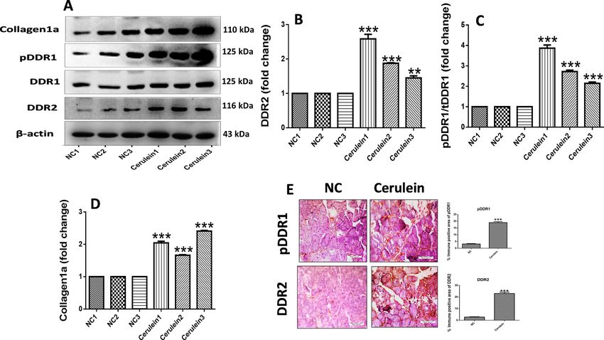
www.nature.com/scientificreports/
Figure 1. Experimental design for development and treatment of CP in mice. Male Swiss albino mice were
randomly divided into 6 groups (7 mice per group): Normal control, IMT alone, Cerulein, Cerulein + IMT
low dose (IMT LD—1 mg/kg), Cerulein + IMT mid dose (IMT MD—3 mg/kg) and Cerulein + IMT high dose
(IMT HD—10 mg/kg). CP was induced by 6-hourly intraperitoneal (i.p.) injections of cerulein (50 μg/kg), 3
alternative days per week, for the period of 3 weeks. Cerulein + IMT group mice were administered IMT (1, 3
and 10 mg/kg) orally, every other day after cerulein exposures for 3 weeks. Normal control group mice received
normal saline same as cerulein group. IMT alone group mice were administrated with IMT (10 mg/kg) orally 4
alterative days in the week for 3 weeks.
(CPCSEA), which is the approval body for animal experimentation in India. The CPCSEA certified Institutional
Animal Ethical Committee (IAEC) of National Institute of Pharmaceutical Education and Research (NIPER)-
Hyderabad, has reviewed the animal protocol and approved it (IAEC Protocol Approval No.: NIP/01/2019/
RT/365).
Male Swiss albino mice (age: 6–8 weeks, body weight 25–30 g) were purchased from Palamur Biosciences
Pvt. Ltd, Mahabubnagar, India. All mice were randomly divided into 6 groups (7 mice per group, n = 42): Normal
control, IMT alone, Cerulein, Cerulein + IMT low dose (LD—1 mg/kg), Cerulein + IMT mid dose (MD—3 mg/
kg) and Cerulein + IMT high dose (HD—10 mg/kg). CP was induced by 6-hourly exposures of cerulein (50 μg/
kg/intraperitoneal (i.p.), 3 alternative days per week, for the period of 3 weeks as per our previously published
protocol20. Cerulein + IMT group mice were administered IMT (1, 3 and 10 mg/kg) orally, every other day after
cerulein exposures for 3 weeks. Normal control animals received i.p. injection of sterile normal saline same as
cerulein group. IMT alone group mice were administrated with IMT (10 mg/kg) orally 4 alterative days in the
week for 3 weeks (Fig. 1). Mice were sacrificed via inhalation euthanasia (CO2 asphyxiation) after completion
of 21 days. Doses of IMT were selected on the basis of previous l iterature21. IMT was dissolved in distilled water
and administrated orally 4 alterative days in the week for 3 weeks. As cerulein needed minimum 12 h to produce
its maximum inflammatory response, IMT was given every other day after cerulein exposures for a period of
3 weeks20.
Blood collection and plasma analysis. Blood samples were collected by cardiac puncture on the day of
sacrifice in heparin containing tubes and plasma was separated after centrifugation. Amylase and lipase levels
were assessed in plasma and measured by using Accurex kinetic enzymatic kits and values are expressed as IU/
L20,22.
Collagen estimation by Sircol assay. Pancreatic tissues were homogenized in PBS and then superna-
tants were incubated with collagen binding dye picrosirius red for 1 h at 37 °C. Next, samples were centrifuged
and obtained pellet was resuspended in 100% ethanol followed by centrifugation at 9168×g for 10 min. Then
pellet was mixed with 0.5 M NaOH solution and incubated for 30 min at 37 °C. The final absorbance was taken
rotein23.
at 540 nm and values were expressed as relative collagen content per milligram of p
Enzyme‑linked immunosorbent assay. Cytokine concentrations in pancreas were determined by eBio-
science ELISA kits for IL-6, IL-1β, TNF-α and TGF-β1 as per the previously described protocol24. Concentration
of cytokines was expressed as pg per milligram of protein. Total protein was estimated by Bicinchoninic acid
assay kit (Sigma Aldrich, USA).
Histopathology and immunohistochemistry analysis. Pancreas were fixed in 10% formaldehyde
solution and paraffin-embedded pancreatic sections were taken at 5 µm using microtome (Leica, Germany). The
pancreatic sections were stained with H & E, picrosirius red (PSR) and Masson’s trichrome (MT) as per previ-
ously described protocol25. For immunostaining, antigen retrieval was carried out by citrate buffer. After wash-
ing, sections were incubated with 3% hydrogen peroxide for 15 min followed by blocking with 3% BSA. The pri-
mary antibodies against α-SMA, collagen1a, pDDR1 and DDR2 (1:100) were added and kept at 4 ℃ overnight.
The HRP-conjugated secondary antibodies were added and sections were stained with 3, 3-diaminobenzidine
(DAB) reagent. Further, sections were counterstained with hematoxylin and images were observed using a light
Scientific Reports | (2021) 11:12894 | https://doi.org/10.1038/s41598-021-92461-z 3
Vol.:(0123456789)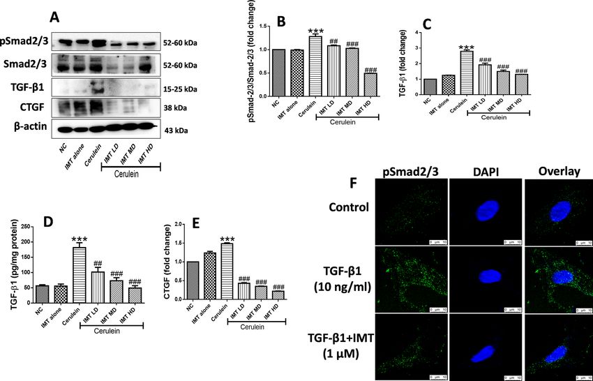
www.nature.com/scientificreports/
Figure 2. Upregulation of DDR1 and DDR2 in pancreatic fibrosis. (A–D) Western blot and corresponding
densitometric analysis of protein expression of phosphorylated DDR1, total DDR1, DDR2 and collagen1a in
pancreatic tissue. (E) Representative photographs of immunohistochemistry of phosphorylated DDR1 and
DDR2 in the pancreatic tissue. β-actin was used as loading control. All values are represented as mean ± SEM
(n = 3–10). Statistical significance was established using Student’s t-test where **P < 0.01, ***P < 0.001 vs Normal
control. Full-length blots are presented in Supplementary Fig. S1.
microscope (CX21i Olympus, India)26. Quantitative analysis of fibrotic and immunopositive area were analyzed
using ImageJ software (NIH, USA).
Immunoblotting. To analyze the pancreas protein expression, immunoblotting technique was used as
described earlier27,28. Small portions of pancreatic tissues were homogenized in RIPA lysis buffer. Then equal
amounts of proteins were separated using SDS-PAGE gel electrophoresis and transferred to a nitrocellulose
membrane. The protein blots were then detected using ECL (Bio-Rad Laboratories, USA) and densitometric
analysis of respective protein bands was carried out by ImageJ software (NIH, USA).
Statistical analysis. All results are given as mean ± SEM. Student’s t-test was used to determine the differ-
ence between two groups. However, differences among more than two groups were analyzed using nonparamet-
ric test one-way analysis of variance by GraphPad Prism scientific software version 6.01. Value of P less than 0.05
was considered statistically significant.
Results
Upregulation of DDR1 and DDR2 expression in pancreatic fibrosis. We evaluated the expression
of DDR1 and DDR2 via western blot and immunohistochemical analysis in CP mice. Interestingly, we found
marked upregulation of phosphorylated DDR1, DDR2 and collagen1a expression in pancreatic tissue of CP mice
(Fig. 2A–D, Supplemental Fig. S1). In addition, immunohistochemical (IHC) staining and quantification analy-
sis demonstrated that expression of DDR1 and DDR2 was significantly increased in CP model mice as compared
to the normal control mice (Fig. 2E). These results indicated the upregulation of DDR1 and DDR2 receptors in
the progression of CP and associated pancreatic fibrosis.
Inhibition of DDR1 and DDR2 prevents progression of pancreatic fibrosis. Next, to investigate
whether collagen receptors DDR1 and DDR2 play key role in the development of CP and associated fibrosis or
not, we evaluated the effects of DDR1/DDR2 inhibitor, IMT in cerulein-induced CP model. Treatment with
IMT (1, 3 and 10 mg/kg) showed significant downregulation of phosphorylated DDR1 and DDR2 expression in
a dose-dependent manner (Fig. 3A–C, Supplemental Fig. S2). In addition, we found that cerulein-treated mice
demonstrated significantly higher collagen expression, whereas oral administration of IMT dose-dependently
reduced collagen expression as compared to cerulein-control mice (Fig. 3D,E). We further assessed the effects
of IMT on TGF-β1-induced PSCs activation by confocal microscopy. Our results demonstrated that TGF-β1-
treated PSCs showed notably upregulated pDDR1, DDR2 and collagen1a expression and the expression of these
proteins was markedly inhibited by IMT treatment (Fig. 3F,G). Together, these results suggest that inhibition of
DDR1 and DDR2 expression suppressed collagen deposition and pancreatic fibrosis.
Scientific Reports | (2021) 11:12894 | https://doi.org/10.1038/s41598-021-92461-z 4
Vol:.(1234567890)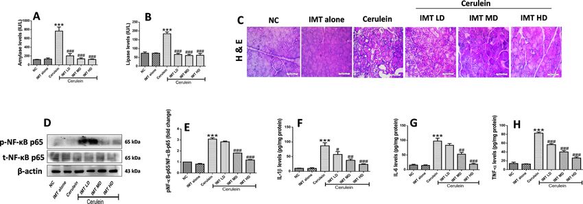
www.nature.com/scientificreports/
Figure 3. Inhibition of DDR1 and DDR2 expression prevents progression of pancreatic fibrosis. (A–E)
Western blot and corresponding densitometric analysis of protein expression of phosphorylated DDR1, total
DDR1, DDR2, collagen1a and collagen3a in pancreatic tissue. (F–H) Immunofluorescence (IF) staining for
phosphorylated DDR1, DDR2 and collagen1a in TGF-β1-induced PSCs. β-actin was used as loading control.
All values are represented as mean ± SEM (n = 3). Statistical significance was established using one-way ANOVA
followed by Tukey’s multiple comparisons test where ***P < 0.001 vs Normal control, ###P < 0.001 vs Cerulein.
Full-length blots are presented in Supplementary Fig. S2.
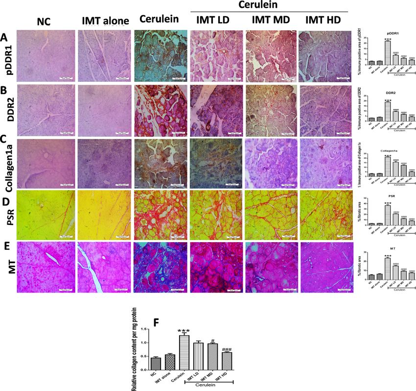
IMT controls collagen deposition in the pancreas. IHC staining and their quantification analysis
showed that the expression of DDR1, DDR2 and collagen1a were strongly increased in CP mice. However, IMT
treatment prevented the elevated pancreatic DDR1, DDR2, and collagen1a expression as confirmed by IHC
analysis (Fig. 4A–C). Further, pancreatic sections of cerulein-treated mice showed marked pancreatic fibrosis
illustrated by red and blue color in PSR and MT staining, respectively around the pancreatic ducts. Interestingly,
treatment with DDRs inhibitor, IMT significantly attenuated collagen deposition in the pancreas (Fig. 4D,E).
In addition, Sircol assay was performed to measure total collagen content in the pancreas. Our results demon-
strated that CP mice demonstrated significantly increased collagen content and this collagen content was effec-
tively reduced by IMT treatment (Fig. 4F).
IMT attenuates cerulein‑induced pancreatic injury and inflammation. Next, we investigated the
protective effects of IMT against cerulein-induced CP model. Cerulein challenged mice demonstrated signif-
icant elevation in plasma amylase and lipase levels, while IMT administration effectively reduced these lev-
els (Fig. 5A,B). Further, to evaluate the anti-inflammatory effects of IMT, histopathological examination was
performed in the pancreatic tissue by H&E staining. Our results revealed that the hallmark characteristics of
CP including acinar cell atrophy, vacuolization, inflammatory cells infiltration and collagen deposition were
observed in the cerulein control pancreas. On the other hand, pharmacological intervention with IMT mark-
edly ameliorated cerulein-induced histopathological alterations as shown in Fig. 5C. These results suggest that
IMT attenuated cerulein-induced pancreatic injury in CP mice. Furthermore, we examined the effect of IMT
on NF-κB p65 expression by western blot analysis. We found that the expression of phosphorylated NF-κB p65
was significantly increased in cerulein challenged mice, while IMT mid dose and high dose mice demonstrated
significant inhibition of phosphorylated NF-κB expression in the pancreatic tissues (Fig. 5D,E, Supplemental
Fig. S3). In addition to this, we observed that repetitive cerulein exposure significantly increased the cytokine
production in the pancreas, while IMT treatment markedly suppressed these cytokines levels, indicating down-
regulation of local production of pro-inflammatory cytokines (Fig. 5F–H). Collectively, our results indicated
that IMT shows potent anti-inflammatory effects and prevent pancreatic injury in CP mice.
IMT inhibits the activation of PSCs studied in‑vivo and in‑vitro model. PSCs activation is a major
event involved in the synthesis and accumulation of ECM proteins resulting in the development of pancreatic
fibrosis. In response to chronic injury, quiescent PSCs convert to an activated PSCs synthesizing excessive ECM
proteins. Our in-vivo results showed that the expression of α-SMA was profoundly increased in cerulein-treated
CP mice, indicating increased number of activated PSCs during CP. On the other hand, pharmacological treat-
ment with IMT significantly inhibited the PSCs activation as demonstrated by downregulation of α-SMA expres-
sion in the pancreas (Fig. 6A–D, Supplemental Fig. S4). Consistent with the in-vivo results, we observed marked
upregulation of α-SMA expression in TGF-β1-stimulated mouse PSCs. However, treatment with IMT success-
Scientific Reports | (2021) 11:12894 | https://doi.org/10.1038/s41598-021-92461-z 5
Vol.:(0123456789)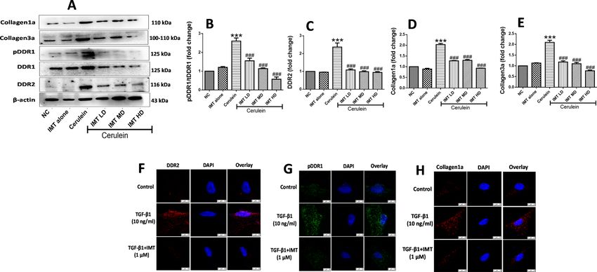
www.nature.com/scientificreports/
Figure 4. IMT controls collagen deposition in the pancreas (A–C) Representative photographs and
quantitative analysis of immunohistochemistry of phosphorylated DDR1, DDR2 and collagen1a protein,
(D,E) Representative photographs and quantitative analysis of PSR and MT staining in the pancreatic tissue,
(F) Relative collagen content in pancreas estimated by Sircol assay. All values are represented as mean ± SEM
(n = 10). Statistical significance was established using one-way ANOVA followed by Tukey’s multiple
comparisons test where ***P < 0.001 vs Normal control, #P < 0.05, ###P < 0.001 vs Cerulein.
fully decreased α-SMA expression, indicating the effective inhibition of PSCs activation by IMT (Fig. 6E). These
results clearly indicated that IMT successfully inhibited PSCs activation.
IMT inhibits TGF‑β/Smad signaling. TGF-β/Smad is a well-established fibrotic signaling pathway which
gets activated in response to the injuries (Fig. 7A, Supplemental Fig. S5). Further, we found that IMT treatment
significantly downregulated the expression of phosphorylated Smad2/3 in CP mice (Fig. 7B). In addition to
this, our results demonstrated that there was a significant increase in TGF-β1 expression in cerulein- chal-
lenged mice. Treatment with IMT significantly inhibited expression of TGF-β1 in a dose-dependent manner
(Fig. 7C,D). Furthermore, our western blot results revealed the expression of CTGF was markedly increased
in cerulein-induced CP mice, which was effectively inhibited by IMT treatment (Fig. 7E). On the other hand,
TGF-β1-induced PSCs also showed marked increase in phosphorylated Smad2/3 expression and this expression
was effectively downregulated by IMT treatment (Fig. 7F). Taken together, these results indicated that IMT sig-
nificantly inhibited TGF-β/Smad signaling in CP model and in PSCs.
Discussion
CP is a fibro-inflammatory disease which is majorly associated with abnormal synthesis and deposition of ECM
in the pancreas leading to the development of pancreatic fi brosis18,20,25. Although, there are multiple signaling
pathways involved in the pathogenesis of CP, activation of collagen-producing PSCs is the foremost event in
the progression of pancreatic fibrosis. PSCs are the main cells of pancreas involved in the ECM deposition and
Scientific Reports | (2021) 11:12894 | https://doi.org/10.1038/s41598-021-92461-z 6
Vol:.(1234567890)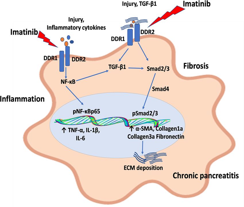
www.nature.com/scientificreports/
Figure 5. Effect of IMT on cerulein-induced pancreatic injury and inflammation. (A,B) Plasma amylase
and lipase levels. (C) Representative photographs of H & E staining of pancreas at 400X magnification. (D,E)
Western blot and corresponding densitometric analysis of protein expression of total and phosphorylated
NF-κB in pancreas, (F–H) Pancreatic IL-1β, IL-6 and TNF-α levels studied by ELSA. β-actin was used as loading
control. All values are represented as mean ± SEM (n = 7). Statistical significance was established using one-
way ANOVA followed by Tukey’s multiple comparisons test where ***P < 0.001 vs Normal control, #P < 0.05,
##
P < 0.01, ###P < 0.001 vs Cerulein. Full-length blots are presented in Supplementary Fig. S3.
Figure 6. Effect of IMT on pancreatic stellate cells activation (PSCs). (A,B) Representative photographs of
immunohistochemistry and quantitative analysis of α-SMA protein in the pancreatic tissue, (C,D) Western
blot and corresponding densitometric analysis of protein expression of α-SMA, (E) Immunofluorescence (IF)
staining for α-SMA in TGF-β1-induced PSCs. β-actin was used as loading control. All values are represented
as mean ± SEM (n = 3–10). Statistical significance was established using one-way ANOVA followed by Tukey’s
multiple comparisons test where ***P < 0.001 vs Normal control, ##P < 0.01, ###P < 0.001 vs Cerulein. Full-length
blots are presented in Supplementary Fig. S4.
activation of fibrotic signaling during pancreatic fibrosis. It is well-known that TGF-β1, a profibrotic cytokine
induces the activation of PSCs resulting in excess synthesis and secretion of collagen by PSCs consequently
leading to pancreatic fibrosis29.
Collagen-activated receptors, DDRs gained a considerable amount of attention because of their involvement
in the multiple pathological conditions and are classified into 2 types: DDR1 and D DR230–32. These receptors
33
are majorly involved in both physiological as well as pathological p rocesses . Recently, studies have reported
that collagen receptors DDR1 and DDR2 play a central role in the regulation of inflammation and fibrotic
conditions34,35. Apart from this, DDR1 and DDR2 receptors also play a decisive role in the activation of myofi-
broblasts and TGF-β/Smad signaling which is majorly involved in the initiation and progression of fi brosis36.
The hallmark feature of DDRs is that they are directly activated upon binding to components of ECM proteins
which then activates inflammatory and fibrotic signaling pathways37. Notwithstanding, the exact involvement
of DDR1 and DDR2 in CP and associated pancreatic fibrosis remains unclear and has not been studied yet. In
CP, repetitive injury of the pancreas trigger release of variety of cytokines and chemokines, which results in the
activation of collagen-producing PSCs accompanied by excessive accumulation of ECM.
Scientific Reports | (2021) 11:12894 | https://doi.org/10.1038/s41598-021-92461-z 7
Vol.:(0123456789)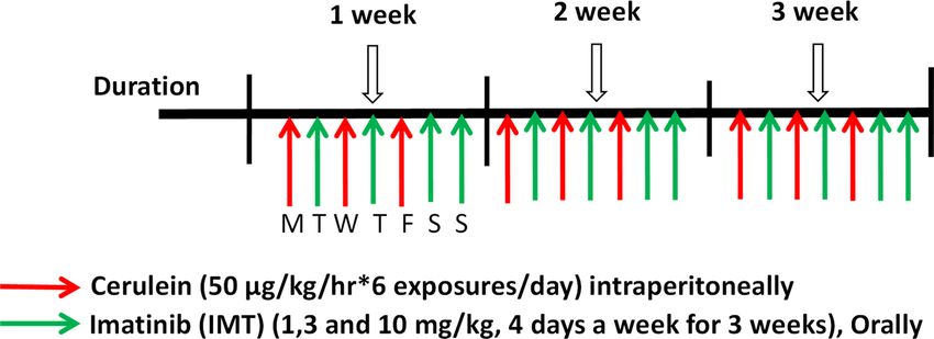
www.nature.com/scientificreports/
Figure 7. Effect of IMT on TGF-β1/Smad signaling pathway. (A–D) Western blot and corresponding
densitometric analysis of protein expression of phosphorylated and total Smad2/3, TGF-β1 and CTGF in
pancreatic tissue, (E) Pancreatic TGF-β1 levels studied by ELISA. (F) IF staining for phosphorylated Smad2/3
in TGF-β1-induced PSCs. β-actin was used as loading control. All values are represented as mean ± SEM (n = 3).
Statistical significance was established using one-way ANOVA followed by Tukey’s multiple comparisons test
where ***P < 0.001 vs Normal control, ##P < 0.01, ###P < 0.001 vs Cerulein. Full-length blots are presented in
Supplementary Fig. S5.
The significant upregulation of DDRs was observed in the present study which suggested that these receptors
play a major role in the development of pancreatic fibrosis. The results were consistent with previous literature
where activation of DDR1 and DDR2 was found to be involved in the progression of fibrosis in kidney and
lungs8,34. In a recent study, Ruggeri et al. reported that overexpression of DDR1 plays a significant role during pan-
creatic injury, tumor development, and tumor p rogression38. Based on our observations of increased expression of
DDR1 and DDR2 in CP conditions, we have selected IMT as a potential inhibitor of these receptors on the basis
of its well proven inhibitory effects on these two receptors14. Moreover, being clinically available FDA approved
drug, it may be relatively easy to develop this drug as possible therapy against CP. The significant inhibition of
DDR1 and DDR2 by IMT observed in the present study suggested a strong link between DDRs and pancreatic
fibrosis. Our results were found to be in correlation with previous studies showing role of DDRs in fibrosis, e.g.,
Moll et al., reported that pharmacological inhibition of DDR1 significantly inhibited fibrotic and inflammatory
protein expression in the human crescentic g lomerulonephritis39. Another study demonstrated the reduction
in the deposition of collagen in the kidneys of DDR1 null mice. In addition, Li et al. demonstrated that deletion
of DDR2 significantly alleviated renal interstitial fibrosis induced by unilateral ureteral obstruction40. In light of
the results of present as well as previous studies, it was evident that DDRs play a major role in the progression of
pancreatic fibrosis and the inhibition of these receptors could become a novel approach for the fibrotic disorders.
The inflammation is one of the most important factors involved in the initiation and progression of the
fibrosis in different organs. In context of pancreatic injury, the inflammatory cells infiltration along with acinar
cell atrophy are major players in the CP41. Although, the DDRs are essential for normal development and tissue
homeostasis; the overexpression of these receptors is associated with tissue i njury42. Similarly, we also observed
marked pancreatic injury and histopathological alterations including atrophy of acinar cells, vacuolization, infil-
tration of inflammatory cells along with induced expression of DDRs. However, the inhibition of DDR1 and
DDR2 resulted in reduced pancreatic injury and histopathological alterations in the present study. Our results
were consistent with the earlier reports where DDRs were reported to be involved in the regulation of tissue
homeostasis, cell proliferation, migration and remodeling of the ECM in the injured p ancreas38. In addition, we
found that CP mice showed activated NF-κB signaling accompanied with elevation of inflammatory mediators
namely, TNF-α, IL-1β and IL-6 in the pancreas. There is a high possibility that this increased inflammatory
Scientific Reports | (2021) 11:12894 | https://doi.org/10.1038/s41598-021-92461-z 8
Vol:.(1234567890)www.nature.com/scientificreports/
Figure 8. Schematic diagram of the molecular mechanisms of discoidin domain receptors (DDR1/DDR2)
in the activation of pancreatic stellate cells (PSCs) and progression of pancreatic fibrosis. During pancreatic
injury, release of variety of inflammatory mediators and transforming growth factor (TGF)-β promotes the
upregulation of expression of DDR1 and DDR2, which results in the activation of TGF-β1 downstream pathway
and NF-κB signaling, consequently leading to excessive ECM deposition and pancreatic fibrosis. Moreover, IMT
produced its protective effects by inhibiting DDR1/DDR2 signaling pathways in CP and associated fibrosis.
process could be due to the elevated DDR1 and DDR2 expression as the previous reports showed that DDR1
and DDR2 activation was accompanied by the induction of pro-inflammatory c ytokines42,43. It is highly likely
that DDR1 and DDR2 could specifically initiate the release of several inflammatory mediators by the activation
of NF-κB in CP pancreas. On the other hand, the inhibition of DDR1 and DDR2 showed anti-inflammatory
potential via successful inhibition of the inflammatory signaling in CP mice further supporting the association
of DDRs with inflammation35,44.
PSCs are the major effector cells which play a central role in the progression and development of pancreatic
fibrosis in response to persistent injury45. PSCs activate and proliferate in response to profibrotic cytokines
such as TGF-β1, CTGF and P DGF46. Activated PSCs show induced expression of α-SMA and increases the
accumulation of ECM proteins including collagens type I and III, and fibronectin47. In fact, the activation of
PSCs has been observed in pancreatic injury of humans as well as animals. Haber et al., demonstrated that PSCs
are activated in the experimental and human pancreatic fibrosis48. In the light of previous study, we observed
activation of PSCs in CP mice which could be due to the upregulation of DDR1 and DDR2 proteins. Grow-
ing evidences showed that excessive deposition of collagen type I and III activates DDR1 and DDR2 signaling
which then induces proliferation and activation of myofibroblasts49,50. Similarly, we found that pharmacological
inhibition of DDR1 and DDR2 significantly blunted PSCs activation and ECM deposition. Our results were
complying with the earlier literature, where inhibition of DDR1 and DDR2 prevented the fibroblast prolifera-
tion and migration30,51. These results suggest that the suppression of PSCs activation and ECM markers might
be the result of the downregulated DDRs’ expression, which ultimately resulted in the attenuation of pancreatic
fibrosis. Our findings imply that inhibition of DDR1 and DDR2 could be an attractive molecular target for the
prevention of PSCs activation and pancreatic fibrosis.
Growing number of evidences report that TGF-β1 plays a phenomenal role in all types of fibrotic diseases
including pancreatic fibrosis. It is the main mediator for the activation of PSCs which subsequently increases
deposition of ECM p roteins29. TGF-β1 binds to its receptors expressed by PSCs and initiates its fibrotic responses
through the activation of Smad pathway. In addition, our earlier studies reported that activation of TGF-β/Smad
stimulates PSCs activation and consequently results in the progression of the pancreatic fi brosis18,25. In context
with CP, researchers have documented that there is an activation of TGF-β1/Smad signaling in response to the
overexpression of DDR1/DDR252, but its role in the regulation of TGF-β1/Smad signaling and PSCs activation
in CP has not been explored. In accordance with the earlier reports, we observed the activation of TGF-β1 and
its downstream mediators, which might be due to the upregulation of DDR1 and DDR2 expression in CP. Our
results are in accordance with the previous study showing that DDR1 interacts with TGF-β1 pathway to restrict
calcifying extracellular vesicle-mediated mineralization and fibrosis in vascular smooth muscle c ells53. Further,
studies have also demonstrated that genetic deletion of DDR1 is directly associated with the downregulation of
TGF-β and CTGF in renal fibrosis43,54. In addition, Zhao et al., reported that upregulation of DDR2 activates the
TGF-β signaling, while DDR2 knockdown resulted in the inhibition of TGF-β signaling in lung fi broblasts34.
Consistent with these findings, we observed that pharmacological inhibition of DDR1 and DDR2 effectively
inhibited TGF-β1 and subsequent Smad signaling. Here, our results suggested that DDRs are involved in the
activation of TGF-β/Smad signaling and pharmacological inhibition of DDRs attenuated pancreatic fibrosis by
inhibiting TGF-β/Smad signaling (Fig. 8). The limitation of our study is that due to unavailability of selective
Scientific Reports | (2021) 11:12894 | https://doi.org/10.1038/s41598-021-92461-z 9
Vol.:(0123456789)www.nature.com/scientificreports/
inhibitors, we have chosen FDA approved clinically available IMT as a model inhibitor of DDR1/2 receptors,
which also processes inhibitory effects on other tyrosine kinases. Nevertheless, the use of highly selective agents
against DDR1 and 2 receptors along with gene silence and knockout models may provide better molecular
understanding on these novel targets to explore for chronic pancreatitis therapy.
Conclusion
Taken together, the data from this study strongly provides evidences that activation of DDR1 and DDR2 are
predominantly implicated in the progression of CP and associated fibrosis. Further, upregulation of DDR1
and DDR2 is involved in the enhancement of pancreatic injury, PSCs activation and TGF-β/Smad signaling.
In addition, pharmacological inhibition of DDRs provided promising protective effects against CP and associ-
ated fibrosis. In the light of our results, it was suggested that DDRs play a significant role in CP progression and
targeting of DDRs could become a potential target for halting the progression of CP.
Received: 17 December 2020; Accepted: 10 June 2021
References
1. Lew, D., Afghani, E. & Pandol, S. Chronic pancreatitis: Current status and challenges for prevention and treatment. Digest. Dis.
Sci. 62(7), 1702–1712 (2017).
2. Majumder, S. & Chari, S. T. Chronic pancreatitis. Lancet 387(10031), 1957–1966 (2016).
3. Pham, A. & Forsmark, C. Chronic pancreatitis: Review and update of etiology, risk factors, and management. F1000Research 7,
607 (2018).
4. Komar, H. M. et al. Inhibition of Jak/STAT signaling reduces the activation of pancreatic stellate cells in vitro and limits caerulein-
induced chronic pancreatitis in vivo. Sci. Rep. 7(1), 1787 (2017).
5. Ramakrishnan, P. et al. Selective phytochemicals targeting pancreatic stellate cells as new anti-fibrotic agents for chronic pancreatitis
and pancreatic cancer. Acta Pharm. Sin. B 10(3), 399–413 (2020).
6. Hebron, M. et al. Discoidin domain receptor inhibition reduces neuropathology and attenuates inflammation in neurodegenera-
tion models. J. Neuroimmunol. 311, 1–9 (2017).
7. Leitinger, B. Molecular analysis of collagen binding by the human discoidin domain receptors, DDR1 and DDR2. Identification
of collagen binding sites in DDR2. J. Biol. Chem. 278(19), 16761–16769 (2003).
8. Dorison, A., Dussaule, J. C. & Chatziantoniou, C. The role of discoidin domain receptor 1 in inflammation, fibrosis and renal
disease. Nephron 137(3), 212–220 (2017).
9. Dorison, A. & Chantziantoniou, C. DDR1: A major player in renal diseases. Cell Adhes. Migr. 12(4), 299–304 (2018).
10. Moll, S. et al. DDR1 role in fibrosis and its pharmacological targeting. Biochim. Et Biophys. Acta Mol. Cell Res. 1866(11), 118474
(2019).
11. Kerroch, M. et al. Protective effects of genetic inhibition of discoidin domain receptor 1 in experimental renal disease. Sci. Rep. 6,
21262 (2016).
12. Xie, R. et al. DDR1 enhances invasion and metastasis of gastric cancer via epithelial-mesenchymal transition. Tumour Biol. 37(9),
12049–12059 (2016).
13. Kothiwale, S. et al. Discoidin domain receptor 1 (DDR1) kinase as target for structure-based drug discovery. Drug Discov. Today
20(2), 255–261 (2015).
14. Day, E. et al. Inhibition of collagen-induced discoidin domain receptor 1 and 2 activation by imatinib, nilotinib and dasatinib. Eur.
J. Pharmacol. 599(1–3), 44–53 (2008).
15. Cao, F. et al. HES 1 is essential for chemoresistance induced by stellate cells and is associated with poor prognosis in pancreatic
cancer. Oncol. Rep. 33(4), 1883–1889 (2015).
16. Apte, M. V. et al. Periacinar stellate shaped cells in rat pancreas: Identification, isolation, and culture. Gut 43(1), 128–133 (1998).
17. Bachem, M. G. et al. Identification, culture, and characterization of pancreatic stellate cells in rats and humans. Gastroenterology
115(2), 421–432 (1998).
18. Bansod, S., Doijad, N. & Godugu, C. Berberine attenuates severity of chronic pancreatitis and fibrosis via AMPK-mediated inhibi-
tion of TGF-β1/Smad signaling and M2 polarization. Toxicol. Appl. Pharmacol. 403, 115162 (2020).
19. Percie du Sert, N. et al. The ARRIVE guidelines 2.0: Updated guidelines for reporting animal research. Br. J. Pharmacol. 177(16),
3617–3624 (2020).
20. Bansod, S., Khurana, A. & Godugu, C. Cerulein-induced chronic pancreatitis in Swiss albino mice: An improved short-term model
for pharmacological screening. J. Pharmacol. Toxicol. Methods 96, 46–55 (2019).
21. Vorkapic, E. et al. Imatinib treatment attenuates growth and inflammation of angiotensin II induced abdominal aortic aneurysm.
Atherosclerosis 249, 101–109 (2016).
22. Bansod, S. & Godugu, C. Nimbolide ameliorates pancreatic inflammation and apoptosis by modulating NF-κB/SIRT1 and apoptosis
signaling in acute pancreatitis model. Int. Immunopharmacol. 90, 107246 (2021).
23. Lareu, R. R. et al. Essential modification of the Sircol Collagen Assay for the accurate quantification of collagen content in complex
protein solutions. Acta Biomater. 6(8), 3146–3151 (2010).
24. Saifi, M. A. & Godugu, C. Inhibition of lysyl oxidase ameliorates renal injury by inhibiting CD44-mediated pericyte detachment
and loss of peritubular capillaries. Life Sci. 243, 117294 (2020).
25. Bansod, S. et al. Nimbolide abrogates cerulein-induced chronic pancreatitis by modulating β-catenin/Smad in a sirtuin-dependent
way. Pharmacol. Res. 156, 104756 (2020).
26. Chilvery, S. et al. Piperlongumine attenuates bile duct ligation-induced liver fibrosis in mice via inhibition of TGF-β1/Smad and
EMT pathways. Int. Immunopharmacol. 88, 106909 (2020).
27. Saifi, M. A. et al. Leveraging the pathophysiological alterations of obstructive nephropathy to treat renal fibrosis by cerium oxide
nanoparticles. ACS Biomater. Sci. Eng. 6(6), 3563–3573 (2020).
28. Bansod, S. et al. Borneol protects against cerulein-induced oxidative stress and inflammation in acute pancreatitis mice model.
Environ. Toxicol. 36(4), 530–539 (2020).
29. Apte, M. V. & Wilson, J. S. Mechanisms of pancreatic fibrosis. Dig. Dis. 22(3), 273–279 (2004).
30. Jia, S. et al. Discoidin domain receptor 2 signaling regulates fibroblast apoptosis through PDK1/Akt. Am. J. Respir. Cell Mol. Biol.
59(3), 295–305 (2018).
31. George, M. et al. Molecular basis and functional significance of Angiotensin II-induced increase in discoidin domain receptor 2
gene expression in cardiac fibroblasts. J. Mol. Cell. Cardiol. 90, 59–69 (2016).
Scientific Reports | (2021) 11:12894 | https://doi.org/10.1038/s41598-021-92461-z 10
Vol:.(1234567890)www.nature.com/scientificreports/
32. Coelho, N. M. & McCulloch, C. A. Mechanical signaling through the discoidin domain receptor 1 plays a central role in tissue
fibrosis. Cell Adhes. Migr. 12(4), 348–362 (2018).
33. Rammal, H. et al. Corrigendum: Discoidin domain receptors: Potential actors and targets in cancer. Front. Pharmacol. 7, 346
(2016).
34. Zhao, H. et al. Targeting of discoidin domain receptor 2 (DDR2) prevents myofibroblast activation and neovessel formation during
pulmonary fibrosis. Mol. Ther. 24(10), 1734–1744 (2016).
35. Kerroch, M. et al. Protective effects of genetic inhibition of discoidin domain receptor 1 in experimental renal disease. Sci. Rep.
6(1), 21262 (2016).
36. Rammal, H. et al. Discoidin domain receptors: Potential actors and targets in cancer. Front. Pharmacol. 7, 55 (2016).
37. Leitinger, B. Chapter two—Discoidin domain receptor functions in physiological and pathological conditions. In International
Review of Cell and Molecular Biology (ed. Jeon, K. W.) 39–87 (Academic Press, 2014).
38. Ruggeri, J. M. et al. Discoidin domain receptor 1 (DDR1) is necessary for tissue homeostasis in pancreatic injury and pathogenesis
of pancreatic ductal adenocarcinoma. Am. J. Pathol. 190(8), 1735–1751 (2020).
39. Moll, S. et al. Selective pharmacological inhibition of DDR1 prevents experimentally-induced glomerulonephritis in prevention
and therapeutic regime. J. Transl. Med. 16(1), 148 (2018).
40. Li, X. et al. Deletion of discoidin domain receptor 2 attenuates renal interstitial fibrosis in a murine unilateral ureteral obstruction
model. Ren. Fail. 41(1), 481–488 (2019).
41. Witt, H. et al. Chronic pancreatitis: Challenges and advances in pathogenesis, genetics, diagnosis, and therapy. Gastroenterology
132(4), 1557–1573 (2007).
42. Borza, C. M. & Pozzi, A. Discoidin domain receptors in disease. Matrix Biol. 34, 185–192 (2014).
43. Kerroch, M. et al. Genetic inhibition of discoidin domain receptor 1 protects mice against crescentic glomerulonephritis. FASEB
J. 26(10), 4079–4091 (2012).
44. Mu, N. et al. Blockade of discoidin domain receptor 2 as a strategy for reducing inflammation and joint destruction in rheumatoid
arthritis via altered interleukin-15 and Dkk-1 signaling in fibroblast-like synoviocytes. Arthritis Rheumatol. 72(6), 943–956 (2020).
45. Xue, R. et al. A rising star in pancreatic diseases: Pancreatic stellate cells. Front. Physiol. 9, 754 (2018).
46. Nielsen, M. F. B., Mortensen, M. B. & Detlefsen, S. Identification of markers for quiescent pancreatic stellate cells in the normal
human pancreas. Histochem. Cell Biol. 148(4), 359–380 (2017).
47. Masamune, A. & Shimosegawa, T. Pancreatic stellate cells—Multi-functional cells in the pancreas. Pancreatology 13(2), 102–105
(2013).
48. Haber, P. S. et al. Activation of pancreatic stellate cells in human and experimental pancreatic fibrosis. Am. J. Pathol. 155(4),
1087–1095 (1999).
49. Vogel, W. et al. The discoidin domain receptor tyrosine kinases are activated by collagen. Mol. Cell 1(1), 13–23 (1997).
50. Xu, H. et al. Collagen binding specificity of the discoidin domain receptors: Binding sites on collagens II and III and molecular
determinants for collagen IV recognition by DDR1. Matrix Biol. 30(1), 16–26 (2011).
51. Borza, C. M. et al. Discoidin domain receptor 1 kinase activity is required for regulating collagen IV synthesis. Matrix Biol. 57–58,
258–271 (2017).
52. Câmara, J. & Jarai, G. Epithelial-mesenchymal transition in primary human bronchial epithelial cells is Smad-dependent and
enhanced by fibronectin and TNF-alpha. Fibrogenesis Tissue Repair 3(1), 2 (2010).
53. Krohn, J. B. et al. Discoidin domain receptor-1 regulates calcific extracellular vesicle release in vascular smooth muscle cell fibro-
calcific response via transforming growth factor-β Signaling. Arterioscler. Thromb. Vasc. Biol. 36(3), 525–533 (2016).
54. Rubel, D. et al. Collagen receptors integrin alpha2beta1 and discoidin domain receptor 1 regulate maturation of the glomerular
basement membrane and loss of integrin alpha2beta1 delays kidney fibrosis in COL4A3 knockout mice. Matrix Biol. 34, 13–21
(2014).
Acknowledgements
Authors would like to thank the Department of Pharmaceuticals, Ministry of Chemicals and Fertilizers, Govern-
ment of India and the Director of NIPER-Hyderabad for providing excellent research facilities. This study was
supported by the Department of Biotechnology (DBT), Govt. of India, for the financial support to Dr. Chandraiah
Godugu by North East-Twinning Grant: MAP/2015/58.
Author contributions
S.B. performed the experiments, analyzed the data, wrote and revised the manuscript. M.A.S. performed the
experiments, wrote and revised the manuscript. C.G. designed the study and revised the manuscript.
Competing interests
The authors declare no competing interests.
Additional information
Supplementary Information The online version contains supplementary material available at https://doi.org/
10.1038/s41598-021-92461-z.
Correspondence and requests for materials should be addressed to C.G.
Reprints and permissions information is available at www.nature.com/reprints.
Publisher’s note Springer Nature remains neutral with regard to jurisdictional claims in published maps and
institutional affiliations.
Scientific Reports | (2021) 11:12894 | https://doi.org/10.1038/s41598-021-92461-z 11
Vol.:(0123456789)www.nature.com/scientificreports/
Open Access This article is licensed under a Creative Commons Attribution 4.0 International
License, which permits use, sharing, adaptation, distribution and reproduction in any medium or
format, as long as you give appropriate credit to the original author(s) and the source, provide a link to the
Creative Commons licence, and indicate if changes were made. The images or other third party material in this
article are included in the article’s Creative Commons licence, unless indicated otherwise in a credit line to the
material. If material is not included in the article’s Creative Commons licence and your intended use is not
permitted by statutory regulation or exceeds the permitted use, you will need to obtain permission directly from
the copyright holder. To view a copy of this licence, visit http://creativecommons.org/licenses/by/4.0/.
© The Author(s) 2021
Scientific Reports | (2021) 11:12894 | https://doi.org/10.1038/s41598-021-92461-z 12
Vol:.(1234567890)You can also read