Improving tissue characterization, differentiation and diagnosis in gynecology with the narrow band imaging technique: A systematic review ...
←
→
Page content transcription
If your browser does not render page correctly, please read the page content below
EXPERIMENTAL AND THERAPEUTIC MEDICINE 23: 36, 2022

                Improving tissue characterization, differentiation
                and diagnosis in gynecology with the narrow‑band
                     imaging technique: A systematic review
        PANAGIOTIS PEITSIDIS1,2, NIKOLAOS VRACHNIS3,4, STAVROS SIFAKIS5, CHRISTOS KATSETOS1,
                       PANAGIOTIS TSIKOURAS6, NIKOLAOS ANTONAKOPOULOS3,
                     EVAGGELOS ALEXOPOULOS7 and KONSTANTINOS KALMANTIS7

1
 National Public Health Organization of Greece, Department of Obstetrics and Gynecology, Tzaneio Hospital, 18536 Piraeus;
       2
        Faculty of Midwifery, University of West Attica, 12243 Athens; 33rd Department of Obstetrics and Gynecology,
          National and Kapodistrian University of Athens Medical School, Attikon Hospital, 12462 Athens, Greece;
  4
    Department of Vascular Biology, Molecular and Clinical Sciences Research Institute, St. George's University of London,
           London SW17 0QT, UK; 5Department of Obstetrics and Gynecology, Mitera Hospital, 71202 Heraklion;
    6
      Department of Obstetrics and Gynecology, Democritus University of Thrace Medical School, 68100 Alexandroupolis;
              7
               Department of Obstetrics and Gynecology, Alexandra Maternity Hospital, 11528 Athens, Greece

                                        Received February 18, 2021; Accepted May 18, 2021

                                                     DOI: 10.3892/etm.2021.10958

Abstract. Narrow‑band imaging (NBI), an on‑demand, real‑time           this method requires apparatus which is expensive; concerns are
endoscopic imaging technique, was developed to enhance                 the long training and experience of staff required and the long
visualization of the mucosal vascular network and surface              learning curve.
texture. The present article provides a systematic review of
studies that assessed the use of NBI in gynecological endoscopy.       Introduction
The following electronic databases were searched: PubMed
(1950‑2020), Google Scholar (2004‑2020) and Cochrane Library           Narrow‑band imaging (NBI), an on‑demand, real‑time,
(2010‑2020). In the initial search, 3,836 entries were identified,     endoscopic imaging technique, was developed to enhance the
of which 31 were finally included in the systematic review. Of         visualization of the mucosal vascular network and surface
the selected studies, 10 (32%) were case reports, 19 (61.2%) were      texture for the purpose of improving tissue differentiation, char‑
prospective studies and 2 (6.4%) were randomized controlled            acterization and diagnosis (1).
trials with control groups. The selected studies reported on               The interaction of particular tissue structures with light is
the use of NBI in hysteroscopy, laparoscopy and colposcopy.            wavelength‑dependent and augmentation of particular mucosal
It was revealed that NBI utilization in hysteroscopy increased         features via NBI is achieved through observation of light trans‑
the accuracy, sensitivity and specificity in detecting malignant       mission at selected wavelengths (or colors) (2).
and premalignant lesions. NBI improved the specificity and                 In the NBI system, selective light transmittance is conducted
sensitivity in the detection of endometriotic lesions and cervical     via optical filtering of white light (WL). Specifically, NBI
lesions. Conventional white light endoscopy in gynecology may          uses two discrete bands of light, a blue band at 415 nm and a
be significantly improved by the use of NBI. Further studies           green band at 540 mm, to create a high‑contrast image of the
with larger cohorts and improved design are required to achieve        tissue surface, which allows enhanced visualization of blood
more reliable results. It is of special interest that utilization of   vessels (3). The two bands correspond to the peak light absorp‑
                                                                       tion of hemoglobin, thus permitting NBI to visualize the blood
                                                                       vessels with greater clarity and accuracy on the surface of the
                                                                       analyzed tissues than observation with WL (3).
                                                                           In clinical practice, there is currently widespread use of
Correspondence to: Dr Panagiotis Peitsidis, National Public            NBI to provide an improved examination of the gastrointestinal
Health Organization of Greece, Department of Obstetrics and
                                                                       system, including the stomach and large intestine, esophagus and
Gynecology, Tzaneio Hospital, Afentouli and Zanni Street,
18536 Piraeus, Greece                                                  pharynx, as well as of the lungs, urinary tract and oropharynx.
E‑mail: peitsidiobgyn@gmail.com                                        This technique, which has been termed ‘optical biopsy’, has
                                                                       brought achievements of earlier diagnosis by substantially
Key words: hysteroscopy, laparoscopy, white light, narrow‑band,        improving the qualitative diagnosis of the depth and grade of
endometriosis, colposcopy                                              invasion of atypical lesions (4).
                                                                           The aim of the present study was to perform a systematic
                                                                       review of all available studies evaluating the use of NBI in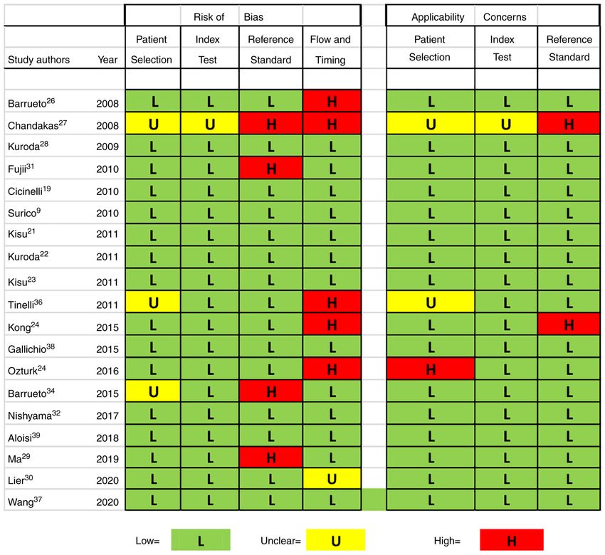
2 PEITSIDIS et al: NARROW-BAND IMAGING IN GYNECOLOGICAL ENDOSCOPY
gynecology clinical practice for the detection of benign and Excel version 16.0 (Microsoft Corporation, 2018). The number
malignant lesions. of publications were calculated per year and per country, and
the median age of participants in the selected studies was also
Materials and methods calculated.
Search strategy. The following electronic databases were Results
searched: PubMed (1950‑2021), Google Scholar (2004‑2021) and
Cochrane Library (2010‑2021). The electronic literature search Literature search and selection. The literature search identi‑
was mostly performed between January 2020 to February 2021. fied a total of 3,836 studies, i.e., 3,800 articles through Google
The search included the following medical subject headings or Scholar, 32 articles through the PubMed database and four
keywords: ‘Narrow‑band imaging’ and ‘gynecology’. The last through the Cochrane Library database. The flowchart of the
search was performed on 08/02/2021. study selection process is displayed in Fig. 1. After removing
The systematic review was performed and the flowchart duplicates and irrelevant articles, 256 articles were considered
diagram was drawn according to the Preferred Reporting Items for further evaluation. Further assessment excluded 215 articles
for Systematic Reviews and Metanalyses statement (5). for various reasons (non‑eligible, non‑English literature, animal
studies and articles providing insufficient information). Finally,
Inclusion criteria. Full‑text articles published in peer‑reviewed 31 studies reporting on a total of 3,128 female patients were
journals and written in the English language were deemed included in the review.
eligible to be included in the review. Studies that did not fulfill the Of the selected studies, 10 (32%) were case reports (9-18), 19
following criteria were excluded from the review: i) Conference (61.2%) were prospective studies (11,12,18-32) and two studies
abstracts and studies not providing sufficient clinical data; and (6.4%) were randomized controlled trials (33,34). The selected
ii) studies reporting narrow‑band imaging utilization in animals, studies had publication dates ranging from 2007 to 2020.
in surgical specimens or in an in vitro environment. The frequency of publications per year and the percentage
All types of studies were included, namely randomized of publications by country are presented in Fig. 2. The years
controlled or observational studies, case series and case reports. with the highest frequency of publications were 2010 and
The selected eligible articles were compared and discrepancies 2011 with 5 studies produced each year and the country
were resolved by discussion. The final decision on eligibility was which produced the highest number of studies was Japan with
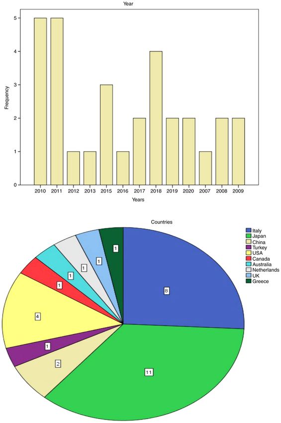
made by the senior investigator (PP) whenever discrepancies had 11 studies (13,18,21,22,28,31-33,35), followed by Italy with
not been resolved through discussion. 8 studies (9,10,15-17,19,20,36).
Data extraction. Two authors (PP and KK) independently Study properties. A total of 11 studies described the use of
extracted information, while SS, CK, VA, PT and NA checked NBI in hysteroscopy for the detection of endometrial patholo‑
the extracted information and tabulated the data. NV checked gies (9,10,19-25,36,37) and another 11 studies reported the
the results and approved the study. use of NBI in laparoscopy for the detection of peritoneal
From the eligible studies, the following clinical data were endometriosis (11-13,26-30,33,34,37,38). Furthermore, three
obtained: Author and year of publication; the time period articles dealt with the use of NBI in digital colposcopy for the
of enrolment of the study population; the country and city in detection of lower genital tract pathologies (14,31,32) and three
which the study was performed; the type of study; the setting articles reported on the use of NBI in laparoscopy for the detec‑
of the hospital (single‑ or multi‑university study); the number of tion of peritoneal metastases associated with ovarian cancer and
patients; the age of the patients; the inclusion criteria for surgery; other gynecological malignancies (15-17).
the interventions performed; the endoscopic system used in each
study; the outcome of the studies; and comments on different NBI in endometrial pathology. Details on the selected studies
studies. The references of the selected studies were scrutinized reporting the use of NBI in hysteroscopy are presented in
for additional information not obtained by the initial search. Table I. A total of 11 studies were included in the review, dating
The Quality Assessment of Diagnostic Accuracy Studies 2 from 2009‑2020 with a combined cohort of 2,424 female
(QUADAS‑2) tool was used to assess the quality of the primary patients (9,10,19-25,36,37). Of these, eight studies were prospec‑
diagnostic accuracy studies. Via this tool, risks for bias may tive studies (Canadian Task Force classification, II‑2) (19,23,36)
be evaluated in the following four key domains: i) Selection of and two studies were case reports (9,10). All studies were designed
participants; ii) index (diagnostic) test; iii) reference standard; in university settings, two studies were performed in multicen‑
and iv) flow and timing of the study (6). tric university settings (20,36) and two studies were reported at
Through the use of the Case Report (CARE) guidelines international congresses as conference reports (10,24). All of the
checklist, the information quality of case reports was evaluated, patients presented with abnormal uterine bleeding (AUB). The
specifically assessing the following items: Patient information, median age of the patients was 45.5±10.5 years. A total of four
the presence of timeline information, diagnostic assessment, studies were performed in outpatient office settings using a vagi‑
clinical findings, therapeutic intervention and outcomes (7,8). noscopic approach without any general anesthesia (19,21,23,36).
Conventional hysteroscopy under general anesthesia was
Statistical analysis. Data obtained from the selected studies performed in seven studies (10,24,25). Operative hysteroscopy
were entered into an Excel v160 spreadsheet (Microsoft was performed in all cases and histology specimens were
Corporation 2018). Descriptive statistical analyses was obtained in each case. Olympus Exera II (Olympus Corporation)
performed using SPSS version 23 (IBM Corporation) and was the main video system used in the majority of studies.EXPERIMENTAL AND THERAPEUTIC MEDICINE 23: 36, 2022 3
Figure 1. Flowchart diagram of the study selection for systematic review according to the Preferred Reporting Items for Systematic Reviews and Metanalyses
guidelines.
NBI hysteroscopy demonstrated increased sensitivity NBI in endometriosis. Details on the selected studies reporting
compared with WL imaging hysteroscopy for the detection of on the use of NBI in laparoscopy for the detection of peritoneal
endometrial cancer in four studies (20,23,24). endometriosis are provided in Table II. A total of 11 studies were
The sensitivity of NBI and WL hysteroscopy for endo‑ included in the present review, dating from 2007 to 2019 with a
metrial cancer reported by individual studies was 78.6 vs. population of 626 female patients in total (11-13,26-30,33,34,38).
63.7%, P4 PEITSIDIS et al: NARROW-BAND IMAGING IN GYNECOLOGICAL ENDOSCOPY Figure 2. Frequency of publications per year and per country. A Histogram and pie chart are provided in order to present the frequency of selected publications per country and per year. by WL imaging (27). The detection rate of endometriotic detection of endometriotic lesions, if a positive diagnosis was lesions with NBI was 100% in a randomized controlled trial made under WL imaging alone (29). NBI and WL combined with 167 female patients (34). Another randomized controlled with 3D imaging was able to increase the sensitivity rate up to trial assessed quality of life in two study groups: One group 91% (13). underwent laparoscopy with WL and the other with NBI. No Olympus Exetera (Olympus Corporation) was the system difference in pain and quality of life was observed between used for NBI in all of the studies. None of the 11 studies reported the two study groups (34). When combined with WL, NBI was any adverse effects or complications related to the surgical tech‑ reported to provide an additional predictive value of 86% for the niques.
Table I. Comparison of studies reporting on the use of hysteroscopy with NBI.

Author     Time     Country,   Type of		             Number of   Age,     Inclusion
(year)     period     city      study   Institutes    subjects   years     criteria Interventions 	 System 	 Outcomes 	 Conclusion                                            (Refs.)

Surico, 2009 Italy,  Case report Single 1 NS AUB Conventional      Olympus Detection of atypical                                                   NBI was able to              (9)
et al		      Novara		            univ.				       hysteroscopy 30˚  Exera II irregular microvessels                                                 increase accuracy
(2009)								                                   4 mm WL&NBI in G2			                                                                              in hysteroscopic
								                                         endometrial CA			                                                                                 detection of
												                                                                                                                                       hyperplasia and
												                                                                                                                                       endometrial CA
Cicinelli, 2008‑ Italy, Prospective Single 395 42.6±10.7 AUB Fluid minihysteroscopy  Olympus                         Increased sensitivity of     NBI increased                (19)
et al      2009  Bari   controlled  univ.				                105˚ 2.7 mm WL & NBI    Exera II                        NBI vs. WL: Proliferating    sensitivity, decreased
(2010)								                                               office setting without		                                endometrium, 0.93 vs. 0.78, false negative biopsies
								                                                     anesthesia compared		                                   P
6

Table I. Continued.

Author     Time       Country,   Type of		             Number of   Age,    Inclusion
(year)     period       city      study   Institutes    subjects   years    criteria Interventions 	 System 	 Outcomes 	 Conclusion                                           (Refs.)

Kisu,  2009‑ Japan, Prospective Single 71 41.9±12.5 AUB Flexible hysterofiberoscope Olympus                          Higher sensitivity for        NBI system allows           (23)
et al  2010  Tokyo		            univ.				               3.1 mm WL&NBI in office Xenon                                hyperplasia and CA of         better view of the
(2011)								                                          setting without anesthesia  Light                            endometrium for all raters    mucosal blood
								                                                compared with histology		                                    with NBI.                     vessels than WL.
								                                                findings		                                                   Average sensitivity was       Larger studies with
											                                                                                                          significantly higher in NBI   randomization design
											                                                                                                          group compared to WL          required to establish
											                                                                                                          (78.6 vs. 63.7%, P
EXPERIMENTAL AND THERAPEUTIC MEDICINE 23: 36, 2022 7
NBI in cervical pathology and peritoneal implants. Details on
AUB, abnormal uterine bleeding, PMB, postmenstrual bleeding; NBI, narrow‑band imaging; WL, white light; HRH, high‑risk hyperplasia; LRH, low‑risk hyperplasia; CA, carcinoma/cancer; univ. university, GA‑General
(Refs.)
(37)
the selected studies reporting on the use of NBI in laparoscopy for
the detection of cervical lesions and gynecological malignancies
are listed in Table III. A total of 9 studies were included in the
particularly those with
review, dating from 2010 to 2020. Of these, four studies were
the skill of trainees,
NBI may increase
Conclusion
previous training
prospective studies (31,32,35,39) and five studies were case
reports (14‑18). The total number of patients was 156 and the
Inclusion
mean age was 43.3±17.25 years. Uterine cervical pathology
was investigated with NBI in five studies (14,18,31,32,35)
and peritoneal implants were investigated with NBI in four
during hysteroscopy
studies (15-17,39).
The vascular pattern of lesions was examined in 21 patients
achieved higher diagnostic
utilization than with WL
with early and in situ cervical adenocarcinoma using NBI (13).
Three levels of trainees
The authors classified the colposcopic lesions according to the
Outcomes
accuracy with NBI
pattern described by Wright (40). The vascular pattern was
visualized in 18 patients (86%); the authors concluded that
NBI colposcopy depicts the vascular pattern on the cervix in
early glandular disease better than conventional colposcopy.
Nishiyama et al (32) proposed a novel microvascular clas‑
sification. They used flexible magnifying endoscopy with NBI
System
Olympus
in 10 patients with cervical lesions; the detection rate was
Exera II
90% (9/10) (32). NBI assisted in the detection of a rare mela‑
noma of the cervix in a case report (14). Furthermore, NBI was
operating channel under
general anesthesia
compared with histology
utilized in laparoscopy for the detection of peritoneal metastasis
in a series of 95 female patients undergoing surgery for various
WL&NBI with a 1.5 mm
gynecological malignancies, as reported by Aloisi et al (39).
Interventions
Hysteroscopy 5 mm
They determined that NBI increased the number of the detected
peritoneal abnormalities; however, no statistically significant
differences were observed in the identification of histologically
confirmed metastatic disease (P=0.18) (39).
A significantly greater number of peritoneal abnormalities
were identified with NBI than with standard WL imaging.
However, no statistically significant differences were observed
symptoms
in the identification of histologically confirmed metastatic
criteria
disease (39). In addition, NBI proved to be useful in the lapa‑
No
roscopic detection of early peritoneal implants in three case
(2020)
reports (15‑17). Kobara et al (18) indicated that NBI increased
the sensitivity, specificity, accuracy and positive predictive value
years
Age,
40.3
univ.
for the detection of cervical intraepithelial lesions‑3 (CIN‑3) in
comparison with conventional colposcopy. NBI with a gastro‑
scope assisted the detection of high‑grade cervical intraepithelial
Number of
subjects
lesions in two patients, while these lesions were not identified
529
by conventional colposcopy (18,35). None of the nine studies
reported any adverse effects or complications related to the
surgical techniques.
Institutes
Single
Quality assessment of case reports. The CARE guidelines
Type of
were followed to perform the quality assessment of the case
Prospective
reports (7). A total of 10 case reports were assessed for their
study
quality (9‑18). The results are presented in Fig. 3. Approximately
30% of the case reports had a low quality in terms of presen‑
tation of demographic information. In total, 55% of the case
Beijing
Country,
reports included had low quality regarding the presentation of
2012‑ China,
city
the patients' history with timeline information. Furthermore,
45% of the case reports were of poor quality in terms of infor‑
Table I. Continued.
mation about differential diagnosis.
period
Time
2014
Quality assessment with QUADAS‑2. The QUADAS‑2 tool
Anesthesia.
was used to assess the risk of bias in four domains. The results
Author
are presented in Fig. 4. In total, 19 studies were assessed. Of
Wang,
(year)
et al8
Table II. Comparison of studies reporting on the use of NBI in patients with endometriosis.
Author Time Type of Number Median Inclusion
(year) period Location study Institutes of subjects age, years criteria Interventions System Outcomes Conclusion (Refs.)
Farrugia, 2007 UK, Case Single 1 NS Suspected Laparoscopy Olympus Identification NBI may help (11)
et al East Kent report univer. endometriosis 0‑degree 10‑mm Exera II of smaller lesions distinguish active
(2007) with WL&NBI; of endometriosis endometriosis from
spectrum, 415 nm; fibrosis and changes
excision of lesions from previous surgery
Barrueto, 2008 USA, Prospective Single 21 36 Suspected Laparoscopy Olympus 14 patients had NBI may be able to (26)
et al Baltimore oilot univer. endometriosis 0‑degree 10‑mm Exera II lesions identified better identify the
(2008) with WL&NBI; by NBI, previously smaller lesions and
spectrum, 415 & not detected by excise a maximum
540 nm; excision of WL. 38 biopsy number of lesions,
lesions specimens were thus delaying the
taken using NBI; recurrence of
20 (53%) endometriosis
confirmed
endometriosis
Chandakas, 2008 Greece, Prospective Multi 95 33.9 Suspected Laparoscopy Olympus Lesions were NBI may detect (27)
et al Athens pilot univer. endometriosis 0‑degree 10‑mm Exera II collected from 95 endometriotic lesions
(2008) with WL&NBI; patients, 82.7% of more easily than WL
spectrum, 415 & the endometriotic
540 nm; excision of lesions detected by
lesions NBI and 55.9%
detected by WL
Kuroda, 2009 Japan, Prospective Single 23 35 Suspected Laparoscopy Olympus 23 patients NBI provides an (28)
et al Tokyo pilot univer. endometriosis 0‑degree 10‑mm Exera II diagnosed accurate objective
(2009) with WL&NBI; with peritoneal evaluation of vascular
measurement of endometriosis. density, particularly
PEITSIDIS et al: NARROW-BAND IMAGING IN GYNECOLOGICAL ENDOSCOPY
vascular density Median difference for the red lesions,
(red, black, white) of vascular which are indicative
lesions in peritoneal density by NBI was of early‑stage
endometriosis with significantly higher endometriosis with
vascular analysis in red lesions angiogenesis
software (4.5%, PTable II. Continued. Author Time Type of Number Median Inclusion (year) period Location study Institutes of subjects age, years criteria Interventions System Outcomes Conclusion (Refs.) Kuroda, 2008 Japan, Prospective Single 73 24‑49 Suspected Laparoscopy 0‑degree Olympus Increased vascular NBI may detect early (33) et al Tokyo control univer. endometriosis 10‑mm with Exera II density detected by vascular lesions in (2010) WL&NBI; Patients NBI in patients with patients with allocated to 4 groups: endometriosis endometriosis i) Endometriosis (P
10

Table II. Continued.

Author         Time		                        Type of		             Number              Median          Inclusion
(year)         period Location                study  Institutes 	 of subjects         age, years        criteria Interventions 	 System Outcomes Conclusion 	            (Refs.)

Ma, et al 2014‑ Australia,   Prospective Single  57  30  Suspected     Laparoscopy 0‑degree  Olympus Total 32 patients      NBI appears                                   (29)
(2019)    2015 Victoria      control     univer.			      endometriosis 5‑mm with WL&NBI.     Exera II with lesions detected beneficial in
								                                                               Excision of lesions		          by WL. 24 of          identifying new areas
										                                                                                            them were positive    of endometriosis
										                                                                                            for Endometriosis.    that may be missed
										                                                                                            Assessment of these if endometriosis is
										                                                                                            24 with NBI led       suspected by WL.
										                                                                                            to diagnosis of       NBI was not
										                                                                                            6 new positions       beneficial if WL was
										                                                                                            of endometriosis.     negative
										                                                                                            Additional predic
										                                                                                            tive value of 86% if
										                                                                                            a positive diagnosis
										                                                                                            was made under
										                                                                                            WL imaging
Kazama,   2018 Japan,        Case        Single   1  44  Pelvic pain   Intestinal endoscopy  NS       Lesions of rectal     NBI may assist in                             (13)
et al		         Saitama      report      univer.			      Occult fecal  with WL&NBI		                  endometriosis         the detection of
(2019)							                                            blood			                                     (Dienogest            small lesions in
										                                                                                            treatment)            cases of intestinal
											                                                                                                                 endometriosis
Lier,     2016‑ Netherlands, Prospective Single  20 34.5 Endometriosis Laparoscopy 0‑degree  Olympus 3D increased the       3D WL combined                                (30)
et al     2017 Amsterdam randomized univer.			           III‑IV        12‑mm with WL, NBI, Exera II   sensitivity rate      with NBI improved
(2020)							                                            (ASRM)        NIR‑ICG and 3D		               (P=0.0016).           the detection rate
								                                                               imaging. Excision of		         Combined 3D and       of peritoneal
                                                                                                                                                                                   PEITSIDIS et al: NARROW-BAND IMAGING IN GYNECOLOGICAL ENDOSCOPY

								                                                               lesions		                      NBI increased the     endometriosis
										                                                                                            sensitivity rate
									                                                                                             91.2% (P
Table III. Comparison of studies reporting on the use of NBI in patients with cervical pathology and peritoneal implants.
Author Time Country, Type of Number of Age, Inclusion
(year) period city study Institute subjects years criteria Interventions System Outcomes Conclusion (Refs.)
Fujii, 2007‑ Japan, Prospective Single 21 36.3 Patients with NBI digital Olympus Vascular pattern was Digital NBI colposcopy (31)
et al 2009 Tokyo pilot univer. diagnosis of colposcopy; biopsy classified as waste depicts vascular pattern
(2010) in situ cervical of lesions and thread‑like pattern, dot‑ on cervix more clearly
adenoCA early immunochemistry like pattern and root‑ than conventional
cervical with CD31 like pattern. Vascular colposcopy, thus
adenoCA antibody; evaluation patterns were displayed diagnosing early
of vascular pattern in 18 (86%) of cases of disease
cervical adenoCA
Fanfani, 2010 Italy, Case report Single 1 NS Borderline Laparoscopy Olympus Identification of NBI in laparoscopy a (15)
et al Rome univer. ovary 0‑degree 10‑mm Exera II peritoneal implants not useful advancement for
(2010) with WL&NBI. clearly visible by WL identifying early
Excision of lesions preneoplastic and
neoplastic lesions and
assisting in making
intraoperative decisions
Fanfani, 2011 Italy, Case report Single 1 NS Cervical CA Laparoscopy Olympus Identification of NBI may a useful tool (16)
et al Rome univer. 0‑degree 10‑mm Exera II peritoneal implants not in the detection of early
(2011) with WL&NBI. clearly visible by WL in neoplastic peritoneal
Excision of lesions a case after laparoscopic implants
hysterectomy for
cervical CA
Gagliardi, 2013 Italy, Case report Single 2 1st Recurrent Laparoscopy Olympus Identification and NBI NBI facilitates (17)
et al Roma univer. 632nd Ovarian CA 1 0‑degree 10‑mm Winter target biopsy of diagnosis of recurrence
(2013) 67 case of FIGO IC with WL&NBI peritoneal implants of of malignant disease
and 1 case of recurrent disease
FIGO IIIC
Nishyama, 2014‑ Japan, Prospective Single 10 39 Patients with Flexible magnifying Olympus Report of NBI‑ME Establishment of a (32)
EXPERIMENTAL AND THERAPEUTIC MEDICINE 23: 36, 2022
et al 2015 Kagawa pilot univer. squamous endoscopy with Exera II microvascular findings novel microvascular
(2017) cervical NBI evaluating as follows: classification system
pathology microvascular ‑Presence of dots in 7 via NBI‑ME of cervical
LGSIL‑cervical patterns. (70 %) lesions
CA Colposcopy and ‑Irregular arrangement
biopsy of lesions of dots in 5 (50%)
‑High‑density of dots in
5 (50%)
‑High vessel caliber in
2 (20%)
‑New tumor vessel in
1 (10%)
‑Detection rate, 90%
11
(9/10 patients)12
Table III. Continued.
Author Time Country, Type of Number of Age, Inclusion
(year) period city study Institute subjects years criteria Interventions System Outcomes Conclusion (Refs.)
Aloisi, 2011‑ USA, Prospective Single 94 55.5 Patients Laparoscopy Olympus Higher number of NBI provides unique (39)
et al 2014 New York Comparative univer. undergoing 0‑degree 10‑mm Exera II peritoneal abnormalities contrast between
(2018) laparoscopic with WL&NBI detected by NBI vs. WL peritoneum and
surgery for (P=0.0239). No microvascular patterns.
gynecologic scientific significance NBI does not provide
CA of all types in the detection of better detection of
peritoneal metastasis peritoneal malignancy
(P=0.18) than WL
Uchita, 2016 Japan Prospective Multi 24 40 Patients NBI with Olympus NBI revealed NBI demonstrated (35)
et al diagnostic univer. underwent gastroscope 3 abnormal findings: 100% sensitivity,
(2018) colposcopy ‑Light white epithelium specificity, accuracy,
and NBI of ‑Heavy white positive predictive
cervix. Patients epithelium value for lesions
underwent ‑Atypical intracapillary >CIN‑3
conisation papillary loop
Anko, 2020 Japan, Case report Single 1 54 Patient with NBI digital NS Report of atypical NBI colposcopy may (14)
et al Kanagawa univer. melanoma of colposcopy vessels and transparent be useful in melanoma
(2019) cervix mucosal surface diagnosis
Kobara, 2020 Japan, Case report Multi 2 39, 37 Patients with NBI with Olympus Detection of atypical NBI may assist in the (18)
et al Kagawa univer. cervical HGSIL gastroscope lesion not identified by diagnosis of HGSIL if
(2020) colposcopy not identified by
PEITSIDIS et al: NARROW-BAND IMAGING IN GYNECOLOGICAL ENDOSCOPY
NBI, narrow‑band imaging; WL, white light; NS, not stated; HGSIL, high‑grade squamous intraepithelial lesion; LGSIL, low‑grade squamous intraepithelial lesion; CA, carcinoma/cancer; FIGO, International Federation of
Gynecology and Obstetrics; univ. university; CIN, cervical intraepithelial neoplasia.EXPERIMENTAL AND THERAPEUTIC MEDICINE 23: 36, 2022 13
Figure 3. Quality assessment of case reports according to Case Report guidelines. Quality is rated as high, low or unclear.
these, 17 had a prospective design lacking randomization and surgery, although exceptions may be made for urgent cases, e.g.
two were randomized controlled trials with control groups. malignancies.
A total of 9 studies were indicated to have a high risk of It has been reported that laparoscopies have a larger
bias (24,25,27,29,30-32,34,36) and 10 studies were determined potential for aerosol spread than hysteroscopies (41‑43), while
to have a low risk of bias (19-21,23,28,30,32,37-39). laparotomies have a lower risk of aerosol spread in comparison
to laparoscopies (41‑43). However, endoscopy is generally
NBI in gynecological endoscopy and special precautions in preferred due to the one‑day stay and the smaller exposure to a
view of the Coronavirus Disease 2019 (COVID‑19) pandemic. hospital environment.
The outbreak of the COVID‑19 pandemic caused by the Severe Strict safety measures regarding the pressure of the gas and
Acute Respiratory Syndrome Coronavirus‑2 (SARS‑CoV‑2) pressure of the fluid should be applied to minimize the possi‑
has produced a public health emergency of international bility of spread. In cases where local anesthesia may be used,
concern, with all of the data demonstrating that the spread of it is expected to be beneficial for the patient, minimizing the
the virus mainly occurs via respiratory droplets during close risk of infection, which may occur after the intubation and the
contact (41,42). During this ongoing pandemic, recommenda‑ extubation procedure.
tions regarding laparoscopy and endoscopy with NBI should Further guidelines issued by the different medical associa‑
follow the general international recommendations for laparos‑ tions such as International Society for Gynecologic Endoscopy
copy. (ISGE; www.isge.org) and American Society of Gynecologic
However, the potential risk of infection through endoscopi‑ Laparoscopists (AAGL; https://www.aagl.org/) (42,43) will
cally generated bioaerosols may possibly be increased as a result provide additional pertinent and vital data during the current
of three important factors pertaining to laparoscopy. These fight against the pandemic.
are the following: i) Use of gas insufflation during entry and
intra‑operatively; ii) generation of bioaerosols via electrosur‑ Strengths and limitations. NBI, which is now regularly used
gery, which is a cornerstone of endoscopy; and iii) the potential in gynecology, is a relatively new optical technology and the
for gas leaks in the operation room, which may lead to elevated present systematic review is, to the best of our knowledge, the
viral loads in the air (41,42). Precautions should be taken during most updated and extensive of its kind that has been provided
gynecological endoscopy with NBI, adhering to the international on this method to date. Due to the relatively small number
recommendations concerning SARS‑CoV‑2 (41‑43). of randomized controlled trials, observational studies were
Regarding elective surgery, it is recommended that universal included in spite of providing a lower level of evidence. While
testing for SARS‑CoV‑2 infection is conducted whenever precise and clear inclusion criteria were employed for the
possible within 40 h preceding surgery. Should a case of preparation of the present review, the small sample size of the
SARS‑CoV‑2 be positively confirmed, it is advisable to delay trials included and the overall absence of definitions of primary14 PEITSIDIS et al: NARROW-BAND IMAGING IN GYNECOLOGICAL ENDOSCOPY
Figure 4. Quality Assessment of Diagnostic Accuracy Studies 2 assessment of studies. Risk of bias is designated as L, H or U. L, low; H, high; U, unclear.
outcomes inevitably reduced the quality of the present review. potential to improve the diagnosis of endometriosis. It may
Furthermore, risk of bias in the flow and timing domains was also have the potential to enhance the diagnosis of prema‑
frequently present. Therefore, it was not possible to perform a lignant and malignant lesions in the fields of hysteroscopy,
pooled data‑analysis or meta‑analysis. While case reports were laparoscopy and colposcopy.
included, the majority of these studies were well‑designed and While Kisu et al (44) performed a review in 2012, the
mostly provided their evidence clearly and accurately, according present systematic review is substantially updated and
to the quality assessment using the CARE (Case report) guide‑ includes quality assessment of the studies according to the
lines. QUADAS‑2 and the CARE guidelines (6,7).
NBI has proven to be an efficacious approach for the
Discussion diagnosis of endometrial cancer and hyperplasia, while
thorough training improves the trainee's diagnostic skills to
The NBI system for image‑enhanced endoscopy was first an extent depending on their previous hysteroscopic experi‑
conceived and developed in May 1999 and the product was ence (37). However, it must be stressed that NBI laparoscopy
launched by Olympus Corporation in May 2006 (21). was not superior in the detection of peritoneal metastases in
The major advantages of the NBI system are the enhance‑ comparison with standard WL laparoscopy alone, which was
ment of endoscopic visualization of superficial neoplastic reported by Aloisi et al (39) and Schnelldorfer et al (45).
lesions and their microvascular architecture. Conventional A significantly greater number of peritoneal abnormalities
endoscopic diagnosis using WL, by contrast, is based on subtle were identified with NBI than with standard WL. However,
morphological changes, e.g., superficially elevated, flat or no statistically significant differences were observed in the
depressed lesions, and on minimal changes in color such as identification of histologically confirmed metastatic disease.
reddish discoloration (19). In fact, of the eight additional suspicious‑appearing nodules
The present systematic review demonstrated that the visualized with NBI, only three were confirmed as malig‑
application of NBI in gynecological endoscopy has the nant on final pathology, and none of the patients had surfaceEXPERIMENTAL AND THERAPEUTIC MEDICINE 23: 36, 2022 15
malignancies identified with NBI that were not also seen with Acknowledgements
WL, even if in a different area (39). Aloisi et al (39) pointed
out that further exploration of the use of NBI/3D WL imaging The authors thank the Emeritus Professor George Iatrakis of
is required; this will now be further evaluated in a large University of West Attica (Athens, Greece) for assisting us in
randomized clinical trial with clinically relevant endpoints, obtaining the full‑text of articles.
with adequate power, quality control and measures (44). This
will be according to the Idea, Development, Exploration, Funding
Assessment and Long‑term Study (IDEAL) framework,
describing 5 stages of evolution for new surgical therapeutic No funding was received.
interventions. IDEAL is an important driver for future incre‑
mental and evidence‑based modifications (46). Availability of data and materials
Furthermore, another limitation is the extension of the
surgical time, particularly in laparoscopic procedures with All data generated or analyzed during this study are included in
the use of NBI. Lier et al (30) reported a median exten‑ this published article.
sion of surgical time of 30 min with NBI due to thorough
inspection of the peritoneum and histological sampling. The Authors' contributions
clinical question is whether an improved detection of endo‑
metriosis with NBI/3D imaging also affects the long‑term All authors contributed equally to the writing and production of
clinical outcomes after surgery, such as reintervention rates, this manuscript. PP and KK extracted information, SS, CK, EA,
pain‑free interval and quality of life (30). PT and NA interpreted the extracted information and tabulated
A shortcoming, particularly with the methodology the data. NV checked the results and approved the study. PP was
reported by Barrueto et al (34), is the low specificity. This may the main author that formed the conception of the study. EA
result in unnecessary resection of healthy tissue, producing checked for the eligibility of the studies. All authors read and
postoperative neuropathic pain and adhesion formation. approved the final manuscript. PP and NV confirm the authen‑
In the study by Surico et al (20), only a small number ticity of all the raw data.
of patients were recruited, while the study was performed at
a single academic institution. Furthermore, the accuracy of Ethics approval and consent to participate
NBI hysteroscopy in the prediction of histological findings
via analysis of interobserver variability was not assessed (20). Not applicable.
Wang et al (37) reported that the physician who performs
hysteroscopy must be familiar with endometrial lesions, Patient consent for publication
which are influenced by estrogen and progesterone secretion.
Endometrial necrosis may not only be observed in malignant Not applicable.
lesions, but also frequently appears in benign hyperplastic
lesions associated with abnormal uterine bleeding. These Competing interests
factors increase the difficulty of hysteroscopic diagnosis;
therefore, the learning curve for the diagnosis of endometrial The authors declare that they have no competing interests.
neoplasms is relatively long. The authors report that >200
hysteroscopic cases are required to be performed by physi‑ References
cians until proficiency is reached (37).
The drawback of NBI colposcopy is that the system is
1. Vincent BD and Fraig M: A pilot study of narrow‑band imaging
expensive; thus, widespread use of it is limited and is particu‑ compared to white light bronchoscopy for evaluation of normal
larly unsuitable for application in developing countries. It airways and premalignant and malignant airways disease. Chest 131:
may be appropriate to use for educational purposes in cancer 1794‑1199, 2007.
2. Sano Y and Horimatsu T: Magnifying observation of microvascular
center hospitals or university hospitals (31). architecture of colorectal lesions using a narrow‑band imaging
In conclusion, conventional WL imaging in gynecological system. Dig Endosc 18: 44‑51, 2006.
endoscopy is now well‑established as a highly sensitive and 3. Watanabe A and Taniguchi M: The value of narrow band imaging
endoscope for early head and neck cancers. Otolaryngol Head Neck
specific technique for the diagnosis of intrauterine diseases Surg 138: 446‑451, 2008.
and the present study clearly indicated that the NBI system, 4. Machida H, Sano Y, Hamamoto Y, Muto M, Kozu T, Tajiri H and
when applied by an expert and experienced surgeon, is Yoshida S: Narrow‑band imaging in the diagnosis of colorectal
capable of enhancing diagnostic accuracy. Furthermore, mucosal lesions: A pilot study. Endoscopy 36: 1094‑1098, 2004.
5. Liberati A, Altman DG, Tetzlaff J, Mulrow C, Gøtzsche PC,
NBI may increase the diagnostic skills of trainees. Future Ioannidis JP, Clarke M, Devereaux PJ, Kleijnen J and Moher D: The
directions of research should take into consideration the reop‑ PRISMA statement for reporting systematic reviews and meta‑anal‑
eration rates, recurrence and overall cost. Evidence‑based yses of studies that evaluate healthcare interventions: Explanation
and elaboration. BMJ 339: b2700, 2009.
frameworks such as IDEAL should be implemented in order 6. Whiting PF, Rutjes AW, Westwood ME, Mallett S, Deeks JJ,
to improve clinical practice. Certainly, there is a requirement Reitsma JB, Leeflang MM, Sterne JA and Bossuyt PM; QUADAS‑2
for large‑scale, multicenter, randomized trials to substan‑ Group: QUADAS‑2: A revised tool for the quality assessment of
diagnostic accuracy studies. Ann Intern Med 155: 529‑536, 2011.
tiate the present results as to the potential for use of NBI in 7. Gagnier JJ, Kienle G, Altman DG, Moher D, Sox H and Riley D;
gynecology, the application of which may improve patients' CARE Group: The CARE guidelines: Consensus‑based clinical
oncological outcomes and thus their quality of life. case report guideline development. J Clin Epidemiol 67: 46‑51, 2014.16 PEITSIDIS et al: NARROW-BAND IMAGING IN GYNECOLOGICAL ENDOSCOPY
8. Tulandi T and Balayla J: Study designs and the use of the canadian 31. Fujii T, Nakamura M, Kameyama K, Saito M, Nishio H, Ohno A,
task force classification. J Obstet Gynaecol Can 40: 1383‑1384, 2018. Hirao N, Iwata T, Tsukazaki K and Aoki D: Digital colposcopy
9. Surico D, Vigone A and Leo L: Narrow band imaging in endome‑ for the diagnosis of cervical adenocarcinoma using a narrow band
trial lesions. J Minim Invasive Gynecol 16: 9‑10, 2009. imaging system. Int J Gynecol Cancer 20: 605‑610, 2010.
10. Raimondo I, Scarciglia ML, Amadio G, Monterisi AF, Scambia G 32. Nishiyama N, Kanenishi K, Mori H, Kobara H, Fujihara S,
and Masciullo V: Narrow band imaging: А new diagnostic tool in Chiyo T, Kobayashi N, Matsunaga T, Ayaki M, Yachida T, et al:
adenomyosis? Int J Gynaecol Obstet 119: S531‑S867, 2012. Flexible magnifying endoscopy with narrow band imaging for the
11. Farrugia M, Nair MS and Kotronis KV: Narrow band imaging in diagnosis of uterine cervical tumors: A cooperative study among
endometriosis. J Minim Invasive Gynecol 14: 393‑394, 2007. gastrointestinal endoscopists and gynecologists to explore a novel
12. Murnaghan O, Rajakumar C, Bougie O and Singh SS: Use of microvascular classification system. Oncol Lett 14: 355‑362, 2017.
narrowband imaging for the surgical management of endometriosis. 33. Kuroda K, Kitade M, Kikuchi I, Kumakiri J, Matsuoka S, Kuroda M
J Obstet Gynaecol Can 39: 711, 2017. and Takeda S: Peritoneal vascular density assessment using
13. Kazama S, Hiramatsu T, Kuroda K, Hongo K, Watanabe Y, narrow‑band imaging and vascular analysis software, and cyto‑
Tanaka T and Kuriki K: A case of unique endoscopic findings of kine analysis in women with and without endometriosis. J Minim
intestinal endometriosis exposed to the mucosa: Aggregation of Invasive Gynecol 17: 21‑25, 2010.
papillary protruded bulges from the submucosal elevation of the 34. Barrueto FF, Audlin KM, Gallicchio L, Miller C, MacDonald R,
rectum. Clin J Gastroenterol 12: 166‑170, 2019. Alonsozana E, Johnston M and Helzlsouer KJ: Sensitivity of narrow
14. Anko M, Nakamura M, Kobayashi Y, Tsuji K, Nakada S, band imaging compared with white light imaging for the detection
Nakamura Y, Funakoshi T, Banno K and Aoki D: Primary malignant of endometriosis. J Minim Invasive Gynecol 22: 846‑852, 2015.
melanoma of the uterine cervix or vagina which were successfully 35. Uchita K, Kanenishi K, Hirano K, Kobara H, Nishiyama N,
treated with nivolumab. J Obstet Gynaecol Res 46: 190‑195, 2020. Kawada A, Fujihara S, Ibuki E, Haba R, Takahashi Y, et al:
15. Fanfani F, Gallotta V, Rossitto C, Fagotti A and Scambia G: Narrow Characteristic findings of high‑grade cervical intraepithelial
band imaging in borderline ovarian tumor. J Minim Invasive neoplasia or more on magnifying endoscopy with narrow band
Gynecol 17: 146‑147, 2010. imaging. Int J Clin Oncol 23: 707‑714, 2018.
16. Fanfani F, Rossito C, Faggotti A, Gallotta V, Gagliardi ML and 36. Tinelli R, Surico D, Leo L, Pinto V, Surico N, Fusco A, Cicinelli MV,
Scambia G: Narrow‑band imaging in laparoscopic management of Meir YJ and Cicinelli E: Accuracy and efficacy of narrow-band
cervical carcinoma. J Minim Invasive Gynecol 18: 146‑147, 2011. imaging versus white light hysteroscopy for the diagnosis of
17. Gagliardi ML, Polito S, Fagotti A, Fanfani F and Scambia G: endometrial cancer and hyperplasia: a multicenter controlled study.
Narrow‑band imaging in laparoscopic management of recurrent Menopause 18: 1026-1029, 2011.
platinum sensitive ovarian cancer. J Minim Invasive Gynecol 20: 37. Wang W, Chen F, Kong L, Guo Y, Cheng J and Zhang Y: Prospective
10‑12, 2013. evaluation of the accuracy of a training program in image recogni‑
18. Kobara H, Uchita K, Uedo N, Matsuura N, Nishiyama N, tion by narrow‑band imaging guided hysteroscopy of endometrial
Kanenishi K and Masaki T: Uterine cervical neoplasm diagnosed neoplasms. Gynecol Obstet Invest 85: 284‑289, 2020.
by flexible magnifying endoscopy with narrow band imaging. 38. Gallicchio L, Helzlsouer KJ, Audlin KM, Miller C, MacDonald R,
Diagnostics (Basel) 10: 903, 2020. Johnston M and Barrueto FF: Change in pain and quality of life
19. Cicinelli E, Tinelli R, Colafiglio G, Pastore A, Mastrolia S, Lepera A among women enrolled in a trial examining the use of narrow band
and Clevin L: Reliability of narrow‑band imaging (NBI) hysteros‑ imaging during laparoscopic surgery for suspected endometriosis.
copy: A comparative study. Fertil Steril 94: 2303‑2307, 2010. J Minim Invasive Gynecol 22: 1208‑1214, 2015.
20. Surico D, Vigone A, Bonvini D, Tinelli R, Leo L and Surico N: 39. Aloisi A, Sonoda Y, Gardner GJ, Park KJ, Elliott SL, Zhou QC,
Narrow‑band imaging in diagnosis of endometrial cancer and Iasonos A and Abu‑Rustum NR: Prospective comparative study of
hyperplasia: A new option? J Minim Invasive Gynecol 17: 620‑625, laparoscopic narrow band imaging (NBI) versus standard imaging
2010. in gynecologic oncology. Ann Surg Oncol 25: 984‑990, 2018.
21. Kisu I, Banno K, Kobayashi Y, Ono A, Masuda K, Ueki A, 40. Wright VC: Cervical glandular disease: Adenocarcinoma in situ
Nomura H, Hirasawa A, Abe T, Kouyama K, et al: Flexible hyster‑ and adenocarcinoma. In: Colposcopy Principles and Practice.
oscopy with narrow band imaging (NBI) for endoscopic diagnosis Barbara S, Apgar GL and Spitzer M (eds). 2nd edition. Saunders
of malignant endometrial lesions. Int J Oncol 38: 613‑618, 2011. Elsevier, Philadelphia, PA, 283Y310, 2008.
22. Kuroda K, Kitade M, Kikuchi I, Kumakiri J, Matsuoka S, Tokita S, 41. Carugno J, Di Spiezio Sardo A, Alonso L, Haimovich S, Campo R,
Kuroda M and Takeda S: A new instrument: A flexible hysteroscope De Angelis C, Bradley L, Bettocchi S, Arias A, Isaacson K, et al:
with narrow band imaging system: Optical quality comparison COVID‑19 pandemic. Impact on hysteroscopic procedures: A
between a flexible and a rigid hysteroscope. Minim Invasive Ther consensus statement from the global congress of hysteroscopy
Allied Technol 20: 263‑266, 2011. scientific committee. J Minim Invasive Gynecol 27: 988‑992, 2020.
23. Kisu I, Banno K, Susumu N, Aoki D. Magnifying hysteroscopy with 42. Thomas V, Maillard C, Barnard A, Snyman L, Chrysostomou A,
narrow-band imaging for visualization of endometrial lesions. Int J Shimange‑Matsose L and Van Herendael B: International Society
Gynaecol Obstet. 115 (1): 74-5, 2011. for Gynecologic Endoscopy (ISGE) guidelines and recommenda‑
24. Kong L, Duan H, Zhang Y, Wang Y and Guo Y: Application of tions on gynecological endoscopy during the evolutionary phases of
narrow‑band imaging in the diagnosis of endometrial lesions. the SARS‑CoV‑2 pandemic. Eur J Obstet Gynecol Reprod Biol 253:
J Minim Invasive Gynecol 22: S45, 2015. 133‑140, 2020.
25. Ozturk M, Ulubay M, Alanbay I, Keskin U, Karasahin E and 43. American Association of Gynecologic Laparoscopists. COVID‑19:
Yenen MC: Using narrow‑band imaging with conventional hyster‑ Joint Society Statement on Elective Surgery. Available from:
oscopy increases the detection of chronic endometritis in abnormal http://www. aagl.org/news/covid‑19‑joint‑statement‑on‑
uterine bleeding and postmenopausal bleeding. J Obstet Gynaecol elective‑surgeries/.
Res 42: 67‑71, 2016. 44. Kisu I, Banno K, Tsuji K, Masuda K, Ueki A, Kobayashi Y,
26. Barrueto FF and Audlin KM: The use of narrowband imaging Yamagami W, Susumu N and Aoki D: Narrow band imaging in
for identification of endometriosis. J Minim Invasive Gynecol 15: gynecology: A new diagnostic approach with improved visual iden‑
636‑639, 2008. tification (Review). Int J Oncol 40: 350‑356, 2012.
27. Chandakas S, Salamalekis E and Erian J: New narrow band imaging 45. Schnelldorfer T, Jenkins RL, Birkett DH, Wright VJ, Price LL and
endoscopic system for the detectionof surface pathology including Georgakoudi I: Laparoscopic narrow band imaging for detection
endometriosis: A series of 95 patients. J Minim Invasive Gynecol 15: of occult cancer metastases: A randomized feasibility trial. Surg
S1‑S159, 2008. Endosc 30: 1656‑1661, 2016.
28. Kuroda K, Kitade M, Kikuchi I, Kumakiri J, Matsuoka S, Jinushi M, 46. Hirst A, Philippou Y, Blazeby J, Campbell B, Campbell M,
Shirai Y, Kuroda M and Takeda S: Vascular density of peritoneal Feinberg J, Rovers M, Blencowe N, Pennell C, Quinn T, et al: No
endometriosis using narrow‑band imaging system and vascular Surgical innovation without evaluation: Evolution and further
analysis software. J Minim Invasive Gynecol 16: 618‑621, 2009. development of the IDEAL framework and recommendations. Ann
29. Ma T, Chowdary P, Eskander A, Ellett L, McIlwaine K, Manwaring J, Surg 269: 211‑220, 2019.
Readman E and Maher P: Can narrowband imaging improve the
laparoscopic identification of superficial endometriosis? A prospec‑ This work is licensed under a Creative Commons
tive cohort trial. J Minim Invasive Gynecol 26: 427‑433, 2019. Attribution-NonCommercial-NoDerivatives 4.0
30. Lier MC, Vlek SL, Ankersmit M, van de Ven PM, Dekker JJML, International (CC BY-NC-ND 4.0) License.
Bleeker MCG, Mijatovic V and Tuynman JB: Comparison of
enhanced laparoscopic imaging techniques in endometriosis
surgery: A diagnostic accuracy study. Surg Endosc 34: 96‑104, 2020.You can also read