Impact of the Amyotrophic Lateral Sclerosis Disease on the Biomechanical Properties and Oxidative Stress Metabolism of the Lung Tissue Correlated ...
←
→
Page content transcription
If your browser does not render page correctly, please read the page content below
ORIGINAL RESEARCH
                                                                                                                                              published: 25 February 2022
                                                                                                                                          doi: 10.3389/fbioe.2022.810243

                                             Impact of the Amyotrophic Lateral
                                             Sclerosis Disease on the
                                             Biomechanical Properties and
                                             Oxidative Stress Metabolism of the
                                             Lung Tissue Correlated With the
                                             Human Mutant SOD1G93A Protein
                                             Accumulation
                                             Duygu Aydemir 1,2, Anjum Naeem Malik 3, Ibrahim Kulac 4, Ayse Nazli Basak 5, Ismail Lazoglu 3
                                             and Nuriye Nuray Ulusu 1,2*
                          Edited by:
               Dario Loureiro Santos,        1
                                               Department of Medical Biochemistry, School of Medicine, Koc University, Istanbul, Turkey, 2Koc University Research Center for
   Universidade de Trás-os-Montes e          Translational Medicine (KUTTAM), Istanbul, Turkey, 3Manufacturing and Automation Research Center, Department of Mechanical
                 Alto Douro, Portugal        Engineering, Koc University, Istanbul, Turkey, 4Department of Pathology, Koc University School of Medicine, Istanbul, Turkey,
                         Reviewed by:
                                             5
                                               Suna and İnan Kirac Foundation, Neurodegeneration Research Laboratory, NDAL-KUTTAM, School of Medicine, Koc
                 Nemany A. N. Hanafy,        University, Istanbul, Turkey
         Kafrelsheikh University, Egypt
                 Sanjeev Kumar Mahto,
 Indian Institute of Technology (BHU),
                                             Amyotrophic lateral sclerosis (ALS) is the most common motor neuron disease, and ALS
                                    India    incidence is increasing worldwide. Patients with ALS have respiratory failure at the
                  Ludovica Cacopardo,
                                             disease’s end stages, leading to death; thus, the lung is one of the most affected
                 University of Pisa, Italy
                                             organs during disease progression. Tissue stiffness increases in various lung diseases
                   *Correspondence:
                    Nuriye Nuray Ulusu       because of impaired extracellular matrix (ECM) homeostasis leading to tissue damage and
                     nulusu@ku.edu.tr        dysfunction at the end. According to the literature, oxidative stress is the major contributor
                                             to ECM dysregulation, and mutant protein accumulation in ALS have been reported as
                    Specialty section:
         This article was submitted to       causative to tissue damage and oxidative stress. In this study, we used SOD1G93A and
                        Biomechanics,        SOD1WT rats and measured lung stiffness of rats by using a custom-built stretcher, where
                a section of the journal
                                             H&E staining is used to evaluate histopathological changes in the lung tissue. Oxidative
       Frontiers in Bioengineering and
                         Biotechnology       stress status of lung tissues was assessed by measuring glucose 6-phosphate
       Received: 06 November 2021            dehydrogenase (G6PD), 6-phosphogluconate dehydrogenase (6-PGD), glutathione
         Accepted: 31 January 2022           reductase (GR), glutathione s-transferase (GST), catalase (CAT), and superoxide
        Published: 25 February 2022
                                             dismutase 1 (SOD1) levels. Western blot experiments were performed to evaluate the
                             Citation:
         Aydemir D, Malik AN, Kulac I,       accumulation of the SOD1G93A mutated protein. As a result, increased lung stiffness,
  Basak AN, Lazoglu I and Ulusu NN           decreased antioxidant status, elevated levels of oxidative stress, impaired mineral and
    (2022) Impact of the Amyotrophic
                                             trace element homeostasis, and mutated SOD1G93A protein accumulation have been
      Lateral Sclerosis Disease on the
        Biomechanical Properties and         found in the mutated rats even at the earlier stages, which can be possible causative of
   Oxidative Stress Metabolism of the        increased lung stiffness and tissue damage in ALS. Since lung damage has altered at the
      Lung Tissue Correlated With the
             Human Mutant SOD1G93A           very early stages, possible therapeutic approaches can be used to treat ALS or improve
                Protein Accumulation.        the life quality of patients with ALS.
Front. Bioeng. Biotechnol. 10:810243.
      doi: 10.3389/fbioe.2022.810243         Keywords: ALS, biomechanical test, stiffness, oxidative stress, SOD1

Frontiers in Bioengineering and Biotechnology | www.frontiersin.org                  1                                         February 2022 | Volume 10 | Article 810243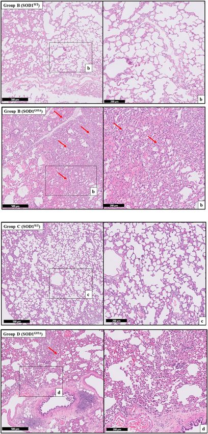
Aydemir et al.                                                                                           Dysfunction of Lung Tissue in ALS Disease

INTRODUCTION                                                              rats (SOD1WT) by eliminating the effects of the aging first time in
                                                                          the literature. We hypothesized that SOD1G93A mutation causes
Amyotrophic lateral sclerosis (ALS) is the most common and                biomechanical impairment in the lung tissue due to elevated
severe motor neuron disease affecting upper and lower motor               oxidative stress. Our study may help to explain possible
neurons in the brain, brain stem, and spinal cord. ALS is                 mechanisms behind the lung dysfunction in the ALS disease
categorized as sporadic ALS (sALS) with no known cause and                that can be used as potential therapeutic approaches in the future
familial ALS (sALS) resulting from the genetic inheritance                to improve the life quality and survival time of patients with ALS.
(Aydemir and Ulusu, 2020b; Burlando et al., 2020). Several
genes have been identified as causative or disease-modifying
fALS and sALS, such as C9ORF72, SOD1, TARDBP, and FUS.                    MATERIALS AND METHODS
D1 is the most studied gene mutation in ALS disease and one of
the essential antioxidant enzymes involved in redox sensing,              Animal Studies
oxidative stress metabolism, and signal transduction (Jawaid              Six male and six female hemizygous SOD1G93A mutated albino
et al., 2010; Gill et al., 2019; Cassina et al., 2021).                   rats were purchased from Taconic with catalog number NTac:SD-
    The lung is one of the most affected organs during ALS                Tg(SOD1G93A) L26H. Animals were inbred at the Animal
progression, and patients with ALS have respiratory failure at            Research Facility of Koc University, and 70 male rats weighing
the end stages of the disease leading to death (Aydemir and               140–650 g were used during the experiments. Power analysis was
Ulusu, 2020b). Therefore, handling respiratory failure aims to            performed to determine the number of animals used in the study.
prolong survival and improve quality of life during ALS                   Animals were housed in the polycarbonate cages with stainless
progression (Niedermeyer et al., 2019). Lung tissue consists of           steel covers in an air-conditioned room (12 h light/dark cycle
elastin, collagen, and proteoglycans representing the connective          with a temperature of 22 ± 2°C and relative humidity of 50 ± 5%).
tissue, giving specific mechanical properties to the lung.                 Animals had free access to tap water and standard rat pellet food.
Biomechanical properties of the lung tissue such as stiffness,            The Ethics Committee approved all experimental procedures and
force, or elastic modulus impaired in various diseases including          animal use of Koc University with the number
Parkinson’s Disease, pulmonary fibrosis, cancer, asthma,                   2019.HADYEK.006.
emphysema, chronic obstructive pulmonary disease (COPD),                     Animals were followed every week by weighing and
and pulmonary arterial hypertension (PAH) disease; however,               controlling their movements, indicating disease progression.
biomechanical changes associated with the histopathological               First, we have divided our animals into ten groups based on age
alterations of the lung tissue have not been studied in the ALS           as 0 (40–45 days old), A (70–75 days old), B (90–95 days old),
disease (Mariappan et al., 2011; Burgstaller et al., 2017). Elastic       C (110–115 days old), and D (130–135 days old), and each
recoil and mechanical stability are essential for healthy lung            group is divided into two subgroups according to their
function regulated by the biochemical and biomechanical                   mutation status respectively as SOD1WT and SOD1G93A.
signals (Hynes, 2009). However, aging leads to impaired lung              Group C accounts for the early stage, and group D refers to
function, such as tissue stiffness, because of alterations in the         the late stage of the ALS disease. We have started to observe
biomechanical and biochemical properties of the lung tissue               disease symptoms in group C, and therefore we have indicated
(Sicard et al., 2018; Racca et al., 2020).                                group C as early-stage and group D as the late stage. Animals
    Tissue stiffness regulates tissue function, homeostasis, and          were anesthetized under isoflurane, and then blood was taken
cellular activity, thus decreased or increased tissue stiffness is        from the heart of the rats. Animals were sacrificed under
associated with dysregulated cell behaviors and disease                   anesthesia via guillotine to minimize the pain and suffering
progressions such as pulmonary fibrosis, liver cirrhosis, ALS,             of animals. All tissues were removed and washed with ice-cold
and scleroderma (Discher et al., 2009; Liu et al., 2015; Guimarães        sterile saline solution to wash out blood following sacrification.
et al., 2020). Lung tissue stiffening can result from several             Half of the lung tissue was frozen in the dry ice and stored at
mechanisms,        including      extracellular    matrix   (ECM)         −80°C, where the rest of the lung was placed in the 10%
dysregulation, protein aggregation, aging, impaired mineral               formalin to perform histopathological examinations.
metabolism, and elevated levels of trace elements (White,
2015). ECM metabolism and homeostasis are affected by the                 Genotyping of SOD1WT and SOD1G93A Rats
tissue’s redox status, and elevated levels of oxidative stress are        25 mg of rat tail tissue was used to isolate DNA via DNeasy Blood
tightly associated with ECM dysregulation (Watson et al., 2016).          & Tissue Kit (QIAGEN, Germany). 20 ng/ul of DNA was used to
Since the SOD1 enzyme is one of the major antioxidant enzymes,            perform PCR with forward and reverse primers (Forward primer:
a mutation in this gene causes impaired oxidative stress                  5′ GTG GCA TCA GCC CTA ATC CA 3′ and Reverse primer: 5′
metabolism and tissue damage (Hayashi et al., 2016; Gill et al.,          CAC CAG TGT GCG GCC AAT GA 3′). Cycling conditions were
2019; Martínez-Palma et al., 2019).                                       denaturation at 95°C for 1 min, extension at 95°C 15s, annealing
    Biomechanical and biochemical aspects of the lung are                 62.1°C 15s and then at 72°C 20s, final extension 72°C for 8 min 34
affected by the oxidative stress status of tissue (Brent et al.,          cycles. After PCR was done, products were loaded into the 2%
2020); thus, we evaluated histopathological, biomechanical,               agarose gel to evaluate the corresponding bands. NDAL
and biochemical changes of the lung tissues of SOD1 G93A                  Laboratory conducted genotyping of SOD1 rats at the Koc
mutated rats (SOD1G93A) male rats comparing with the wild type            University Hospital.

Frontiers in Bioengineering and Biotechnology | www.frontiersin.org   2                                 February 2022 | Volume 10 | Article 810243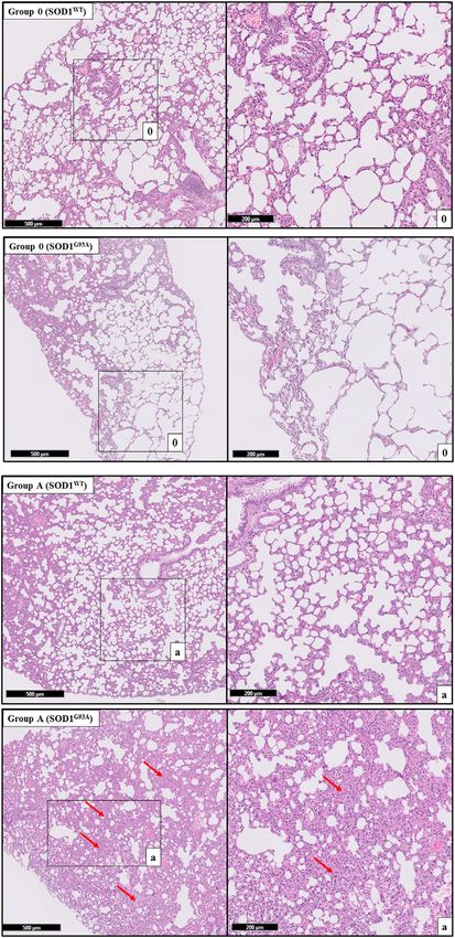
Aydemir et al.                                                                                                                 Dysfunction of Lung Tissue in ALS Disease

  FIGURE 1 | The computer-aided design (CAD) of the sample holding clamps and the dissected lung sample of the rat. (A) The locking of the lung sample between
  the force sensor side and the linear actuator side clamps. (B) CAD model of the top and bottom mount of the clamps. (C) Zoomed view of the saw-tooth structure on the
  clamps along with the dimensions. (D) Dissected lung sample of the rat. (E) The schematics illustrate the dimensions of the studied lung sample. (F) The custom-built
  tensile testing experimental setup indicates the laser displacement sensor, the force sensor, the linear actuator, and the data acquisition system.

Experimental Setup of the Biomechanical                                                 displacement sensor was interfaced with the NI-DAQ through
Tests                                                                                   a laser controller (Figure 1).
Uniaxial tensile testing using a custom-built experimental                                 The clamps were developed by first creating a computer-aided
setup which was comprised of a pre-calibrated                                           design (CAD) model in Siemens NX 12.0 software. The CAD
                                                                                        models of the clamps were then processed using slicing software
piezoelectric-based precise force sensor (Kistler 9256C1), a
                                                                                        before manufacturing. The additive manufacturing was
laser type displacement sensor (Keyence LK-H052), an
                                                                                        performed using a 3D printer, employing the fused filament
electromagnetic-based linear actuator (Dunkermotoren                                    deposition (FFD) technique and the polylactic acid (PLA) type
STA-25), a pair of additively manufactured tissue holding                               filament. The layer thickness of 100 microns and the brass type
clamps and a high-resolution digital camera for the                                     nozzle having a tip diameter of 0.4 mm were selected to
subsequent slippage analysis of the lung samples. The force                             manufacture clamps. Figure 1 illustrates the CAD model of
sensor was interfaced with the data acquisition module (NI-                             the clamps from different orientations and the dissected lung
DAQ 9235) through a charge amplifier, whereas; the laser                                 sample of a rat. The additively manufactured tissue holding

Frontiers in Bioengineering and Biotechnology | www.frontiersin.org                 3                                         February 2022 | Volume 10 | Article 810243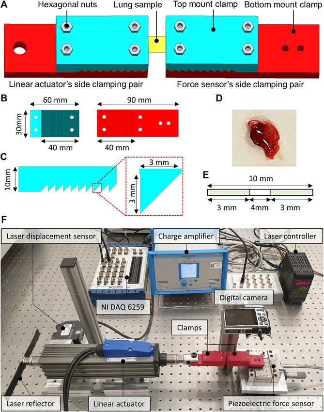
Aydemir et al.                                                                                              Dysfunction of Lung Tissue in ALS Disease

clamps comprised a pair of top and bottom mount structures with            Microwave Digestion
a saw-tooth type locking arrangement to keep the lung samples              Acidic treatment was used to dissolve serum samples of the rats for
firmly when locked. The clamping area was 10-teeth, each having             ICP-MS analysis. A microwave digestion system (Milestone START
a height and width equal to 3 mm. The top and the bottom                   D) only equipped with the temperature control sensor was used to
mounts were locked into each other with the help of the                    dissolve serum samples of SOD1WT and SOD1G93A rats. 100 µl of
hexagonal nuts enabling the reusability of the additively                  serum sample was dissolved in 10 ml of 65% SUPRAPURE® nitric
manufactured clamps for testing multiple samples. One pair of              acid (HNO3) (MERCK, Germany) as described previously in detail
clamps was mounted on the force sensor, whereas the second pair            (Aydemir et al., 2020c).
of clamps were attached to the moving shaft of the linear actuator,
as depicted in Figure 1. The two pairs of clamps were then                 ICP-MS
positioned to be 4 mm apart from each other. The lung sample               Samples were diluted at a 1/10 ratio after acidic digestion via
was fixed between the two clamps, as demonstrated in Figure 1,              microwave digestion and used for the ICP-MS analysis. The trace
and the hexagonal nuts were tightened enough to avoid slippage             and mineral element levels of the rat serum samples were evaluated
and sample breakage.                                                       using Agilent 7700x ICP-MS (Agilent Technologies Inc., Tokyo,
                                                                           Japan). External calibration solutions were prepared using the Spex
                                                                           Certiprep Multi-element calibration standard (2A). Data analysis
Experimental Protocol of the Biomechanical                                 was performed via MassHunter software (Aydemir et al., 2020c).
Tests
The uniaxial tensile testing of all the samples was performed              Tissue Preparation and Mitochondria
under ambient conditions of the test room. The test room
temperature and relative humidity were maintained at 22–25°C
                                                                           Isolation
                                                                           Mitochondria isolation was performed according to the modified
and 60%–65% RH, respectively. At the start of every experiment,
                                                                           method described by Clayton and Shadel (Clayton and Shadel, 2014).
the laser displacement and the force sensor offsets were zeroed
                                                                           The buffer solution was prepared with 1 M sucrose and 100 mM Tris-
through the laser control module and the charge amplifier. The
                                                                           HCl (pH 7.4) in the ultrapure water. pH was set up to 7.4, and
experiment started by linearly displacing the shaft of the linear
actuator at a speed of 0.5 mm/s in the direction opposite to that of                                                      ™
                                                                           protease inhibitor cocktail Roche cOmplete , EDTA-free (Merck,
                                                                           Germany) was added into the buffer freshly just before
the force sensor to apply uniaxial tension on the lung sample. The
                                                                           homogenization. Tissue samples weighing 40–100 mg were
force sensor remained fixed throughout the experiment. The
                                                                           homogenized in this buffer and then centrifuged at 7,000 g for
experiment is completed once the full tearing of the lung
                                                                           20 min at 4°C. The pellet containing mitochondria was separated
sample is observed. The tensile force was measured with the
                                                                           into the clean tubes and stored at 80°C. The supernatant of each
help of a precise piezoelectric force sensor. The charge output of
                                                                           sample was collected and centrifuged at 105,000 ×g for 65 min at 4°C,
the force sensor was converted into voltages through a charge
                                                                           and the supernatant was stored as the cytosolic fraction at 80°C.
amplifier. The laser displacement sensor measured the axial
                                                                           200 mM sodium phosphate buffer (pH 7.4) containing protease
displacement. The force and the laser sensor data were
                                                                           inhibitor cocktail was added into the mitochondria lysate after
acquired at a sampling rate of 1,000 samples/seconds and
                                                                           thawing and sonicated via Branson W250 at 70% amplitude for
stored in the comma-separated file format for subsequent
                                                                           three cycles of 4 s each. Samples were centrifuged at 105,000×g for
analysis.
                                                                           65 min at 4°C, and the supernatant was stored as the mitochondrial
                                                                           fraction of the lung at 80°C (Aydemir et al., 2019c). Protease inhibitor
Hematoxylin and Eosin Staining                                             cocktails were used to prepare mitochondrial and cytosolic fractions
Tissues were collected and washed with ice-cold saline buffer and          to prevent protease activity since many proteases can degrade
fixed in 10% formalin and processed by an automated system                  proteins of interest (Ryan and Henehan, 2017; Dayal et al., 2020).
then embedded in paraffin Sakura Tissue Tek VIP Tissue
Processor & Tissue Embedding Station, Leica) Paraffin                       Protein Determination
(FFPE—formalin-fixed paraffin-embedded) blocks were                          According to the kit instructions, a Pierce BCA Protein Assay kit
prepared and cut by 4 µm thick sections on glass slides for                (Thermo Scientific, USA) was used to evaluate the soluble protein
hematoxylin and eosin (H&E) staining. Before staining tissues,             concentration in all samples by using albumin as standard.
sections on the slides were deparaffinized via xylene by heating at         Samples were pipetted into the 96 well plates and read at
60°C for 20 min and then rehydrated with firstly alcohol (96%)              562 nm using Synergy H1, BIOTEK.
and then PBS (phosphate-buffered saline). Slides were dipped in
Mayer’s hematoxylin for 1 min and washed with distilled water.
After that, slides were immersed in eosin for 10 s and washed              Enzyme-Linked Immunosorbent Assay for
multiple times with ethanol (70%, 96%, and absolute). Slides were          Catalase and SOD1
dipped in xylene, and mounting was performed using an                      Prepared tissue lysates were used to evaluate cytosolic CAT and
automated slide mounting system (Tissue-Tek Prisma, Sakura                 SOD1 activity using the Abbkine Rat Catalase ELISA kit (Abbkine,
Finetek USA). This system uses only xylene and a special film for           China). Samples were pipetted on the 96-well plate of the ELISA kit
mounting and does not need other mounting media.                           and, after incubated with HRP-conjugated antibody wells, were

Frontiers in Bioengineering and Biotechnology | www.frontiersin.org    4                                   February 2022 | Volume 10 | Article 810243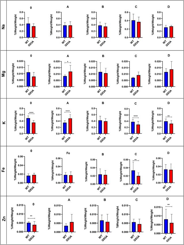
Aydemir et al.                                                                                                                   Dysfunction of Lung Tissue in ALS Disease

  FIGURE 2 | Force, displacement, and stiffness values of lung tissues belonging to SOD1G93A and SOD1WT rats. (a) The stiffness value of lung tissue is calculated as
  represented by the figure via using displacement and force values. *p ≤ 0.05, **p ≤ 0.001 and ***p ≤ 0.000.

washed and treated with chromogen solutions according to the kit                          reductase (GR), and glutathione s-transferase (GST) enzyme
instructions. After the experimental procedure was done, the                              activities were measured in both cytosol and mitochondria
absorbance of the samples was read at 450 nm.                                             fractions of lung samples as described previously (Aydemir
                                                                                          et al., 2019a; Aydemir et al., 2019b; Aydemir et al., 2019c).

Evaluation of Oxidative Stress Enzyme                                                     Western Blot
Activities                                                                                Cytosolic and mitochondrial fractions of the lung tissues were used
Glucose    6-phosphate  dehydrogenase (G6PD),    6-                                       for western blot analysis to evaluate the SOD1G93A protein
phosphogluconate dehydrogenase (6-PGD), glutathione                                       accumulation after protein quantification. B8H10 antibody

Frontiers in Bioengineering and Biotechnology | www.frontiersin.org                   5                                         February 2022 | Volume 10 | Article 810243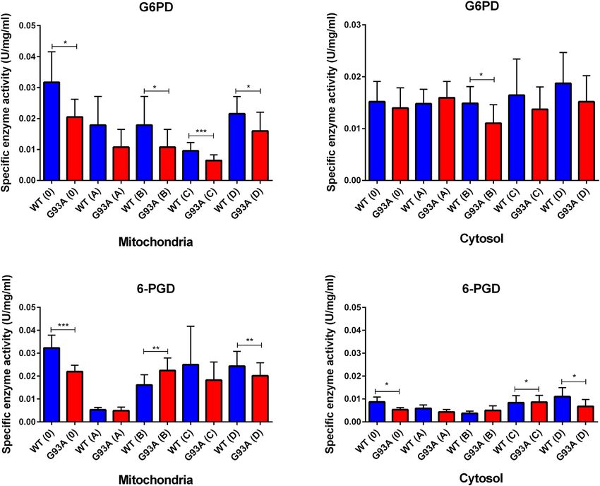
Aydemir et al.                                                                                                            Dysfunction of Lung Tissue in ALS Disease

TABLE 1 | Lung stiffness of SOD1G93A (SOD1) mutated, and wild-type (WT) rats               rats. All tissues were examined after H&E staining, and
are represented in the table as mean ± SD.
                                                                                           representative pictures were chosen to demonstrate
                                         Group                Stiffness [N/mm]             histopathological changes in the lung. A pathologist performed
                                                                                           the histopathological examination and detected the percentage of
MALE                WT                   0                       0.26   ±   0.06
                                                                                           atelectasis by comparing wild-type and mutated lung samples. No
                                         A                       0.23   ±   0.04
                                         B                       0.23   ±   0.09           histopathological changes have been observed between SOD1WT
                                         C                       0.16   ±   0.13           and SOD1G93A rats in group 0 (Figure 6); on the other hand,
                                         D                       0.24   ±   0.06           substantial atelectasis (~30%–60%) and some emphysemas were
                    SOD1                 0                       0.21   ±   0.19           observed in the SOD1G93A rats of groups A (Figure 4), B
                                         A                       0.34   ±   0.11
                                         B                       0.47   ±   0.07
                                                                                           (Figure 5), C and D (Figure 6) more common than the
                                         C                       0.23   ±   0.09           SOD1WT group.
                                         D                       0.39   ±   0.13

                                                                                           Evaluation of the Trace Element and Mineral
(MEDIMABS, Montreal, CANADA) was used to detect human                                      Levels in the Lung Tissue
SOD1G93A aggregates in the tissue of interest. After protein                               Following acidic digestion, the lung tissue’s trace element and
quantification as described above, 30–70 μg protein from each                               mineral levels were evaluated via ICP-MS. Mg levels slightly
sample was diluted with 4x Laemmli buffer (10% SDS, 50%                                    decreased in the SOD1G93A rats compared to the SOD1WT rats
glycerol, 0.3 M Tris-HCl (pH 6.8), 0.05% bromphenol blue in 50 ml                          except for group D; however, this decrease was insignificant
Mili-Q water) containing 10% of beta-mercaptoethanol and samples                           (Figure 6). Mg levels increased in the mutated rats in
were incubated at 95°C for 2 min in thermoblock. Then samples were                         comparison with wild-type rats in groups A, C, and D, and
separated on 10% SDS-PAGE with 5% stacking gel using PowerPac
Universal Power Supply at 90 V for ~2.5 h. After gel electrophoresis,
                                                                                   ™       this change was significant in group A (*p ≤ 0.05) (Figure 6). K
                                                                                           levels decreased in the mutated rats of groups A (***p ≤ 0.0001), C
proteins were transferred to the PVDF membrane for an hour at                              (***p ≤ 0.0001), and D (**p ≤ 0.001) in comparison with wild-type
150 mA with Bio-Rad wet transfer apparatus at 4°C room. Following                          rats, where K level decreased in the SOD1G93A rats in group 0
that, PVDF membranes were blocked with 5% BSA prepared in                                  (***p ≤ 0.0001) compared to the SOD1WT rats (Figure 6). Fe
phosphate-buffered saline/0.1% Tween (PBS-T) for an hour and                               levels did not show any significant differences between the two
probed with primary SOD1 antibody (diluted in 1% BSA in PBST                               groups except in group C, where Fe levels significantly decreased
to 1:250) at 4°C overnight as described previously (Otahal et al., 2020).                  in the mutated rats (**p ≤ 0.001) compared to the wild-type rats
                                                                                           (Figure 6). On the other hand, Zn levels reduced in the mutated
Statistical Analysis                                                                       rats of groups 0, B, C, and D; however, this decrease was
The statistical significance of the clinical data was evaluated via                         significant for group 0 (**p ≤ 0.001) and D (**p ≤ 0.001).
GraphPad Software, Inc., USA. Mann Whitney test was used to
compare the two groups. All data were represented as the mean ±
                                                                                           Human SOD1G93A Mutated Protein
standard deviation (SD).
                                                                                           Accumulation in the Cytosol and
                                                                                           Mitochondria of the Lung Tissues
RESULTS                                                                                    SOD1G93A mutated protein accumulation (~16 kDa) in the
                                                                                           cytosolic and mitochondrial fractions of the lung tissues
Biomechanical Changes in the Lung Tissue                                                   belonging to rats was evaluated via B8H10 antibody
Displacement, force, and stiffness of the SOD1WT and SOD1G93A                              recognizing mutant human SOD1G93A protein expressed by in
rats were measured by a custom-built stretcher device                                      the SOD1G93A rat model. Our data showed that human mutant
represented in Figure 1. Displacement and force have been                                  SOD1G93A protein was accumulated in the cytosol and
measured in the lung samples of all rats, and the slope in the                             mitochondria of lung tissues belonging to the SOD1G93A rats;
linear region of the force versus displacement curve depicts the                           however, we did not observe any accumulation in the wild-type
stiffness of the lung sample (Figure 2) (Wolfla et al., 2010). Lung                         rats (Figure 6).
stiffness increased in the SOD1G93A rats compared to SOD1WT
rats in all groups except 0 (Table 1) (Figure 2). Increase in the
                                                                                           Evaluation of the Oxidative Stress
lung stiffness was not significant in the mutated rats of groups A,
B, C and D, however this increase was almost double in the                                 Metabolism and Pentose Phosphate
indicated groups in comparison with wild type rats (Table 1).                              Pathway Enzymes in the Cytosol and
                                                                                           Mitochondria of the Lung Tissues
                                                                                           G6PD enzyme activity decreased in the cytosolic and
Histopathological Changes in the Lung                                                      mitochondrial fractions of lung tissues belonging to the
Tissue                                                                                     SOD1G93A rats in comparison with SOD1WT rats, indicated
H&E staining was performed to evaluate histopathological                                   decrease was significant in the mitochondria of the groups 0
changes in the lung tissues of both SOD1WT and SOD1G93A                                    (*p ≤ 0.05), B (*p ≤ 0.05), C (***p ≤ 0.0001) and D (*p ≤ 0.05),

Frontiers in Bioengineering and Biotechnology | www.frontiersin.org                    6                                 February 2022 | Volume 10 | Article 810243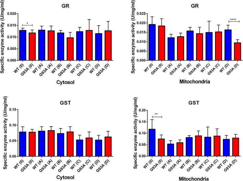
Aydemir et al.                                                                                                   Dysfunction of Lung Tissue in ALS Disease

wherein the cytosol for the group B (*p ≤ 0.05) (Figure 7). Same
as G6PD, 6-PGD enzyme activity decreased in both cytosolic and
mitochondrial fractions of the mutated rats compared to the wild-
type rats. For the cytosolic fractions of the lung, 6-PGD enzyme
activity significantly reduced in the groups 0 (***p ≤ 0.0001), B
(**p ≤ 0.001), and D (**p ≤ 0.001), where this decrease was
significant in the mitochondria of the groups C (*p ≤ 0.05) and D
(*p ≤ 0.05) (Figure 7).
   GR enzyme activity decreased in the mitochondrial fractions
of the mutated rats compared to the wild-type rats, and this
decrease was significant for group D (***p ≤ 0.0001). For the
cytosol, GR activity decreased in the SOD1G93A rats of the groups
0, A, and B; this decrease was significant only for group 0 (*p ≤
0.05). On the other hand, GR activity slightly increased in the
mutated rats belonging to groups C and D (Figure 8). GST
enzyme activity increased in the cytosolic fraction of lung samples
belonging to the SOD1G93A rats; however, this increase was
insignificant in any group. In the mitochondria of lung tissues,
GST enzyme activity decreased in the mutated rats (*p ≤ 0.05 for
group 0), on the other hand slightly increased in groups B, C, and
D (Figure 8).
   We further measured SOD1 and CAT levels to evaluate
oxidative stress status in the cytosol of the lung tissues. SOD1
levels decreased in the SOD1G93A rats of group A (*p ≤ 0.05),
where they increased in group C (insignificant) and D (**p ≤
0.001). CAT levels decreased in the mutated rats of groups 0 (*p ≤
0.05), A (insignificant), and B (insignificant); however, increased
in groups C (*p ≤ 0.05) and D (*p ≤ 0.05) (Figure 9).

DISCUSSION
ALS is the most common motor neuron disease, and the
incidence is increasing worldwide every year. Since there is no
treatment against ALS, therapeutic approaches are urgently
needed to cure people with ALS (Aydemir and Ulusu, 2020b).
The lung is one of the most affected organs during ALS
progression; however, no studies address possible mechanisms
behind the lung impairment in patients with ALS that can be used
as a promising targeted therapy approach in the future (Brent
et al., 2020). Biomechanical and biochemical properties of the
lung have been impaired in several diseases such as Parkinson’s
Disease, pulmonary fibrosis, cancer, asthma, emphysema, chronic
obstructive pulmonary disease (COPD), and pulmonary arterial
hypertension (PAH) disease according to the literature; however,             FIGURE 3 | H&E-stained lung tissue sections of lung tissues.
no published data addressing possible mechanisms contributing                Representative images of SOD1G93A and SOD1WT rats of groups 0 and A
to the ALS progression (Burgstaller et al., 2017). Therefore, in this        were taken from each group. Scale lines on the left row indicate 500 μm and
                                                                             the second row 200 μm. Red arrows indicate the area with atelectasis of
study, we aimed to investigate the impact of ALS on the                      SOD1G93A rats.
biomechanical changes correlated with oxidative stress
metabolism and mutant SOD1G93A protein accumulation in
the lung tissue. Since aging directly contributes to tissue
dysregulation, such as increased stiffening, we used control                Hirani, 2008), we first measured lung stiffness to evaluate
animals for each age group to eliminate the effects of aging as             biomechanical changes during ALS progression by eliminating
described in the section of animal studies (Sicard et al., 2018).           effects of aging. The stiffness of the biological specimens can be
    Since increased lung stiffness has been observed in several             estimated experimentally using tensile testing equipment. The
diseases due to impaired elasticity, altered biochemical and                modulus of elasticity can also be used to frame the mechanical
biomechanical homeostasis of the lung tissue (Wells and                     behavior of the biological samples using tensile testing

Frontiers in Bioengineering and Biotechnology | www.frontiersin.org     7                                       February 2022 | Volume 10 | Article 810243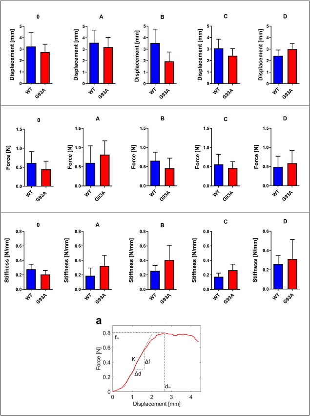
Aydemir et al.                                                                                                      Dysfunction of Lung Tissue in ALS Disease

                                                                                    samples, it is challenging to estimate the exact cross-sectional area
                                                                                    to calculate elastic modulus. Therefore, in this study, only the
                                                                                    stiffness measurements are considered for subsequent analysis
                                                                                    and comparison via a custom-built-up stretcher device (De Kegel
                                                                                    et al., 2018; Aydın et al., 2019; Sezgin et al., 2019). For the first
                                                                                    time in the literature, we showed that lung stiffness increased in
                                                                                    the SOD1G93A mutated male rats compared to the SOD1WT rats
                                                                                    even at the very early stages of life, indicating impaired tissue
                                                                                    function (Figure 2).
                                                                                        Increased stiffness causes impairment in lung tissue’s
                                                                                    biomechanical properties, which can be evaluated by the
                                                                                    histopathological evaluation; therefore, we performed H&E
                                                                                    staining for all lung samples to compare mutated and wild-
                                                                                    type rats (Cacopardo et al., 2021). Each lung sample was
                                                                                    evaluated after H&E staining and compared between groups
                                                                                    by a pathologist. Substantial atelectasis (~30%–60%) and some
                                                                                    emphysemas were observed in the lung tissue of mutated rats of
                                                                                    all groups except groups 0 according to our data (Figures 3–5).
                                                                                    Atelectasis leads to the impairment in the lung elasticity and
                                                                                    blood oxygenation, causing inflammation, impairment in the
                                                                                    alveolar-capillary barrier, tissue damage, and respiratory
                                                                                    function at the end (Zeng et al., 2021). Atelectasis and tissue
                                                                                    stiffness started at the early stages of ALS progression, indicating
                                                                                    tissue damage even at the pre-symptomatic stages (Figures 3–5).
                                                                                    Increased tissue stiffness and histopathological changes
                                                                                    indicating tissue dysfunction have been found in the lung
                                                                                    samples of SOD1G93A rats compared to SOD1WT rats (Figures
                                                                                    2–5). Elasticity, homeostasis, and stiffness of lung tissue are
                                                                                    mainly regulated by ECM regulation. According to the
                                                                                    literature, extracellular matrix (ECM) dysregulation, protein
                                                                                    aggregation, impaired mineral metabolism, aging, and trace
                                                                                    element accumulation are essential mechanisms contributing
                                                                                    to lung stiffness (White, 2015). Moreover, ECM regulation is
                                                                                    affected by mineral composition, matrix modifying enzymes,
                                                                                    oxidative stress, and remodeling processes (Liu et al., 2010;
                                                                                    Parameswaran et al., 2011; White, 2015).
                                                                                        Trace element and mineral levels are vital for tissue
                                                                                    homeostasis, cellular functions, oxidative stress metabolism,
                                                                                    and aging (Aydemir et al., 2020a); thus, we have evaluated Na,
                                                                                    Mg, K, Fe, and Zn levels of the lung tissue. Zn levels decreased in
                                                                                    the mutated rats of groups B, C, and D compared to the wild-type
                                                                                    rats (Figure 6). Zn plays a role in the matrix metalloproteinase
                                                                                    (MMP) enzyme activities regulating ECM homeostasis and
                                                                                    oxidative stress metabolism as integrating into the structure of
                                                                                    the oxidative stress enzymes (Kostov and Halacheva, 2018). On
  FIGURE 4 | H&E-stained sections of the lung tissue samples belonging
  to the SOD1WT and SOD1G93A rats of groups B and C. Representative images
                                                                                    the other hand, Na and K levels fluctuated in the SOD1G93A rats
  were taken from each group. Scale lines on the left row indicate 500 μm and       compared to SOD1WT rats indicating impaired electrolyte levels
  the second row 200 μm. Red arrows indicate the area with atelectasis of           and impaired mineral metabolism in the lung tissue (Figure 6).
  SOD1G93A rats.                                                                    According to the literature, Na, K-ATPase function has altered
                                                                                    the alveolar epithelial barrier leading to tissue damage in the lung
                                                                                    (Vadász et al., 2007), and Na+ pumps are responsible for the
equipment. The force divided by the cross-sectional area of the                     creating Na+ gradient to keep the balance of pulmonary liquid
sample is termed the stress, and the displacement divided by the                    balance. Additionally, Na+ pumps and balance are involved in the
unstressed length of the specimen is termed the strain. The slope                   epithelial stretch, vital for breathing and oxygenation (Fisher and
in the linear region of the stress versus strain curve is defined as a               Margulies, 2002). K levels significantly decreased in the mutated
modulus of elasticity of the test specimen (Maikos et al., 2008).                   rats, where Na levels fluctuated in the lung samples of SOD1G93A
Due to the small size and non-uniform symmetry of the lungs                         rats during ALS progression, according to our data (Figure 6). As

Frontiers in Bioengineering and Biotechnology | www.frontiersin.org             8                                  February 2022 | Volume 10 | Article 810243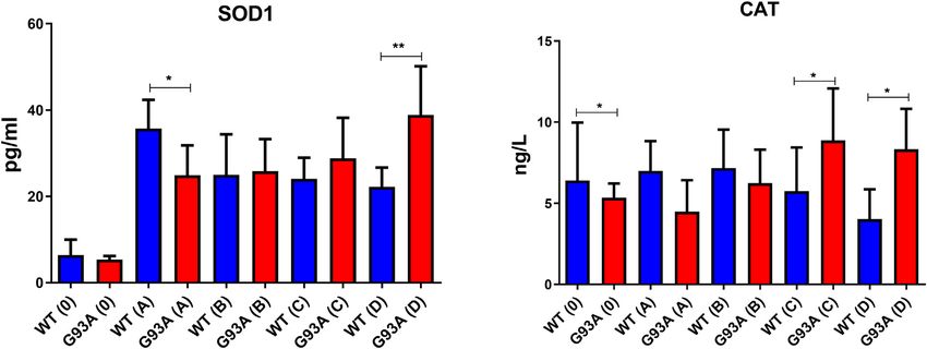
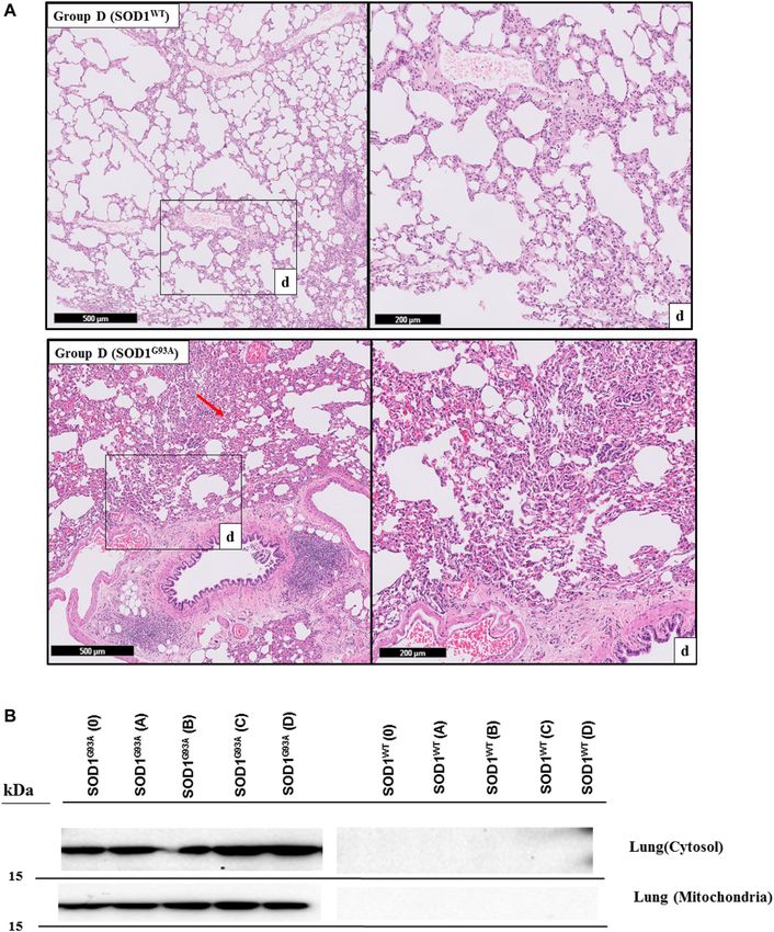
Aydemir et al.                                                                                                                  Dysfunction of Lung Tissue in ALS Disease

  FIGURE 5 | H&E-stained sections of the lung tissue samples belonging to the SOD1WT and SOD1G93A rats of group D. All lung tissues of rats (n = 90) were stained
  and evaluated; among them, representative images were taken from each group. Scale lines on the left row indicate 500 μm and the second row 200 μm. Red arrows
  indicate the area with atelectasis and emphysema of SOD1G93A rats (A). SOD1G93A protein accumulation was evaluated in the cytosolic and mitochondrial fractions of the
  lung tissue. Accumulation of human mutant SOD1G93A has been found in all SOD1G93A groups, where SOD1WT did not show any mutant protein in the lung
  tissue (B).

Frontiers in Bioengineering and Biotechnology | www.frontiersin.org                  9                                         February 2022 | Volume 10 | Article 810243
Aydemir et al.                                                                                                         Dysfunction of Lung Tissue in ALS Disease

  FIGURE 6 | Trace element and mineral levels of SOD1G93A and SOD1WT rats. *p ≤ 0.05, **p ≤ 0.001 and ***p ≤ 0.0001.

Frontiers in Bioengineering and Biotechnology | www.frontiersin.org           10                                       February 2022 | Volume 10 | Article 810243
Aydemir et al.                                                                                                      Dysfunction of Lung Tissue in ALS Disease

  FIGURE 7 | G6PD and 6-PGD enzymes belonging to the PPP were evaluated in both cytosol and mitochondria of lung tissues of SOD1WT (WT) and SOD1G93A
  (G93A) rats. Data were given as mean ± SD of n = 8 animals for each group. Notes: *p ≤ 0.05, **p ≤ 0.001 and ***p ≤ 0.0001.

a result, Zn, K, Mg, and Na levels are impaired in the SOD1G93A                   tissue homeostasis (Watson et al., 2016). On the other hand,
rats compared to the SOD1WT rats indicating altered homeostasis                   pulmonary endothelial barrier in the lung tissue is affected by
in the MMP activity, ECM regulation, oxidative stress                             elevated levels of oxidative stress resulting in the dysregulation of
metabolism, and Na, K-ATPase function (Figure 6) that                             biomechanical changes of the lung (Haak et al., 2018). During
contribute to the impaired oxidative stress metabolism and                        breathing cycle, blood passes through pulmonary vessels and
lung function.                                                                    distension occurs in the lung leading to the formation of
   Several theories address possible mechanisms behind ALS                        biomechanical forces such as fluid shear stress, pressure and
pathogenesis; among them, protein aggregation has been                            cyclic stress. Indicated biomechanical changes cause formation of
tightly correlated with dysfunction in the cellular function,                     oxidative stress in the lung, thus maintenance of anti-oxidant
oxidative stress metabolism, and tissue damage (Zhao et al.,                      defense and redox metabolism play vital role to protect lung
2010; Hayashi et al., 2016; Kaur et al., 2016). Additionally,                     homeostasis (Zemskov et al., 2019). Oxidative stress is elevated by
impaired Na, K, Zn, and Mg levels can be considered                               both ageing and diseases causing to the impaired inflammatory
indicators of impaired oxidative stress metabolism (Aydemir                       function, pulmonary endothelial barrier function, apoptosis and
et al., 2018). Thus, we hypothesized that tissue damage and                       ECM dysregulation in the lung. On the other hand, oxidative
dysfunction could result from elevated oxidative stress levels                    stress is considered is one of the major hallmarks several lung
induced by the accumulation of human mutant SOD1G93A                              diseases including chronic obstructive pulmonary disease
protein in both mitochondria and the cytosol of lung tissues                      (COPD), acute respiratory distress syndrome (ARDS) and
(Figure 6) since oxidative stress impairs ECM regulation and                      idiopathic pulmonary fibrosis (IPF). Since we have proven that

Frontiers in Bioengineering and Biotechnology | www.frontiersin.org          11                                    February 2022 | Volume 10 | Article 810243
Aydemir et al.                                                                                                            Dysfunction of Lung Tissue in ALS Disease

  FIGURE 8 | GR and GST enzymes were evaluated in both cytosol and mitochondria of lung tissues of SOD1WT (WT) and SOD1G93A (G93A) rats. Data were given as
  mean ± SD of n = 8 animals for each group. Notes: *p ≤ 0.05, **p ≤ 0.001 and ***p ≤ 0.0001.

  FIGURE 9 | SOD1 and CAT levels in the cytosol of lung tissues belonging to both SOD1WT (WT) and SOD1G93A (G93A) samples were evaluated via ELISA assay.
  Data were given as mean ± SD of n = 8 animals for each group. Notes: *p ≤ 0.05, **p ≤ 0.001 and ***p ≤ 0.000.1.

Frontiers in Bioengineering and Biotechnology | www.frontiersin.org             12                                       February 2022 | Volume 10 | Article 810243
Aydemir et al.                                                                                             Dysfunction of Lung Tissue in ALS Disease

mutant SOD1G93A protein accumulated and histopathological                   targeted therapy approaches can be tried to improve lung
changes have arisen in the lung tissue, oxidative stress parameters         function to prolong survival and improve the life quality of
have evaluated as major cause of the impairment in the lung                 ALS patients.
function during ALS progression (Hecker, 2018).
    Oxidative stress is produced in all aerobic cells; however,
neurons are more susceptible to oxidative stress. Antioxidant               CONCLUSION
protection of the organism is maintained by three enzymes,
including the SOD enzyme family, catalase, and GSH-                         Decreased antioxidant status, elevated levels of oxidative stress,
dependent enzymes such as glutathione reductase (GR),                       and mutant SOD1G93A protein accumulation have been found in
glutathione-s transferase (GST), and glutathione peroxidases                the lung samples of SOD1G93A mutated rats even at the earlier
(GPx). The redox metabolism in the organism is regulated by                 stages of life first time in the literature, which can be possible
reduced glutathione (GSH), oxidized glutathione (GSSG), GSSG/               causative of increased stiffness and impaired morphology of lung
GSH ratio, catalase (CAT), superoxide dismutase family (SOD),               tissue. All these changes can result from the accumulation of
NAD+/NADH, NADP+/NADPH, glucose-6 phosphate                                 SOD1G93A rats in mitochondria and cytosol, causing impaired
dehydrogenase (G6PD), 6-phosphogluconate dehydrogenase                      energy and oxidative stress metabolism in the lung. Since
(6-PGD), GR, GPx and GST. NADPH is a reducing agent and                     indicated alterations have started at the early stages of ALS
plays a vital role in the detoxifying processes by eliminating              progression, even in the pre-symptomatic stages, possible
oxidative radicals and peroxides (Ozcan et al., 2021).                      therapeutic approaches can be used to treat ALS or improve
Additionally, it involves various metabolic processes, including            the life quality of patients with ALS.
cell proliferation, survival and senescence, energy metabolism,
mitochondrial functions, calcium homeostasis, antioxidant/
generation of oxidative stress, gene expression, fatty acid, and            DATA AVAILABILITY STATEMENT
steroid synthesis. On the other hand, NADPH is required for the
amino acid synthesis involved in the cytochrome P450 systems                The original contributions presented in the study are included in
enabling detoxification of drugs, redox balance, immunological               the article/Supplementary Material, further inquiries can be
homeostasis, aging, and cell death. Decreased levels of G6PD and            directed to the corresponding author.
6-PGD are the indicators of impaired glucose and energy
metabolisms in both cytosol and mitochondria as well
(Aydemir et al., 2019a; Aydemir and Ulusu, 2020a; Aydemir                   ETHICS STATEMENT
et al., 2020b).
    NADPH enables the reduction of GSSG to its reduced form                 The animal study was reviewed and approved by the Ethics
GSH, which is a major scavenger of ROS and required for many                Committee of Koc University with the number
reduction systems. G6PD and 6-PGD enzymes reduce NADP                       2019.HADYEK.006.
into NADPH, so they remain NADPH pool in the cells and keep
redox balance. Imbalance or malfunction in the redox pathways
of antioxidant enzymes causes impairment in the antioxidant                 AUTHOR CONTRIBUTIONS
metabolism leading to elevated levels of oxidative stress (Aydemir
et al., 2018; Aydemir et al., 2019a; Aydemir and Ulusu, 2020a;              Conceptualization, NU and DA; Methodology, DA, NU, IL, ANB,
Aydemir and Ulusu, 2020b; Aydemir et al., 2021). G6PD and 6-                and ANM, Investigation, DA and ANM; Resources, NU, ANB,
PGD levels decreased in SOD1G93A mutated rats in both cytosol               and IL; Data Curation, DA; Writing—Original Draft, DA and
and mitochondria (Figure 7), which may lead to the reduced                  ANM; Writing—Review & Editing, DA, NU, IL, and ANM;
antioxidant status and elevated levels of oxidative stress.                 Visualization, DA and ANM; Supervision, NU, ANB, and IL.
Additionally, GR and GST levels increased in the SOD1G93A
rats compared to the SOD1WT rats indicating elevated levels of
oxidative stress (Figure 8)). We further evaluated CAT and SOD1             ACKNOWLEDGMENTS
levels to prove high levels of oxidative stress and showed that
CAT and SOD1 levels significantly increased in the SOD1G93A                  The authors gratefully acknowledge the use of the services and
rats compared to the SOD1WT rats (Figure 9). As a result, we                facilities of the Koc University Research Center for Translational
found that oxidative stress metabolism is impaired in the mutated           Medicine (KUTTAM), funded by the Presidency of Turkey,
rats because of the accumulation of mutant SOD1G93A protein                 Presidency of Strategy and Budget. The content is solely the
that can be considered a possible mechanism behind increased                authors’ responsibility and does not necessarily represent the
lung stiffness and impaired tissue function. In the future, possible        official views of the Presidency of Strategy and Budget.

Frontiers in Bioengineering and Biotechnology | www.frontiersin.org    13                                 February 2022 | Volume 10 | Article 810243
Aydemir et al. Dysfunction of Lung Tissue in ALS Disease
REFERENCES Dayal, A. A., Medvedeva, N. v., Nekrasova, T. M., Duhalin, S. D., Surin, A. K., and
Minin, A. A. (2020). Desmin Interacts Directly with Mitochondria. Ijms 21,
8122. doi:10.3390/ijms21218122
Aydemir, D., Dağlıoğlu, G., Candevir, A., Kurtaran, B., Bozdogan, S. T., Inal, T. C., De Kegel, D., Vastmans, J., Fehervary, H., Depreitere, B., Vander Sloten, J., and
et al. (2021). COVID-19 May Enhance Risk of Thrombosis and Hemolysis in Famaey, N. (2018). Biomechanical Characterization of Human Dura Mater.
the G6PD Deficient Patients. Nucleosides, Nucleotides & Nucleic Acids 40, J. Mech. Behav. Biomed. Mater. 79, 122–134. doi:10.1016/j.jmbbm.2017.12.023
505–517. doi:10.1080/15257770.2021.1897457 Discher, D. E., Mooney, D. J., and Zandstra, P. W. (2009). Growth Factors,
Aydemir, D., Hashemkhani, M., Acar, H. Y., and Ulusu, N. N. (2020a). Evaluation Matrices, and Forces Combine and Control Stem Cells. Science 324,
of the Biocompatibility of the GSH-Coated Ag2S Quantum Dots In Vitro: a 1673–1677. doi:10.1126/science.1171643
Perfect Example for the Non-toxic Optical Probes. Mol. Biol. Rep. 47, Fisher, J. L., and Margulies, S. S. (2002). Na+-K+-ATPase Activity in Alveolar
4117–4129. doi:10.1007/s11033-020-05522-3 Epithelial Cells Increases with Cyclic Stretch. Am. J. Physiology-Lung Cell Mol.
Aydemir, D., Hashemkhani, M., Acar, H. Y., and Ulusu, N. N. (2019a). In Vitro Physiol. 283, L737–L746. doi:10.1152/ajplung.00030.2001
interaction of Glutathione S-transferase-pi Enzyme with Glutathione-coated Silver Gill, C., Phelan, J. P., Hatzipetros, T., Kidd, J. D., Tassinari, V. R., Levine, B., et al.
Sulfide Quantum Dots: A Novel Method for Biodetection of Glutathione S- (2019). SOD1-positive Aggregate Accumulation in the CNS Predicts Slower
transferase Enzyme. Chem. Biol. Drug Des. 94, 2094–2102. doi:10.1111/cbdd.13614 Disease Progression and Increased Longevity in a Mutant SOD1 Mouse Model
Aydemir, D., Karabulut, G., Gok, M., Barlas, N., and Ulusu, N. N. (2019b). Data the of ALS. Sci. Rep. 9, 6724. doi:10.1038/s41598-019-43164-z
DEHP Induced Changes on the Trace Element and mineral Levels in the Brain Guimarães, C. F., Gasperini, L., Marques, A. P., and Reis, R. L. (2020). The Stiffness
and Testis Tissues of Rats. Data in Brief 26, 104526. doi:10.1016/j.dib.2019. of Living Tissues and its Implications for Tissue Engineering. Nat. Rev. Mater.
104526 5, 351–370. doi:10.1038/s41578-019-0169-1
Aydemir, D., Karabulut, G., Şimşek, G., Gok, M., Barlas, N., and Ulusu, N. N. Haak, A. J., Tan, Q., and Tschumperlin, D. J. (2018). Matrix Biomechanics and
(2018). Impact of the Di(2-Ethylhexyl) Phthalate Administration on Dynamics in Pulmonary Fibrosis. Matrix Biol. 73, 64–76. doi:10.1016/j.matbio.
Trace Element and Mineral Levels in Relation of Kidney and Liver 2017.12.004
Damage in Rats. Biol. Trace Elem. Res. 186, 474–488. doi:10.1007/ Hayashi, Y., Homma, K., and Ichijo, H. (2016). SOD1 in Neurotoxicity and its
s12011-018-1331-0 Controversial Roles in SOD1 Mutation-Negative ALS. Adv. Biol. Regul. 60,
Aydemir, D., Öztaşcı, B., Barlas, N., and Ulusu, N. N. (2019c). Effects of 95–104. doi:10.1016/j.jbior.2015.10.006
Butylparaben on Antioxidant Enzyme Activities and Histopathological Hecker, L. (2018). Mechanisms and Consequences of Oxidative Stress in Lung
Changes in Rat Tissues. Arch. Ind. Hyg. Toxicol. 70, 315–324. doi:10.2478/ Disease: Therapeutic Implications for an Aging Populace. Am.
aiht-2019-70-3342 J. Physiology-Lung Cell Mol. Physiol. 314, L642–L653. doi:10.1152/ajplung.
Aydemir, D., Sarayloo, E., and Nuray, U. N. (2020b). Rosiglitazone-induced 00275.2017
Changes in the Oxidative Stress Metabolism and Fatty Acid Composition in Hynes, R. O. (2009). The Extracellular Matrix: Not Just Pretty Fibrils. Science 326,
Relation with Trace Element Status in the Primary Adipocytes. J. Med. Biochem. 1216–1219. doi:10.1126/science.1176009
39 (3), 267–275. doi:10.2478/jomb-2019-0041 Jawaid, A., Salamone, A. R., Strutt, A. M., Murthy, S. B., Wheaton, M., McDowell,
Aydemir, D., Simsek, G., and Ulusu, N. N. (2020c). Dataset of the Analyzing Trace E. J., et al. (2010). ALS Disease Onset May Occur Later in Patients with Pre-
Elements and Minerals via ICP-MS: Method Validation for the Mammalian morbid Diabetes Mellitus. Eur. J. Neurol. 17, 733–739. doi:10.1111/j.1468-1331.
Tissue and Serum Samples. Data in Brief 29, 105218. doi:10.1016/j.dib.2020. 2009.02923.x
105218 Kaur, S. J., McKeown, S. R., and Rashid, S. (2016). Mutant SOD1 Mediated
Aydemir, D., and Ulusu, N. N. (2020a). Comment on the: Molecular Mechanism of Pathogenesis of Amyotrophic Lateral Sclerosis. Gene 577, 109–118. doi:10.
CAT and SOD Activity Change under MPA-CdTe Quantum Dots Induced 1016/j.gene.2015.11.049
Oxidative Stress in the Mouse Primary Hepatocytes (Spectrochim Acta A Mol Kostov, K., and Halacheva, L. (2018). Role of Magnesium Deficiency in Promoting
Biomol Spectrosc. 2019 Sep 5; 220:117104). Spectrochimica Acta A: Mol. Atherosclerosis, Endothelial Dysfunction, and Arterial Stiffening as Risk
Biomol. Spectrosc. 229, 117792. doi:10.1016/j.saa.2019.117792 Factors for Hypertension. Ijms 19, 1724. doi:10.3390/ijms19061724
Aydemir, D., and Ulusu, N. N. (2020b). Importance of the Serum Biochemical Liu, F., Mih, J. D., Shea, B. S., Kho, A. T., Sharif, A. S., Tager, A. M., et al. (2010).
Parameters as Potential Biomarkers for Rapid Diagnosis and Evaluating Feedback Amplification of Fibrosis through Matrix Stiffening and COX-2
Preclinical Stage of ALS. Med. Hypotheses 141, 109736. doi:10.1016/j.mehy. Suppression. J. Cel Biol. 190, 693–706. doi:10.1083/jcb.201004082
2020.109736 Liu, Y.-J., Ju, T.-C., Chen, H.-M., Jang, Y.-S., Lee, L.-M., Lai, H.-L., et al. (2015).
Aydın, H. E., Kızmazoglu, C., Kaya, I., Husemoglu, B., Sozer, G., Havıtcıoglu, H., Activation of AMP-Activated Protein Kinase α1 Mediates Mislocalization of
et al. (2019). Biomechanical Properties of the Cranial Dura Mater with TDP-43 in Amyotrophic Lateral Sclerosis. Hum. Mol. Genet. 24, 787–801.
Puncture Defects : An In Vitro Study. J. Korean Neurosurg. Soc. 62, doi:10.1093/hmg/ddu497
382–388. doi:10.3340/jkns.2018.0130 Maikos, J. T., Elias, R. A. I., and Shreiber, D. I. (2008). Mechanical Properties of
Brent, J. R., Franz, C. K., Coleman, J. M., and Ajroud-Driss, S. (2020). ALS. Neurol. Dura Mater from the Rat Brain and Spinal Cord. J. Neurotrauma 25, 38–51.
Clin. 38, 565–575. doi:10.1016/j.ncl.2020.03.013 doi:10.1089/neu.2007.0348
Burgstaller, G., Oehrle, B., Gerckens, M., White, E. S., Schiller, H. B., and Mariappan, Y. K., Glaser, K. J., Hubmayr, R. D., Manduca, A., Ehman, R. L., and
Eickelberg, O. (2017). The Instructive Extracellular Matrix of the Lung: McGee, K. P. (2011). MR Elastography of Human Lung Parenchyma: Technical
Basic Composition and Alterations in Chronic Lung Disease. Eur. Respir. J. Development, Theoretical Modeling and In Vivo Validation. J. Magn. Reson.
50, 1601805. doi:10.1183/13993003.01805-2016 Imaging 33, 1351–1361. doi:10.1002/jmri.22550
Burlando, B., Milanese, M., Giordano, G., Bonifacino, T., Ravera, S., Blanchini, F., Martínez-Palma, L., Miquel, E., Lagos-Rodríguez, V., Barbeito, L., Cassina, A., and
et al. (2020). A Multistationary Loop Model of ALS Unveils Critical Molecular Cassina, P. (2019). Mitochondrial Modulation by Dichloroacetate Reduces
Interactions Involving Mitochondria and Glucose Metabolism. Plos One 15, Toxicity of Aberrant Glial Cells and Gliosis in the SOD1G93A Rat Model of
e0244234. doi:10.1371/journal.pone.0244234 Amyotrophic Lateral Sclerosis. Neurotherapeutics 16, 203–215. doi:10.1007/
Cacopardo, L., Guazzelli, N., and Ahluwalia, A. (2021). Characterizing and s13311-018-0659-7
Engineering Biomimetic Materials for Viscoelastic Mechanotransduction Niedermeyer, S., Murn, M., and Choi, P. J. (2019). Respiratory Failure in Amyotrophic
Studies. Tissue Eng. B: Rev. doi:10.1089/ten.teb.2021.0151 Lateral Sclerosis. Chest 155, 401–408. doi:10.1016/j.chest.2018.06.035
Cassina, P., Miquel, E., Martínez-Palma, L., and Cassina, A. (2021). Glial Metabolic Otahal, A., Aydemir, D., Tomasich, E., and Minichsdorfer, C. (2020). Delineation
Reprogramming in Amyotrophic Lateral Sclerosis. Neuroimmunomodulation of Cell Death Mechanisms Induced by Synergistic Effects of Statins and
28, 204–212. doi:10.1159/000516926 Erlotinib in Non-small Cell Lung Cancer Cell (NSCLC) Lines. Sci. Rep. 10,
Clayton, D. A., and Shadel, G. S. (2014). Purification of Mitochondria by Sucrose 959. doi:10.1038/s41598-020-57707-2
Step Density Gradient Centrifugation. Cold Spring Harb Protoc. 2014, Ozcan, M., Aydemir, D., Bacanlı, M., Anlar, H. G., Ulusu, N. N., and Aksoy, Y.
pdb.prot080028. doi:10.1101/pdb.prot080028 (2021). Protective Effects of Antioxidant Chlorophyllin in Chemically Induced
Frontiers in Bioengineering and Biotechnology | www.frontiersin.org 14 February 2022 | Volume 10 | Article 810243Aydemir et al. Dysfunction of Lung Tissue in ALS Disease Breast Cancer Model In Vivo. Biol. Trace Elem. Res. 199, 4475–4488. doi:10. Wolfla, C., Stemper, B., Board, D., and Yoganandan, N. (2010). Biomechanical 1007/s12011-021-02585-6 Properties of Human Thoracic Spine Disc Segments. J. Craniovert Jun Spine 1, Parameswaran, H., Majumdar, A., and Suki, B. (2011). Linking Microscopic Spatial 18. doi:10.4103/0974-8237.65477 Patterns of Tissue Destruction in Emphysema to Macroscopic Decline in Zemskov, E. A., Lu, Q., Ornatowski, W., Klinger, C. N., Desai, A. A., Maltepe, E., et al. Stiffness Using a 3D Computational Model. Plos Comput. Biol. 7, e1001125. (2019). Biomechanical Forces and Oxidative Stress: Implications for Pulmonary doi:10.1371/journal.pcbi.1001125 Vascular Disease. Antioxid. Redox Signaling 31, 819–842. doi:10.1089/ars.2018.7720 Racca, F., Vianello, A., Mongini, T., Ruggeri, P., Versaci, A., Vita, G. L., et al. (2020). Zeng, C., Lagier, D., Lee, J.-W., and Vidal Melo, M. F. (2021). Perioperative Practical Approach to Respiratory Emergencies in Neurological Diseases. Pulmonary Atelectasis: Part I. Biology and Mechanisms. Anesthesiology Neurol. Sci. 41, 497–508. doi:10.1007/s10072-019-04163-0 136, 181–205. doi:10.1097/ALN.0000000000003943 Ryan, B. J., and Henehan, G. T. (2017). Avoiding Proteolysis during Protein Zhao, W., Beers, D. R., Henkel, J. S., Zhang, W., Urushitani, M., Julien, J.-P., et al. Purification. Methods Mol Biol. 1485, 53–69. doi:10.1007/978-1-4939- (2010). Extracellular Mutant SOD1 Induces Microglial-Mediated Motoneuron 6412-3_4 Injury. Glia 58, 231–243. doi:10.1002/glia.20919 Sezgin, B., Guney, K., Lazoglu, I., Tatar, S., Layegh, E., Ozel, M., et al. (2019). Defining a New Variable that May Impact Long-Term Postoperative Nasal Tip Conflict of Interest: The authors declare that the research was conducted in the Support. Ann. Plast. Surg. 82, 445–451. doi:10.1097/SAP.0000000000001600 absence of any commercial or financial relationships that could be construed as a Sicard, D., Haak, A. J., Choi, K. M., Craig, A. R., Fredenburgh, L. E., and potential conflict of interest. Tschumperlin, D. J. (2018). Aging and Anatomical Variations in Lung Tissue Stiffness. Am. J. Physiology-Lung Cell Mol. Physiol. 314, L946–L955. Publisher’s Note: All claims expressed in this article are solely those of the authors doi:10.1152/ajplung.00415.2017 and do not necessarily represent those of their affiliated organizations, or those of Vadász, I., Raviv, S., and Sznajder, J. I. (2007). Alveolar Epithelium and Na,K-ATPase in the publisher, the editors and the reviewers. Any product that may be evaluated in Acute Lung Injury. Intensive Care Med. 33, 1243–1251. doi:10.1007/s00134-007- this article, or claim that may be made by its manufacturer, is not guaranteed or 0661-8 endorsed by the publisher. Watson, W. H., Ritzenthaler, J. D., and Roman, J. (2016). Lung Extracellular Matrix and Redox Regulation. Redox Biol. 8, 305–315. doi:10.1016/j.redox.2016.02.005 Copyright © 2022 Aydemir, Malik, Kulac, Basak, Lazoglu and Ulusu. This is an Wells, A. U., and Hirani, N. (2008). Interstitial Lung Disease Guideline: The British open-access article distributed under the terms of the Creative Commons Attribution Thoracic Society in collaboration with the Thoracic Society of Australia and License (CC BY). The use, distribution or reproduction in other forums is permitted, New Zealand and the Irish Thoracic Society. Thorax 63, 1–58. doi:10.1136/thx. provided the original author(s) and the copyright owner(s) are credited and that the 2008.101691 original publication in this journal is cited, in accordance with accepted academic White, E. S. (2015). Lung Extracellular Matrix and Fibroblast Function. Ann. ATS practice. No use, distribution or reproduction is permitted which does not comply 12, S30–S33. doi:10.1513/AnnalsATS.201406-240MG with these terms. Frontiers in Bioengineering and Biotechnology | www.frontiersin.org 15 February 2022 | Volume 10 | Article 810243
You can also read