Immunocompatibility of a new dual modality contrast agent based on radiolabeled iron oxide nanoparticles - Nature
←
→
Page content transcription
If your browser does not render page correctly, please read the page content below
www.nature.com/scientificreports
OPEN Immunocompatibility of a new
dual modality contrast agent
based on radiolabeled iron‑oxide
nanoparticles
Maria‑Argyro Karageorgou1,2 & Dimosthenis Stamopoulos1,3*
Radiolabeled magnetic nanoparticles are promising candidates as dual-modality-contrast-agents
(DMCA) for diagnostic applications. The immunocompatibility of a new DMCA is a prerequisite
for subsequent in vivo applications. Here, a new DMCA, namely Fe3O4 nanoparticles radiolabeled
with 68Ga, is subjected to immunocompatibility tests both in vitro and in vivo. The in vitro
immunocompatibility of the DMCA relied on incubation with donated human WBCs and PLTs (five
healthy individuals). Optical microscopy (OM) and atomic force microscopy (AFM) were employed
for the investigation of the morphological characteristics of WBCs and PLTs. A standard hematology
analyzer (HA) provided information on complete blood count. The in vivo immunocompatibility of
the DMCA was assessed through its biodistribution among the basic organs of the mononuclear
phagocyte system in normal and immunodeficient mice (nine in each group). In addition, Magnetic
Resonance Imaging (MRI) data were acquired in normal mice (three). The combined OM, AFM and
HA in vitro data showed that although the DMCA promoted noticeable activation of WBCs and PLTs,
neither degradation nor clustering were observed. The in vivo data showed no difference of the DMCA
biodistribution between the normal and immunodeficient mice, while the MRI data prove the efficacy
of the particular DMCA when compared to the non-radiolabeled, parent CA. The combined in vitro and
in vivo data prove that the particular DMCA is a promising candidate for future in vivo applications.
Superparamagnetic iron oxide nanoparticles (IONPs), namely magnetite (Fe3O4) and/or maghemite (γ-Fe2O3),
are commonly used in various biomedical applications, such as magnetic resonance imaging,1,2 hyperthermia
cancer treatment,3,4 cell s eparation5 etc. due to their exceptional nanoscale physicochemical properties. Among
their numerous applications, the use of IONPs in dual modality imaging has attracted significant attention in
ecade6,7. Particularly, dual modality contrast agents (DMCA), such as radiolabeled IONPs, are promis-
the last d
ing candidates in imaging applications since they combine the advantages of two different imaging modalities,
for example of Positron Emission Tomography (PET) and/or Single-Photon Emission Computed Tomography
(SPECT) with Magnetic Resonance Imaging (MRI) in a way that the obtained results are superior to the sum of
those of the two former imaging techniques.
For diagnostic purposes, intravenous injection of a DMCΑ constitutes the most common route of admin-
istration. However, following the intravenous injection into blood circulation, plasma proteins are immedi-
ately adsorbed onto the surface of the DMCA, resulting in the recognition and interaction of the DMCA with
immune cells, namely white blood cells (WBCs)8. Apart from WBCs, the circulating DMCA interacts with other
important blood constituents namely the coagulation blood cells, that is platelets (PLTs)9, whose major contribu-
tion is on hemostasis. Thus, before the use of a new DMCA in diagnostic applications, the investigation of its
biocompatibility with the aforementioned human blood cells should be a principal concern, in order to avoid
any potential side effect that could be hazardous for in vivo use. For this reason, various immunological studies
have been reported in the literature highlighting the impact of nanoparticles with different physico-chemical
properties (i.e. size, shape, surface charge etc.) on the WBCs and PLTs8–16. Particularly, nanoparticles interfere
with the biological functions of WBCs (i.e. phagocytosis, oxidative burst etc.), leading to cytotoxic effects such
1
Department of Physics, National and Kapodistrian University of Athens, Zografou Panepistimioupolis,
15784 Athens, Greece. 2Institute of Nuclear and Radiological Sciences and Technology, Energy and Safety, National
Center for Scientific Research “Demokritos”, Ag. Paraskevi, 15310 Athens, Greece. 3Institute of Nanoscience
and Nanotechnology, National Center for Scientific Research “Demokritos”, Ag. Paraskevi, 15310 Athens,
Greece. *email: densta@phys.uoa.gr
Scientific Reports | (2021) 11:9753 | https://doi.org/10.1038/s41598-021-89117-3 1
Vol.:(0123456789)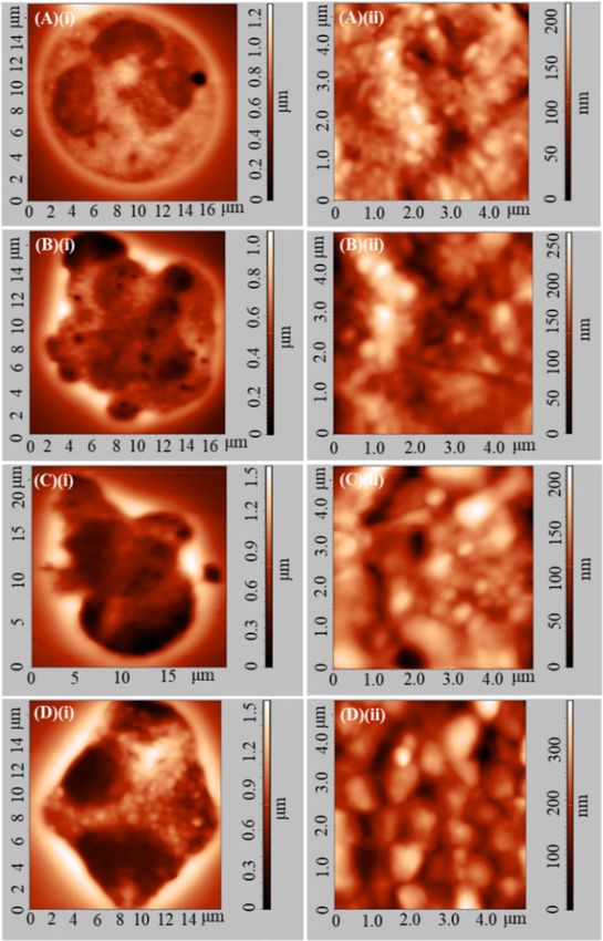
www.nature.com/scientificreports/
as cellular membrane damage, degranulation, morphological transformation, or even a poptosis15–20. Moreover,
nanoparticles can hamper the physiological function of PLTs by altering activation and aggregation processes,
resulting in acute thrombosis or other detrimental health e ffects8,21–26.
To that effect, the current experimental study refers to the evaluation of the immunocompatibility of a new
DMCA that is based on radiolabeled IONPs. Specifically, F e3O4 NPs are surface-functionalized with 2,3-dicar-
boxypropane-1,1-diphosphonic acid (DPD) and subsequently radiolabeled with Gallium-68 (68Ga) so that the
68
Ga-DPD–Fe3O4 complex is ultimately formed27. The immunocompatibility of this DMCA is evaluated here
for the first time, both in vitro and in vivo. Referring to the in vitro evaluation, the immunocompatibility was
tested upon incubation with human WBCs and PLTs isolated from donated peripheral blood. Referring to the
in vivo evaluation, comparative biodistribution studies were performed in normal and immunodeficient mice
models. Additionally, in vivo MRI imaging was performed in normal mice. Previously reported s tudies28–31 on
relevant IONPs have recruited both optical microscopy (OM) and atomic force microscopy (AFM), in order to
investigate, at the microscopic and at the nanoscopic level respectively, the overall morphological and surface
membrane characteristics of isolated red blood cells (RBCs). In the same context31, samples of both WBCs and
PLTs, isolated from donated peripheral blood of five healthy individuals, were incubated at room temperature
at tolerable ( CDMCA = 0.1 mg/ml) and extreme ( CDMCA = 1 mg/ml) concentrations26 of 68Ga-DPD-Fe3O4 DMCA,
up to 120 min. OM and AFM were recruited to investigate in vitro the morphological characteristics of WBCs
and PLTs at the microscopic (10–6 m) and at the nanoscopic (10–9 m) level, since morphological transforma-
tion in both shape and size is a key factor that indicates the activation process for both blood cells. In addition,
a standard hematology analyzer (HA), employed in every day clinical practice, was used to obtain standard
information on complete blood count for comparative reasons with the results originating from the microscopy
techniques. Referring to the in vivo evaluation, we studied the accumulation of the DMCA on the basic organs
of the mononuclear phagocyte system, namely liver, spleen and lungs, by injecting a standard DMCA dose via
the tail vein in nine normal and nine immunodeficient mice.
In brief, the combined OM and AFM results proved that the presence of the 68Ga-DPD-Fe3O4 DMCA
enhances the activation of WBCs and PLTs, as observed at the microscopic level by morphological transforma-
tion referring to both their shape and size. Specifically, both WBCs and PLTs undergo a shape change from a
completely spherical to a polarized and/or ruffled form with the occasional appearance of pseudopods in the
case of PLTs. In addition, the WBCs exhibit a statistically significant size change. Interestingly, at the nanoscopic
level we observed that the size of the WBCs’ granules was increased in the activated state. Although WBCs and
PLTs are somehow activated by the DMCA, neither degradation nor clustering were observed. Furthermore, the
HA data indicated that despite their obvious activation, all standard WBCs’ and PLTs’ indices remained in the
physiological range. The results obtained from the in vivo biodistribution study performed between normal and
immunodeficient mice practically showed no difference of the DMCA accumulation among the basic organs of
the mononuclear phagocyte system. Thus, we can assume that the particular DMCA could potentially evade the
immune system. In vivo MRI imaging data were in accordance with the respective in vivo biodistribution data
and revealed that the contrast effect produced by the particular DMCA was sufficient enough compared to the
one produced by the non-radiolabeled CA, however being concentration-dependent.
Taking into account both the in vitro and in vivo experimental results we propose that the particular DMCA
exhibits noticeable potential for future use in diagnostic applications.
Results and discussion
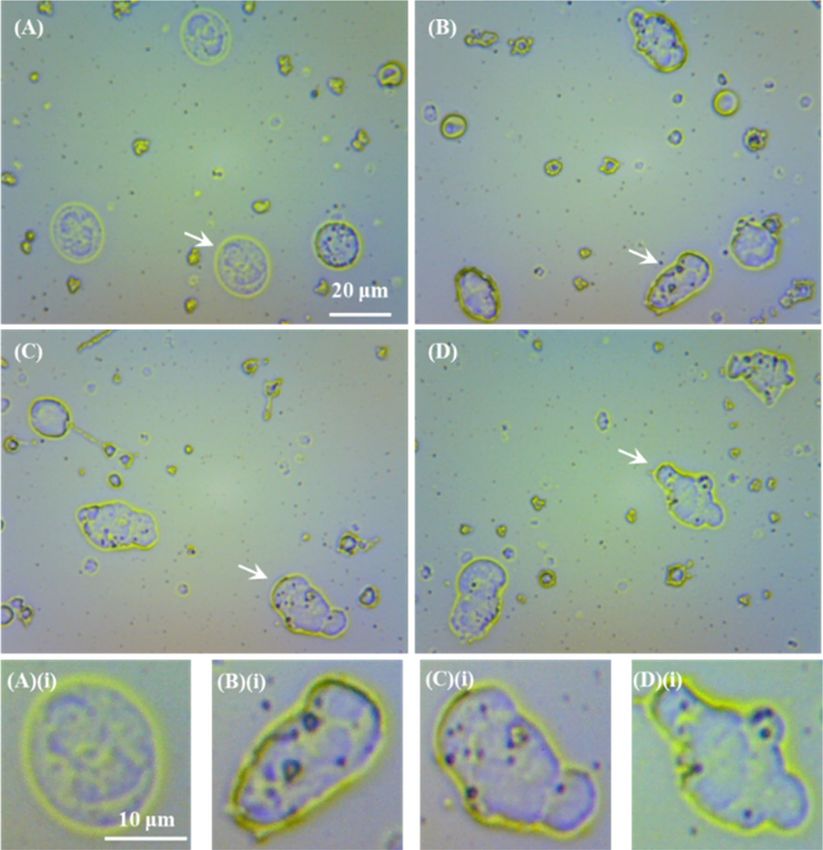
Figure 1A–D show representative OM images of WBC films coming from donor E for DMCA-free prior to (panel
(A)) and after 120 min incubation (panel (B)) and DMCA-incubated WBCs for 120 min at room temperature
in tolerable and extreme concentrations, C DMCA = 0.1 mg/ml (panel (C)) and C DMCA = 1 mg/ml (panel (D)),
respectively. The direct comparison of these images shows that before the incubation process (panel (A)) the
WBCs are in resting form, while after the 120 min incubation without (panel (B)) and with the presence of the
DMCA (panels (C) and (D)) the WBCs are activated, exhibiting a change of their morphological characteristics,
namely shape and size.
In particular, referring to their shape change, panel (A)(i) focuses on a WBC of panel (A) that is in resting
state thus preserving its spherical shape, coming from a DMCA-free sample prior to (0 min) incubation. On
the contrary, panels (B)(i)–(D)(i) focus on WBCs in activated state of the respective panels (B)–(D), com-
ing from DMCA-free after 120 min incubation (panel (B)(i)) and DMCA-incubated for 120 min samples at
CDMCA = 0.1 mg/ml(panel (C)(i)) and at C DMCA = 1 mg/ml (panel (D)(i)) that show the progression of shape
transformation from a simple polarized form (panel (B)(i)) to a complex ruffled shape (panel (D)(i)). The same
behavior was recorded in all five donors studied here.
Figure 2 quantitatively summarizes the OM results referring to the shape change of the activated
WBCs observed in the DMCA-free and DMCA-incubated samples with different DMCA concentrations
(CDMCA = 0.1 mg/ml; low concentration—LC and CDMCA = 1 mg/ml; high concentration—HC). Specifically, we
observed that the WBCs showed a statistically significant activation during 120 min incubation both with-
out DMCA (27.6 ± 13.8% prior to versus 57.6 ± 15.5% after incubation) and with DMCA (79.2 ± 3.3% for
CDMCA = 0.1 mg/ml and 78.8 ± 5.4% for CDMCA = 1 mg/ml). Thus, we can assume that at the microscopic level
the activation of WBCs is influenced by the incubation process, whereas it is enhanced by the presence of the
DMCA, however without being concentration dependent.
Referring to the size change of WBCs, Fig. 3 quantitatively summarizes the ‘mean diameter’ of the
WBCs measured in the DMCA-free and DMCA-incubated samples with different DMCA concentrations
(CDMCA = 0.1 mg/ml; low concentration—LC and CDMCA = 1 mg/ml; high concentration—HC). First, we clar-
ify that in activated, elliptical-like WBCs the parameter ‘mean diameter’ refers to the mean value of the two
Scientific Reports | (2021) 11:9753 | https://doi.org/10.1038/s41598-021-89117-3 2
Vol:.(1234567890)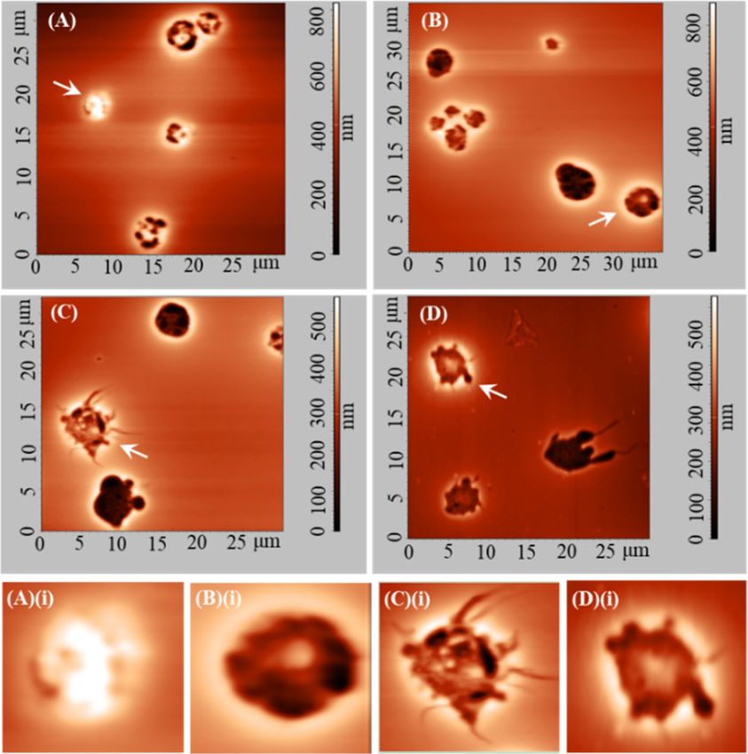
www.nature.com/scientificreports/
Figure 1. Representative optical microscopy (OM) images of WBCs incubated with and without the DMCA.
(A–D) OM images of WBC films referring to (A) DMCA-free prior to incubation (0 min), (B) DMCA-free
after incubation (120 min), (C) DMCA-incubated at CDMCA = 0.1 mg/ml(120 min) and (D) DMCA-incubated
at CDMCA = 1 mg/ml (120 min) (objective lens × 20, camera × 13 in all cases). (A)(i)–(D)(i) Bottom figures show
magnification of specific WBCs indicated by the arrows in the respective top figures, in (A)(i) resting (spherical
shape) and in (B)(i)–(D)(i) activated state (polarized and/or ruffled shape).
diameters as measured along the basic axis of elongation and the normal direction. Thus, in non-activated,
spherical WBCs the ‘mean diameter’ coincides to the typical diameter (for details, see the “Definition of activated
cells” in “Methods”). We observed that the ‘mean diameter’ of the DMCA-free WBCs remains practically identi-
cal upon incubation (14.38 ± 1.84 μm prior to incubation and 14.53 ± 2.05 μm upon 120 min incubation). The
respective ‘mean diameter’ of the DMCA-incubated WBCs exhibits a statistically significant increase in respect
to that of the DMCA-free ones (16.13 ± 2.73 μm for C DMCA = 0.1 mg/ml and 16.52 ± 2.68 μm for CDMCA = 1 mg/
ml, both upon 120 min incubation). Finally, the ‘mean diameter’ of the DMCA-incubated WBCs exhibits a
borderline statistically significant increase.
Both DMCA concentrations used in our study exceed the safety levels recommended for the intravenous
injection of iron agents in treatment of iron-deficiency anemia (i.e. CFe(III) = 0.016 mg/ml). The OM data refer
to many hundreds of WBCs (approximately 720 for each donor) and indicate that, for the conditions studied
here, the presence of the 68Ga-DPD-Fe3O4 DMCA promotes WBC activation by size and shape change from a
spherical to a polarized and/or ruffled form.
Figure 4A–D show representative two-dimensional topography AFM images of WBCs in (A)(i) resting state
(coming from a DMCA-free prior to incubation (0 min) sample) and in (B)(i)–D(i) activated state. Panels (B)
(i), (C)(i) and D(i) refer to a DMCA-free sample after incubation (120 min), a DMCA-incubated sample for
Scientific Reports | (2021) 11:9753 | https://doi.org/10.1038/s41598-021-89117-3 3
Vol.:(0123456789)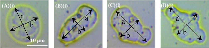
www.nature.com/scientificreports/
Figure 2. Percentage [%] of activated WBCs’ observed in samples incubated with and without the DMCA.
Mean value of the activated WBCs’ percentage [%] observed in DMCA-free (prior to (0 min) and after
(120 min) incubation) and DMCA-incubated ( CDMCA = 0.1 mg/ml; low concentration—LC and CDMCA = 1 mg/
ml; high concentration—HC) samples. In all panels the results are expressed as the overall MV ± SD of the data
coming from all five donors.
Figure 3. Mean value of the WBCs’ ‘mean diameter’ measured in all five donors by OM. Mean value of
the WBCs’ ‘mean diameter’, measured in DMCA-free (prior to (0 min) and after (120 min) incubation) and
DMCA-incubated (CDMCA = 0.1 mg/ml; low concentration—LC and CDMCA = 1 mg/ml; high concentration—HC)
samples. In both panels the results are expressed as the overall MV ± SD of the data coming from all five donors.
CDMCA = 0.1 mg/ml (120 min) and a DMCA-incubated sample for C DMCA = 1 mg/ml (120 min), respectively. In all
cases the morphological characteristics are recorded in detail both qualitatively and quantitatively at the nano-
scopic level by means of AFM. As indicated in panel (A)(i) a resting WBC exhibits a completely spherical shape,
whereas activated WBCs (panels (B)(i)–D(i)) undergo a noticeable morphological transformation, showing a
polarized and/or ruffled shape. The panels (A)(ii)–(D)(ii) present surface granules of the respective WBCs at a
scanned area of 5 × 5 μm2. A direct comparison of these panels shows that the granules of the WBCs increase in
the activated state. These AFM data exhibit the typical behavior observed in all five donors.
Figure 5A–D show representative OM images of PLT films coming from donor B for DMCA-free prior to
(panel (A)) and after 120 min incubation (panel (B)) and DMCA-incubated samples for 120 min at room tem-
perature in tolerable and extreme concentrations, C DMCA = 0.1 mg/ml (panel (C)) and C DMCA = 1 mg/ml (panel
(D)), respectively. The direct comparison of these images shows that, at the microscopic level, the shape of PLTs
progressively changes by the presence of the DMCA from a nearly spherical shape (panel (A)) to an irregular one
with the degree of activation. Indeed, after 120 min incubation without (panel (B)) and with the DMCA (panels
(C) and (D)) PLTs present a polarized and/or ruffled shape with the occasional appearance of pseudopods. The
same behavior was recorded in all five donors studied here.
Scientific Reports | (2021) 11:9753 | https://doi.org/10.1038/s41598-021-89117-3 4
Vol:.(1234567890)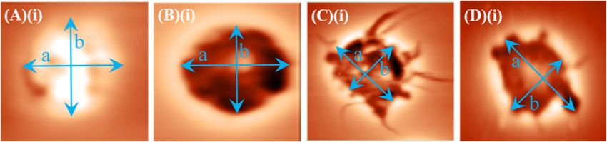
www.nature.com/scientificreports/
Figure 4. Representative two-dimensional atomic force microscopy (AFM) images of WBCs in resting and
activated state. (A)(i)–(D)(i) Two-dimensional topography of WBCs referring to (A)(i) DMCA-free prior to
incubation (0 min), (B)(i) DMCA-free after incubation (120 min), (C)(i) DMCA-incubated at C DMCA = 0.1 mg/
ml (120 min) and (D)(i) DMCA-incubated at CDMCA = 1 mg/ml (120 min) samples. Panel (A)(i) shows a WBC
in resting state, while panels (B)(i)–(D)(i) show WBCs in activated state. Panels (A)(ii)–(D)(ii) present surface
granules of WBCs shown in (A)(i)–(D)(i) respectively, at a scanned area of 5 × 5 μm2. A typical comparison of
(A)(ii)–(D)(ii) images reveals that the size of the WBC granules is increased in the activated state.
Scientific Reports | (2021) 11:9753 | https://doi.org/10.1038/s41598-021-89117-3 5
Vol.:(0123456789)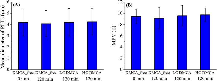
www.nature.com/scientificreports/
Figure 5. Representative optical microscopy (OM) images of PLTs incubated with and without the DMCA.
(A)–(D) OM images of PLTs films referring to (A) DMCA-free prior to incubation (0 min), (B) DMCA-free
after incubation (120 min), (C) DMCA-incubated at CDMCA = 0.1 mg/ml (120 min) and (D) DMCA-incubated
at CDMCA = 1 mg/ml (120 min) (objective lens × 80, camera × 13 in all cases). A typical comparison of (A)–(D)
images reveals that the shape of the PLTs progressively change in the presence of the DMCA from a nearly
spherical form to an irregular one with the degree of activation (polarized and/or ruffled shape with the
occasional appearance of pseudopods).
Figure 6A–D show two-dimensional AFM images of PLTs referring to DMCA-free prior to incubation
(0 min) (panel (A)), DMCA-free after incubation (120 min) (panel (B)), DMCA-incubated at C DMCA = 0.1 mg/
ml(120 min) (panel (C)) and DMCA-incubated at C DMCA = 1 mg/ml (120 min) samples (panel (D)). Interestingly,
the AFM images provide detailed and accurate information of the PLT shape morphology, which is recorded
both qualitatively and quantitatively at the nanoscopic level. Panels (A)(i)–(D)(i) show in detail the change in
shape morphology of specific PLTs, indicated by the arrows in the respective top images (panels (A)–(D)) upon
activation. In particular, a PLT in resting state (panel (A)(i)) presents a nearly spherical shape, whereas it changes
by the presence of DMCA to a polarized and/or ruffled one with the occasional appearance of pseudopods (panels
(B)(i)–(D)(i)) upon activation. These data show the mean behavior that resulted from all five donors.
The AFM data refer to many hundreds of PLTs (approximately 320 for each donor) and indicate that the
presence of the 68Ga-DPD-Fe3O4 DMCA promotes the PLTs’ activation by a shape change from a spherical to a
polarized and/or ruffled form with the occasional appearance of pseudopods.
Figure 7 quantitatively summarizes the AFM results referring to the shape change of the activated
PLTs observed in the DMCA-free and DMCA-incubated samples with different DMCA concentrations
(CDMCA = 0.1 mg/ml; low concentration—LC and C DMCA = 1 mg/ml; high concentration—HC). Specifically,
we observed that the PLTs are not activated during 120 min incubation without DMCA (68.8 ± 13.9% prior
to versus 66.6 ± 14.9% after), whereas 120 min incubation with DMCA motivates activation (86.2 ± 7.7% for
CDMCA = 0.1 mg/ml and 82.8 ± 9.4% for CDMCA = 1 mg/ml), without being concentration dependent.
Thus, from the obtained OM and AFM data, we can assume that both at the microscopic and at the nanoscopic
level the activation of PLTs is promoted by the presence of the DMCA, as evidenced by a shape change from a
nearly spherical to an irregular form (polarized and/or ruffled shape with the occasional appearance of pseu-
dopods). We stress that despite the observed activation, no PLTs clustering was observed in any of the samples.
Figure 8 quantitatively summarizes the AFM results of the size characteristics, mean diameter (panel A) and
mean platelet volume (MPV) (panel B), of the PLTs measured in the DMCA-free and DMCA-incubated samples
with different DMCA concentrations ( CDMCA = 0.1 mg/ml; low concentration—LC and C DMCA = 1 mg/ml; high
concentration—HC). First, we clarify that in an activated PLT the parameter ‘mean diameter’ refers to the mean
value of the two diameters as measured along the basic axis of elongation and the normal direction. Thus, in
Scientific Reports | (2021) 11:9753 | https://doi.org/10.1038/s41598-021-89117-3 6
Vol:.(1234567890)www.nature.com/scientificreports/
Figure 6. Representative two-dimensional atomic force microscopy (AFM) images of PLTs incubated with
and without the DMCA. (A–D) Two-dimensional AFM images of PLTs referring to (A) DMCA-free prior
to incubation (0 min), (B) DMCA-free after incubation (120 min), (C) DMCA-incubated at CDMCA = 0.1 mg/
ml(120 min) and (D) DMCA-incubated at CDMCA = 1 mg/ml (120 min). (A)(i)–(D)(i) Bottom figures show
magnification of specific PLTs indicated by the arrows in the respective top images, in (A)(i) resting (spherical
shape) and in (B)(i)–(D)(i) activated state. A typical comparison of (A)–(D) and (A)(i)–(D)(i) images reveals
that the shape of the PLTs changes in the presence of DMCA from a nearly spherical form to an irregular one
with the degree of activation (polarized and/or ruffled shape with the occasional appearance of pseudopods).
non-activated state, the ‘mean diameter’ coincides to the typical diameter. On the other hand, the mean platelet
volume (MPV) of a PLT was calculated based on the AFM data (for details, see the “Definition of activated cells”
in “Methods”).
Referring to the diameter (panel (A)) of PLTs measured in all samples, we observed that it is practically identi-
cal (4.19 ± 1.19 for DMCA_free (0 min), 4.09 ± 1.16 for DMCA_free (120 min), 4.20 ± 1.23 for CDMCA = 0.1 mg/ml
and 4.27 ± 1.19 for CDMCA = 1 mg/ml). The same result stands for the MPV (9.44 ± 1.20 for DMCA_free (0 min),
9.12 ± 1.91 for DMCA_free (120 min), 9.57 ± 1.84 for C DMCA = 0.1 mg/ml and 9.76 ± 1.18 for CDMCA = 1 mg/ml).
Thus, we can assume that the PLTs’ overall size characteristics (diameter and volume) do not change upon
activation.
Finally, we would like to comment on the relatively high initial value of PLTs’ activation percentage (67%)
observed in the DMCA-free samples prior to incubation (0 min) as evidenced in Fig. 7. This fact can be ascribed
to the non-ideal conditions that hold during every in vitro process. First, PLTs are stored at room temperature
(20–25 °C) that differs substantially from the ideal body temperature (36–37 °C). Second, when left on the
workbench the PLTs are at rest that is they are not subjected to the ideal flow conditions that experience in
the cardiovascular system. Third, other causes such as the strength of centrifugation during the stage of PLTs
Scientific Reports | (2021) 11:9753 | https://doi.org/10.1038/s41598-021-89117-3 7
Vol.:(0123456789)www.nature.com/scientificreports/
Figure 7. Percentage [%] of activated PLTs’ observed in samples incubated with and without the DMCA.
Mean value of the activated PLTs’ percentage [%] observed in DMCA-free (prior to (0 min) and after (120 min)
incubation) and DMCA-incubated (CDMCA = 0.1 mg/ml; low concentration—LC and CDMCA = 1 mg/ml; high
concentration—HC) samples. In all panels the results are shown as the overall MV ± SD of the data coming from
all five donors.
Figure 8. Quantitative data of PLTs’ size characteristics measured in all five donors by AFM. Mean value
of the PLTs’ size characteristics, namely (A) diameter and (B) mean platelet volume (MPV), measured in
DMCA-free (prior to (0 min) and after (120 min) incubation) and DMCA-incubated ( CDMCA = 0.1 mg/ml; low
concentration—LC and CDMCA = 1 mg/ml; high concentration—HC) samples. In both panels the results are
shown as the overall MV ± SD of the data coming from all five donors.
isolation may play an important role. The non ideal temperature storage32–34, absence of agitation (as well as type
and strength of agitation)35–37 and strength of c entrifugation38,39 may all influence the status of PLTs, ultimately
leading to a relatively high baseline activation. However, the aim of our study is to observe if any pre-existing
activation of PLTs is promoted by the presence of the DMCA. Indeed, this is evidenced by the data of Fig. 7.
To obtain further information for the status of many thousands of WBCs and PLTs, we recruited an HA,
which is employed in every day clinical practice. The Table 1 presents this information for DMCA-free (prior
to and after incubation) and DMCA-incubated (CDMCA = 0.1 mg/ml; low concentration—LC and CDMCA = 1 mg/
ml; high concentration—HC) samples. The complete blood count showed that all standard WBCs and PLTs
indices remained in the physiological range, even in the DMCA-incubated sample with the high concentration
of CDMCA = 1 mg/ml. Thus, we can assume that despite the observed morphological activation by means of OM
and AFM, the presence of 68Ga-Fe3O4-DPD DMCA had no intense impact on WBCs and PLTs.
Combining all the OM, AFM and HA data presented above for both WBCs and PLTs, we can conclude that
although the presence of the DMCA promoted their activation, neither degradation nor clustering were observed,
hence making this DMCA a potential candidate for diagnostic imaging. However, these results are exclusively
in vitro. Clearly, in vivo data is highly necessitated, so that safer conclusions can be drawn. To this effect, we
studied nine Swiss and nine SCID mice on a comparative basis under administration of a standard DMCA dose
(see subsection “In vivo biodistribution study on normal and immunodeficient mice” of section “Methods”).
Scientific Reports | (2021) 11:9753 | https://doi.org/10.1038/s41598-021-89117-3 8
Vol:.(1234567890)www.nature.com/scientificreports/
DMCA-free (t = 0 min) DMCA-free (t = 120 min) LC DMCA (t = 120 min) HC DMCA (t = 120 min)
WBC 4.0–10.0 (K/μL) 6.0 ± 1.0 4.8 ± 1.6 5.5 ± 1.5 5.9 ± 1.1
PLT 150–400 (K/μL) 266 ± 50 268 ± 35 240 ± 92 254 ± 68
PCT 0.16–0.35 (%) 0.26 ± 0.06 0.27 ± 0.09 0.23 ± 0.11 0.25 ± 0.07
MPV 7.2–11.0 (fl) 9.7 ± 2.0 10.0 ± 2.2 9.8 ± 2.5 10.2 ± 2.4
Table 1. Main WBCs’ and PLTs’ indices obtained by a clinical hematology analyzer (HA). HA data referring
to standard WBCs’ and PLTs’ indices, namely concentration of white blood cells (WBC), concentration of
platelets (PLT), plateletcrit (PCT) and mean platelet volume (MPV), of the DMCA-free (prior to and after
incubation) and DMCA-incubated (CDMCA = 0.1 mg/ml; low concentration—LC and CDMCA = 1 mg/ml;
high concentration—HC) samples for 120 min at room temperature. The results are expressed as the mean
value ± standard deviation (MV ± SD) of five healthy donors.
Figure 9. Accumulation of the DMCA in the blood and the MPS organs of normal Swiss and immunodeficient
SCID mice. The accumulation of the DMCA in the blood and in the organs of the mononuclear phagocyte
system (MPS) as percentage injected activity per gram of tissue (%IA/g) at 30, 60 and 120 min post injection in
(a) Swiss and in (b) SCID mice. For each time point three mice were studied, thus the results are expressed as
the MV ± SD.
Figure 9 presents quantitative data of the accumulation of the DMCA in the blood and in the main organs
of the mononuclear phagocyte system, namely liver, spleen and lungs, as percentage of the injected activity per
gram of tissue (%IA/g) at 30, 60 and 120 min post injection in Swiss (panel (A)) and in SCID (panel (B)) mice.
Specifically, the blood retention of the DMCA in Swiss mice exhibits an overall statistically significant difference
when compared to the one in SCID mice (2.00 ± 0.51, 2.19 ± 0.42 and 1.49 ± 0.92 versus 6.89 ± 0.04, 4.36 ± 0.31
and 4.10 ± 0.27, at 30, 60 and 120 min post injection, respectively). On the contrary, the %IA/gof the DMCA
in the liver in both Swiss and SCID mice is practically identical (32.42 ± 16.12, 42.52 ± 1.95 and 44.66 ± 22.86
versus 51.00 ± 7.93, 47.74 ± 4.56 and 45.42 ± 9.38, at 30, 60 and 120 min post injection). The same holds for the
%IA/g of the DMCA in the spleen in both Swiss and SCID mice (13.98 ± 2.49, 16.48 ± 3.58 and 16.21 ± 1.74 versus
20.04 ± 9.62, 22.08 ± 20.75 and 19.57 ± 3.10). Finally, referring to the %IA/g of the DMCA in the lungs, we see
Scientific Reports | (2021) 11:9753 | https://doi.org/10.1038/s41598-021-89117-3 9
Vol.:(0123456789)www.nature.com/scientificreports/
Figure 10. Representative T1-weighted coronal and T 2-weighted axial MRI data of normal Swiss mice. (A)(i)–
(C)(i) T1-weighted coronal MRI data of n = 3 normal Swiss mice injected with (A)(i)–(B)(i) 68Ga-DPD-Fe3O4
DMCA at different concentrations: (A)(i) CDMCA = 0.01 mg/ml and (B)(i) CDMCA = 0.1 mg/ml, respectively and
with (C)(i) non-radiolabeled DPD-Fe3O4 CA at a concentration of C CA = 0.3 mg/ml. (A)(ii)–(C)(ii) T2-weighted
axial MRI data of n = 3 normal Swiss mice injected with (A)(ii)–(B)(ii) 68Ga-DPD-Fe3O4 DMCA at different
concentrations: (A)(ii) CDMCA = 0.01 mg/ml and (B)(ii) CDMCA = 0.1 mg/ml, respectively and with (C)(ii) non-
radiolabeled DPD-Fe3O4 CA at a concentration of C CA = 0.3 mg/ml. All MRI images were acquired 6 h p.i.
that the respective values in Swiss mice exhibits an overall statistically significant difference when compared
to the one in SCID mice (2.10 ± 0.49, 1.57 ± 0.43 and 1.32 ± 0.69 versus 3.99 ± 1.24, 2.93 ± 0.21 and 2.47 ± 0.47).
The above data evidenced high and practically equal uptake of the DMCA in the liver and spleen in both
animal models. To explain these data we recall that the immune system is divided into the innate and the adap-
tive. The former is the first defense mechanism exhibiting a non-specific response against a pathogen, while the
latter is the second defense mechanism and is characterized by antigen specificity and immunologic memory.
In addition, organs rich in phagocytic cells, such as liver and spleen, constitute an important part of the innate
immune system, while organs rich in T and B lymphocytes, such as spleen and lungs, are major components of the
adaptive40. Also, we recall that the nanoparticles are quickly opsonized upon their administration into the blood-
stream, which is the main reason of their recognition and elimination by the phagocytic cells of innate system.
In our case, normal Swiss mice show physiological function of the entire immune system both innate and
adaptive, compared to the ones bearing the SCID mutation, which are devoid of T and B cells, hence the adap-
tive system. Accordingly, the comparative results obtained in our study demonstrate that the particular DMCA
encounters the activation of the innate immune system through the action of phagocytic cells, at least in the
mice models studied here. This is an encouraging first indication that the 68Ga-DPD-Fe3O4 DMCA studied here
may be a safe candidate for further in vivo applications in humans.
In vivo MRI experiments were performed on three normal Swiss mice to evaluate the efficacy of the DMCA,
68
Ga-DPD-Fe3O4, on a comparative basis to that of the parent non-radiolabeled CA, DPD-Fe3O4. Figure 10
illustrates representative T 1-weighted coronal (panels (A)(i)–(C)(i)) and T 2-weighted axial (panels (A)(ii)–(C)
(ii)) MRI data of n = 3 normal Swiss mice focused on the area of interest (namely, liver and spleen). The mice
were injected with the 68Ga-DPD-Fe3O4 DMCA at two different concentrations, CDMCA = 0.01 mg/ml (panels
(A)(i) and (A)(ii)) and CDMCA = 0.1 mg/ml (panels (B)(i) and (B)(ii)), respectively, and with the parent, non-
radiolabeled DPD-Fe3O4 CA at a concentration of C CA = 0.3 mg/ml (panels (C)(i) and (C)(ii)). All MRI data
were acquired 6 h p.i. A comparison of the MRI data (A)(i)/(A)(ii) with the respective ones (B)(i)/(B)(ii) reveals
that the contrast effect of the DMCA is clearly enhanced (loss of signal) upon increase of its dose. This is due
to the accumulation of the DMCA in the liver and spleen and indicates that the contrast effect of the DMCA is
concentration-dependent, as expected. This finding is in nice agreement to the respective in vivo biodistribution
Scientific Reports | (2021) 11:9753 | https://doi.org/10.1038/s41598-021-89117-3 10
Vol:.(1234567890)www.nature.com/scientificreports/
data presented above in Fig. 9A. Also, a comparison of the MRI data (B)(i)/(B)(ii) with the respective ones
(C)(i)/(C)(ii) reveals that the contrast effect of the DMCA, 68Ga-DPD-Fe3O4, at the tolerable concentration
CDMCA = 0.1 mg/ml, is comparable to that of the parent, non-radiolabeled CA, DPD-Fe3O4, at a three-times
higher concentration CCA = 0.3 mg/ml. This result is significant since it proves that the magnetic properties of
the parent CA, DPD-Fe3O4, are not suppressed by the radiolabeling process. Accordingly, the resulted DMCA
is clearly efficient for both PET (Ref.27) and MRI (this work).
Conclusion
In the current study, we examined the biocompatibility of a new DMCA showing in vitro biopsy results on
immune and coagulation cells of human blood and comparative in vivo biodistribution data on normal and
immunodeficient mice.
Referring to the in vitro experiments, donated WBCs and PLTs from five healthy individuals were incubated
at room temperature for 120 min with two different concentrations of the DMCA (tolerable/low concentration,
CDMCA = 0.1 mg/ml and extreme/high concentration, C DMCA = 1 mg/ml). The evaluation of the shape/size char-
acteristics of WBCs and PLTs was performed by means of OM and AFM, in order to obtain information from
both the microscopic and the nanoscopic level. The results showed that the shape and size of WBCs, as well as
the shape of PLTs change at the microscopic/nanoscopic level in the presence of the DMCA. Specifically, at the
microscopic level, we observed that WBCs are noticeably activated during 120 min incubation without DMCA
(27.6 ± 13.8% prior to versus 57.6 ± 15.5% after), whereas 120 min incubation with DMCA enhances activation
(79.2 ± 3.3% for CDMCA = 0.1 mg/ml and 78.8 ± 5.4% for CDMCA = 1 mg/ml), as proved by a shape change from a
completely spherical to a polarized and/or ruffled form. Furthermore, the ‘mean diameter’ of WBCs changes
with their activation due to the presence of the DMCA (14.38 ± 1.84 μm (prior to) and 14.53 ± 2.05 μm (after
incubation), versus 16.13 ± 2.73 μm for C DMCA = 0.1 mg/ml and 16.52 ± 2.68 μm for CDMCA = 1 mg/ml).
At the nanoscopic level, we observed that the size of the WBCs granules is increased in the activation state.
Concerning the PLTs, at the microscopic level, we observed that they are not activated during 120 min incuba-
tion without DMCA (68.8 ± 13.9% prior to versus 66.6 ± 14.9% after), whereas 120 min incubation with DMCA
motivates activation (86.2 ± 7.7% for C DMCA = 0.1 mg/ml and 82.8 ± 9.4% for CDMCA = 1 mg/ml), as proved by a
shape change from a completely spherical to a polarized and/or ruffled form with the occasional appearance of
pseudopods. However, despite the activation of both the WBCs and PLTs, neither degradation nor clustering
was observed. These findings were also enhanced by the HA data, which showed that all standard WBC and
PLT indices remained in the physiological range. The in vitro results indicate adequate biocompatibility of the
particular DMCA with human WBCs and PLTs.
Referring to the in vivo experiments, comparative biodistribution studies were performed between normal
Swiss and SCID mice to evaluate the accumulation of the DMCA at the main organs of the mononuclear phago-
cyte system. The overall results indicated high accumulation of the DMCA in organs rich in phagocytic cells,
namely liver and spleen, without any statistical difference between the two mice groups. In vivo MRI imaging
studies in normal Swiss mice were performed to evaluate the efficacy of the DMCA. The results indicated that
the contrast effect produced by the particular DMCA was concentration-dependent, as expected. More impor-
tantly, the contrast efficacy of the DMCA, 68Ga-DPD-Fe3O4, was clearly comparable to that of the parent, non-
radiolabeled CA, DPD-Fe3O4.
The overall in vitro and in vivo data presented here prove that the DMCA studied here is a promising can-
didate for future applications.
Methods
Preparation of 68Ga‑DPD‑Fe3O4 DMCA. The synthesis and radiolabeling of 68Ga-DPD-Fe3O4 DMCA
have been reported e lsewhere27. For obvious safety reasons all experiments with 68Ga-DPD-Fe3O4 DMCA were
conducted after the decay of Gallium-68 to the nonradioactive isotope Zinc-68.
Healthy donors and blood collection. All the donors participated in this study provided their written,
informed consent. The donors underwent blood sampling following the approval of the Ethics Committee of the
NCSR “Demokritos”. All experiments were carried out in accordance with relevant guidelines and regulations.
In brief, five donors coming from the authors and colleagues participated this study after a screening complete
blood count was conducted (sex: 2 men and 3 women and age: 20–60 years old)31. One donor was hypertensive
and followed relevant medication by the time of the study (Preterax, 5 mg/1.25 mg). The other four donors had
no indication/confirmation of any current/chronic disease. None of the five donors had any hematological dis-
ease. Accordingly, all five donors participating this study are considered to be healthy.
According to our study, 15 ml of whole blood was drawn by venipuncture from each donor, which was equally
deposited in five anticoagulant-containing ethylenediaminetetraacetic acid (EDTA) Vacutainer K3 test tubes
(BD, Franklin Lakes, NJ, USA).Since our experimental protocol requires all blood samples to be fresh, the whole
blood was used directly after its collection, without being s tored28–31.
Isolation of the buffy coat, incubation process and preparation of WBC and PLT films. For
the effective study of the WBCs and PLTs with the OM and AFM techniques, single layered films of appropriate
characteristics should be prepared28–30. To this end, an amount of 15 ml of whole blood was drawn by veni-
puncture from each donor and was equally distributed in five EDTA test tubes that were left on the work bench
at room temperature for 1 h so that blood cells were mildly fractionated under the gravity force. As a result, a
precipitate of RBCs was formed at the bottom of the tube, while the supernatant plasma was rich primarily in
PLTs and secondarily in WBCs (platelets rich plasma) with a rich reservoir primarily of WBCs and secondarily
Scientific Reports | (2021) 11:9753 | https://doi.org/10.1038/s41598-021-89117-3 11
Vol.:(0123456789)www.nature.com/scientificreports/
Figure 11. Quantitative criterion of polarization for resting and activated WBCs. (A)(i)–(D)(i) OM images of
WBCs in (A)(i) resting (spherical shape) and in (B)(i)–(D)(i) activated state, where the long (a) and short (b)
axes of the cells are indicated by the arrows for each case. The quantitative criterion of polarization was based on
the dimensionless ratio (a − b)/a and was found to be 0.15 for (A)(i), 0.52 for (B)(i), 0.44 for (C)(i) and 0.35 for
(D)(i).
of PLTs agglomerated at the interface of these two fractions (buffy coat). Then, the buffy coat was collected via
careful aspiration.
After the isolation of the buffy coat, ten parts of each sample was incubated with one part of 68Ga-DPD-Fe3O4
DMCA at low ( CDMCA = 0.1 mg/ml) and high ( CDMCA = 1 mg/ml) concentrations31. The incubation process was
performed under mild conditions (20 rounds per minute) at room temperature (T = 25 °C) for 120 min. We note
that this process exerts minimum mechanical stress on WBCs and PLTs, thus preserving their morphological
characteristics at the maximum degree possible. DMCA-free WBCs and PLTs prior to and after incubation (under
the same conditions with the DMCA-loaded samples) served as control samples.
Accordingly, four distinct samples’ categories were prepared and studied for each one of the donors: DMCA-
free WBCs and PLTs prior to incubation, DMCA-free WBCs and PLTs after incubation, DMCA-incubated WBCs
and PLTs at tolerable/low concentration (CDMCA = 0.1 mg/ml) and DMCA-incubated WBCs and PLTs at extreme/
high concentration (CDMCA = 1 mg/ml). For each one of these categories three copies of dry films were prepared
onto standard glass slides as described e lsewhere28–30 for examination with OM and AFM.
Optical microscopy. OM images of WBC and PLT films were acquired by using a standard light micro-
scope (LEICA DMRXP, Leica, Wetzlar, Germany) in transmission mode operation with an overall magnifica-
tion up to 1000 ×. From the obtained OM images microscopic-level information was extracted on the overall
shape morphology of the WBCs and PLTs. For each donor, approximately n = 180 WBCs and n = 150 PLTs were
observed per sample. Given that four different categories of samples were studied in each case this makes a total
population of n = 720 WBCs and n = 600 PLTs for each donor.
Atomic force microscopy. AFM images of WBC and PLT films were recorded using a scanning probe
microscope (Solver PRO, NT-MDT Co, Moscow, Russia) performing in non-contact scanning mode. Detailed
information on the AFM instrumentation can be found i n31. The optimum imaging results were acquired by set-
ting the following scanning parameters: line frequency = 1.5–4 Hz, area = 0.5 × 0.5–150 × 150 μm2 and lines per
image = 256–51231. For each donor, approximately n = 15 WBCs and n = 80 PLTs were scanned per sample and
detailed information on morphological characteristics, mainly shape and size, were obtained at the nanoscopic
level. Given that four different categories of samples were studied in each case this makes a total population of
n = 60 WBCs and n = 320 PLTs for each donor.
Definition of activated cells. The activated WBCs were counted according to their shape change, which
was evident by the OM. The activated WBCs were defined by using the quantitative criterion of polarization
(else flattening/ellipticity) based on the dimensionless ratio (a − b)/a, where a and b are the long and short axes
of the cell. In Fig. 11, the procedure for the cells already presented in Fig. 1(A)(i)–(D)(i), is demonstrated. The
results on polarization are 0.15, 0.52, 0.44 and 0.35 for each case. Taking into account standard sources of error,
the threshold criterion we used for distinguishing non-activated and activated cells is the value 0.2; for polariza-
tion below 0.2 the cell was considered as non-activated, while when the polarization exceeded 0.2 the cell was
considered activated.
The activated PLTs were counted according to their shape change, which was evident by the AFM. The acti-
vated PLTs were defined by using the same quantitative criterion of polarization as in the case of WBCs, namely
the ratio (a − b)/a. In Fig. 12, the procedure for the cells already presented in Fig. 6(A)(i)–(D)(i), is demonstrated.
The results on polarization are 0.03, 0.19, 0.37 and 0.46, for each case. Taking into account standard sources of
error, the threshold criterion we used for distinguishing non-activated and activated PLTs is the value 0.13; for
polarization below 0.13 the cell was considered as non-activated, while when the polarization exceeded 0.13
the cell was considered activated. On the other hand, the mean platelet volume (MPV) of a PLT was calculated
based on the quantitative data of AFM.
We have minimized the possibly subjective nature of the quantitative analysis of the OM and AFM results
due to the following two reasons: (i) the exact quantitative criterion on the polarization mentioned above that
Scientific Reports | (2021) 11:9753 | https://doi.org/10.1038/s41598-021-89117-3 12
Vol:.(1234567890)www.nature.com/scientificreports/
Figure 12. Quantitative criterion of polarization for resting and activated PLTs. (A)(i)–(D)(i) OM images of
PLTs in (A)(i) resting (spherical shape) and in (B)(i)–(D)(i) activated state, where the long (a) and short (b) axes
of the cells are indicated by the arrows for each case. The quantitative criterion of polarization was based on the
dimensionless ratio (a − b)/a and was found to be 0.03 for (A)(i), 0.19 for (B)(i), 0.37 for (C)(i) and 0.46 for (D)
(i).
was used in common for all sample series guarantees an objective comparative basis and (ii) the former quanti-
tative analysis was conducted independently by the three individuals and the results were practically identical.
Clinical hematology analyzer. Reference samples of WBCs and PLTs incubated with and without the
DMCA underwent complete blood count by means of a standard clinical hematology analyzer (ADVIA 560
Hematology System, Siemens Healthcare GmbH, Erlangen, Germany). Given that the population of WBCs and
PLTs examined by means of OM and AFM was inevitably limited (800–1000 cells), the utilization of an HA was
crucial for confirmation reasons. Accordingly, the HA provided information concerning all standard WBC and
PLT indices that stems from the examination of many thousands of WBCs and PLTs31.
To test the reliability of the HA and to reject any possible interference with the DMCA nanoparticles, control
tests were performed on representative dummy samples. Specifically, blood plasma, DMCA-loaded blood plasma
and DMCA-loaded distilled H 2O were examined. We safely concluded that the size of the DMCA nanoparticles
(< 200 nm)27 is well below the detection limit of the HA, thus their presence does not influence the recorded data.
In vivo biodistribution study on normal and immunodeficient mice. Animal experiments were
carried out according to European and national regulations. These studies have been further approved by the
Ethics Committee of the NCSR “Demokritos” and animal care and procedures followed are in accordance with
institutional guidelines and licenses issued by the Department of Agriculture and Veterinary Policies of the Pre-
fecture of Attiki (Registration numbers: EL 25 BIO 022 and EL 25 BIO 021). Normal Swiss and immunodeficient
SCID mice were obtained from the breeding facilities of the Institute of Biosciences and Applications, NCSR
“Demokritos.” The study protocol was approved by the Department of Agriculture and Veterinary Policies of the
Prefecture of Attiki (Protocol Number: 875110/11-11-2020).
The in vivo biodistribution of the DMCA was evaluated both in 9 Swiss mice (weight 23–27 g) and in 9
SCID mice (weight 17–19 g). To avoid venous thrombosis, prior to tail injection the sample of 68Ga-DPD-Fe3O4
DMCA was loaded onto a size exclusion PD-10 column, containing Sephadex G-25 resin and eluted with phos-
phate buffer saline (PBS), in order to eliminate aggregated 68Ga-DPD-Fe3O4 DMCA. For this reason, ten 0.5 ml
fractions were collected, and the radioactivity of each fraction was measured using a dose calibrator (Capintec,
Ramsey, NJ). The fractions containing the highest radioactivity were pooled and used for the study. Specifically,
intravenous administration of 100 μl PBS suspension (11.11 μg/100 μl DMCA per mouse) of DMCA was per-
formed via the tail vein. The animals were sacrificed at 30, 60, and 120 min post injection (3 Swiss and 3 SCID
mice per time-point). Then, samples of blood and organs of interest that belong to the mononuclear phagocyte
system, namely liver, spleen and lungs, were excised, weighed, and measured for radioactivity in a Gamma
scintillation counter (Cobra II, Canberra, Packard, Downers Grove, Illinois). The remaining radioactivity in the
tail, as well as background counts was subtracted, and the radioactivity decay was autocorrected by the counter.
Then, the accumulation of the DMCA in each organ was expressed as the percentage injected activity per gram
of tissue (%IA/g ± SD) and calculated compared to the activities of a standard dose of the injected solution.
In vivo MRI imaging study on normal Swiss mice. The MRI imaging was conducted at the Alphavet
Veterinary Diagnostic Imaging Center, Athens, Greece, using a 1.5 T MRI Unit. In brief, n = 3 normal Swiss mice
(weight 31–33 g) were used for the MRI study, which were intravenously injected via the tail vein as follows:
mouse (1) was injected with 100 μl (0.0221 mg) 68Ga-DPD-Fe3O4 DMCA, leading to a final body concentration
of CDMCA = 0.01 mg/ml, mouse (2) was injected with 100 μl (0.229 mg) 68Ga-DPD-Fe3O4 DMCA, leading to a
final body concentration of CDMCA = 0.1 mg/ml and mouse (3) was injected with 100 μl (0.8 mg) non-radiola-
beled, DPD-Fe3O4 CA, leading to a final body concentration of CCA = 0.3 mg/ml. The calculations of the final
concentrations were based on the assumption that the blood content is approximately 7% of the total weight of
each mouse. After 6 h p.i., the mice were anesthetized with an intraperitoneal injection of ketamine (75 mg/kg)
and xylazine (5 mg/kg) and placed for imaging, where standard T1-weighted coronal and T2-weighted axial MRI
data were acquired.
Scientific Reports | (2021) 11:9753 | https://doi.org/10.1038/s41598-021-89117-3 13
Vol.:(0123456789)www.nature.com/scientificreports/
Statistical analysis. Statistical analysis of the obtained results was performed by Pair Sample t test. Data are
presented as mean value ± standard deviation (MV ± SD). Differences were considered not significant at P > 0.05
and statistically significant at P < 0.05 (**).
Received: 31 July 2020; Accepted: 30 March 2021
References
1. Na, H. B., Song, I. C. & Hyeon, T. Inorganic nanoparticles for MRI contrast agents. Adv. Mater. 21, 2133–2148 (2009).
2. Nedyalkova, M. et al. Iron oxide nanoparticles—In vivo/in vitro biomedical applications and in silico studies. Adv. Colloid Interface
Sci. 249, 192–212 (2017).
3. Dennis, C. L. et al. The influence of magnetic and physiological behaviour on the effectiveness of iron oxide nanoparticles for
hyperthermia. J. Phys. D Appl. Phys. 41, 134020 (2008).
4. Silva, A. C. et al. Application of hyperthermia induced by superparamagnetic iron oxide nanoparticles in glioma treatment. Int.
J. Nanomed. 6, 591–603 (2011).
5. Plouffe, B. D., Murthy, S. K. & Lewis, L. H. Fundamentals and application of magnetic particles in cell isolation and enrichment:
A review. Rep. Prog. Phys. 78, 016601 (2015).
6. Thomas, R., Park, I.-K. & Jeong, Y. Y. Magnetic iron oxide nanoparticles for multimodal imaging and therapy of cancer. Int. J. Mol.
Sci. 14, 15910–15930 (2013).
7. Ai, F., Ferreira, C. A., Chen, F. & Cai, W. Engineering of radiolabeled iron oxide nanoparticles for dual modality imaging. WIREs
Nanomed. Nanobiotechnol. 8, 619–630 (2016).
8. Huang, R. B., Mocherla, S., Heslinga, M. J., Charoenphol, P. & Eniola-Adefeso, O. Dynamic and cellular interactions of nanoparticles
in vascular-targeted drug delivery. Mol. Membr. Biol. 27, 190–205 (2010).
9. Ilinskaya, A. N. & Dobrovolskaia, M. A. Nanoparticles and the blood coagulation system. Part I: Benefits of nanotechnology.
Nanomedicine 8, 773–784 (2013).
10. Mayer, A. et al. The role of nanoparticle size in hemocompatibility. Toxicology 258, 139–147 (2009).
11. Bihari, P. et al. Single-walled carbon nanotubes activate platelets and accelerate thrombus formation in the microcirculation.
Toxicology 269, 148–154 (2010).
12. Miyamoto, M., Sasakawa, S., Ozawa, T., Kawaguchi, H. & Ohtsuka, Y. Platelet aggregation induced particles. Biomaterials 10,
251–257 (1989).
13. Farace, C. et al. Immune cell impact of three differently coated lipid nanocapsules: Pluronic, chitosan and polyethylene glycol. Sci.
Rep. 6, 18423 (2016).
14. Borgognoni, C. F., Kim, J. H., Zucolotto, V., Fuchs, H. & Riehemann, K. Human macrophage responses to metal-oxide nanopar-
ticles: A review. Artif. Cells Nanomed. Biotechnol. 46, 694–703 (2018).
15. Durocher, I., Noël, C., Lavastre, V. & Girard, D. Evaluation of the in vitro and in vivo proinflammatory activities of gold (+) and
gold (−) nanoparticles. Inflamm. Res. 66, 981–992 (2017).
16. Pujari-Palmer, S. et al. In vivo and in vitro evaluation of hydroxyapatite nanoparticle morphology on the acute inflammatory
response. Biomaterials 90, 1–11 (2016).
17. Gonçalves, D. M., Chiasson, S. & Girard, D. Activation of human neutrophils by titanium dioxide ( TiO2) nanoparticles. Toxicol.
In Vitro 24, 1002–1008 (2010).
18. Babin, K., Antoine, F., Gonçalves, D. M. & Girard, D. TiO2, CeO2 and ZnO nanoparticles and modulation of the degranulation
process in human neutrophils. Toxicol. Lett. 221, 57–63 (2013).
19. Couto, D. et al. Interaction of polyacrylic acid coated and non-coated iron oxide nanoparticles with human neutrophils. Toxicol.
Lett. 225, 57–65 (2014).
20. Noël, C., Simard, J.-C. & Girard, D. Gold nanoparticles induce apoptosis, endoplasmic reticulum stress events and cleavage of
cytoskeletal proteins in human neutrophils. Toxicol. In Vitro 31, 12–22 (2016).
21. Radomski, A. et al. Nanoparticle-induced platelet aggregation and vascular thrombosis. Br. J. Pharmacol. 146, 882–893 (2005).
22. Corbalan, J. J., Medina, C., Jacoby, A., Malinski, T. & Radomski, M. W. Amorphous silica nanoparticles aggregate human platelets:
Potential implications for vascular homeostasis. Int. J. Nanomed. 7, 631–639 (2012).
23. Šimundić, M. et al. Effect of engineered T iO2 and ZnO nanoparticles on erythrocytes, platelet-rich plasma and giant unilamelar
phospholipid vesicles. BMC Vet. Res. 9, 7 (2013).
24. Krajewski, S. et al. Hemocompatibility evaluation of different silver nanoparticle concentrations employing a modified Chandler-
loop in vitro assay on human blood. Acta Biomater. 9, 7460–7468 (2013).
25. He, Z. et al. The effects of gold nanoparticles on the human blood functions. Artif. Cells Nanomed. Biotechnol. 46, 720–726 (2018).
26. Lozano-Fernández, T., Dobrovolskaia, M., Camacho, T., González-Fernández, A. & Simón-Vázquez, R. Interference of metal oxide
nanoparticles with coagulation cascade and interaction with blood components. Part. Part. Syst. Charact. 36, 1800547 (2019).
27. Karageorgou, M. A. et al. Gallium-68 labeled iron oxide nanoparticles coated with 2,3-dicarboxypropane-1,1-diphosphonic acid
as a potential PET/MR imaging agent: A proof-of-concept study. CMMI 6951240 (2017).
28. Stamopoulos, D. et al. Bare and protein conjugated Fe3O4 ferromagnetic nanoparticles for utilization in magnetically assisted
hemodialysis: Biocompatibility with human blood cells. Nanotechnology 19, 505101 (2008).
29. Stamopoulos, D. et al. Biocompatibility and solubility of Fe3O4–BSA conjugates with human blood. Curr. Nanosci. 5, 177–181
(2009).
30. Stamopoulos, D., Manios, E., Gogola, V., Niarchos, D. & Pissas, M. On the biocompatibility of Fe3O4 ferromagnetic nanoparticles
with human blood cells. J. Nanosci. Nanotechnol. 10, 6110–6115 (2010).
31. Karageorgou, M. A., Bouziotis, P., Vranješ-Djurić, S. & Stamopoulos, D. Hemocompatibility of gallium-68 labeled iron oxide
nanoparticles coated with 2,3-dicarboxypropane-1,1-diphosphonic acid. Mater. Sci. Eng. C. 115, 111121 (2020).
32. Maurer-Spurej, E. et al. Room temperature activates human blood platelets. Lab. Investig. 81, 581 (2001).
33. Huskens, D. et al. Standardization and reference ranges for whole blood platelet function measurements using a flow cytometric
platelet activation test. PLoS ONE 13, e0192079 (2018).
34. Morrof, G., Holme, S., George, V. M. & Heaton, W. A. Effect on platelet properties of exposure to temperatures below 20°C for
short periods during storage at 20 to 24°C. Transfusion 34, 317–321 (1994).
35. Wallvik, L., Stenke, L. & Åkerblom, O. The effect of different agitation modes on platelet metabolism, thromboxane formation,
and alpha-granular release during platelet storage. Transfusion 30, 639–643 (1990).
36. Kalmin, N. D., Wilson, M. J. & Liles, B. A. In vitro assessment of platelet damage during rotator storage. Am. J. Clin. Pathol. 79,
719–721 (1983).
37. Holme, S., Vaidja, K. & Murphy, S. Platelet storage at 22 degrees C: Effect of type of agitation on morphology, viability, and function
in vitro. Blood 52, 425–435 (1978).
38. Fijnheer, R. et al. Platelet activation during preparation of platelet concentrates: A comparison of the platelet-rich plasma and the
buffy coat methods. Transfusion 30, 634–638 (1990).
Scientific Reports | (2021) 11:9753 | https://doi.org/10.1038/s41598-021-89117-3 14
Vol:.(1234567890)www.nature.com/scientificreports/
39. Merolla, M., Nardi, M. A. & Berger, J. S. Centrifugation speed affects light transmission aggregometry. Int. J. Lab. Hematol. 34,
81–85 (2012).
40. Bancroft, G. J. & Kelly, J. P. Macrophage activation and innate resistance to infection in SCID mice. Immunobiology 191, 424–431
(1994).
Acknowledgements
The authors would like to thank Dr. Sanja Vranješ-Djurić for providing the non-radiolabeled DPD-Fe3O4 samples,
Dr. Penelope Bouziotis for use of radiolabeling and animal experimentation facilities, Mr. Stavros Xanthopoulos
for assistance in the animal models, Dr. Panagiotis Apostolopoulos for providing the hematology analyzer data
and Dr. Ioannis Panopoulos for assistance in MRI imaging.
Author contributions
M.A.: investigation, methodology, formal analysis, roles/writing–original draft, writing–review and editing. D.S.:
conceptualization, methodology, validation, formal analysis, project administration, resources, supervision, roles/
writing—original draft, writing–review and editing.
Competing interests
The authors declare no competing interests.
Additional information
Correspondence and requests for materials should be addressed to D.S.
Reprints and permissions information is available at www.nature.com/reprints.
Publisher’s note Springer Nature remains neutral with regard to jurisdictional claims in published maps and
institutional affiliations.
Open Access This article is licensed under a Creative Commons Attribution 4.0 International
License, which permits use, sharing, adaptation, distribution and reproduction in any medium or
format, as long as you give appropriate credit to the original author(s) and the source, provide a link to the
Creative Commons licence, and indicate if changes were made. The images or other third party material in this
article are included in the article’s Creative Commons licence, unless indicated otherwise in a credit line to the
material. If material is not included in the article’s Creative Commons licence and your intended use is not
permitted by statutory regulation or exceeds the permitted use, you will need to obtain permission directly from
the copyright holder. To view a copy of this licence, visit http://creativecommons.org/licenses/by/4.0/.
© The Author(s) 2021
Scientific Reports | (2021) 11:9753 | https://doi.org/10.1038/s41598-021-89117-3 15
Vol.:(0123456789)You can also read