IHC PANEL MARKERS Ovarian Cancer - BioGenex
←
→
Page content transcription
If your browser does not render page correctly, please read the page content below
IHC PANEL MARKERS
                               O v a r i a n                       C a n c e r

BioGenex offers wide-ranging antibodies for several IHC panel for initial differentiation, tumor
origin, treatment methods, and prognosis. All BioGenex antibodies are validated on human
tissues to ensure sensitivity and specificity. BioGenex comprehensive IHC panels include a
range of mouse monoclonal, rabbit monoclonal, and polyclonal antibodies to choose from.

BioGenex offers a vast spectrum of high-quality antibodies for both diagnostic and reference
laboratories. BioGenex strives to support efforts in clinical diagnostics and drug discovery
development as we continue to expand our antibody product line offering in both
ready-to-use and concentrated formats for both manual and automation systems.

                            Antibodies for Ovarian Cancer
 CK7, bFGF, Estradiol, CA125, ER, Vimentin, CK20, Her2, Mesothelin, C-jun, NSE, FSH, CEA, PLAP,
 WT1, beta-HCG, Inhibin, AFP, EGFR, Calretenin, p53, Napsin A, PR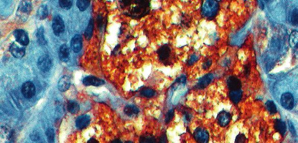
IHC PANEL MARKERS - Ovarian Cancer
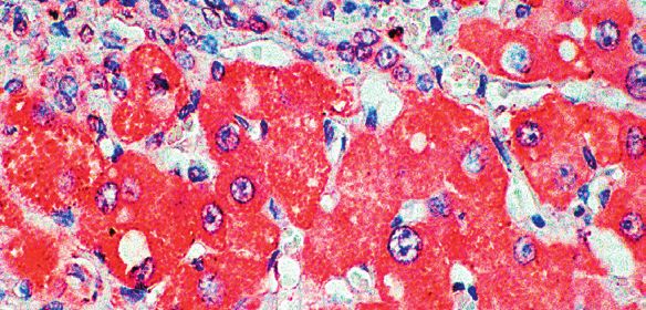
Cytokeratin 7
Cytokeratin 7 is a 54 kD marker of simple epithelium. Antibody to
Cytokeratin 7 strongly stains all cell layers of the urinary bladder transitional
epithelium. However, Cytokeratin 7 is absent from gastrointestinal epithelium,
hepatocytes, proximal and distal tubules of the kidney, and myoepithe-
lium, and also cannot be detected in the stratified epithelia of the skin,
tongue, esophagus, or cervix. Cytokeratin 7 recognizes specific subtypes of
adenocarcinomas and can be used to differentiate between Cytokeratin 7-pos-
itive tissues such as ovarian carcinomas and transitional cell carcinomas and
Cytokeratin 7-negative tissues such as carcinomas of the gastrointestinal tract
and prostate cancers.
Antibody Clone Localization Catalog Family
Cytokeratin 7 OVTL12/30 Cytoplasm AM255, AX255, MU255
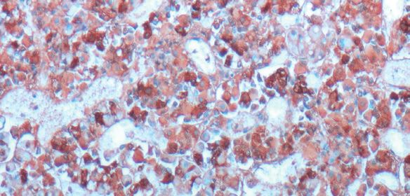
bFGF
bFGF is a pro-angiogenic cytokine which is present in diverse
tissues. It is known to function as an autocrine mediator of mitogenesis of
endothelial cells in vivo, resulting in angiogenesis. It also increases fibroblast
production of plasminogen activator and collagenase. bFGF is a heparin binding
cytokine that is found inside cells and in extracellular stores bound to heparin or
heparin sulfate proteoglycans. bFGF may be released to mediate tissue repair
since expression is known to be high in mast cells responding to injury. The
monoclonal antibody to bFGF can be used for the study of myometrial smooth
muscle cells, uterine leiomyomas, cardiac myocytes, arterial endothelium,
gastric carcinoma, and invasive or metastatic melanoma. This antibody stains
bFGF in cytoplasm of many diverse cell types.
Antibody Clone Localization Catalog Family
bFGF bFGF88 Cytoplasm AM359, AX359, MU359
Estradiol
Estradiol plays an important role in the genesis and development of
human breast cancer and endometrial carcinoma. It is synthesized primarily
in the ovary, but also in the placenta, testis, and possibly the adrenal cortex.
Estradiol is also produced by testicular Leydig tumors, as well as by
Sertoli tumors of the testis and ovary. It is also produced in mammary gland
carcinoma, and carcinoma of the adrenal cortex.
Antibody Clone Localization Catalog Family
Estradiol Polyclonal Nucleus AR038, AW038
CA125
Monoclonal antibody Ov185:1 reacts with repetitive protein determinant
expressed in the protein core of the CA125 human ovarian cancer
antigen. This marker is usually associated with ovarian epithelial malignancies.
Immunohistochemistry with CA125 antibody in conjunction with other
markers was found to be useful in tracing the origin of adeno carcinoma of
unknown origin. This antibody stains membrane in ovarian cancer cells.
Antibody Clone Localization Catalog Family
CA125 oV185:1 Membrane and Cytoplasm AM429, AX429, MU429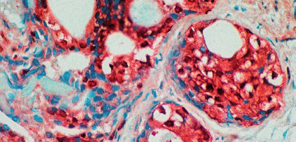
IHC PANEL MARKERS - Ovarian Cancer

Estrogen Receptor
                       Estrogen receptor (ER) content of breast cancer tissue is an important
                       parameter in the prediction of prognosis and response to endocrine therapy.
                       Highly specific monoclonal antibodies to ER have allowed the determination
                       of receptor status of breast tumors to be carried out. This antibody stains the
                       nucleus of receptor positive cells.

Antibody               Clone            Localization                  Catalog Family
Estrogen Receptor      EP1              Nuclear                       AN710, AY710, MU710
Estrogen Receptor      Er88             Nuclear                       AM368, AX368, MU368

Vimentin
                       Vimentin is the major intermediate filament in a variety of mesenchymal
                       or mesenchymally derived non-muscle cell types. Vimentin is found in all
                       types of sarcomas and lymphomas. Positive staining for vimentin is seen in
                       most cells of fibrosarcomas, liposarcomas, malignant fibrous histocytomas,
                       angiosarcomas, chondrosarcomas and lymphomas. When the vimentin
                       antibody is used in combination with other antibodies as a panel, it can aid in
                       the histological classification of normal and malignant tissues. This antibody
                       immunohistochemically labels a variety of mesenchymal cells.

Antibody               Clone            Localization                  Catalog Family
Vimentin               V9               Cytoplasm                     AM074, AX074, MU074

Cytokeratin 20
                       Cytokeratin 20 (46kD) is relatively less acidic than other type I keratins. This
                       antibody reacts with certain types of carcinomas such as adeno carcinomas of
                       the colon, transitional cell carcinomas of the bladder and Merkel cell tumors
                       of the skin. It does not stain breast, lung and endometrial adenocarcinomas.
                       The differential staining pattern of this antibody makes it very useful for tumor
                       evaluation when used in conjunction with cytokeratin 7 staining.

Antibody               Clone            Localization                  Catalog Family
Cytokeratin 20         IT.ks20.8        Cytoplasm                     AM315, AX315, MU315
Cytokeratin 20         EP28             Cytoplasm                     AN849, AY849, MU849

Her2
                       HER2 (human epidermal growth factor receptor 2), also known as Neu,
                       ErbB-2, CD340 (cluster of differentiation 340) or p185, is a protein that in
                       humans is encoded by the ERBB2 gene. HER2 is a member of the epidermal
                       growth factor receptor (EGFR/ErbB) family. Breast cancers with HER2 gene
                       amplification or HER2 protein overexpression are called HER2-positive, which
                       represent about 25% breast cancer.

Antibody               Clone            Localization                   Catalog Family
c-erbB-2 (HER-2/neu)   SP101            Membrane and cytoplasm         AN752, AY752, NU752
c-erbB-2 (HER-2/neu)   SP3              Membrane and cytoplasm         AN753, AY753, NU753
c-erbB-2 (HER-2/neu)   CB11             Membrane and cytoplasm         AM134, AX134, MU134
c-erbB-2 (HER-2/neu)   EP3              Membrane and cytoplasm         AN726, AY726, NU726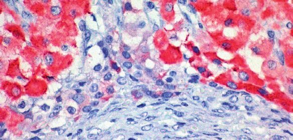
IHC PANEL MARKERS - Ovarian Cancer

C-Jun
                 c-Jun is a component of the transcription factor Activator Protein 1 (AP-1)
                 that binds and activates transcription at TPA-responsive element (TRE/AP-1)
                 elements and appears to be a major downstream target of the Stress-ac-
                 tivated protein kinases/Jun amino-terminal kinases (SAPK/JNK) signaling
                 pathway. The transcriptional activity of c-Jun is regulated by phosphorylation
                 due to extracellular signals including growth factors, transforming oncopro-
                 teins, and UV irradiation that stimulates phosphorylation at Ser63/73 and
                 activates c-Jun dependent transcription. c-Jun antibodies are used to study the
                 signal-transducing transcription factor of the AP1 family. c-Jun has been
                 implicated in several areas of cell biology including cell cycle progression
                 through the G1 phase, transformation, and differentiation and has recently
                 been linked to apoptosis. c-Jun is a known proto-oncogene and is found to be
                 significantly overexpressed in lung, breast and ovarian cancers, making it a
                 viable tumor marker.

Antibody         Clone            Localization                   Catalog Family
C-Jun            4H9              Nucleus                        AM958, AX958, MU958

NSE
                 NSE is a gene which encodes for a protein found in matured
                 neurons and is used in panels along with chromogranin, synaptophysin and
                 neurofilament. Elevated NSE concentrations are observed in patients with
                 neuroblastoma, pancreatic islet cell carcinoma, medullary thyroid carcinoma,
                 pheochromocytoma, and other neuroendocrine tumors as well as certain benign
                 conditions. NSE is specific for such proteins, and aids in detection of neural and
                 neuroendocrine lineage.

Antibody         Clone            Localization                   Catalog Family
NSE              MIG-N3           Cytoplasm                      AM055, AX055, MU055

FSH
                 Follicle stimulating hormone enables ovarian folliculogenesis to the antral
                 follicle stage and is essential for Sertoli cell proliferation and mainte-
                 nance of sperm quality in the testis. Members of the pituitary glycoprotein
                 hormone family, of which FSH is one (see also luteinizing hormone, chorionic
                 gonadotropin, and thyroid stimulating hormone),consist of a shared alpha
                 chain and a beta chain encoded by a separate gene. The FSHB gene encodes
                 the beta subunit of follicle stimulating hormone.Tumors that do not consist of
                 adenohypophysial cells neither produce nor contain pituitary hormone, and
                 thus immunoperoxidase techniques are helpful in distinguishing them from
                 those pituitary tumors that store various hormones in the cell cytoplasm.
                 FSH, a glycoprotein hormone, stimulates the graafian follicles of the ovary and
                 assists subsequently in follicular maturation and the secretion of estradiol.
                 In the male, it stimulates the epithelium of the seminiferous tubules and is
                 partially responsible for inducing spermatogenesis.

Antibody         Clone            Localization                   Catalog Family
FSH              Polyclonal       Cytoplasm                      AR766, AW766, PU766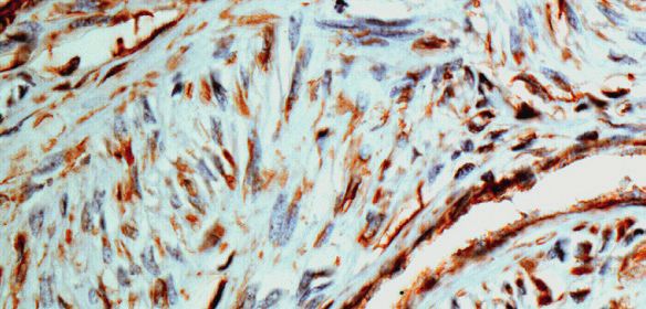
IHC PANEL MARKERS - Ovarian Cancer

CEA
           CEA is demonstrated as a linear labeling of the apical poles of cells lining
           the glandular lumen and, occasionally, as weak staining near the apex of
           colonic epithelial cells. CEA, however, should not be used as a marker of
           differentiation because many colon and lung tumors actually show
           increased staining with differentiation. Pancreatic carcinomas, testicular tumor,
           gallbladder neoplasms and granular cell myoblastomas stain positive, whereas
           malignant tumors of brain, prostate, skin, lymphoreticular tissues, hepatocellular
           carcinomas, oesophageal squamous cell carcinomas, and mesothelioma fail
           to stain for CEA.

Antibody   Clone            Localization                   Catalog Family
CEA        Polyclonal       Cytoplasm                      AR009, AW009
CEA        CEA88            Cytoplasm                      AM365, AX365, MU365

PLAP
           Human Placental Alkaline Phosphatase (PLAP), a 60-70 kD oncofetal antigen,
           is a member of a family of membrane bound alkaline phosphatase enzymes
           and isoenzymes. PLAP and/or PLAP-like isoenzymes have been found to be
           expressed by malignant tumors of germ cell and non-germ cell origin. The
           antibody reacts with PLAP in syncytiotrophoblasts in placenta and also reacts
           with human germ cell tumors. This antibody stains positive in the cytoplasmic
           membrane and cytoplasm of positive cells.

Antibody   Clone            Localization                   Catalog Family
PLAP       PL8-F6           Cytoplasm                      AM228, AX228, MU228

WT1
           WT-1 monoclonal antibody recognizes a 47-55 kDa tumor suppressor
           protein, identified as Wilm’s Tumor (WT1) protein. The antibody reacts with all
           isoforms of the full-length WT1 and also identifies WT1 lacking exon 2-encod-
           ed amino acids, frequently found in subsets of sporadic Wilm’s tumors.WT1,
           a sporadic and familial pediatric kidney tumor, is genetically heterogeneous.
           Wilm’s tumor is associated with mutations of WT1, a zinc-finger transcription
           factor that is essential for the development of the metanephric kidney and the
           urogenital system. The WT1 gene is normally expressed in fetal kidney and
           mesothelium, and its expression has been suggested as a marker for Wilm’s
           tumor and mesothelioma. WT1 protein has been identified in proliferative
           mesothelial cells, malignant mesothelioma, ovarian carcinoma, gonadoblasto-
           ma, nephroblastoma, and desmoplastic small round cell tumor. WT1 protein
           expression in mesothelial cells has become a reliable marker for the diagnosis
           of mesotheliomas.

Antibody   Clone            Localization                   Catalog Family
WT1        WT1/1434R        Nucleus & Cytoplasm            AN940, AX940, MU940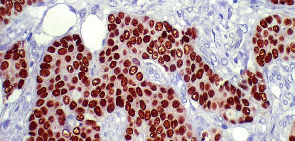
IHC PANEL MARKERS - Ovarian Cancer

B-HCG
                 Human Chorionic Gonadotropin (hCG) is a 40 kD glycoprotein secreted in
                 large quantities by the placenta and normally circulates at readily detectable
                 levels only during gestation. Immunohistochemical studies reveal localization
                 of hCG in syncytiotrophoblasts. Isolated clusters of giant cells may be found
                 in association with certain components of germ cell tumors but are most fre-
                 quently associated with embryonic carcinoma, endodermal sinus tumor, and
                 germinoma. This antibody stains the cytoplasm of positive cells.

Antibody         Clone             Localization                  Catalog Family
B-HCG            M94318            Cytoplasm                     AM395, AX395, MU395

Inhibin Alpha
                 Inhibins are dimeric gonadal protein hormones that negatively regulate pitu-
                 itary FSH synthesis and secretion. Inhibin contains an alpha and beta subunit
                 linked by disulfide bonds. Two forms of inhibin differ in their beta subunits (A
                 or B), while their alpha subunits are identical. Inhibin B is comprised of the
                 Inhibin alpha subunit disulfide linked to the Inhibin beta subunit. Initial stud-
                 ies indicated that Inhibin is a critical negative regulator of gonadal stromal cell
                 proliferation and was the first secreted protein identified to have tumor-sup-
                 pressor activity. Inhibin alpha-subunit immunoreactivity has been detected in
                 Sertoli cells, spermatocytes, and in some Leydig cells.

Antibody         Clone             Localization                  Catalog Family
Inhibin Alpha    R1                Cytoplasm                     AM446, AX446, MU446

AFP
                 Alpha-Fetoprotein (AFP) is a 64 kD tumor-associated embryonal antigen
                 produced by fetal liver, hepatoma, yolk sac, and several germ cell tumors
                 of testicular and ovarian origin. Of the germ cell tumors, only embryonal
                 carcinoma and endodermal sinus tumors stain positive for AFP and not
                 teratomas. The positive results are useful in distinguishing embryonal
                 carcinoma from seminoma. AFP is present in the mononuclear embryonal
                 carcinoma cell and in the intracellular or extracellular hyaline droplets. This
                 antibody stains positive for alpha fetoprotein in the cytoplasm of positive cells.

Antibody         Clone             Localization                  Catalog Family
AFP              C3                Cytoplasm                     AM008, AX008, MU008

EGFR
                 Epidermal growth factor receptor (EGFR) is a 170 kDa transmembrane
                 glycoprotein receptor tyrosine kinase that, activated by epidermal growth
                 factor (EGF), affects cell growth and differentiation. EGFR can be a prognostic
                 marker for ovarian cancer. The antibody detects both EGFR phosphorylated
                 on Tyr1068 of the nature human isoform 1 (corresponding to Y1092 from the
                 precursor form P00533-1/p170), and also unphosphorylated EGFR. It is asso-
                 ciated with a number of cancers, including lung cancer, anal cancers[7] and
                 glioblastoma multiforme. In breast cancer, EGFR is predorminately expressed
                 in basal cell-like carcinoma; it has been recommendated for identification of
                 basal-like breast carcinoma along with Cytokeratin 5/6.

Antibody         Clone             Localization                  Catalog Family
EGFR             EP22              Membrane & Cytoplasm          AN781, AX781, MU781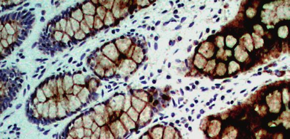
IHC PANEL MARKERS - Ovarian Cancer

Calretenin
                        This antibody recognizes a protein of 31.5kDa, identified as Calretinin.
                        Calretinin is an intracellular calcium-binding protein belonging to the troponin
                        C superfamily characterized by a structural motif described as the EF-hand
                        domain. It is abundantly expressed in central and peripheral neural tissues,
                        particularly in the retina and in the neurons of the sensory pathways, and
                        calretinin may play an important role in the survival of nerve cells during
                        disturbances in calcium homeostasis. Calretinin is also expressed by both
                        normal and neoplastic mesothelial cells, and it has been suggested as a useful
                        marker for the identification of malignant mesotheliomas of the epithelial type
                        and for the differentiation of these malignancies of lung adenocarcinoma.

Antibody                Clone            Localization                  Catalog Family
Calretenin              SP13             Cytoplasm & Membrane          AN747, AX747, MU747

p53
                        p53 is a tumor suppressor gene product identified in a wide variety of
                        tumors. p53 protein is present in low concentration in normal cells, but elevated
                        levels of mutant p53 have been found in many common cancers. Accumulation
                        of mutant p53 detected by immunohistochemical staining has been reported
                        in breast, lung, colon, stomach, bladder, and testis carcinomas, soft-tissue
                        sarcomas, and melanomas. This antibody stains positive in nucleus of a
                        variety of tumor cells.

Antibody                Clone            Localization                  Catalog Family
p53                     DO7              Nuclear                       AM239, AX239, MU237

Napsin-A
                        Napsin A has specific function in normal alveolar epithelium and is proposed
                        to play a role in the protelytic processing of surfactant precursors. Napsin
                        A is reported to be predominantly expressed in lamellar bodies of type II
                        pneumocutes, secondary lysosymes of alveolar macrophages, respiratory
                        epithelium of terminal and respiratory bronchioles, plasma cells within a
                        subset of lymphocytes in normal lung, as well as in epithellial cells of renal
                        tubiles in normal kidney and is weakly expressed in normal spleen.

Antibody                Clone            Localization                  Catalog Family
Napsin-A                IP64             Cytoplasm                     AM701, AX701, MU701

Progesterone Receptor
                        The use of monoclonal antibodies to determine Progesterone Receptor
                        status increases the predictive value of immunohistochemical analysis with
                        respect to the response of human tumors to hormonal modulation. Historically,
                        estrogen receptorpositive/ progesterone receptor-positive breast carcinoma
                        patients have demonstrated a better response to endocrine therapy than
                        estrogen receptor-positive/progesterone receptor-negative patients. This
                        antibody stains positive in nucleus of the receptor positive cells.

Antibody                Clone            Localization                   Catalog Family
Progesterone Receptor   1A6              Nucleus                        AM172, AX172, MU172
Progesterone Receptor   PR88             Nucleus                        AM328, AX328, MU328
Progesterone Receptor   EP2              Nucleus                        AN711, AY711, NU711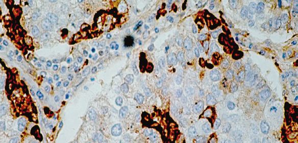
IHC PANEL MARKERS - Ovarian Cancer

BioGenex Primary Antibody Format and Pack Size

BioGenex antibodies are optimized to provide a maximum signal with the minimum background
for immunohistochemical staining. All our antibodies are optimized and recommended for use
with all Super Sensitive™ Detection Systems to provide optimum staining.

BioGenex Ready-to-Use (RTU) antibodies are fully optimized for use with BioGenex Detection
Systems without the need for further dilution or titration. BioGenex concentrated antibodies
are provided with recommended dilutions for optimal use with BioGenex Detection Systems,
allowing rapid titration and testing.

 Prefix        Type                Species                            Suffix                      Volume and Format

 AM/AN         Monoclonal          AM-Mouse/AN-Rabbit                 -5M/5ME                     6 mL - Ready-to-use (manual)

 AM/AN         Monoclonal          AM-Mouse/AN-Rabbit                 -10M/10ME                   10 mL - Ready-to-use (i6000™)

                                                                      -YCD/YCDE and
 AX/AY         Monoclonal          AX-Mouse/AY-Rabbit                                             16 mL and 5 mL Ready-to-use (Xmatrx®)
                                                                      -50D/50DE

 AR            Polyclonal          Rabbit                             -5R/5RE                     6 mL - Ready-to-use (manual)

 AR            Polyclonal          Rabbit                             -10R/10RE                   10 mL - Ready-to-use (i6000™)

                                                                      -YCD/YCDE and
 AW            Polyclonal          Rabbit                                                         16 mL and 5 mL Ready-to-use (Xmatrx®)
                                                                      -50D/50DE
                                                                      -UC/UCE and
 MU/NU         Monoclonal          AM- Mouse/AN-Rabbit                                            1 mL and 0.5 mL Concentrate
                                                                      -5UC/5UCE
                                                                      -UC/UCE and
 PU            Polyclonal          Rabbit                                                         1 mL and 0.5 mL Concentrate
                                                                      -5UC/5UCE

                                                     Other Panel Markers from BioGenex
 Breast cancer panel                                                               Pancreas tumor
 B&T cell Associated Lymphoma                                                      Liver cancer
 Cervical cancer                                                                   Kidney cancer
 Colorectal and stomach cancer                                                     Head & neck cancer
 Lung cancer                                                                       Bladder cancer
 Melanoma                                                                          Germ cell tumor
 Muscle cancer                                                                     Vascular tumor
 Prostate/Testicular cancer                                                        Pituitary gland
 Neuroendocrine tumor                                                              Esophagus cancer

For specific information on the individual antibody, please refer to the datasheets available
on www.biogenex.com or call BioGenex Technical Support at 1(800)421-4149 or write to
support@biogenex.com.

                                                                                            Customer Service
                                                                                            US:     customerservice@biogenex.com
In the U.S., call +1 (800) 421-4149                                                         India: indiacs@biogenex.com
Outside the U.S., call +91-40-27185500                       www.biogenex.com               Global: internationalcs@biogenex.com

© 2019 BioGenex Laboratories, Inc. All Rights Reserved, Doc No: 907-4060.0 Rev B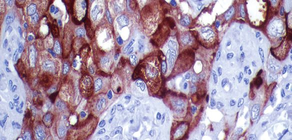
You can also read