High fiber diets attenuate emphysema development via modulation of gut microbiota and metabolism - Nature
←
→
Page content transcription
If your browser does not render page correctly, please read the page content below
www.nature.com/scientificreports
OPEN High‑fiber diets attenuate
emphysema development
via modulation of gut microbiota
and metabolism
Yoon Ok Jang1,2, Ock‑Hwa Kim1, Su Jung Kim2, Se Hee Lee1,3, Sunmi Yun4, Se Eun Lim4,
Hyun Ju Yoo2, Yong Shin5 & Sei Won Lee1*
Dietary fiber functions as a prebiotic to determine the gut microbe composition. The gut microbiota
influences the metabolic functions and immune responses in human health. The gut microbiota and
metabolites produced by various dietary components not only modulate immunity but also impact
various organs. Although recent findings have suggested that microbial dysbiosis is associated with
several respiratory diseases, including asthma, cystic fibrosis, and allergy, the role of microbiota
and metabolites produced by dietary nutrients with respect to pulmonary disease remains unclear.
Therefore, we explored whether the gut microbiota and metabolites produced by dietary fiber
components could influence a cigarette smoking (CS)-exposed emphysema model. In this study, it
was demonstrated that a high-fiber diet including non-fermentable cellulose and fermentable pectin
attenuated the pathological changes associated with emphysema progression and the inflammatory
response in CS-exposed emphysema mice. Moreover, we observed that different types of dietary fiber
could modulate the diversity of gut microbiota and differentially impacted anabolism including the
generation of short-chain fatty acids, bile acids, and sphingolipids. Overall, the results of this study
indicate that high-fiber diets play a beneficial role in the gut microbiota-metabolite modulation and
substantially affect CS-exposed emphysema mice. Furthermore, this study suggests the therapeutic
potential of gut microbiota and metabolites from a high-fiber diet in emphysema via local and
systemic inflammation inhibition, which may be useful in the development of a new COPD treatment
plan.
Chronic obstructive pulmonary disease (COPD) is a progressive respiratory disease characterized by persistent
airflow obstruction and abnormal inflammatory r esponses1,2. Cigarette smoking (CS) is a major risk factor for
respiratory and systemic diseases including COPD, cardiovascular disease, and cancer due to the pathogenic-
ity of circulating toxic constituents, metabolites and their associated inflammatory r esponse3,4. Moreover, CS
can directly or indirectly influence the microbiome through immune homeostasis, biofilm formation, oxygen
deprivation, and other potential m echanisms5,6.
Many studies have revealed that the gut microbial community (gut microbiota) affects metabolic function,
inflammation, and physiological processes that modulate local and systemic inflammatory responses. Several
scientific findings have highlighted the correlation between the gut microbiota and lung immunity in the gut-
lung axis7,8. The constituents of the gut microbiota change in response to dietary nutrients, which are associated
with immune responses and homeostasis. This interaction between gut microbiota and nutrients is also associ-
ated with respiratory diseases, mostly studies in asthma and allergy, along with gut microbiome d ysbiosis9,10.
Among nutrients working as prebiotics, dietary fibers are associated with a more diversified gut microbiota
1
Department of Pulmonary and Critical Care Medicine, and Clinical Research Center for Chronic Obstructive
Airway Diseases, Asan Medical Center, University of Ulsan College of Medicine, 88, Olympic‑ro 43‑gil, Songpa‑gu,
Seoul 05505, Republic of Korea. 2Department of Convergence Medicine, Asan Medical Center, Asan Institute for
Life Sciences, University of Ulsan College of Medicine, Seoul, Republic of Korea. 3Department of Pulmonology,
Allergy and Critical Care Medicine, CHA Bundang Medical Center, CHA University, Seongnam‑si, Republic
of Korea. 4Metagenome Service Department, Macrogen, Inc., Seoul, Republic of Korea. 5Department of
Biotechnology, College of Life Science and Biotechnology, Yonsei University, 50 Yonsei‑ro, Seodaemun‑gu, Seoul,
Republic of Korea. *email: seiwon@amc.seoul.kr
Scientific Reports | (2021) 11:7008 | https://doi.org/10.1038/s41598-021-86404-x 1
Vol.:(0123456789)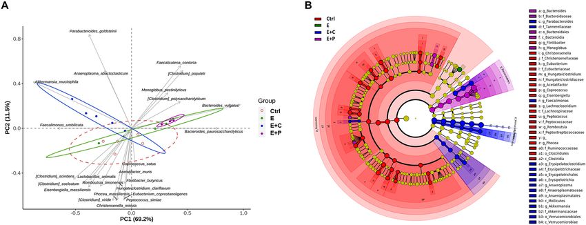
www.nature.com/scientificreports/
that releases metabolites with multiple functions in the host, and they are known to reduce the risk of chronic
inflammatory diseases.
The metabolites produced by the gut microbiome regulate the host’s immune system and influence various
organs. In accordance with these findings, the physiological influence of gut microbiota alteration and subsequent
nutrient variation are governed by the differences in microbial metabolome profiles. Furthermore, metabolic
byproducts derived from the bacterial fermentation of dietary fibers are important local and systemic signal-
ing molecules that sustain immune and tissue h omeostasis11. Among them, short-chain fatty acids (SCFAs) are
metabolites that have been studied extensively, and they have immunomodulatory functions within the diverse
hysiology12. SCFAs are produced by the fermentation of dietary fiber by specific microbes in the
aspects of host p
intestinal colon and exhibit anti-inflammatory p roperties13,14. Bile acids (BAs) are essential components of the
digestive system and it reflects the amount of those escaping extraction from the portal b lood15. Interestingly,
they have also been associated with lung injury16–19 and lung inflammation20–22. Sphingolipids (SLs) can be rapidly
generated or converted into each other, and they play pivotal roles in various cellular processes23. Moreover, SLs
are increasingly recognized as critical mediators in major pulmonary diseases and are associated with COPD
phenotypes23,24. Among them, ceramide, an intermediate product of sphingomyelin (SM) metabolism, is associ-
ated with COPD25 and the inflammatory response26.
In accordance with these findings, gut microbiota, metabolites, and nutrients are closely associated with
the increased incidence of various inflammatory diseases. We previously performed a study to show that fecal
microbiota transplantation and high fiber diet attenuated emphysema development and changed the gut micro-
biota composition27. Despite the growing knowledge on the immunomodulatory effects of the gut microbiota
on systemic diseases, microbiota-metabolite interaction induced by dietary nutrients in the gut-lung axis in
emphysema needs to be investigated further. Therefore, in this study, we investigated the effect of high-fiber
diet on emphysema and the impact of gut microbiota-metabolite interaction by different types of dietary fiber
compositions in a CS-exposed emphysema model. Furthermore, the relationship between the gut microbiota
and metabolites was investigated based on the high-fiber diet components by analyzing the altered microbiota
and metabolome profiles in a CS-exposed emphysema model.
Results
Dietary fiber improves alveolar destruction and inflammation during emphysema progres-
sion. To determine whether dietary fiber influence emphysema, the effect of high-fiber dietary modification
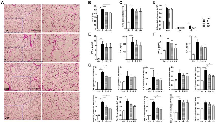
on pulmonary inflammatory responses in CS-exposure-induced emphysema was first analyzed. Histological
analysis showed that alveolar destruction and airspace enlargement were most severe in the emphysema group
and less severe in the emphysema with high-fiber diet group (Fig. 1A). This observation was confirmed by the
mean linear intercept (MLI) measurement, which was significantly lower in the emphysema with high-fiber diet
group than in the emphysema group (Fig. 1B). The alveolar structures and forms of histological emphysema were
more improved in the high-pectin diet group compared to those in the high-cellulose diet group (Fig. 1A,B).
Moreover, the bronchoalveolar lavage fluid (BALF) for immune infiltration to the inflammatory response was
analyzed. Inflammatory cell infiltration was higher in the BALF of all CS-exposed groups (Fig. 1C). This inflam-
mation was characterized by increased macrophage infiltration, which was the highest in the emphysema group.
The number of macrophages and neutrophils in the BALF was significantly lower in the emphysema with high-
fiber diet group than in the emphysema group (Fig. 1D). The results of the analysis of the inflammatory cells
from the BALF indicated a marginal decrease in the number of macrophages and neutrophils in the high-pectin
diet group compared to the high-cellulose diet group. Moreover, the inflammatory cytokine levels in the BALF
and serum were further evaluated. The levels of interleukin-6 (IL-6) and interferon-γ (IFN-γ) were significantly
increased in the BALF and serum of the emphysema group, which is presumably associated with increased mac-
rophage infiltration. The IL-6 and IFN-γ levels in the BALF and serum were lower in the high-pectin diet group
than those in the emphysema group (Fig. 1E,F). The inflammatory cytokine levels in the BALF and serum were
lower in the high-pectin diet group than those in the high-cellulose diet group. In accordance with the above
results, the mRNA expression levels of representative pro-inflammatory mediators and immune modulators,
including IFN-γ, IL-1β, IL-6, IL-8, IL-18, IRF-5, MMP-12, TNF-α, TGF-β, and cathepsin S, were lower in the
emphysema with high-fiber diet group than those in the emphysema group. The expression of inflammatory
markers was lowest in the high-pectin diet group than in all other groups (Fig. 1G). These results indicated
that the high-fiber diet attenuated the pathological changes during emphysema progression and influenced an
inflammatory response associated with CS-exposed emphysema. Moreover, less inflammation was observed in
emphysema mice supplemented with fermentable fiber content (pectin) than in emphysema mice supplemented
with non-fermentable fiber content (cellulose). Furthermore, these results indicate that a prebiotic high-fiber
diet helps to prevent CS-exposure-induced emphysema progression and suggests that immune modulation by
dietary fibers can attenuate the local and systemic effects of emphysema.
Dietary fiber modulates the microbial community structure. Diet has a major impact on gut
microbiota composition, diversity, and richness. Different components of diet shape the gut bacterial communi-
ties. Considering that different dietary fiber components have different impacts on emphysema, different types
of dietary fiber components were hypothesized to influence the microbial composition and are related to the
immunity in the gut-lung axis. To confirm this hypothesis, the microbial analysis of the fecal samples was per-
formed. A principal coordinate analysis based on the relative abundance of genera represents the distinct fecal
microbial community structures. Different components of dietary fiber were significantly associated with differ-
ent microbial community structures in the gut (Fig. 2A). Moreover, changes in the microbial composition of the
fecal samples were analyzed using the linear discriminant analysis effect size (LEfSe) cladogram (Fig. 2B) and
Scientific Reports | (2021) 11:7008 | https://doi.org/10.1038/s41598-021-86404-x 2
Vol:.(1234567890)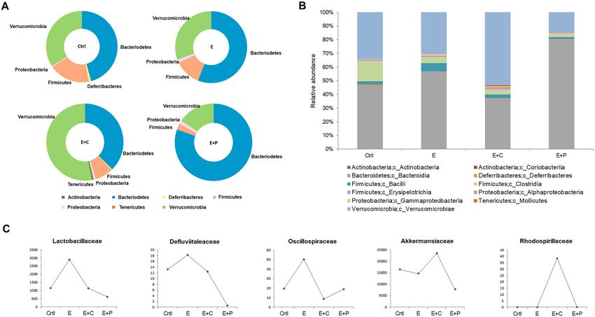
www.nature.com/scientificreports/

                                  Figure 1.  High-fiber diet attenuates inflammation and the degree of alveolar destruction. (A) Representative
                                  H&E-stained lung tissues from mice in the four groups (magnification: ×100 and ×400). (B) The mean linear
                                  intercept (MLI) of lung tissues from each group. (C) Total number of cells in the bronchoalveolar lavage fluid
                                  (BALF) infiltrating the airways. (D) Differential cell numbers of BALF in each group. (E,F) The levels of the
                                  cytokines IL-6 and IFN-γ in the BALF (E) and serum (F) were measured using ELISA. (G) The relative mRNA
                                  levels of IFN-γ, IL-1β, IL-6, IL-8, IL-18, IRF-5, MMP-12, TNF-α, TGF-β, and cathepsin S in lung tissues.
                                  (n = 5–6 mice per group). Values are expressed as the mean ± SE. *P < 0.05 and **P < 0.01. Ctrl control group, E
                                  emphysema group, E + C emphysema with high-cellulose diet group, E + P emphysema with high-pectin diet
                                  group.

                                  Figure 2.  High-fiber diet alters the composition of the gut microbial community. (A) Principal coordinate
                                  analysis (PCoA) biplot of the fecal microbiota composition. (B) The cladogram of the linear discriminant
                                  analysis effect size (LEfSe) analysis for differentially abundant taxa of fecal microbiotas in the four groups; Ctrl
                                  (red), control group; E (green), emphysema group; E + C (blue), emphysema with high-cellulose diet group;
                                  E + P (violet), emphysema with high-pectin diet group. (n = 5 mice per group).

                                  histogram (Fig. S1). Furthermore, the differences in the microbial composition were evaluated based on the phy-
                                  lum, class, and family levels (Fig. 3A-C). The Bacteroidetes phylum level was the most abundant in the emphy-
                                  sema with high-pectin diet group compared to the other groups (Fig. 3A). At the family level, pyrosequencing
                                  showed that the emphysema group had increased Lactobacillaceae, Defluviitaleaceae, and Oscillospiraceae family
                                  members compared to the control group. On the other hand, the high-fiber diet group had decreased amounts of

Scientific Reports |   (2021) 11:7008 |                https://doi.org/10.1038/s41598-021-86404-x                                                        3

                                                                                                                                                 Vol.:(0123456789)
www.nature.com/scientificreports/
Figure 3. High-fiber diet changes the gut microbiota composition. Taxonomic plots showing the relative
abundance across the different groups. (A,B) Phylum- (A) and class- (B) level relative abundance of the fecal
microbiome of each group. (C) Family level bacteria-specific relative compositional changes in all groups. (n = 5
mice per group). Ctrl control group, E emphysema group, E + C emphysema with high-cellulose diet group, E + P
emphysema with high-pectin diet group.
Lactobacillaceae, Defluviitaleaceae, and Oscillospiraceae compared to the emphysema group and Lactobacillaceae
and Defluviitaleaceae family levels were the lowest in the high-pectin diet group (Fig. 3C). The emphysema with
high-cellulose diet group had the highest abundance of the Verrucomicrobia phyla (genus Akkermansia) and
Alphaproteobacteria class (Fig. 3A). Furthermore, within the Alphaproteobacteria class, the Rhodospirillaceae
family was observed only in the high-cellulose diet group (Fig. 3C). Based on these findings, it is observed that
different types of dietary fiber components can regulate the changes in the gut microbial composition. These
results provide a mechanistic insight into the protective nature of dietary-modulated microbiota during the
progression of emphysema and suggest a beneficial effect on dietary-microbiota-immunity.
Dietary fiber regulates the global metabolomic profile. To confirm the association between metab-
olites and emphysema or fiber, global metabolomic profiles were analyzed using MetaboAnalyst 4.0. The four
groups in the feces, serum, and lung were classified based on the partial least squares discriminant analysis (PLS-
DA). The optimal number of metabolic features for classification was selected based on the best predictive ability
of the model. Five unidentified metabolites in the feces, three unidentified metabolites and L-phenylalanine
in the serum, and one unidentified metabolite and AMP in the lung were used as basis for the classification,
respectively (Fig. S2). A pathway analysis was performed using a KEGG library with significantly changed meta-
bolic features. Based on the P value < 0.05 in the pathway analysis and impact value threshold (0.10), various
pathways induced by CS-exposed emphysema and fiber diet were identified. The comparison between the two
groups was further analyzed in three ways: (1) control vs. emphysema, (2) emphysema vs. emphysema with
high-cellulose diet, and (3) emphysema vs. emphysema with high-pectin diet for feces, affected by microbiota
ecology primarily. β-alanine metabolism and histidine metabolism had the highest impact (0.40) on grouping
control and emphysema. Several common pathways were significantly affected by high-cellulose and -pectin
diet. Linoleic acid, primary BA synthesis, phenylalanine, tyrosine and tryptophan biosynthesis, steroid hormone
biosynthesis, and SL metabolisms exhibit a significant effect. Among them, linoleic acid metabolism showed the
highest impact value (1.0) in both comparisons (Fig. 4), and primary BA biosynthesis had the most significant
impact with a P value of 2.28 × 10⁻⁶. Linoleic acid metabolism and d-glutamate metabolism showed the high-
est impact values (1.0) in the lungs based on the pathway topology analysis (Fig. S3). Caffeine metabolism and
linoleic acid metabolism had the highest impact in the serum. Arachidonic acid metabolism was the pathway
with significant impact in both feces (Fig. 4) and serum (Fig. S4). The metabolic patterns provide an overview of
the metabolic differences in the four groups. The top metabolites representing the differences in the feces, lung,
and serum are visually illustrated by heatmaps (Fig. S5). Furthermore, the targeted metabolites were different in
the four groups.
Scientific Reports | (2021) 11:7008 | https://doi.org/10.1038/s41598-021-86404-x 4
Vol:.(1234567890)www.nature.com/scientificreports/
Figure 4. Summary of pathway analysis with MetaboAnalyst. (A–C) Metabolic pathway analysis of the feces in
E (A), E + C (B), and E + P (C) were performed with significantly changed metabolic features (P < 0.05). Potential
target pathways were labeled based on the impact-value threshold (0.10) and P-value (0.05). Ctrl control group,
E emphysema group, E + C emphysema with high-cellulose diet group, E + P emphysema with high-pectin diet
group.
Figure 5. Dietary fiber regulates the short chain fatty acid (SCFA) metabolism. (A,B) The concentrations of the
SCFA metabolites (acetic, propionic, butyric, valeric, 2-methylbutyric, caproic, and 2,3,4-methylvaleric acids)
were measured within the content of the fecal metabolite profiling. (n = 4 mice per group; n = 5 mice in E + P).
Values are expressed as the mean ± SE. *P < 0.05 and **P < 0.01. Ctrl control group, E emphysema group, E + C
emphysema with high-cellulose diet group, E + P emphysema with high-pectin diet group.
Dietary fiber regulates targeted metabolomic profiles. Further analysis with targeted metabolomic
profiles was performed for SCFAs, BAs, and SLs, and detailed results were described in the supplementary
results. Overall, the acetate, propionate, and butyrate concentrations were the lowest in the emphysema group
compared to the other groups. The local concentrations of SCFAs (acetate, propionate, and butyrate) were nota-
bly higher in the emphysema with high-fiber (cellulose and pectin) diet group than in the emphysema group.
The SCFA (acetate, propionate, and butyrate) concentrations were increased in the high-pectin diet group com-
pared to the high-cellulose diet group (Fig. 5). The results are similar to those previously reported28. The primary
BA concentrations, such as cholic acid (CA) and chenodeoxycholic acid (CDCA), in the feces were higher in the
control group than in the emphysema group (Fig. 6A). The secondary BA concentrations, such as deoxycholic
acid (DCA) and lithocholic acid (LCA), in the feces, serums, and lung tissues were higher in the emphysema
group than in the control group (Fig. 6A-C). Interestingly, the CA, CDCA, and ursodeoxycholic acid (UDCA)
Scientific Reports | (2021) 11:7008 | https://doi.org/10.1038/s41598-021-86404-x 5
Vol.:(0123456789)www.nature.com/scientificreports/
Figure 6. Dietary fiber alters the bile acid (BA) metabolism. (A–C) The levels of the BA metabolites (CA, DCA,
CDCA, UDCA, and LCA) in the feces (A), serum (B), and lung (C) were measured using metabolite profiling.
(n = 6 mice per group). Values are expressed as the mean ± SE. *P < 0.05 and **P < 0.01. Ctrl control group, E
emphysema group, E + C emphysema with high-cellulose diet group, E + P, emphysema with high-pectin diet
group, CA cholic acid, DCA deoxycholic acid, CDCA chenodexycholic acid, UDCA ursodeoxycholic acid, LCA
lithocholic acid.
concentrations in the feces were higher in the high-pectin diet group than in the emphysema group. Mean-
while, the CDCA, DCA, LCA, CA, and UDCA concentrations in the lung tissues and serums were higher in
the emphysema group than in the other groups (Fig. 6). These results are similar to those previously reported in
respiratory disease29.
Discussion
The gut microbiota, via various dietary nutrients, is involved in the production of metabolites that play a major
role in human immunity. Metabolites like SCFAs significantly influence systemic immunity and the circulatory
systems30. Therefore, it is reasonable that gut microbiota dysbiosis via malnutrition or unhealthy diet is associ-
ated with dysregulated inflammation affecting autoimmune or respiratory d iseases31. However, the interac-
tions between dietary fiber components and the regulatory mechanisms of respiratory diseases have not been
investigated.
This study explored whether dietary fibers acting as prebiotics directly interact with gut microbes and play a
role in the production of key metabolites, including SCFAs, BAs, and SL. This study also discussed how dietary
fibers impact gut microbial ecology, host physiology, and immunity. The therapeutic potential of modulating
gut microbiota and metabolites by a high-fiber diet was investigated in a CS-exposed emphysema model. CS is a
major risk factor for COPD, which is impacted by the significant pathogenicity of circulating toxic constituents
or metabolites of absorbed C S4,32. A CS-induced emphysema model was used based on general pathologies and
mechanisms of human emphysema. In this model, a high-cellulose and high-pectin diet significantly suppressed
the inflammation and attenuated the pathological changes in emphysema mice. The number of immune cells,
macrophages, and neutrophils was lower in the high-fiber (cellulose and pectin) diet group than in the emphy-
sema group. These results confirm that inflammatory cytokine levels and mRNA levels were decreased in the
high-fiber (cellulose and pectin) diet group and that dietary fiber strongly inhibits inflammation. The effect of
high fiber diet on lung function and COPD is also proven in epidemiologic studies33,34, although the underlying
mechanism is not clear. Extensive studies have found that the gut microbiota is crucial in maintaining host home-
ostasis via immune system i nteractions35,36. Low gut microbial diversity is associated with increased risk of lung
disease37. The interactions between dietary fiber components and gut microbial communities were investigated
Scientific Reports | (2021) 11:7008 | https://doi.org/10.1038/s41598-021-86404-x 6
Vol:.(1234567890)www.nature.com/scientificreports/
in an emphysema model. The present study showed that the emphysema group had increased Lactobacillaceae,
Defluviitaleaceae, and Oscillospiraceae family members compared to the control group. The high-cellulose and
high-pectin diet groups had lower Lactobacillaceae, Defluviitaleaceae, and Oscillospiraceae family members com-
pared to the emphysema group. The increase of Lactobacillaceae in COPD was also identified in human studies
and it was also associated with local inflammation38,39. The role of the Defluviitaleaceae and Oscillospiraceae
family members (Clostridia class) in the respiratory diseases has not yet been investigated, our study suggests
the necessity to further confirm their roles. Interestingly, the Rhodospirillaceae family (Alphaproteobacteria
class) was observed only in the high-cellulose diet group. Alphaproteobacteria is a diverse class of organisms
within the Proteobacteria phylum with many biological roles and cellulose usually increases their n umbers40,41.
Bacteroidetes are major producers of SCFAs, and the proportion of Bacteroidetes is significantly decreased in
OPD42. Accordingly, the emphysema with high-pectin diet group had the highest abundance of the
cases of C
Bacteroidetes phyla compared to the other groups in this study. The emphysema with high-cellulose diet group
had the highest abundance of the Verrucomicrobia phyla (genus Akkermansia) and the Alphaproteobacteria class
(phylum Proteobacteria), which has many important biological roles. These data indicate that both pectin and
cellulose supplementation modulates the microbial community structures and effectively alter the composition
of the gut microbiota. The protective effects of high-fiber diet against emphysema progression may be influenced
by the direct interactions of microbiota, dietary fiber and immune system.
Various metabolites have been extensively investigated in terms of their beneficial immunomodulatory
roles43,44. Specifically, SCFAs reduce colitis by inducing colonic regulatory T c ells45 and driving colonocytes
towards mitochondrial beta oxidation of fatty a cids46. SCFAs also prevent asthma p athology28. Malnutrition
adversely affects the gut microbiota composition and decreases the production of physiologically active byprod-
ucts that are key driving factors in the prevalence of inflammatory disease44. Previous studies have reported that
dietary fiber-induced microbial SCFAs not only suppress the ILC2-dependent airway inflammation47 but also
modulate immunity28. Similarly, in the present study, the feces from the emphysema with high-fiber (cellulose
and pectin) diet group were significantly enriched with acetate, propionate, and butyrate and had large alterations
in the gut microbiota. Moreover, the SCFAs concentrations were increased in the high-pectin diet group than
in the high-cellulose diet group. Moreover, the valeric acid and 2,3,4-methylvaleric acid concentrations were
higher in emphysema mice with high-pectin diet than in emphysema mice. We confirmed that high-pectin diet
was associated with an abundance of SCFA with anti-inflammatory properties in an emphysema model. These
results show that different types of dietary fiber ingredients (fermentable or non-fermentable) induce different
changes in metabolite levels and regulate the levels of different SCFA metabolites. In addition, the protective
effects of dietary fiber induced SCFAs through the interactions between the microbiota and immune system may
inhibit the progression of emphysema.
Among the various endogenous metabolites produced by host-gut microbiota metabolism, BAs are gaining
increased attention48. They serve as nutrient signaling hormones, play a role in inflammation, and are associated
with COPD and lung disease i nflammation16–20. We also explored the regulation of BA metabolites by different
dietary fiber components in an emphysema model. In this study, we showed that the DCA and LCA concentra-
tions in the feces, serums, and lung were higher in emphysema mice than in control mice. Moreover, the DCA,
LCA, and CDCA concentrations in serums and lung were lower in the high-pectin diet and high-cellulose diet
groups than in the emphysema group. The secondary BA concentrations (DCA, UDCA, and LCA) in the feces
were higher in the high-pectin diet group compared to other groups. A previous review article suggested that
dietary fibers bind conjugated primary BAs and may serve as a platform for gut bacteria, thus leading to decon-
jugation which is a perquisite for biotransformation to secondary B As49. Therefore, this indicates that different
types of dietary fiber components can affect various BA metabolites. Moreover, this study suggests that BAs, via
dietary-metabolic regulation, are associated with local and systemic inflammatory responses in an emphysema
model.
SLs are constituents of cellular membranes and participate in cellular responses to stress, thereby ensuring cell
survival. Therefore, the proper functioning of the SL metabolic pathway is essential for cellular h omeostasis50.
Moreover, the role of SLs as mediators of inflammation may have significant implications in a range of pulmo-
nary diseases in which inflammation is a central process of the p athogenesis51. The extensive alterations in SL
metabolism induced by CS exposure are controlled by a complex regulatory program, which, concomitantly, may
also mediate other adaptive and destructive tissue responses. Moreover, changes in SL metabolism are intrinsic
to the broad response of the tissue to CS exposure and dietary fiber intake, including oxidative stress and xeno-
biotic stress responses, immune response, and other metabolic alterations. As smoking-related inflammation
and a high-fiber diet impact SL metabolism, the association between SLs and inflammation in COPD can be
determined. Further metabolomics studies are needed to investigate the potential diagnostic and therapeutic
candidates in emphysema.
umans52. Omega-3 fatty acids,
The fatty acids including alpha-linolenic acid and linoleic acid are essential for h
such as plant-derived alpha-linolenic acid and its forms primarily found in fish, give rise to anti-inflammatory
and pro-resolving m ediators53. Previous studies have reported that the higher dietary intake of omega-3 fatty
acids leads to the decrease in cardiovascular risk/atherosclerosis54, autoimmune disease a ctivity55, morbidity in
asthma56, COPD57, and other chronic diseases58. Global metabolomics profile analysis revealed that the linoleic
acid metabolism pathway had the highest impact on the emphysema with high-cellulose and high-pectin diet
groups. Therefore, these results suggest that anti-inflammatory mechanisms including the linoleic acid pathway
prevent the COPD development.
These results suggested that the protection exhibited by high-cellulose or high-pectin diet against emphy-
sema may depend on the microbiota-metabolism-immunity interactions. However, the complex relationship
among these biological systems and prebiotics in emphysema needs further investigation. Additionally, the
bioavailability of nutrients and bioactive compounds in fruits and vegetables is an extremely important area of
Scientific Reports | (2021) 11:7008 | https://doi.org/10.1038/s41598-021-86404-x 7
Vol.:(0123456789)www.nature.com/scientificreports/
nutrition and medical research. Water-soluble dietary fibers can impair the bioavailability of beta-carotene in
the gastrointestinal t ract59–62. Thus, further research is required to understand the dietary-fiber-associated bio-
availability and functions of carotenoids.
The important regulatory roles of naturally occurring deuterium in fundamental biological processes and the
antitumor effect of deuterium depletion have been demonstrated in multiple s tudies63–67. Deuterium depletion
occurs through returning low deuterium ketogenic propionic acid to the circulation for terminal oxidation into
CO2 and deuterium-depleted metabolic water in the mitochondria/peroxisomes of host cells68. Hence, the corre-
lation among emphysema, deuterium-depleted metabolites, and metabolizing organisms in the gut microbiome-
SCFAs digesting dietary fiber needs to be investigated further. New insights into the role of deuterium depletion
in emphysema may facilitate the development of novel emphysema treatments.
The findings of this study indicate that compositions of the gut microbiota and metabolites can be modu-
lated using a high-fiber diet, consequently attenuating emphysema development via inhibition of local and sys-
temic inflammation, which may provide future therapeutic applications for preventing or delaying emphysema
progression.
Materials and methods
Emphysema mouse model. Female C57BL/6 mice (8 weeks old) were purchased from Orient Bio Inc.
(Seongnam, Republic of Korea) and maintained at room temperature (RT, 25 °C) in a 12/12-h light/dark cycle.
The animal model of emphysema was exposed with CS according to a previously reported protocol27. This study
complied with guidelines of the 8th edition of the Guide for the Care and Use of Laboratory Animals. All animal
experiments were approved by the Institutional Animal Care and Use Committee of Asan Medical Center, Seoul,
Republic of Korea (2019-14-069), and were performed according to the ARRIVE (Animal Research: Reporting
In vivo Experiments) guidelines69.
Diet modifications. The animals were randomly divided into four groups (n = 6 mice/group): Group I
(control group, Ctrl), Group II (emphysema group, E, CS exposure only), Group III (E + C, CS exposure with a
high-cellulose diet), and Group IV (E + P, CS exposure with a high-pectin diet). The mice were fed ad libitum
with the standard AIN-76A diet supplied by Daehan Biolink Co., LTD (Chungbuk, Republic of Korea). The two
types of dietary modifications based on the standard AIN 76A diet were as follows: the high-cellulose diet (20%
cellulose), which was modified with cellulose from vegetable supplement, and the high-pectin diet (20% pectin),
which was modified with pectin from citrus supplement. Groups I and II were given the standard AIN-76A diet
for 4 weeks. Group III and IV were given the AIN-76A diet for 1 week and were administered with a modified
diet for the last 3 weeks of the experiment period.
Preparation of samples. After 4 weeks, the animals were anesthetized through isoflurane inhalation, and
the blood samples were collected by heart puncture. The trachea was catheterized and perfused with 1.5 mL
of PBS. The cellular and liquid fractions of the BALF were separated by centrifugation at 2200 rpm for 5 min
at 4 °C. The cell pellet was suspended in PBS, seeded onto a slide, and stained with Diff-Quick (Sysmex, Kobe,
Japan). Inflammatory cells were observed under a light microscope (Leica, USA). All specimens were collected,
fixed, immediately frozen, and stored at − 80 °C until analysis.
Histomorphological assessment. The left lung was inflated with 0.5% low-melting-point agarose at a
pressure of 15 cm H2O. The left lobe was embedded in paraffin, cut into 5-μm-thick sections, and stained with
hematoxylin and eosin. Emphysematous changes were observed under a light microscope (Leica, USA) and were
evaluated by measuring the MLI. MLI is a measurement of the mean interalveolar septal wall distance deter-
mined by the number of interruptions in 1 mm lines of the alveolar wall. Four lines were drawn in each field, and
at least five random fields were examined per mouse.
Cytokine level quantification. The levels of the cytokines IL-6 and IFN-γ in the BALF and serum were
measured using a commercially available ELISA kit (R&D Systems, Minneapolis, MN, USA) based on the manu-
facturer’s instructions.
Quantitative real‑time PCR analysis. Total RNA was extracted from lung tissues using TRIzol reagent
(Thermo Fisher Scientific, Waltham, MA, USA) in accordance with the manufacturer’s protocol as previously
described27. Transcription levels were measured using real-time PCR with sequence-specific primers for IFN-γ,
IL-1β, IL-6, IL-8, IL-18, MMP-12, TNF-α, TGF-β, cathepsin S, and IRF-5 (Table S1).
DNA extraction and microbiota bacterial composition analysis. DNA was extracted from fecal
samples using a DNA isolation kit (MO BIO, Carlsbad, CA, USA) in accordance with the manufacturer’s instruc-
tions. DNA was amplified with 16S_V3_F and 16S_V4_R primers specific to the V3-V4 hypervariable regions
of the 16S rDNA gene (Supplementary Table S1). The libraries were prepared using PCR products in accordance
with the MiSeq System Guide (Illumina, USA) and quantified using the QIAxpert System (QIAGEN, Germany).
Each amplicon was quantified, and an equimolar ratio was pooled and sequenced on a MiSeq (Illumina) in
accordance with the manufacturer’s instructions. Microbiota bacterial composition analysis was performed, fol-
lowing the procedure previously described27.
Scientific Reports | (2021) 11:7008 | https://doi.org/10.1038/s41598-021-86404-x 8
Vol:.(1234567890)www.nature.com/scientificreports/
Quantitative metabolome measurement. Standard metabolites and internal standards were pur-
chased from Sigma-Aldrich (St. Louis, MO, USA), and all solvents were purchased from J. T. Baker (Center Val-
ley, PA, USA). The SCFA metabolites in feces and BA metabolites were measured using a previously described
procedure70,71. Mouse serum (20 µL) was mixed well with 500 μL of cold acetonitrile and 50 μL of internal
standard solution (50 μL of 1 μM cholic acid-d5 solution). For the SL metabolites, the Folch method was used to
extract the lipids from 20 to 30 mg of mouse lung after adding 50 μL of internal standard solution (50 nM of C17
ceramide solution)72. For the 30 μL of mouse serum, the lipids was extracted using the Bligh and Dyer method
after adding the internal standard s olution73. The organic layer containing sphingolipids was dried using a vac-
uum centrifuge and stored at − 20 °C until liquid chromatography-tandem mass spectrometry (LC–MS/MS)
analysis. The dried matter from the feces, serum, and lung was stored at − 20 °C and reconstituted with methanol
(MeOH) prior to LC–MS/MS analysis for all the analyses performed. Furthermore, the samples were analyzed
using a LC–MS/MS system equipped with a 1290 HPLC (Agilent, Waldbronn, Germany) and QTRAP5500 mass
spectrometry (AB Sciex, Toronto, Canada). Additional methods are described in the data supplement.
Global metabolome profiling. The metabolites were extracted using conventional liquid–liquid extrac-
tion procedures72,73. Briefly, 3–4 volumes of chloroform/methanol (2/1, v/v) were added to freeze-dried feces,
lung, and serum and then centrifuged for 15 min. Nonpolar metabolites containing lipids were collected from
the lower organic phase, and polar metabolites were collected in the upper aqueous phase. The LC–MS was
equipped with Ultimate3000 (Dionex) and Orbitrap XL (Thermo Fisher). A reverse phase column (Pursuit 5;
150 × 2.0 mm) and HILIC (HILIC Plus; 100 × 2.1 mm) were used for nonpolar and polar metabolites, respec-
tively. LC–MS analysis was conducted for each sample solution in positive and negative ion modes. Data nor-
malization was performed using log transformation and auto-scaling. Metabolite features, including neutral
mass and retention time values, were determined using the Compound Discoverer 2.1. Moreover, metabolite
features showing statistically significant changes (P < 0.05) were chosen and identified using the KEGG database
(http://www.kegg.jp/kegg/kegg1.html) with a mass accuracy of 10 ppm. Statistical analyses including the PLS-
DA were conducted using MetaboAnalyst 4.0.
Statistical analysis. Data were analyzed using the Kruskal–Wallis H test, Mann–Whitney U test, and one-
way ANOVA followed by Tukey’s test with the IBM SPSS version 25 (https://www.ibm.com/analytics/spss-stati
stics-software). All values were expressed as the mean ± standard error (SE). Statistical significance was set at
P < 0.05.
Received: 27 November 2020; Accepted: 12 March 2021
References
1. Vogelmeier, C. F. et al. Global strategy for the diagnosis, management, and prevention of chronic obstructive lung disease 2017
report. GOLD executive summary. Am. J. Respir. Crit. Care Med. 195, 557–582. https://doi.org/10.1164/rccm.201701-0218PP
(2017).
2. Chronic obstructive pulmonary disease. Nat. Rev. Dis. Primers 1, 15079. https://doi.org/10.1038/nrdp.2015.79 (2015).
3. Barnes, P. J. Chronic obstructive pulmonary disease. N. Engl. J. Med. 343, 269–280. https://doi.org/10.1056/NEJM20000727343
0407 (2000).
4. Arcavi, L. & Benowitz, N. L. Cigarette smoking and infection. Arch. Intern. Med. 164, 2206–2216. https://doi.org/10.1001/archi
nte.164.20.2206 (2004).
5. Macgregor, I. D. Effects of smoking on oral ecology. A review of the literature. Clin. Prev. Dent. 11, 3–7 (1989).
6. Huang, C. & Shi, G. Smoking and microbiome in oral, airway, gut and some systemic diseases. J. Transl. Med. 17, 225. https://doi.
org/10.1186/s12967-019-1971-7 (2019).
7. Marsland, B. J., Trompette, A. & Gollwitzer, E. S. The gut-lung axis in respiratory disease. Ann. Am. Thorac. Soc. 12(Suppl 2),
S150-156. https://doi.org/10.1513/AnnalsATS.201503-133AW (2015).
8. Ubags, N. D. J. & Marsland, B. J. Mechanistic insight into the function of the microbiome in lung diseases. Eur. Respir. J. https://
doi.org/10.1183/13993003.02467-2016 (2017).
9. Turnbaugh, P. J., Backhed, F., Fulton, L. & Gordon, J. I. Diet-induced obesity is linked to marked but reversible alterations in the
mouse distal gut microbiome. Cell Host Microbe 3, 213–223. https://doi.org/10.1016/j.chom.2008.02.015 (2008).
10. Wood, L. G., Shivappa, N., Berthon, B. S., Gibson, P. G. & Hebert, J. R. Dietary inflammatory index is related to asthma risk, lung
function and systemic inflammation in asthma. Clin. Exp. Allergy 45, 177–183. https://doi.org/10.1111/cea.12323 (2015).
11. Maslowski, K. M. et al. Regulation of inflammatory responses by gut microbiota and chemoattractant receptor GPR43. Nature
461, 1282–1286. https://doi.org/10.1038/nature08530 (2009).
12. Koh, A., De Vadder, F., Kovatcheva-Datchary, P. & Backhed, F. From dietary fiber to host physiology: short-chain fatty acids as
key bacterial metabolites. Cell 165, 1332–1345. https://doi.org/10.1016/j.cell.2016.05.041 (2016).
13. McNabney, S. M. & Henagan, T. M. Short chain fatty acids in the colon and peripheral tissues: a focus on butyrate, colon cancer,
obesity and insulin resistance. Nutrients https://doi.org/10.3390/nu9121348 (2017).
14. Canani, R. B. et al. Potential beneficial effects of butyrate in intestinal and extraintestinal diseases. World J. Gastroenterol. 17,
1519–1528. https://doi.org/10.3748/wjg.v17.i12.1519 (2011).
15. Kim, M. J. & Suh, D. J. Profiles of serum bile acids in liver diseases. Korean J. Intern. Med. 1, 37–42. https://doi.org/10.3904/kjim.
1986.1.1.37 (1986).
16. Zecca, E. et al. Bile acid pneumonia: A “new” form of neonatal respiratory distress syndrome?. Pediatrics 114, 269–272. https://
doi.org/10.1542/peds.114.1.269 (2004).
17. Su, K. C. et al. Bile acids increase alveolar epithelial permeability via mitogen-activated protein kinase, cytosolic phospholipase A2,
cyclooxygenase-2, prostaglandin E2 and junctional proteins. Respirology 18, 848–856. https://doi.org/10.1111/resp.12086 (2013).
18. Wu, Y. C. et al. Bile acid aspiration in suspected ventilator-associated pneumonia. Chest 136, 118–124. https://doi.org/10.1378/
chest.08-2668 (2009).
Scientific Reports | (2021) 11:7008 | https://doi.org/10.1038/s41598-021-86404-x 9
Vol.:(0123456789)www.nature.com/scientificreports/
19. Savarino, E. et al. Gastro-oesophageal reflux and gastric aspiration in idiopathic pulmonary fibrosis patients. Eur. Respir. J. 42,
1322–1331. https://doi.org/10.1183/09031936.00101212 (2013).
20. Perng, D. W. et al. Exposure of airway epithelium to bile acids associated with gastroesophageal reflux symptoms: a relation to
transforming growth factor-beta1 production and fibroblast proliferation. Chest 132, 1548–1556. https://doi.org/10.1378/chest.
07-1373 (2007).
21. D’Ovidio, F. et al. The effect of reflux and bile acid aspiration on the lung allograft and its surfactant and innate immunity molecules
SP-A and SP-D. Am. J. Transpl. 6, 1930–1938. https://doi.org/10.1111/j.1600-6143.2006.01357.x (2006).
22. D’Ovidio, F. et al. Bile acid aspiration and the development of bronchiolitis obliterans after lung transplantation. J. Thorac. Car-
diovasc. Surg. 129, 1144–1152. https://doi.org/10.1016/j.jtcvs.2004.10.035 (2005).
23. Yang, Y. & Uhlig, S. The role of sphingolipids in respiratory disease. Ther. Adv. Respir. Dis. 5, 325–344. https://doi.org/10.1177/
1753465811406772 (2011).
24. Bowler, R. P. et al. Plasma sphingolipids associated with chronic obstructive pulmonary disease phenotypes. Am. J. Respir. Crit.
Care Med. 191, 275–284. https://doi.org/10.1164/rccm.201410-1771OC (2015).
25. Nobakht, M. G. B. F., Aliannejad, R., Rezaei-Tavirani, M., Taheri, S. & Oskouie, A. A. The metabolomics of airway diseases, includ-
ing COPD, asthma and cystic fibrosis. Biomarkers 20, 5–16. https://doi.org/10.3109/1354750X.2014.983167 (2015).
26. Bodas, M., Pehote, G., Silverberg, D., Gulbins, E. & Vij, N. Autophagy augmentation alleviates cigarette smoke-induced CFTR-
dysfunction, ceramide-accumulation and COPD-emphysema pathogenesis. Free Radic. Biol. Med. 131, 81–97. https://doi.org/10.
1016/j.freeradbiomed.2018.11.023 (2019).
27. Jang, Y. O. et al. Fecal microbial transplantation and a high fiber diet attenuates emphysema development by suppressing inflam-
mation and apoptosis. Exp. Mol. Med. https://doi.org/10.1038/s12276-020-0469-y (2020).
28. Trompette, A. et al. Gut microbiota metabolism of dietary fiber influences allergic airway disease and hematopoiesis. Nat Med 20,
159–166. https://doi.org/10.1038/nm.3444 (2014).
29. Aldhahrani, A., Verdon, B., Ward, C. & Pearson, J. Effects of bile acids on human airway epithelial cells: implications for aerodiges-
tive diseases. ERJ Open Res. https://doi.org/10.1183/23120541.00107-2016 (2017).
30. Anand, S. & Mande, S. S. Diet, microbiota and gut-lung connection. Front. Microbiol. 9, 2147. https://doi.org/10.3389/fmicb.2018.
02147 (2018).
31. Ege, M. J. et al. Exposure to environmental microorganisms and childhood asthma. N. Engl. J. Med. 364, 701–709. https://doi.org/
10.1056/NEJMoa1007302 (2011).
32. Cruickshank-Quinn, C. I. et al. Transient and persistent metabolomic changes in plasma following chronic cigarette smoke expo-
sure in a mouse model. PLoS ONE 9, e101855. https://doi.org/10.1371/journal.pone.0101855 (2014).
33. Kan, H., Stevens, J., Heiss, G., Rose, K. M. & London, S. J. Dietary fiber, lung function, and chronic obstructive pulmonary disease
in the atherosclerosis risk in communities study. Am. J. Epidemiol. 167, 570–578. https://doi.org/10.1093/aje/kwm343 (2008).
34. Varraso, R., Willett, W. C. & Camargo, C. A. Jr. Prospective study of dietary fiber and risk of chronic obstructive pulmonary disease
among US women and men. Am. J. Epidemiol. 171, 776–784. https://doi.org/10.1093/aje/kwp455 (2010).
35. Hooper, L. V., Littman, D. R. & Macpherson, A. J. Interactions between the microbiota and the immune system. Science 336,
1268–1273. https://doi.org/10.1126/science.1223490 (2012).
36. Shreiner, A. B., Kao, J. Y. & Young, V. B. The gut microbiome in health and in disease. Curr. Opin. Gastroenterol. 31, 69–75. https://
doi.org/10.1097/MOG.0000000000000139 (2015).
37. Abrahamsson, T. R. et al. Low gut microbiota diversity in early infancy precedes asthma at school age. Clin. Exp. Allergy 44,
842–850. https://doi.org/10.1111/cea.12253 (2014).
38. Bruzzese, E. et al. Disrupted intestinal microbiota and intestinal inflammation in children with cystic fibrosis and its restoration
with Lactobacillus GG: a randomised clinical trial. PLoS ONE 9, e87796. https://doi.org/10.1371/journal.pone.0087796 (2014).
39. Kim, H. J. et al. The microbiome of the lung and its extracellular vesicles in nonsmokers, healthy smokers and COPD patients.
Exp. Mol. Med. 49, e316. https://doi.org/10.1038/emm.2017.7 (2017).
40. Deng, M. et al. The effect of different carbon sources on water quality, microbial community and structure of biofloc systems.
Aquaculture 482, 103–110. https://doi.org/10.1016/j.aquaculture.2017.09.030 (2018).
41. Pankratov, T. A., Dedysh, S. N. & Zavarzin, G. A. The leading role of actinobacteria in aerobic cellulose degradation in Sphagnum
peat bogs. Dokl. Biol. Sci. 410, 428–430. https://doi.org/10.1134/s0012496606050243 (2006).
42. Sze, M. A. et al. The lung tissue microbiome in chronic obstructive pulmonary disease. Am. J. Respir. Crit. Care Med. 185, 1073–
1080. https://doi.org/10.1164/rccm.201111-2075OC (2012).
43. Kosiewicz, M. M., Zirnheld, A. L. & Alard, P. Gut microbiota, immunity, and disease: a complex relationship. Front. Microbiol. 2,
180. https://doi.org/10.3389/fmicb.2011.00180 (2011).
44. Maslowski, K. M. & Mackay, C. R. Diet, gut microbiota and immune responses. Nat. Immunol. 12, 5–9. https://doi.org/10.1038/
ni0111-5 (2011).
45. Smith, P. M. et al. The microbial metabolites, short-chain fatty acids, regulate colonic treg cell homeostasis. Science 341, 569–573.
https://doi.org/10.1126/science.1241165 (2013).
46. Litvak, Y., Byndloss, M. X. & Baumler, A. J. Colonocyte metabolism shapes the gut microbiota. Science https://doi.org/10.1126/
science.aat9076 (2018).
47. Lewis, G. et al. Dietary fiber-induced microbial short chain fatty acids suppress ILC2-dependent airway inflammation. Front.
Immunol. 10, 2051. https://doi.org/10.3389/fimmu.2019.02051 (2019).
48. Zarrinpar, A. et al. Antibiotic-induced microbiome depletion alters metabolic homeostasis by affecting gut signaling and colonic
metabolism. Nat. Commun. 9, 2872. https://doi.org/10.1038/s41467-018-05336-9 (2018).
49. Makki, K., Deehan, E. C., Walter, J. & Backhed, F. The impact of dietary fiber on gut microbiota in host health and disease. Cell
Host Microbe 23, 705–715. https://doi.org/10.1016/j.chom.2018.05.012 (2018).
50. Petrache, I. & Berdyshev, E. V. Ceramide signaling and metabolism in pathophysiological states of the lung. Annu. Rev. Physiol.
78, 463–480. https://doi.org/10.1146/annurev-physiol-021115-105221 (2016).
51. Chakinala, R. C., Khatri, A., Gupta, K., Koike, K. & Epelbaum, O. Sphingolipids in COPD. Eur. Respir. Rev. https://d oi.o
rg/1 0.1 183/
16000617.0047-2019 (2019).
52. Alagawany, M. et al. Omega-3 and omega-6 fatty acids in poultry nutrition: effect on production performance and health. Animals
(Basel) 9, 1. https://doi.org/10.3390/ani9080573 (2019).
53. Duvall, M. G. & Levy, B. D. DHA- and EPA-derived resolvins, protectins, and maresins in airway inflammation. Eur. J. Pharmacol.
785, 144–155. https://doi.org/10.1016/j.ejphar.2015.11.001 (2016).
54. Jain, A. P., Aggarwal, K. K. & Zhang, P. Y. Omega-3 fatty acids and cardiovascular disease. Eur. Rev. Med. Pharmacol. Sci. 19,
441–445 (2015).
55. Simopoulos, A. P. Omega-3 fatty acids in inflammation and autoimmune diseases. J. Am. Coll. Nutr. 21, 495–505. https://doi.org/
10.1080/07315724.2002.10719248 (2002).
56. Papamichael, M. M., Shrestha, S. K., Itsiopoulos, C. & Erbas, B. The role of fish intake on asthma in children: a meta-analysis of
observational studies. Pediatr. Allergy Immunol. 29, 350–360. https://doi.org/10.1111/pai.12889 (2018).
57. Wood, L. G. Omega-3 polyunsaturated fatty acids and chronic obstructive pulmonary disease. Curr. Opin. Clin. Nutr. Metab. Care
18, 128–132. https://doi.org/10.1097/MCO.0000000000000142 (2015).
Scientific Reports | (2021) 11:7008 | https://doi.org/10.1038/s41598-021-86404-x 10
Vol:.(1234567890)www.nature.com/scientificreports/
58. Hwang, Y.-Y. & Ho, Y.-S. Nutraceutical support for respiratory diseases. Food Sci. Hum. Wellness 7, 205–208. https://doi.org/10.
1016/j.fshw.2018.09.001 (2018).
59. Zanutto, M. E., Jordao Junior, A. A., Meirelles, M. S., Favaro, R. M. & Vannucchi, H. Effect of citric pectin on beta-carotene bio-
availability in rats. Int. J. Vitam. Nutr. Res. 72, 199–203. https://doi.org/10.1024/0300-9831.72.4.199 (2002).
60. Palafox-Carlos, H., Ayala-Zavala, J. F. & Gonzalez-Aguilar, G. A. The role of dietary fiber in the bioaccessibility and bioavailability
of fruit and vegetable antioxidants. J. Food Sci. 76, R6–R15. https://doi.org/10.1111/j.1750-3841.2010.01957.x (2011).
61. Rodriguez-Amaya, D. B. Food carotenoids: analysis, composition and alterations during storage and processing of foods. Forum
Nutr. 56, 35–37 (2003).
62. van Het Hof, K. H., West, C. E., Weststrate, J. A. & Hautvast, J. G. Dietary factors that affect the bioavailability of carotenoids. J.
Nutr. 130, 503–506. https://doi.org/10.1093/jn/130.3.503 (2000).
63. Boros, L. G. et al. Submolecular regulation of cell transformation by deuterium depleting water exchange reactions in the tricar-
boxylic acid substrate cycle. Med. Hypotheses 87, 69–74. https://doi.org/10.1016/j.mehy.2015.11.016 (2016).
64. Cong, F. S. et al. Deuterium-depleted water inhibits human lung carcinoma cell growth by apoptosis. Exp. Ther. Med. 1, 277–283.
https://doi.org/10.3892/etm_00000043 (2010).
65. Kotyk, A., Dvorakova, M. & Koryta, J. Deuterons cannot replace protons in active transport processes in yeast. FEBS Lett. 264,
203–205. https://doi.org/10.1016/0014-5793(90)80248-h (1990).
66. Somlyai, G. et al. Naturally occurring deuterium is essential for the normal growth rate of cells. FEBS Lett. 317, 1–4. https://doi.
org/10.1016/0014-5793(93)81479-j (1993).
67. Somlyai, G. et al. Effect of systemic subnormal deuterium level on metabolic syndrome related and other blood parameters in
humans: a preliminary study. Molecules https://doi.org/10.3390/molecules25061376 (2020).
68. Scheiman, J. et al. Meta-omics analysis of elite athletes identifies a performance-enhancing microbe that functions via lactate
metabolism. Nat. Med. 25, 1104–1109. https://doi.org/10.1038/s41591-019-0485-4 (2019).
69. Kilkenny, C., Browne, W. J., Cuthill, I. C., Emerson, M. & Altman, D. G. Improving bioscience research reporting: the ARRIVE
guidelines for reporting animal research. PLoS Biol. 8, e1000412. https://doi.org/10.1371/journal.pbio.1000412 (2010).
70. Jang, J. E. et al. Protective role of endogenous plasmalogens against hepatic steatosis and steatohepatitis in mice. Hepatology 66,
416–431. https://doi.org/10.1002/hep.29039 (2017).
71. Song, H. E., Lee, H. Y., Kim, S. J., Back, S. H. & Yoo, H. J. A facile profiling method of short chain fatty acids using liquid chroma-
tography-mass spectrometry. Metabolites https://doi.org/10.3390/metabo9090173 (2019).
72. Folch, J., Lees, M. & Sloane Stanley, G. H. A simple method for the isolation and purification of total lipides from animal tissues.
J. Biol. Chem. 226, 497–509 (1957).
73. Bligh, E. G. & Dyer, W. J. A rapid method of total lipid extraction and purification. Can. J. Biochem. Physiol. 37, 911–917. https://
doi.org/10.1139/o59-099 (1959).
Acknowledgements
This study was supported by the National Research Foundation of Korea (NRF) grant funded by the Korean
government (MSIT) (No. 2020R1A2C1008431, S.W.L.).
Author contributions
S.W.L. conceived the study. S.W.L. and Y.O.J. contributed to the design of the study and the writing of the
manuscript. Y.O.J. and S.J.K. performed the experiments. Y.O.J., O.K., S.J.K., and H.J.Y. analyzed the data. S.Y.
and S.E.L. performed microbiome analysis. S.W.L., Y.O.J., S.H.L., H.J.Y., and Y.S. provided critical analysis and
discussions. All authors reviewed and corrected the manuscript.
Competing interests
The authors declare no competing interests.
Additional information
Supplementary Information The online version contains supplementary material available at https://doi.org/
10.1038/s41598-021-86404-x.
Correspondence and requests for materials should be addressed to S.W.L.
Reprints and permissions information is available at www.nature.com/reprints.
Publisher’s note Springer Nature remains neutral with regard to jurisdictional claims in published maps and
institutional affiliations.
Open Access This article is licensed under a Creative Commons Attribution 4.0 International
License, which permits use, sharing, adaptation, distribution and reproduction in any medium or
format, as long as you give appropriate credit to the original author(s) and the source, provide a link to the
Creative Commons licence, and indicate if changes were made. The images or other third party material in this
article are included in the article’s Creative Commons licence, unless indicated otherwise in a credit line to the
material. If material is not included in the article’s Creative Commons licence and your intended use is not
permitted by statutory regulation or exceeds the permitted use, you will need to obtain permission directly from
the copyright holder. To view a copy of this licence, visit http://creativecommons.org/licenses/by/4.0/.
© The Author(s) 2021
Scientific Reports | (2021) 11:7008 | https://doi.org/10.1038/s41598-021-86404-x 11
Vol.:(0123456789)You can also read