Happy Hypoxia: Higher NO in red blood cells of COVID-19 patients - Research Square
←
→
Page content transcription
If your browser does not render page correctly, please read the page content below
Happy Hypoxia: Higher NO in red blood cells of
COVID-19 patients
Esmaeil Moratz
NRITLD
Majid Malekmohammad ( mmalekmohammad@yahoo.com )
NRITLD
Hamidreza Jamaati
NRITLD
Parisa Adimi Naghan
NRITLD
Mohammadreza Hashemian
NRITLD
Payam Tabarsi
NRITLD
Maohammad Varahram
NRITLD
Hamidreza Zaheri
NRITLD
Efsun Gonca Uğur Chousein
Pulmonology
Gert Folkerts
UIPS
Ian Adcock
Imperial London College
Research Article
Keywords: COVID-19, RBC, NO, Cytokines, IL-6
DOI: https://doi.org/10.21203/rs.3.rs-49770/v1
License: This work is licensed under a Creative Commons Attribution 4.0 International License.
Read Full License
Page 1/8Abstract
Background: Severe acute respiratory syndrome coronavirus 2 (SARS-CoV-2) that causes coronavirus
disease 2019 (COVID-19) has spread to almost 100 countries, infected over 10M patients and resulted in
505K deaths worldwide as of 30th June 2020. The major clinical feature of severe COVID-19 requiring
ventilation is acute Respiratory Distress Syndrome (ARDS) with multi-functional failure as a result of a
cytokine storm with increased serum levels of cytokines. The pathogenesis of the respiratory failure in
COVID-19 is yet unknown, but diffuse alveolar damage with interstitial thickening leading to
compromised gas exchange is a plausible mechanism. Hypoxia has been seen in the COVID-19 patients
however, patients present with a distinct phenotype. Intracellular levels of NO playing important role in the
vasodilation of small vessels.
Objectives: To elucidate the intracellular levels of NO inside of RBCs in COVID-19 patients compared with
that of healthy control subjects.
Methods: We recruited 14 COVID-19 infected cases who had pulmonary involvement of their disease, 4
non-COVID-19 healthy controls (without pulmonary involvement and were not hypoxic) and 2 hypoxic
non-COVID-19 patients subjects who presented at the Masih Daneshvari Hospital of Tehran, Iran between
March-May 2020. Whole blood samples were harvested from patients and intracellular levels of NO in 1
million red blood cells (RBC) was measured by DAF staining using ow cytometry (FACS Calibour, BD, CA,
USA).
Results: The Mean orescent of intensity for NO was signi cantly enhanced in COVID-19 patients
compared with healthy control subjects (P≤0.05). As a further control for whether hypoxia induced this
higher intracellular NO, we evaluated the levels of NO inside RBC of hypoxic patients. No signi cant
differences in NO levels were seen between the hypoxic and non-hypoxic control group.
Conclusions: This pilot study demonstrates increased levels of intracellular NO in RBCs from COVID-19
patients. Future studies should examine whether intracellular NO would be increased in large number of
COVID-19 patients for usage of possible NO therapy in severe patients.
Introduction
The coronavirus SARS-CoV-19 that causes coronavirus disease 2019 (COVID-19) has spread to almost
100 countries, infected over 10 million patients and resulted in 505K deaths worldwide as of 30th June
2020 (1). The major clinical feature is acute Respiratory Distress Syndrome (ARDS) with a key
complication being heart and multi-functional failure abnormal blood oxygen saturation is at least 95% in
most lung diseases, such as pneumonia. Whilst decreasing oxygen saturation accompanies other
change, such as stiff or oedematous lungs, increasing levels of carbon dioxide are usually seen in COVID-
19 patients with pneumonia (2). Thus, many COVID-19-infected patients with pulmonary involvement
have hypoxia and dyspnea as important hallmarks of disease. In COVID-19 patients, despite the
Page 2/8respiratory system insu ciently oxygenating the blood, these patients are often alert and feeling
relatively well and can easily talk (3).
Red blood cells (RBCs) are highly adapted cells for blood gas transport. At the high oxygen tensions
(PO2) prevailing in the pulmonary system, the blood is normally completely saturated with oxygen and
hemoglobin (Hb) will formed an R structure. When the blood enters the microcirculation, the PO2 is
attenuated promoting oxygen dissociation from hemoglobin and a shift to the T form (4).
Clinical examination of severe cases of COVID-19 patients shows a decreased ratio of arterial oxygen
partial pressure to fractional inspired oxygen (PaO2:FiO2 ratio) with concomitant hypoxia and tachypnea
in most cases (5). Nitric oxide (NO) plays a key role in controlling the vascular system by regulating
vascular tone and blood ow following activation of soluble guanylate cyclase (sGC) within the vascular
smooth muscle. NO also controls mitochondrial oxygen consumption by inhibiting cytochrome c oxidase
(6). RBCs have long been considered as powerful scavengers of endothelial cell-derived NO, participating
in systemic NO metabolism mainly by limiting NO bioavailability (7). RBCs passing through the
microcirculation sense tissue oxygen conditions via their degree of deoxygenation and couple this
information to the release of vasodilatory compounds including ATP and NO to enhance blood ow to
hypoxic tissues (8). NO is a free radical and has a critical pathophysiological role in infectious diseases.
RBC intracellular NO is derived from three sources: a) entry from the cell exterior by binding to the highly
conserved β-globin chain cysteine 93 residue to form bioactive S-nitrosohemoglobin (SNO–Hb) (9), b)
formation from nitrite entering RBC due to the reductive potential of deoxyhemoglobin (10) and c)
intracellular production of NO by RBC derived from an active and functional eNOS-like enzyme (RBC
NOS). This is localized in the RBC membrane and cytoplasm and has similar properties to eNOS in terms
of phosphorylation sites controlling enzymatic activity and its activity dependence on intracellular
calcium and L-arginine concentrations (11).
Transfer of NO from SNO–Hb to the membrane-bound anion exchanger (AE1) is required for transfer of
NO out of the RBC and is dependent on both the SNO–Hb state (T or R) and the SNO–Hb concentration.
Therefore, the ability of SNO–Hb to transfer NO to AE1 or other proteins (e.g., glutathione) are limiting
factors in respiratory e ciency. The kinetics and allosteric regulation of Hb nitrosylation by oxygen and
pH are consistent with the physiologic mechanisms that modulate tissue blood ow, namely acidosis,
hypoxemia and tissue hypoxia lead to NO generation by the RBC via SNO–protein transfer of NO activity
(12). In addition, insults such as cellular stress activates RBC NOS, leading to NO release and
vasodilation of vessel segments under hypoxic conditions. Together, this supports a prominent role of
RBC-derived NO in the regulation of local blood ow (13).
Therefore, the erythrocrine function of RBCs i.e. the release of bioactive molecules including NO, NO
metabolites, and ATP are likely to be important in tissue protection and regulation of cardiovascular
homeostasis by RBCs. Despite this clear role of NO in vasodilation, there is little evidence regarding the
role of NO in COVID-19 particularly in ‘happy hypoxic’ patients. To examine the hypothesis that NO is
Page 3/8important in regulating vasodilation during hypoxia in these subjects we studied intracellular levels of NO
in COVID-19 patients.
Materials And Methods
We examined the 14 COVID-19 infected cases who had lung involvement of their disease , 4 non-COVID-
19 healthy control (without lungs involvement and not hypoxic) and 2 hypoxic-nonCOVID-19 patients
subjects who presented at the Masih Daneshvari Hospital of Tehran, Iran between March-May 2020. All
COVID-19 infected cases were diagnosed based on the World Health Organization (WHO) interim
guidance. Patients were con rmed positive for COVID-19 nucleic acid in the respiratory samples via real-
time reverse-transcriptase polymerase-chain-reaction (RT-PCR) or serum speci c antibodies and chest
imaging including chest X ray and CT sacn. Demographic data of all participants presented in Table 1.
Red blood cells were isolated from 3 ml whole blood cells with EDTA used as an anticoagulant. Whole
blood was diluted 400 x with FACS buffer (BSA and PBS) and then stained with the NO-speci c probe, 4-
amino-5-methylamino-2′, 7′-di uoro uorescein (DAF-FM DA) (BD Pharmingin, catalog 566663, USA).
Intracellular NO was detected within RBCs by ow cytometry (FCM, BD FACS Calibour) using DAF-FM DA
dye as urochrome. Following incubation of RBC cell suspensions for 30 min with the dye (10μM), a
uorescent signal was detected that corresponded to the level of NO.
Results
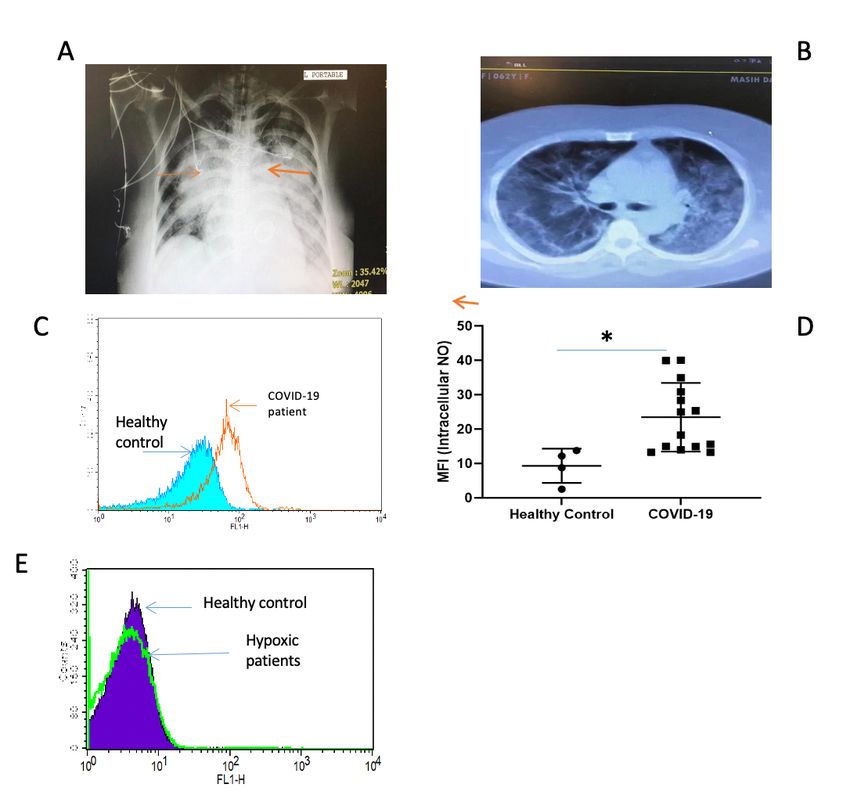
Chest X ray and CT imaging of the lungs shows signi cant changes in the lungs with bilateral alveolar
diffusion (Fig. 1 A and B).
After isolation of RBC, they were stained with DAF and intracellular NO determined. COVID-19 patients
had a greater shift re ecting higher intracellular levels of NO (Fig. 1C). The mean orescent intensity
(MFI) was calculated for each subject and plotted as a histogram (Fig. 1D). This showed a signi cant
increase in intracellular NO levels in RBCs from COVID-19 subjects (P≤0.05).
To determine whether hypoxia may be responsible for the increased levels of intracellular NO in COVID-19
patient’s RBC, we examined intracellular NO levels of RBC from hypoxic patients without COVID-19 (due
to COPD and emphysema. As depicted Fig. 1E, no signi cant increase of intracellular NO was seen in
hypoxic patients compared to non-hypoxic controls.
Discussion
We demonstrate increased levels of intracellular NO in RBC from COVOD-19 subjects. This is not due to
the presence of hypoxia per se but may afford protection against the hypoxia seen in COVID-19 patients.
During health, constitutive NO production in RBCs is largely NOS-dependent, whereas in hypoxic
conditions NO production may involve nitrite reduction by deoxyhemoglobin carbonic anydrase and/or
eNOS itself (14).
Page 4/8RBC-derived NO causes the vasodilation of small vessels in tissues allowing oxygen to be readily
released to tissues. In our study, intracellular RBC NO of COVID-19 patients is signi cantly higher than in
healthy controls and this may enable the release of oxygen to tissues resulting in the clinical
manifestation of happy hypoxia in these patients. Since NO is a pulmonary vasodilator and also as
antiviral activity against coronavirus strains it is likely that NO treatment may be effective in COVOD-19
subjects. There is no evidence that direct oxygen therapy is bene cial in the management of
breathlessness in severe COVID-19 patients but our data suggests that NO therapy may be bene cial in
COVID-19 patients with hypoxia (15).
In summary, COVID-19 patients show higher levels of NO inside RBC compared to non-COVID-19
hypoxemic patients. Whether higher levels of intracellular NO inside RBC of COVID-19 infected patients
drive the unexpected silent hypoxia phenotype needs to be examined in future clinical studies using NO
donors in hypoxemic COVID-19 patients.
Conclusions
This pilot study shows that elevated levels of intracellular NO may mask the effects of hypoxia in COVID-
19 patients which present as a ‘happy’ hypoxic state. Further studies are required to con rm this but the
data suggests that trials of NO therapy or NO donors may be useful in treating severe COVID-19 patients
with hypoxia.
List Of Abbreviations
ARDS; Acute Respiratory Distress Syndrome (ARDS)
COVID-19; coronavirus disease 2019 (COVID-19
FACS; Fluorescence-activated cell sorting
MFI; mean orescent of intensity
NO; Nitric oxide
RBC; Red blood cells
Declarations
Ethics approval and consent to participate: The study was approved by Ethical committee of Masih
Daneshvari Hospital IR.SBMU.NRITLD.REC.1399.123 and consent obtained from study participants was
written.
Consent for publication: All authors approved the submission.
Page 5/8Availability of data and materials: The data will be available upon written request.
Funding: This study was supported by the authors own funds.
Competing Interests: The authors have no con icts of interest to declare.
Authors' contributions: EM performed the experiments and initial data analysis. MM, HRJ, PANN, SMRH,
PT, MV and HZ provided the patients and samples. EGC, GF and IMA critically reviewed and revised the
manuscript. All authors reviewed the nal version and approved submission.
Acknowledgement: We acknowledge all study participants who are alive and remember those patients
who died of COVID-19.
References
1. https://www.worldometers.info/coronavirus/
2. Luciano Gattinoni1, Davide Chiumello, Pietro Caironi, Mattia Busana, Federica Romitti, Luca Brazzi
and Luigi Camporota. COVID-19 pneumonia: different respiratory treatments for different
phenotypes? Intensive Care Med https://doi.org/10.1007/s00134-020-06033-2.
3. https://www.livescience.com/silent-hypoxia-killing-covid-19-coronavirus-patients.html.
4. Miriam M. Cortese-Krott, Ana Rodriguez-Mateos, Roberto Sansone, Gunter G. C. Kuhnle, Sivatharsini
Thasian-Sivarajah, et al. . Human red blood cells at work: identi cation and visualization of
erythrocytic eNOS activity in health and disease. 2012;120(20): 4229-4237.
5. Wang M., Zhou Y., Zong Z., Liang Z., Cao Y., Tang H. A precision medicine approach to managing
2019 novel coronavirus pneumonia. Precis Clin Med. 2020;3:14–21.
6. Buerk DG. Nitric oxide regulation of microvascular oxygen. Antioxid Redox Signal. 2007;9:829–843.
7. Miriam M. Cortese-Krott and Malte Kelm. Endothelial nitric oxide synthase in red blood cells: Key to a
new erythrocrine function? Redox Biol. 2014; 2: 251–258.
8. Ellsworth, M. L., Forrester, T., Ellis, C. G. and Dietrich, H. H. (1995). The erythrocyte as a regulator of
vascular tone. Am. J. Physiol. 269, H2155-H2161.
9. Pawloski JR, Hess DT, Stamler JS. Export by red blood cells of nitric oxide bioactivity. 2001;409:622–
626.
10. Gladwin MT, Schechter AN. NO contest: nitrite versus S-nitroso-hemoglobin. Circ Res. 2004;94:851–
855.
11. Kleinbongard P, Schulz R, Rassaf T, Lauer T, Dejam A, Jax T, et al. Red blood cells express a
functional endothelial nitric oxide synthase. 2006;107:2943–2951].
12. Doctor A, Platt R, Sheram ML, Eischeid A, McMahon T, Maxey T, et al. Hemoglobin conformation
couples erythrocyte S-nitrosothiol content to O2 Proc Natl Acad Sci USA. 2005;102:5709–5714.
Page 6/813. Ulker P, Gunduz F, Meiselman HJ, Baskurt OK. Nitric oxide generated by red blood cells following
exposure to shear stress dilates isolated small mesenteric arteries under hypoxic conditions. Clin
Hemorheol Microcirc. 2013;54:357–369.
14. Zweier JL, Wang P, Samouilov A, et al. Enzyme-independent formation of nitric oxide in biological
tissues. Nat Med. 1995;1(8):804-809.
15. Jennifer Couzin-Frankel. The mystery of the pandemic's ‘happy hypoxia. Science, 368, (6490),455-
456.
Table
Table.1. Demographic information of all participants (COVID-19 and control groups)
Abbreviation used: MV: Mechanical Ventilation; NIV: Non-invasive Ventilation
In radiology: - , negative; +, Unilateral Ground glass opacity (GGO)/Consolidation, ++, Bilateral GGO/Consolidation; +++ , ARDS
G IgM RT-PCR Mortalility Radialogy Ventilation PCO2 O2/S Sex AGE
COVID-19
- - - - - 41 92/97 M 39 Control
- - - - - 40 98/97 M 65 Control
- - - - - 40 97/98 F 44 Control
- - - - - 42 94/98 F 54 Control
+ + ++ MV 56 69/86 F 80 Patient 1
+ + +++ MV 28 34/69 M 77 Patient 2
+ - ++ - 47 61/87 M 34 Patient 3
+ + ++ MV 44 26/51 F 62 Patient 4
+ + ++ MV 47 43/65 M 72 Patient 5
+ - + ++ - 63 36/63 M 69 Patient 6
+ - + ++ MV 47.5 47/82 M 60 Patient 7
+ - - ++ - 55 48/80 M 41 Patient 8
+ - - ++ MV 58.6 36/69 M 66 Patient 9
+ - - ++ - 45 98/98 M 61 Patient 10
+ - + ++ NIV 41.4 81/82.5 F 74 Patient 11
+ - - ++ - 41 40.5/74.6 F 58 Patient 12
+ - - ++ - 44 84/97 F 81 Patient 13
- + + ++ MV 49 44/78 F 21 Patient 14
Figures
Page 7/8Figure 1 (A) Representative chest X ray of a COVID-19 patient on mechanical ventilation showing bilateral consolidations (red arrows). (B) Spiral CT scan of a representative COVID-19 patient indicating multiple bilateral patchy ground glass in ltration. (C) Red blood cells were washed and preincubated with 5 mM of DAF for 20 min at 37°C in PBS containing 1% BSA, in the dark. The generation of intracellular NO was determined by FACS analysis as described in the Materials and Methods section. A representative histograph from one out of 14 COVID-19 positive patients and 4 healthy controls is shown. (D)The mean uorescent intensity (MFI) of all the subjects in each group is presented (*p
You can also read