Handling keloid and hypertrophic scars by long-pulsed Nd: YAG laser: Evaluating effectiveness
←
→
Page content transcription
If your browser does not render page correctly, please read the page content below
How to Cite:
Ramadan, H., Saber, M., Salah, M., & Samy, N. (2022). Handling keloid and hypertrophic
scars by long-pulsed Nd: YAG laser: Evaluating effectiveness. International Journal of
Health Sciences, 6(S2), 10076–10083. https://doi.org/10.53730/ijhs.v6nS2.7628

Handling keloid and hypertrophic scars by long-
pulsed Nd: YAG laser: Evaluating effectiveness

Heba Ramadan
MSc of Dermatology, Tanta, Egypt

Mahmoud Saber
MD of Surgery, Surgery Unit, Medical Application of Laser (MAL) Department,
National Institute of Laser and Enhanced Sciences, Cairo University, Giza, Egypt

Manal Salah
MD of Dermatology, Dermatology Unit, Medical Application of Laser (MAL)
Department, National Institute of Laser and Enhanced Sciences, Cairo University,
Giza, Egypt

Nevien Samy
MD of Dermatology, Dermatology Unit, Medical Application of Laser (MAL)
Department, National Institute of Laser and Enhanced Sciences, Cairo University,
Giza, Egypt

               Abstract---Background and objectives: Keloids and HTs appear to
               have an abundant healing response that sets a challenge for
               physicians. Patients and methods: Patients with keloids and HTs were
               handled by a long-pulsed 1064 nm Nd: YAG laser every 4 weeks for
               five sessions. The number of patients was twenty. The scars were
               rated by VSS, consisting of 4 ingredients: vascularity, pigmentation,
               pliability, and height. Moreover, histopathological assessment by
               hematoxylin and eosin stain and Masson trichrome stain. After 6
               months of finishing the treatment, the lesion was estimated to
               evaluate the recurrence rate. Results: According to VSS, there was a
               considerable improvement in vascularity, pigmentation, pliability, and
               height after the treatment than before the treatment. The score of the
               scar assessment dropped off from 9.40 to 3.75 after treatment.
               Hematoxylin and eosin staining and Elastica Masson-Goldner staining
               appeared to change the structure of the tissue collagen. Collagen
               bundles lost their whirl structure, and the thickness of the collagen
               layer decreased. The Wall of blood vessels was thinner, and the
               number of blood vessels was decreased. Conclusion: The treatment of
               keloids and HTs showed great results with a long-pulsed Nd-YAG
               laser.

International Journal of Health Sciences ISSN 2550-6978 E-ISSN 2550-696X © 2022.
Manuscript submitted: 27 March 2022, Manuscript revised: 18 April 2022, Accepted for publication: 9 May 2022
10076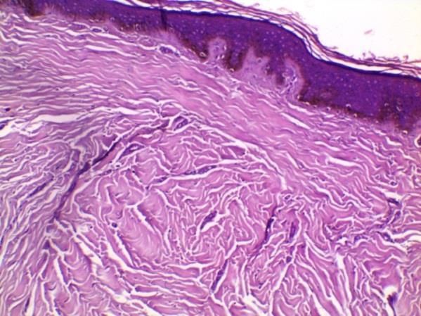
10077

           Keywords---long pulsed Nd: YAG laser, keloid, hypertrophic scar,
           vancouver scar scale.

Introduction

Hypertrophic scarring is a popular condition that manifests on a wound that rises
by deep injury, burns, or poor recovery from surgical proceedings. [1] Clinical
manifestations of the budding HTs appear that tissue is elevated than the
surrounding skin stays within the boundaries of the main wound, it is dark red or
purple in most cases, and it is firmer than normal skin in many lesions and
sometimes attached by localburning soreness and pruritus. These symptoms may
continue for many months or even years before imperceptible declination. [2]
Keloids and HTs evolve from an inappropriate equilibrium between deposition and
degeneration of extracellular matrix (ECM) ingredients, especially collagen. A
defect produces the overflowing collagen in fibroblasts production due to
increased density and activation of growth factor receptors. [3] Additionally,
transforming growth factor- beta (TGF-β) is implicated in fibroblast proliferation
and chemotaxis, collagen synthesis, and the deposition and remodeling of the
novel ECM of the wound. Normally, TGF-β activity is stopped when wound
healing is done. In keloids and HTs, TGF-β levels (especially TGF-β1 isoform) are
raised and constant. [4]

The handling process of HTs stays a troublesome unsolved problem. Numerous
therapeutic methods have been termed, including invasive and non-invasive
medication. The invasive medication includes, eg. intralesional admission of
steroid, 5- fluorouracil (5-FU), or bleomycin, cryotherapy, and laser [5]. The non-
invasive medication usually includes e.g. Pressure therapy and silicone gel. These
modalities are documented with different efficacy on HTs and keloid [6]. Non-
ablative laser processes like pulsed dye laser, Nd: YAG, and Erbium glass can also
be used in curing keloids and HTs. The Erbium glass fiber laser appears to be a
curative therapy for atrophic scars, stretch marks, and acne scars.
Oxyhemoglobin is the goal chromophore of the pulsed dye laser (PDL) that works
by making intravascular coagulation and, therefore, the devastation of the
microvascular lattice. Hypoperfusion and hypoxemia, therefore, lead to lowered
tissue expression of pro-inflammatory factors such as TGF- β1 with each other;
increase the effects of antiproliferative factors like matrix metallopeptidases
(MMP), extracellular signal-regulated kinase (EPK) and p38 kinase. The
mechanism of work of the Nd: YAG laser nearly matches that of PDL. Due to its
wavelength (1,064 nm), the depth of penetration of the Nd: YAG laser is so good,
an attribute shown to be valuable in the handling of HTs and keloids [7]. The
influence of long-pulsed Nd YAG laser in the cure of keloids and HTs generates
warmth, which initiates inflammation and lifts vascular permeability, matrix
metalloproteinase (MMP) production, and decomposition of collagen fiber
fascicle.[8]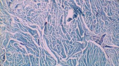
10078

Patients and Methods

Study layout

This randomized prospective study was performed in the Medical Laser Center,
the National Institution of Laser Enhanced Science, Cairo University. Twenty
patients with HTs or keloids yielded from multiple causes (burn, injury, trauma,
etc.) were involved in the study and their pre-study approval was obtained for this
work. Patients who had already been cured within the past 6 months, those
under 18 years and pregnant and lactating patients were excluded from the
study. The patients were cured with pulsed- 1064nm Nd: YAG laser. A laser
regimen of the HTs or the keloids was performed using a long-pulsed Nd: YAG
laser (1064 nm) (Cool Glide Excell; Altus Medical Burlingame, CA) on the lesion a
whole. The parameters of laser therapy were made 4 weeks apart between
sessions: the power of 45 J, the pulse duration of 5 ms, and the beam diameter of
5 to 10mm.

Clinical Estimation

The echo of the pathological scars to the medication was subjectively evaluated by
Vancouver Scar Assessment Scale (VSS), which owned four ingredients;
vascularity, pigmentation, pliability, and height. Histological Estimation:To
demonstrate the mechanism by which this treatment modality works, punch
biopsies were acquired from volunteer patients before treatment as a baseline and
after 3 days from termination of the treatment. In addition, the histopathology of
the specimens was checked up by hematoxylin and eosin staining (H&E) and
Masson trichrome staining.

Results

In this random work, twenty patients with HTs and keloids were involved in the
standing work. The average age of patients was 28.40 ± 9.79. The average lesion
duration; per month; was 3.60 ± 2.19. The scars were solitary (55%) and denovo
(85%) in many patients. According to VSS, there was a considerable improvement
in vascularity, pigmentation, pliability, and height than before the treatment. The
vascularity was normal before the treatment in 0 (0%), pink in 5(25.0%), red in 5
(25.0%) and purple in 10 (50.0%). After the treatment, the vascularity was normal
in 10(50.0%), pink in 10(50.0%), red in 0 (0%) and purple in 0 (0%). There was
significant difference (p=0.001).

Before the treatment, the pigmentation was normal in 13 (65.0%), hypopigmented
in 1 (5.0%), hyperpigmented in 1(5.0%) and mixed hyper pigmented in 5 (25.0%).
After the treatment, the pigmentation was normal in 8 (40.0%), hypopigmented in
6 (30.0%), hyperpigmented in 6(30.0%) and mixed hyperpigmented in 0 (0%).
here was a significant difference (p=0.004). Before the treatment, the pliability
was normal in 0 (0%), supple in 1(5.0%), yielding in 1 (5.0%), firm in 4 (20.0%),
ropes in 9 (45.0%) and contracture in 5 (25.0%). After the treatment, the
pliability was normal in 5 (25.0%), supple in 8(40.0%), yielding in 4 (20.0%), firm
in 3 (15.0%), ropes in 0 (0%) and contracture in 0 (0%). There was significant
difference (p=0.001). Before the treatment, the height was flat in 0(0%), < 2mm in
10079
1 (5.0%), 2-5 mm in 19 (95.0%) and > 5mm in 0 (0%). After the treatment, the
height was flat in 4(20%), < 2mm in 10 (50.0%), 2-5 mm in 6 (30.0%) and > 5mm
in 0 (0%). There was a considerable difference (p=0.001). The average value of VSS
total score pre-treatment was 9.40, and that post-treatment was 3.75, with a
significant difference (P=0.001).
Table 1: score of VSS before and after the treatment
t.
Scope Mean ± S. D p. value
test
Tot Before 4 – 14 9.4 ± 2.76
53.545 0.001*
VSS After 0 – 7 3.75 ± 2.07
Clinical Results
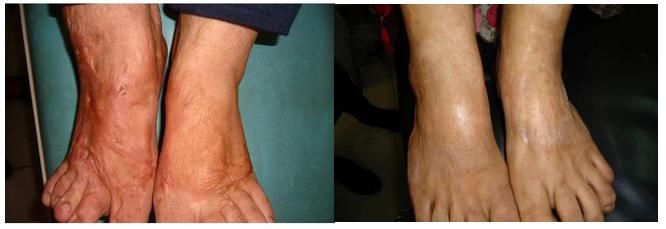
Figure 1: Keloid scar on foot of female patient showed good recovery after
treatment
Figure 2: Hypertrophic scar on female patient's chest appeared to have a partial
reduction after treatment
Histopathological result
Hematoxylin and eosin staining (H&E) and Elastica Masson-Goldner staining
(EM) of keloids and HTs explained that treatment induced a change in the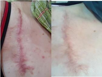
10080
structure of the collagen of tissue (illustrated in the following pictures). (From
figure 3 to 6)
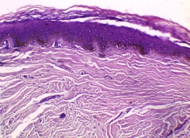
Figure 3: The histopathology of hypertrophic scar tissue before treatment is
identified by alteration of the papillary and reticular dermis by lesion tissue with
distinguishedperpendicularly oriented blood vessels. The fibrous bundles are
analogous and horizontal in the upper dermis. (H&E,200×)
Figure 4: Hypertrophic scar tissue after treatment is recognized by fewer blood
vessels, and collagen bundles become more coordinated. (H&E, 200×)
Figure 5: Hypertrophic scar tissues before treatment are identified by horizontal
fibrous bundles and parallel with distinguished vertically oriented blood vessels
(Elastica masson, 200×)10081
Figure 6: Hypertrophic scar tissue after treatment displayed that wall of blood
vessels and collagen bundles became thinner. (Elastica Masson, 200×)
Recurrence
The recurrence rate was 9 (45.0%) in a lesion evaluated after 6 months of
termination of the therapy
Figure 7: recurrence after the treatment.
Discussion
The deep-seated inflammation recognizes keloids and HTs, and capillary vessels
are massive, making erythematous scars. [9]. Pathological scar evolution is
probably caused by interactions between mechanical power capacity and
inflammation, collagen output, and angiogenesis; this proves that 1064 nm Nd:
YAG laser therapy is likely helpful in handling pathological scars. [10] There has
been a significant advance in lasers over the last three decades in the treatment
of keloids and HTs. The vascular lasers work by getting absorbed by haemoglobin
which produces heat and coagulation necrosis resulting in hypoperfusion and
tissue hypoxia; this leads to neocollagenesis. In addition, the Nd: YAG laser gets
absorbed by the haemoglobion and transforms it into black methaemoglobin,
which acts like a chromophore for the Nd: YAG laser10082
Kumar et al. [11] handled 17 keloids by Nd: YAG laser and proved that 10 scars
(58.8%) were treated, and 7 lesions (41.2%) appeared with only a little reduction.
Sherman and Rosenfeld; 1988 treated 17 keloids by Nd: YAG laser and said that
there were hopeful outcomes in all the keloids. Also, Abergel et al. [12] produced
research in 8 patients with keloids utilization Nd: YAG laser and showed perfect
outcomes. Numerous laser sessions are recommended for an excellent reaction,
but lower fluences are proposed to prevent adverse effects in patients with dark
skin. However, some authors have documented that a better tissue response
supports higher fluences. [13,14,15] In another review study by Bouzari et al., it
was concluded that various lasers are efficient in curing and the prohibition of
HTs and keloids. [16]. Akaishi S (2012) [17] said that the average total scar
estimation score improved significantly. To conclude, hopeful results have been
provided in treating keloids and HTs by the long-pulsed Nd-YAG laser.
References
1. Kant Bevan den Kerckhove E, Colla C, et al. A new treatment of hypertrophic
and keloid scars with combined triamcinolone and verapamil: a retrospective
study. Eur J Plast Surg. 2018;41: 69-80.
2. Lee DE, Ryan M, Ayoub NT. Metalloproteinases, and Vitamin D in Keloids
and Hypertrophic Scars. PRS Global Open. 2015; 16:9-12.
3. Lee G, Hunter-Smith DJ, Rozen WM. Autologous fat grafting in keloids and
hypertrophic scars: a review. Scars Burn Heal. 2017; 3:1-6.Penn JW,
Grobbelaar AO, Rolfe KJ. The role of the TGF-β family in wound healing,
burns and scarring: a review. Int J Burns Trauma. 2012; 2:18-28.
4. Espana A, Solano T, Quintanilla E. Bleomycin in the treatment of keloids and
hypertrophic scars by multiple needle punctures. Dermatol Surg. 2001; 27:
23-27.
5. Guo Feng, Wang Xi-Qiao. The Review of Most Current Therapy for Large Area
of Hypertrophic Scar and Keloid. Journal of Advanced Plastic Surgery
Research.2015;1: 29-33.
6. Poetschke J, Gauglitz GG. Current options for the treatment of pathological
scarring. JDDG. 2016; 3:6-13.
7. Akaishi S, Koike S, Dohi T, et al. Nd:YAG Laser Treatment of Keloids and
Hypertrophic Scars. Eplasty. 2012; 12:5–17.
8. Ogawa R, Akaishi S , Izumi M. Histologic analysis of keloids and hypertrophic
scars. Ann Plast Surg. 2009; 62:104–105.
9. Koike S, Akaishi S, Nagashima Y, et al. Nd:YAG Laser Treatment for Keloids
and Hypertrophic Scars: An Analysis of 102 Cases. Plast Reconstr Surg Glob
Open. 2014;2: 272-280.
10. Kumar K, Kapoor BS, Rai P, Shukla HS. In‑situ irradiation of keloid scars
with Nd:YAG laser. J Wound Care. 2000; 9:213-215.
11. Abergel RP, Dwyer RM, Meeker CA, et al. Laser treatment of keloids: A clinical
trial and an in vitro study with Nd:YAG laser. Lasers Surg Med. 1984; 4:291-
295.
12. Dierickx C, Goldman MP, Fitzpatrick RE. Laser treatment of
erythematous/hypertrophic and pigmented scars in 26 patients. Plast
Reconstr Surg. 1995; 95:84-90.
13. Goldman MP, Fitzpatrick RE. Laser treatment of scars. Dermatol Surg. 1995;
21:685- 687.10083
14. Reiken SR, Wolfort SF, Berthiaume F, et al. Control of hypertrophic scar
growth using selective photo thermolysis. Lasers Surg Med. 1997; 21:7‑12.
15. Bouzari N, Davis SC, Nouri K. Laser treatment of keloids and hypertrophic
scars. Int J Dermatol. 2007; 46:80‑88.
16. Akaishi S, Koike S, Dohi T, et al. Nd:YAG Laser Treatment of Keloids and
Hypertrophic Scars. Eplasty. 2012; 12:5-17.You can also read