Giardia intestinalis thymidine kinase is a high-affinity enzyme crucial for DNA synthesis and an exploitable target for drug discovery
←
→
Page content transcription
If your browser does not render page correctly, please read the page content below
RESEARCH ARTICLE

Giardia intestinalis thymidine kinase is a high-affinity enzyme
crucial for DNA synthesis and an exploitable target for drug
discovery
Received for publication, February 25, 2022, and in revised form, May 6, 2022 Published, Papers in Press, May 11, 2022,
https://doi.org/10.1016/j.jbc.2022.102028
Sascha Krakovka1,‡ , Farahnaz Ranjbarian2,‡, Lucas A. Luján3 , Alicia Saura3, Nicolai B. Larsen4 ,
Alejandro Jiménez-González1 , Anna Reggenti2, Hugo D. Luján3, Staffan G. Svärd1, *, and Anders Hofer2, *
From the 1Department of Cell and Molecular Biology, BMC, Uppsala University, Uppsala, Sweden; 2Department of Medical
Biochemistry and Biophysics, Umeå University, Umeå, Sweden; 3Centro de Investigación y Desarrollo en Immunología y
Enfermedades Infecciosas (CIDIE), Consejo Nacional de Investigaciones Científicas y Técnicas (CONICET)/Universidad Católica de
Córdoba (UCC), Cordoba, Argentina; 4Statens Serum Institut, København S, Denmark
Edited by Ruma Banerjee

   Giardiasis is a diarrheal disease caused by the unicellular                   adjusted life years lost (1). G. intestinalis is a noninvasive,
parasite Giardia intestinalis, for which metronidazole is the                    unicellular, and binuclear parasite, and it is spread as infectious
main treatment option. The parasite is dependent on exoge-                       cysts from drinking water and contaminated surfaces. After
nous deoxyribonucleosides for DNA replication and thus is                        passage through the stomach, the parasites excyst, forming the
also potentially vulnerable to deoxyribonucleoside analogs.                      trophozoite form that attaches to the upper small intestine and
Here, we characterized the G. intestinalis thymidine kinase, a                   starts multiplying. Some of the cells detach and move to the
divergent member of the thymidine kinase 1 family that                           lower intestines, where a stress response induces encystation.
consists of two weakly homologous parts within one poly-                         During encystation, the genome is duplicated twice, leading to
peptide. We found that the recombinantly expressed enzyme is                     the cell acquiring a total of four nuclei and 16 genome copies.
monomeric, with 100-fold higher catalytic efficiency for                          The resulting cystic form has low metabolic activity and can
thymidine compared to its second-best substrate, deoxyuridine,                   withstand diverse environmental factors like UV light, low or
and is furthermore subject to feedback inhibition by dTTP.                       high salt concentrations, and low temperatures. It is thereby
This efficient substrate discrimination is in line with the lack                  adapted to spreading between hosts (2). The symptoms of
of thymidylate synthase and dUTPase in the parasite, which                       giardiasis vary from asymptomatic infections to severe diar-
makes deoxy-UMP a dead-end product that is potentially                           rhea and abdominal cramping. In most cases, the immune
harmful if converted to deoxy-UTP. We also found that the                        system clears the parasites after 2 to 4 weeks, but sometimes
antiretroviral drug azidothymidine (AZT) was an equally good                     the infection becomes chronic with long-term effects such as
substrate as thymidine and was active against WT as well as                      food allergies, chronic fatigue syndrome, and irritable bowel
metronidazole-resistant G. intestinalis trophozoites. This drug                  syndrome. These effects can also occur in patients without
inhibited DNA synthesis in the parasite and efficiently                           detectable infection and may persist for over a decade after the
decreased cyst production in vitro, which suggests that it could                 initial disease (2, 3). Also, several recent studies have shown a
reduce infectivity. AZT also showed a good effect in                             connection between G. intestinalis infections and delayed
G. intestinalis–infected gerbils, reducing both the number of                    growth of children in middle- and low-income countries (4–6).
trophozoites in the small intestine and the number of viable                     Both the acute symptoms as well as the long-term effects
cysts in the stool. Taken together, these results suggest that the               mandate rapid treatment of the disease.
absolute dependency of the parasite on thymidine kinase for its                     There is currently no human vaccine providing protection
DNA synthesis can be exploited by AZT, which has promise as                      from G. intestinalis, and chemotherapy with metronidazole
a future medication effective against metronidazole-refractory                   and other 5-nitroimidazoles is the major treatment option.
giardiasis.                                                                      The 5-nitroimidazoles specifically target microaerophilic and
                                                                                 anaerobic cells because the active forms of these drugs are
                                                                                 rapidly inactivated in oxygen-rich environments (7). The major
   Giardia intestinalis (also known as Giardia duodenalis and                    5-nitroimidazole used is metronidazole, which has many side
Giardia lamblia) causes giardiasis and is one of the major                       effects, including nausea, vomiting, metallic taste, and head-
parasitic causes of diarrhea with 190 million symptomatic in-                    aches, and long-term use is associated with neurological
fections per year worldwide, adding up to 171,100 daily                         symptoms and reduced liver function (8, 9). In recent years,
                                                                                 G. intestinalis drug resistance rates of 20 to 40% have been
‡
                                                                                 reported in the clinic, making the search for an alternative to
  These authors contributed equally to this work.
* For correspondence: Anders Hofer, anders.hofer@umu.se; Staffan G. Svärd,       metronidazole all the more urgent (10–12). Crossresistance to
   staffan.svard@icm.uu.se.                                                      other 5-nitroimidazoles is also common and is easily inducible

                                                                                                                    J. Biol. Chem. (2022) 298(6) 102028      1
© 2022 THE AUTHORS. Published by Elsevier Inc on behalf of American Society for Biochemistry and Molecular Biology. This is an open access article under the CC
BY license (http://creativecommons.org/licenses/by/4.0/).
Giardia intestinalis thymidine kinase
in the laboratory (13, 14). This highlights the importance of                  and thymidine kinase 2. This family has a broader substrate
finding new classes of molecules with different modes of action                 specificity and includes members that phosphorylate purines
to treat giardiasis.                                                           as well as pyrimidines. One of the incentives behind the cur-
   The parasitic lifestyle has the advantage that a number of                  rent study was to investigate whether the G. intestinalis
essential metabolites are already present in the host, and                     thymidine kinase has a strict base specificity similar to other
consequently parasites often lack metabolic genes that are                     members of the TK1 family or if this could be the same
needed in free-living organisms (15). DNA biosynthesis                         enzyme as in the fraction that phosphorylated both thymidine
requires the four deoxyribonucleoside triphosphates (dNTPs)                    and deoxycytidine in the initial study (20). Interestingly, we
dCTP, dTTP, dATP, and dGTP, but G. intestinalis has neither                    found that the G. intestinalis thymidine kinase has the most
ribonucleotide reductase nor thymidylate synthase (Fig. 1),                    restrictive base specificity ever reported for any enzyme in the
which are key enzymes for de novo synthesis of dNTPs and                       TK1 family. It had a 100 times higher catalytic efficiency with
dTTP, respectively (16–18). The lack of these two enzymes                      thymidine than to the second-best natural substrate, deoxy-
makes the parasite dependent on salvaging deoxyribonucleo-                     uridine, which could be a way for the parasite to prevent
sides from the host, and this process requires membrane                        deoxyuridine triphosphate (dUTP) production and thus limit
transporters and kinases for the uptake and phosphorylation of                 the consequent misincorporation of this nucleotide into DNA.
the deoxyribonucleosides (19). The addition of the first                        We observed no detectable activity with deoxycytidine. In
phosphate is generally rate limiting and is catalyzed by                       contrast to the strict base specificity, the recognition of the
deoxyribonucleoside kinases (19). The complete dependency                      sugar moiety was more relaxed and azidothymidine (AZT) was
on deoxyribonucleoside salvage might be exploited for drug                     an efficient substrate for inhibiting parasite DNA replication,
development in two different ways, either by blocking one of                   in-vitro cell proliferation, and parasite burden in
the salvage enzymes or by using substrate analogs that upon                    G. intestinalis–infected rodents. We also found encystation to
phosphorylation mimic the natural dNTPs and act as chain                       be efficiently hindered by both AZT and metronidazole but
terminators in DNA synthesis. The dependency of the or-                        with AZT also affecting the DNA content in each cyst. Finally,
ganism on the deoxyribonucleoside kinases for normal DNA                       cell lines resistant to metronidazole and related compounds
replication precludes the ability to acquire drug resistance by                (21) showed no better tolerance toward AZT than
downregulating the enzymes.                                                    metronidazole-susceptible cell lines.
   Two deoxyribonucleoside kinase activities were previously                      All together, we show in this work that AZT appears to be a
purified and characterized from G. intestinalis (20), with one of               good drug candidate for the treatment of giardiasis by
them phosphorylating purines and the other pyrimidines.                        exploiting an essential metabolic pathway in G. intestinalis.
However, at that time the corresponding genes were not                         Moreover, it has a clear mechanism of action (inhibition of
known and subsequent genomic analyses showed that                              DNA replication) affecting cell proliferation as well as encys-
G. intestinalis has three deoxyribonucleoside genes, indicating                tation and does not show crossresistance with the main class of
that the initial studies either missed one of the kinases or that              drugs used today.
two of them were copurified. One of the G. intestinalis
deoxyribonucleoside kinase genes is from the same family as
                                                                               Results
human thymidine kinase 1 (TK1). The members of this family
typically have a restrictive base specificity only recognizing                  General description of the G. intestinalis thymidine kinase
thymidine and deoxyuridine among naturally occurring sub-                         The G. intestinalis thymidine kinase has a theoretical mo-
strates. The other two G. intestinalis genes are from the sec-                 lecular mass of 42 kDa (gene GL50803_8364 in the
ond deoxyribonucleoside kinase family, which includes three                    G. intestinalis WB strain), which is nearly double the size
human enzymes: deoxycytidine kinase, deoxyguanosine kinase,                    compared to the corresponding mammalian (TK1) and
                                                                               Escherichia coli enzymes (Fig. 2A). The large size is compa-
                                                                               rable to the corresponding enzyme from Trypanosoma brucei,
                      NTP                        dNTP           dTTP
                                                                               which is composed of two consecutive thymidine kinase se-
      De novo:                     RNR
                                                                               quences in the same polypeptide but with the first one being
      purine          NDP                        dNDP          dTDP
      pyrimidine
                                                                               inactive due to the lack of some important residues (22). The
                                                          TS
                      NMP                        dNMP          dTMP
                                                                               full-length T. brucei sequence failed to align properly with the
                                                                               other sequences and is not included in Figure 2A. The enzyme
                                                        dNKs/TK                from G. intestinalis was also found to consist of two parts, but
              bases/nucleosides                    dN           dThd           in this case, the first part seemed to be the active kinase
Figure 1. Nucleotide metabolism in G. intestinalis. The parasite lacks         (Fig. 2B). This part is 41% identical to human thymidine kinase
many pathways in nucleotide metabolism including ribonucleotide reduc-         (TK1) and contains the important residues needed for enzyme
tase (RNR) and thymidylate synthase (TS), which are crucial for general dNTP   activity (Fig. 2B). The second part has only weak homology
synthesis and dTTP synthesis, respectively. Consequently, the parasite is
completely dependent on deoxyribonucleoside kinases to make dNTPs.             with other thymidine kinases and lacks many important resi-
There are three such kinases in the G. intestinalis WB genome, including two   dues (Fig. 2B). The murine parasite Giardia muris has the
deoxyribonucleoside kinases from the dCK-dGK-TK2 family (dNKs, ORFs
4558 and 17451) and one from the thymidine kinase 1 family (TK, ORF            same setup with two consecutive TK1 domains and a more
8364).                                                                         conserved N-terminal part, whereas the related salmon

2 J. Biol. Chem. (2022) 298(6) 102028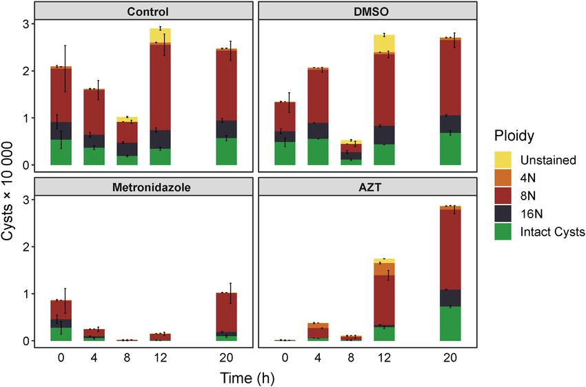
Giardia intestinalis thymidine kinase

Figure 2. Amino acid sequence alignment of thymidine kinases from the TK1 family. A, alignment overview of TK1 family members including
thymidine kinases from two Giardia species and the related parasite Spironucleus salmonicida. Green blocks represent the aligned parts with spaces showing
gaps in the alignment. B, detailed view showing the amino acid sequences from a second alignment performed with three of the thymidine kinases. The
G. intestinalis thymidine kinase sequence was split prior to the alignment into part 1 (amino acids 1–210) and part 2 (amino acids 211–392). The alignments
performed in (A) and (B) were generated with Clustal Omega, and the color code in (B) indicates positive charge (blue), negative charge (red), nonpolar
residues (orange), polar residues (green), and aromatic residues (yellow). The highlighted residues are the NTP consensus sequence needed for ATP binding,
D58, which is needed for interaction with the 30 position of the deoxyribose, R60, which is involved in the formation of the transition state, and E98, which is
needed for abstraction of the hydrogen from the 50 end of the deoxyribose in order to initiate the enzymatic reaction. All of the highlighted residues were
present in the first part of the G. intestinalis thymidine kinase, while many of them were lacking in the second part. C, phylogenetic tree of the eukaryotic
clade of the TK1 family. For a more detailed view of the phylogenetic tree and a description of the parameters used to construct the tree, see Fig. S2.

parasite Spironucleus salmonicida thymidine kinase is shorter                    part originated from a thymidine kinase. BLAST and domain
with only one TK1 domain (Figs. 2A and S1). Studies from                         searches did not pick up any other homologous genes or do-
T. brucei showed that the purpose of the inactive domain is to                   mains. A phylogenetic analysis of G. intestinalis thymidine
stabilize the protein and to help increase the affinity of the                    kinase (Figs. 2C and S2) showed that all diplomonad TK ho-
substrate for the active domain (22). In G. intestinalis, the                    mologs are grouped together in a very divergent cluster
homology between the two domains is much lower (13%                              compared with the rest of the eukaryotic taxa. The tree to-
identity), and it is therefore impossible to be sure if the second               pology and the branch support values also excluded the

                                                                                                                     J. Biol. Chem. (2022) 298(6) 102028      3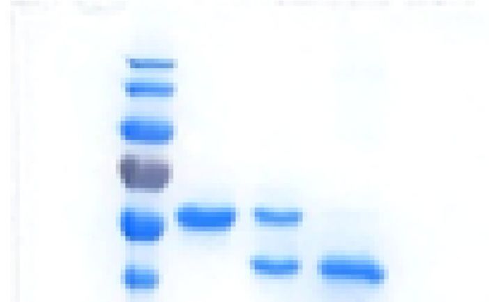
Giardia intestinalis thymidine kinase
possibility of a horizontal gene transfer from the host (98.7/100                                           human enzyme and can be viewed as pseudodimers, although
and 97.3/99, respectively), and this is an advantage for devel-                                             the active part of the enzyme is in opposite orientations in the
oping drugs that specifically target the pathogen.                                                           two cases.

                                                                                                            Phosphate donor specificity of the G. intestinalis thymidine
Cloning, expression, and general characterization of the
                                                                                                            kinase
G. intestinalis thymidine kinase
                                                                                                               The G. intestinalis thymidine kinase had a relaxed phos-
   The G. intestinalis thymidine kinase gene was cloned into a
                                                                                                            phate donor specificity giving high activities with all NTPs
pET-Z vector for expression in E. coli as a fusion construct
                                                                                                            (Table 1). ATP and GTP were the best phosphate donors as
with a cleavable His6-tagged protein Z attached to the
                                                                                                            judged by their catalytic efficiencies (kcat/Km), which were
thymidine kinase. Figure 3A shows an SDS gel from different
                                                                                                            approximately the double as compared to the corresponding
steps during the purification process yielding a homogeneous
                                                                                                            values with the pyrimidine NTPs. Although ATP had a
nontagged protein in the final step. General characterization of
                                                                                                            significantly lower kcat than the other NTPs, this was
the purified thymidine kinase showed that the enzyme activity
                                                                                                            compensated for by a low Km resulting in the high catalytic
was rather insensitive to different salts tested up to 0.2 M
                                                                                                            efficiency. The remaining experiments were performed with
(Fig. 3B) and that DTT had a slightly stimulatory effect
                                                                                                            ATP, which has both the lowest Km value and is generally the
(Fig. S3). The enzyme activity was linear with respect to time
                                                                                                            most abundant nucleotide in cells and is therefore likely to
and enzyme concentration (Fig. S4).
                                                                                                            bind the enzyme most often of the four NTPs under physio-
   Mass photometry analysis showed that the G. intestinalis
                                                                                                            logical conditions.
thymidine kinase is a monomer (Fig. 3C). This is similar to the
T. brucei thymidine kinase, which is also monomeric (22), but
different from the mammalian enzyme (TK1) that is in a                                                      Substrate specificity of the G. intestinalis thymidine kinase
dimer–tetramer equilibrium (23). These results can be                                                         The previous finding that a protein fraction purified from
explained by the polypeptide structures where both the                                                      G. intestinalis cells could phosphorylate both thymidine and
G. intestinalis and T. brucei enzymes are much larger than the                                              deoxycytidine (20) inspired us to test the thymidine kinase

         A                                                            B                                    1.5
                                                                          TK activity (μmol ∙ min ∙ mg )
                                                                          -1

               180
                                                                          -1

               130                                                                                         1.0
               100
                70
                                                                                                           0.5
                                                 HisZ-TK
                55

                40                               TK
                                                                                                           0.0
                                                                                                                      0     0.1      0.2   0.1   0.2   0.1     0.2    0.1   0.2 (M)
                35
                                                                                                                     Ctrl      KAc           NaCl        KCl           AmAc
                                                                      C                                     60
                25                               His-TEV                                                                      43
                                                                                                                            σ 16.2
                                                                                                            50         416 counts (67%)
                                                                                                                       Skewness: 0.000

                                                                                                            40
                                                                                Counts

                15                                                                                          30

                                                                                                            20

                                                                                                            10
                10
                                                                                                             0
                                                                                                                 0            200          400         600           800       1000
                                                                                                                                              Mass [kDa]

Figure 3. Purification and general characterization of the recombinantly expressed G. intestinalis thymidine kinase. A, SDS-PAGE showing the
purified fusion construct of His-tagged protein Z attached to the thymidine kinase (HisZ-TK). Lane 1 shows the full-length construct after nickel agarose
chromatography (theoretically 52 kDa), lane 2 shows the same protein after cleavage, and lane 3 shows the purified nontagged thymidine kinase
(TK, theoretically 42 kDa). The final nontagged protein was collected in the flow-through after cleavage and repurification on nickel agarose to remove
noncleaved protein, HisZ (10 kDa, not visible in the gel), and His-tagged TEV protease (27 kDa). B, the effect of different salts on thymidine kinase enzyme
activity. Standard enzyme assay conditions were used in the assays, and the thymidine concentration was 0.2 mM. The results in (B) represent the average of
three independent experiments with standard errors indicated. C, mass photometry analysis of G. intestinalis thymidine kinase showed that it is a monomer.
The measured molecular mass was close to the theoretical mass of 42 kDa. TEV, tobacco etch virus.

4 J. Biol. Chem. (2022) 298(6) 102028
Giardia intestinalis thymidine kinase Table 1 Kinetic parameters of the G. intestinalis thymidine kinase with different phosphate donors and substrates NTP Km (μM) Vmax/mg (μmol/min) kcat (s−1) kcat/Km (M−1 s−1) ATP 467 ± 29 0.93 ± 0.10 0.65 1.4 × 10³ GTP 1303 ± 78 2.41 ± 0.11 1.69 1.3 × 10³ UTP 1927 ± 269 1.66 ± 0.21 1.16 6.0 × 10² CTP 2396 ± 199 1.70 ± 0.12 1.19 5.0 × 10² Substrate Km (μM) Vmax/mg (μmol/min) kcat (s−1) kcat/Km (M−1 s−1) Thd 0.069 ± 0.007 0.98 ± 0.09 0.67 1.00 × 10⁷ dUrd 5.3 ± 1.3 1.02 ± 0.06 0.67 1.34 × 10⁵ dCyd
Giardia intestinalis thymidine kinase
A B
1.5 0.8
TK activity (μmol ∙ min ∙ mg ) 25 μM Thd
TK activity (μmol ∙ min ∙ mg )
-1
-1
50 μM Thd
0.6
-1
-1
1.0
0.4
0.5
0.2
0.0 0.0
Ctrl dTTP dUTP dCTP dGTP dATP 0 100 200 300 400 500
[dTTP] (μM)
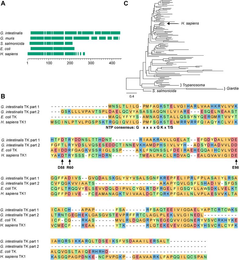
Figure 5. Inhibition of the G. intestinalis thymidine kinase activity with dTTP. A, enzyme activity measured with 200 μM thymidine as the substrate in
the presence of different dNTPs at 200 μM. B, enzyme activity with 25 μM and 50 μM thymidine and variable concentrations of dTTP as the inhibitor. The
curves in (B) were fitted by nonlinear regression to the inhibitor versus response (three parameters) function in GraphPad Prism 9.3.1 (IC50 = 30 ± 3 μM and
R2 = 0.99 for the 25 μM series; IC50 = 43 ± 1 μM and R2 = 0.96 for the 50 μM series). The graphs in (A) and (B) represent the average of three independent
experiments with standard errors, except for the 50 μM series in (B) (n = 2).
(Fig. 5B). The dTTP concentration required to cause 50% in- G. intestinalis DNA synthesis, parasites were incubated with
3
hibition was roughly equal to the substrate concentration in H-labeled deoxycytidine or deoxyadenosine and different
the two series shown in Figure 5B, indicating similar binding concentrations of AZT for 1 h. The radioactivity in DNA is an
strengths for the substrate and the regulator. indicator of the replication rate in this kind of experiment. As
shown in Figure 6, AZT had a strong impact on DNA synthesis
Antigiardial activity of AZT on WT and metronidazole- with more than 50% inhibition already at 2 μM. This con-
resistant strains centration is in a similar range as the EC50 determinations of
A proliferation assay with the WB strain of G. intestinalis G. intestinalis proliferation (Table 2), supporting the obser-
showed that AZT was nearly as efficient as metronidazole and vation that the inhibition of DNA synthesis is the major effect
that the inhibitory effect was independent of whether the cells of the drug. Figure 6 also shows that the results are similar
were resistant to metronidazole or not (Table 2 and Fig. S5). In regardless of the radioactive tracer used (deoxycytidine or
contrast, the two resistant cell lines (M1 and M2) showed clearly deoxyadenosine), which strengthens the conclusion that AZT
elevated EC50 levels in the control experiments with metroni- inhibits DNA synthesis rather than affecting deoxy-
dazole. Also included in Table 2 is an M1-derived revertant that ribonucleoside uptake or metabolism.
has lost metronidazole resistance (M1NR). This cell line was as
sensitive to both drugs as the WB parent strain. These general Efficacy of compounds against encystation
conclusions were similar regardless of the incubation time with The Uppsala encystation protocol (25) was used with
the drug. The main difference between the 48 h and 72 h time G. intestinalis cells to investigate the effect of AZT on cyst
series in Table 2 is that the general sensitivity to the drugs formation. As a final step, cysts were stored in ddH2O for
increased when the incubation was longer. The M1 and M2 cell 3 days to lyse cells that had not initiated the encystation
lines used in this study are not only resistant to metronidazole process and to open the cell membranes of immature cysts
but also against other related antigiardial compounds including to make them permeable to propidium iodide (PI), which
tinidazole, ornidazol and nitazoxanide (21). allowed measurements of their DNA contents. A second dye,
fluorescein diacetate (FDA), detects living cells and stains
Effect of AZT on G. intestinalis DNA synthesis cysts that have completed the encystation process and that
AZT is best known for its ability to block HIV reverse are impermeable to PI (FDA+, PI−). In this way, intact cysts
transcriptase, but it also inhibits mammalian DNA synthesis become clearly separated from immature cysts (FDA−, PI+)
when used at a higher dose. To test the effect of AZT on that are further categorized depending on how many times
Table 2
EC50 values in μM of metronidazole and AZT against G. intestinalis using WT (WB), metronidazole-resistant (M1 and M2), and resistance-
revertant (M1NR) cell lines
WB M1 M2 M1NR
72 h incubation
AZT 4.38 ± 0.16 3.55 ± 0.13 1.63 ± 0.17 4.58 ± 0.34
Metronidazole 2.41 ± 0.24 (4.6%) 13.7 ± 1.5 9.79 ± 0.24 1.94 ± 0.28
48 h incubation
AZT 6.0 ± 1.8 8.40 ± 0.23 3.9 ± 1.6 6.52 ± 0.87
Metronidazole 5.36 ± 0.27 (10%) 30.9 ± 4.6 14.3 ± 3.2 3.70 ± 0.21
The numbers represent the average of three independent experiments with standard errors indicated.
6 J. Biol. Chem. (2022) 298(6) 102028Giardia intestinalis thymidine kinase
A B
100 100
DNA synthesis (% of control)
DNA synthesis (% of control)
50 50
0 0
0 5 10 0 5 10 15 20
[AZT] (μM) [AZT] (μM)
Figure 6. Inhibition of G. intestinalis DNA synthesis with different concentration of AZT. A, the parasites were incubated with 10.5 nM 3H-dCyd and
various concentrations of AZT for 1 h under normal growth conditions. B, similar to (A) but with 13 nM 3H-dAdo as the radioactive tracer and including 2 μM
deoxycoformycin to protect dAdo from deamination. The recorded radioactivity in cellular DNA is shown as the percentage compared to the activity in cells
treated without AZT. The results are based on three and two independent experiments for dCyd and dAdo, respectively, with standard errors indicated. AZT,
azidothymidine.
the genome has been amplified (Fig. S6). In our AZT ex- solvent (dimethyl sulfoxide [DMSO]) as in the drug-treated
periments, the drug was added at certain time points during cells. There was little variation in the number of cysts or
the encystation process, and the results were compared with the staining pattern between the two controls apart from a
similar experiments performed with metronidazole. We also clear dip around 8 h (Fig. 7). At this time point, cells un-
included two controls, one with pure encystation medium dergo membrane reorganization and are particularly sensitive
and one that contained a similar final concentration of the to any disturbance (25). Only at the time points of 8 h and
Figure 7. The effect of AZT and metronidazole on G. intestinalis cyst formation and total cell numbers. Treatment with pure encystation medium
(control), DMSO, AZT, or metronidazole started at different time points during the encystation process. After completion of the encystation period (28 h)
and subsequent treatment, the cells were categorized based on their staining with FDA (measuring metabolic activity) and PI (measuring DNA content). The
PI staining requires a leaky membrane, and different genome amplification levels could therefore not be determined in the intact FDA+ cysts. Numbers
given are normalized to be per 100,000 seeded trophozoites. Intact cysts (FDA+, PI−) are colored green, whereas the PI+ cysts (nearly all of them FDA−) are
categorized according to their ploidy into 4N (orange), 8N (red), and 16N (black). Cysts that did not stain at all are marked in yellow. AZT, azidothymidine;
DMSO, dimethyl sulfoxide; FDA, fluorescein diacetate; PI, propidium iodide.
J. Biol. Chem. (2022) 298(6) 102028 7Giardia intestinalis thymidine kinase 12 h could cells that were not labeled with either dye be therefore became partially categorized as 4N (Figs. 8 and S8). detected at significant levels. These cells either did not These results suggest that the genome amplification to 8N contain DNA or had intact membranes but lacked FDA- was incomplete, which is in line with the mechanism of AZT activating esterases. When comparing AZT-treated cells as a chain terminator of DNA synthesis creating sequence with the two controls, a strong effect was observed if AZT gaps and thereby lowering the DNA content per cell. This was added early during encystation (0–8 h), with almost explains the decreased effect of AZT at later time points complete removal of cysts when treating immediately upon when DNA synthesis is almost completed. induction of encystation (0 h). This is in good agreement Experiments with metronidazole showed a different kind of with the experiments in Figure 6 showing that AZT blocked pattern, and cell numbers were reduced substantially at all DNA synthesis. Another observation was the increased per- time points (Fig. 7). However, the effect was less pronounced centage of cysts with lower genome duplication levels compared to AZT at 0 h and there was no peak shift to the left (notably 4N) when comparing controls to AZT-treated cells (Figs. 8 and S8). This is in line with the respective mode of at all time points except 20 h when most DNA synthesis had drug action in which metronidazole kills anaerobic cells been completed prior to the addition of the drug (Fig. S7). directly via radical induced damage, whereas AZT blocks DNA Further analysis showed that the increased percentage of 4N replication and thereby prevents proliferation and proper cells was because the 8N peak shifted to the left and encystation. Figure 8. PI signals at different time points in the cysts after control, AZT, and metronidazole treatments. Cysts were recovered from cells treated at different time points during encystation with AZT, metronidazole, or pure encystation medium with the PI-staining indicated. The resulting fluorescence profiles were overlaid upon each other and show a clear reduction of cell numbers for both drugs at early time points and a pronounced shift of PI signals from AZT at all but the last time point. The control is in red, AZT treatment is in blue, and metronidazole treatment is in gray. AZT, azidothymidine; PI, propidium iodide. 8 J. Biol. Chem. (2022) 298(6) 102028
Giardia intestinalis thymidine kinase
Antigiardial activity of AZT in a rodent model                                      parasite burden and a drastically reduced period with symp-
   The gerbil model of acute giardiasis was used to assess the                      toms (Fig. 9D) as compared to the control gerbils (Fig. 9A).
activity of AZT on three different G. intestinalis cell lines,                      The numbers of cysts were also drastically reduced upon
including two assemblage A clones (WB-1267 and WB-417)                              treatment, albeit with a delay of a few days (Fig. S9D). The
expressing VSP1267 and VSP414, respectively, and one                                infection gradually leveled off over time also in the control
assemblage B clone (GS/M-H7) expressing VSPH7. Six-week-                            animals without treatment, which is typical for self-healing
old male and female gerbils were orogastrically inoculated with                     infections, but in the case of the AZT-treated gerbils, the ef-
5 × 10⁵ trophozoites of each of the three clones, and the                           fect was much faster and the symptomatic period much
magnitude of infection was verified either by counting the                           shorter. There was no obvious difference between the effects of
trophozoites collected from the upper portions of the gerbils’                      AZT and metronidazole, and both drugs seemed equally effi-
small intestine or by quantification of cysts in the stools over a                   cient in reducing parasitemia and cyst formation (Figs. 9, B–E
30 day period, as previously reported (26). Oral administration                     and S9, B–E).
of 40 mg/kg AZT (treated), 25 mg/kg of metronidazole
(treated control), or PBS alone (untreated controls) for 3 days                     Discussion
as a pretreatment before the infection showed a marked effect                          This work provides insights into the biological role of the
on the infectivity of each of the clones compared to the un-                        G. intestinalis thymidine kinase and how its substrate selectivity
treated controls (Fig. 9, A–C). The number of trophozoites                          can be exploited for drug discovery (Fig. 10). One of the most
within the intestine and the number of cysts in the stool were                      striking findings when studying the isolated enzyme was the
negligible in the animals treated with AZT or metronidazole                         large difference in affinity for deoxyuridine in comparison to
(Figs. 9, B and C and S9, B and C). In experiments where the 3                      thymidine. The G. intestinalis enzyme had a catalytic efficiency
day AZT treatment was instead started at the peak of infection                      that was 100 times higher with thymidine compared to
(day 10 postinfection), this resulted in a rapid decrease in                        deoxyuridine. For comparison, there is only a 14-fold difference

           A                   10                                                    B                     10
                                                                                                                          C            10

                                                                                                                              Trophozoites × 10 6
                               8                                                                           8
         Trophozoites × 10 6

                                                                                     Trophozoites × 10 6

                               6                                                                           6

                                                                                                                                                     Metr.
                               4                                                                           4

                                                                                                                                              0
                               2                                                                           2        AZT                                 0                Time (d.p.i.)        30
                                                 symptoms
                               0                                                                           0
                                    0   5       10        15         20   25   30                                         0                         5        10          15      20      25   30
                                                     Time (d.p.i.)                                                                                           Time (d.p.i.)

           D                   10               AZT                                  E                     10                                       metronidazole

                               8
         Trophozoites × 10 6

                                                                                     Trophozoites × 10 6

                               6
                                                                                                            5
                               4

                               2
                                            symptoms
                               0                                                                            0
                                    0   5       10        15         20   25   30                               0             5                         10          15        20         25   30
                                                     Time (d.p.i.)                                                                                           Time (d.p.i.)
Figure 9. AZT treatment of G. intestinalis–infected gerbils. A, the effect of PBS (control) on the number of trophozoites in the intestines of gerbils
infected with the different G. intestinalis strains GS/M-H7 (●), WB-417 (▴), or WB-1267 (■). B and C, the number of intestinal trophozoites in gerbils treated
with AZT or metronidazole for 3 days prior to the infection. D, the number of intestinal trophozoites in gerbils treated with AZT for 3 days starting at day 10.
E, similar to (D) but with metronidazole treatment. The period of symptoms is also shown in (A) and (D) with a more detailed summary of symptoms in
Table S1. AZT, azidothymidine.

                                                                                                                                                                  J. Biol. Chem. (2022) 298(6) 102028   9
Giardia intestinalis thymidine kinase
A major purpose of the current study was to find an alternative
class of medicines for G. intestinalis infections. Our search was
DNA synthesis Chain terminaon prompted by the realization that there are only two classes of first-
line drugs targeting this parasite: 5-nitroimidazoles (including
metronidazole) and the closely related benzimidazole albenda-
dUTP dTTP AZT-TP
zole (28). The close relation of the two main drug classes against
giardiasis raises the concern of multidrug resistance, especially
based on the recent reports of up to 40% treatment failures by
metronidazole (10). The only other approved drugs, paromo-
dUMP dTMP AZT-MP mycin and quinacrine, have been second lined due to their side
effects, costs, and administration difficulties making them unus-
Thymidine kinase able in resource-poor settings where giardiasis is most severe (28).
An ideal drug against G. intestinalis should fulfill the following
dUrd dThd AZT
criteria:
Figure 10. Role of G. intestinalis thymidine kinase in parasite DNA
synthesis and drug discovery. The widths of the arrows indicate which (1) Strong effect against the parasite and prevention of viable
substrates are preferred by the thymidine kinase, with thymidine and AZT cyst formation and thereby transmission.
being much more efficient than deoxyuridine. Thymidine phosphorylation
leads to the formation of dTTP, which is used as a building block for DNA (2) Easy form of administration to facilitate intake in low-
replication, whereas AZT is a drug that is metabolized in a similar manner as resource settings.
thymidine but acts as a chain terminator in DNA synthesis. The figure also (3) Good safety profile, which means few and well-
shows two enzymes missing in G. intestinalis, dUTPase (dUTP → dUMP) and
thymidylate synthase (dUMP → dTMP). AZT, azidothymidine. characterized side effects.
(4) Low costs.
between the two substrates with the mammalian thymidine ki- The last two points are hard to combine when developing an
nase (TK1) (23). The main difference between the two enzymes antiparasitic drug from scratch (29), and we were therefore
is the affinity for thymidine, as indicated by their Km values of focusing on drugs that have been explored for other diseases
0.07 μM and 0.5 μM, respectively. The stronger discrimination (30). When looking for new treatment options for a given dis-
between thymidine and deoxyuridine in the G.intestinalis ease, it is important to keep in mind in which setting the disease
enzyme is just a consequence of its higher thymidine affinity. is prevalent. Giardiasis is a disease infecting disproportionally
This could be an adaption to be able to grow under conditions more people in the developing world (1), so treatment options
with low thymidine concentrations, but also to survive in the have to be inexpensive if they are to be widely used. Clinical trials
absence of dUTPase, an enzyme that the parasite lacks. The represent a major cost associated with drug development (31),
incorporation of dUTP into DNA is a major cause of abasic sites and thus, it is advisable to establish first whether a drug that has
created by base excision repair (27), and dUTPase is therefore been developed and approved by the Food and Drug Adminis-
needed in most organisms. G. intestinalis is a special case tration for another purpose can be used for the condition instead
because it lacks ribonucleotide reductase making phosphory- (30). In lower-resource settings non-orally administered drugs
lation of deoxyuridine the only source of dUTP. It is also can be harder to promote, thus putting extra emphasis on
important to remember that deoxyuridine monophosphate selecting a drug that can be taken orally. Keeping the low-
(dUMP) formed in G. intestinalis is a dead-end product. In most resource environment in mind, the drug should not require
other organisms, dUMP is the substrate of thymidylate synthase cold storage if possible.
and can thereby be channeled into dTTP production. We set out to identify a potential drug that fulfills these
G. intestinalis lacks this enzyme and cannot use dUMP. Hence, it requirements and found that the well-established anti-HIV
is logical that the enzyme has developed a much stronger drug zidovudine (AZT) was a good substrate for the
selectivity of thymidine over deoxyuridine. G. intestinalis thymidine kinase and efficiently inhibited
The G. intestinalis thymidine kinase and other members parasite DNA synthesis and proliferation. AZT is a thymidine
from the TK1 family are regulated by dTTP, which binds to analog with an azido group replacing the 30 OH group, and it
the active site and competes with thymidine. This kind of remains a choice in HIV treatment today, usually as part of a
regulation seems counterintuitive from a single cell perspec- combination treatment (32). Giardia infections were
tive because it decreases inhibition when there is plenty of frequently seen during the early stages of the HIV pandemic,
thymidine and would thus not seem optimal for controlling but after introduction of combination therapies containing
the intracellular concentration of dTTP at a stable level. We AZT the levels decreased (33). AZT is also widely used for HIV
therefore think the main benefit of this regulation is to save treatment during pregnancy and during birth to prevent
thymidine for the neighboring cells when there are limited mother-to-child transmission (34). The drug is administered
amounts of thymidine in the surroundings. For G. intestinalis, orally, and peak plasma levels as high as 2 to 18 μM are
which is a single-cell organism, it could be an advantage to generally well tolerated (35, 36). Short-term dosing studies
spread the thymidine equally between cells of the same clone. showed that serum concentrations up to 133 μM were safely
10 J. Biol. Chem. (2022) 298(6) 102028Giardia intestinalis thymidine kinase
reached for 48 h, indicating that the therapeutic window is the LGX substitution model. Branch supports were
large (37). Heavy side effects seem limited to long-term use, assessed using ultrafast bootstrap approximation (UFboot)
combination use, and use of AZT in patients with advanced (44) with 1000 bootstrap replicates and SH-like approxi-
HIV-1. In those settings, effects like hematologic toxicity, mate likelihood ratio test (SH-aLRT) (45), for which 1000
myopathy, and hypersensitivity can be observed. However, replicates were used.
over short-term use only mild anemia, headaches, nausea, and
other sorts of intestinal discomfort have been reported. Given Cloning of the G. intestinalis thymidine kinase gene for
that our studies in gerbils showed that the infection resolved expression in E. coli
with a 3-day treatment schedule, long-term side effects will
most likely not be of concern in the treatment of giardiasis The identified G. intestinalis thymidine kinase gene was from
with AZT (38). Median costs during treatment for HIV are assemblage A isolate WB (GL50803_8364) in the Giardia
80 USD per month with the drug being taken twice daily, genomic resource (https://giardiadb.org/giardiadb/). The gene
which corresponds to a price of 1.3 USD per 300 mg tablet was amplified from genomic DNA using the following primers:
(https://apps.who.int/iris/bitstream/handle/10665/104451/97 50 - GCC TCA TGA ACT CCC TTA CGC TCA TAC TGG
89241506755_eng.pdf?sequence=1&isAllowed=y). -30 and 50 -CAT GCT GGT ACC TCA CAG GTC TTC GTT GAT
We conclude that AZT is a potential alternative to metro- CCC TAG CA-30 . To enable cloning, the primers contained the
nidazole because it fulfills all the criteria that we established restriction sites BspH1 and Acc65I, respectively (bold-faced). The
initially for an antigiardial drug to be successful. AZT has a amplified thymidine kinase sequence was cloned into the
different mode of action compared to the 5-nitroimidazoles, expression vector pET-Z. The resulting vector, pET-ZTK, con-
and it efficiently kills the trophozoites of G. intestinalis both tained the 10 kDa fusion partner Z (Z domain of staphylococcal
in vitro and in vivo at a concentration comparable to metro- protein A), a His tag, and a tobacco etch virus (TEV) site prior to
nidazole. AZT also reduces the number of cysts formed the inserted gene. Positive clones were confirmed by DNA
in vitro and in vivo. Furthermore, experiments on cell lines sequencing from both directions of the insert.
resistant to metronidazole, tinidazole, ornidazole and nita-
zoxanide showed that there is no indication for crossresistance Protein expression in E. coli and purification
with metronidazole and related compounds. AZT has the Food The pET-ZTK vector was introduced into E. coli BL21(DE3)
and Drug Administration approval and is priced rather cheaply plys, and the bacteria were grown in 1 l LB medium supple-
due to its widespread use as an anti-HIV medicine. The mented with 50 μg/ml kanamycin and 34 μg/ml chloram-
common use also gives us a rich body of information on side phenicol at 37 C. Expression of the G. intestinalis thymidine
effects, which are expected to be low with the short treatment kinase was induced at A600 ’0.6 with 0.5 mM IPTG overnight at
period proposed here. Another positive aspect of repurposing 18 C. The cells were harvested by centrifugation at 4000g for
an existing drug is the existing production and distribution 15 min. The pellet was washed with 20 mM Tris–HCl pH 7.5
lines reaching many low-resource settings. Finally, the drug and centrifuged at 4000g for 10 min. The supernatant was
can be taken orally and stored at room temperature (RT), thus discarded, and the pellet was resuspended in lysis buffer
enabling its administration in most places without additional (20 mM Tris–HCl pH 7.5, 0.4 M NaCl, and 0.1 mM PMSF). The
equipment or education. In conclusion, AZT appears to be a volume used was then 10 ml/g of pellet. The cell suspension was
promising candidate to treat both 5-nitroimidazole refractory frozen and thawed three times to lyse the cells followed by
and nonrefractory giardiasis. centrifugation at 40,000 rpm for 45 min (Beckman L-90 ultra-
centrifuge Ti70 rotor). The supernatant was loaded onto a 2 ml
Millipore Ni-NTA His⋅Bind resin column (Merck). The puri-
Experimental procedures
fication steps were as follows. The column was first washed with
Phylogenetic analyses 20 mM Tris–HCL pH 7.5, 0.4 M NaCl, and 20 mM imidazole.
G. intestinalis assemblage A isolate WB thymidine kinase The second wash solution was similar but with 50 mM imid-
was used as a BLASTp query against the NCBI nr database azole. The Z-tagged thymidine kinase was finally eluted with
(June 2020), keeping 500 hits with e-valuesGiardia intestinalis thymidine kinase
into 50 to 100 μl fractions because of its tendency to aggregate KOH per liter, solution B contained only 7% methanol, and
after several freeze-thaw cycles. solution C contained 3.52 g/l tetrabutylammonium bromide in
7% methanol. The 10× loading buffer was prepared by mixing
Thymidine kinase assay 500 μl 4× solution C (1.41 g tetrabutylammonium bromide in
G. intestinalis thymidine kinase was incubated in 50 μl of 100 ml water) with 500 μl solution A. The column was run at
buffer containing [50 -3H]-labeled thymidine (Moravek Bio- ambient temperature and equilibrated with 11% solution A,
chemicals), 2 mM ATP, 5 mM MgCl2, 0.5 mM DTT, 100 mM 69% solution B, and 20% solution C. The flow rate was 1 ml/
potassium acetate, and 50 mM Tris–HCl, pH 7.5, at 37 C for min, the loop size was 100 μl, and the UV detector was set at
30 min. In one sample for each concentration of thymidine 270 nm. The deoxyribonucleoside substrates and deoxy-
measured, the enzyme was omitted to determine background nucleoside monophosphate (dNMP) products were eluted
counts. In experiments where we replaced ATP with different isocratically under these conditions. After the last peak of in-
concentrations of phosphate donors (CTP, UTP, GTP, and terest, deoxyadenosine monophosphate (dAMP) had eluted at
ATP), the MgCl2 concentration was increased to 10 mM. After 20 min; the composition was changed to a mixture of 70% A,
the enzyme reaction was completed, the samples were incu- 10% B, and 20% C to elute ATP before returning to initial
bated at 100 C for 2 min to stop the reaction. The remaining conditions. A 10 min equilibration step was included between
procedure was similar to what was described previously (46). the runs. The dNMPs were quantified by comparing the peak
The reaction mix was spotted onto two Whatman DE81 filters heights to a standard with known concentrations and multi-
from GE Healthcare (20 μl was spotted on each filter). Because plied by 10 to compensate for the previous dilution step.
this product was discontinued, we could only use it initially
and in later assays we replaced it with Amersham Hybond-XL Mass photometry analysis
(GE Healthcare), which we cut into 15 × 15 mm squares. The Analyses were performed on a Refeyn 2MP mass photometer
highest usable concentration of phosphate donor to avoid (Refeyn Ltd) calibrated with NativeMark Unstained Protein
saturation of the filter was 5 mM, but it was possible to also Standard (Thermo Fisher Scientific). The thymidine kinase
use up to 10 mM if the sample was diluted twofold prior to was mixed with PBS giving a final protein concentration of
spotting. After applying the samples, the filters were dried for 50 nM prior to analysis.
at least half an hour in darkness to minimize light-induced
background counts. Subsequently, the filters were put into
Cell maintenance of parasites used for in vitro assays
separate scintillation vials, washed three times with 5 ml of
1 mM ammonium formate with agitation for 5 min each time The following G. intestinalis isolates, which were induced
(decanting the liquid after each wash). The final wash was with for in vitro resistance toward metronidazole according to
5 ml EtOH, and after decanting the liquid, we added 1 ml of a Tejman-Yarden et al. (21), were used in the in vitro assays: WB
solution containing 0.1 M HCl and 0.1 M KCl and vortexed (ATCC 50803) clone C6/A11 (WB) and the recently generated
the tubes for 20 s. Finally, 5 ml scintillation liquid was added strains WB-C6/A11-M1 (M1), WB-C6/A11-M2 (M2), and
and the samples were mixed by shaking the tubes before WB-C6/A11-M1NR (M1NR). Cells were cultured in TYI-S-33
radioactivity counting. Similar results were obtained on both medium as described elsewhere (48). The metronidazole-
types of filters. resistant strains were continuously maintained in medium
An alternative way of measuring the thymidine kinase ac- supplemented with metronidazole, which was removed in the
tivity was used in the five-substrate assay, where thymidine, last passage (24 h) before the experiments.
deoxyuridine, deoxycytidine, deoxyadenosine, and deoxy-
guanosine were present simultaneously. In this case, the sub- Drug susceptibility assays
strates were nonradioactive and the amount of product was To establish drug efficiency, EC50 values were determined.
measured by HPLC. The HPLC protocol is a variant of a Briefly, cells were counted and diluted to 1 × 10⁴ cells/ml for
previously described protocol for nucleoside triphosphates metronidazole-sensitive cells and 2 × 10⁴ cells/ml for the
(47) but with a lower concentration of phosphate in the mobile resistant lines and seeded in 40 μl TYI-S-33 per well in 96-well
phase in order to optimize it for the study of monophosphates plates. These values were adjusted to 5 × 10³ cells/ml for
instead of triphosphates and with methanol instead of aceto- sensitive cells and 1 × 10⁴ cells/ml for resistant lines when
nitrile in order to create larger spacing between the peaks. The incubating for 72 h. The 96-well plates were inoculated
assay itself was performed as described previously and anaerobically at 37 C for 2 h to allow the cells to attach. Drugs
included 200 μM of each of the five substrates and 100 ng G were diluted in growth medium from 200 mM stocks in
intestinalis thymidine kinase. After the inactivation step DMSO to their final concentrations. After 2 h, 160 μl TYI-S33
(boiling for 2 min), 20 μl of the reaction mixture was diluted containing the drug at concentrations of 1 to 50 μM for AZT
nine times and mixed with 10× loading buffer (described later). and 1 to 200 μM for metronidazole were added. For every drug
The mixture was loaded onto a 150 mm × 4.6 mm SunShell concentration a control well with corresponding DMSO di-
C18-WP column from ChromaNik Technologies Inc. The lutions was run to correct for solvent effects. Plates were
mobile phase was a ternary mixture of three solutions; A, B, incubated for 48 h or 72 h anaerobically at 37 C before adding
and C. Solution A contained 23 g/l potassium phosphate in 7% 50 μl CellTiter-Glo reagent (Promega). The plates were mixed
(v/v) methanol and was adjusted to pH 5.6 with 1.27 ml 4 M by shaking for 10 min, the mixture was allowed to settle for
12 J. Biol. Chem. (2022) 298(6) 102028Giardia intestinalis thymidine kinase
10 min, and the fluorescence was read out in an Infinite M200 5 days. Cysts were live-stained/dead-stained with FDA and PI
Pro (Tecan Group, Ltd). EC50 values were determined by similarly to what was described for G. muris cysts (53). FDA
fitting the data to Log inhibitor versus Response curves (four was dissolved into a stock solution of 25 mM in acetone and
parameters, variable slope) using GraphPad Prism 9.3.1 kept in the dark. Immediately before incubation, it was diluted
(GraphPad Software Inc). Standard errors were then deter- to a working solution by adding 80 μl to 10 ml PBS pH 6. The
mined as the variation between the individual EC50 values cysts were spun down, resuspended in 60 μl PBS, and stained
from three or four independent experiments. by the addition of 40 μl PI Ready Flow Reagent (Thermo Fisher
Scientific) and 100 μl FDA working solution. Cells were
Inhibition of DNA synthesis incubated for 30 min at RT in the dark before counting them
in a MACSQuant VYB cell counter (Miltenyi Biotec Inc) using
G. intestinalis trophozoites at 70% confluence were either
the blue laser (488 nm) and B1 filter (525 nm/50 nm) for FDA
incubated with 2 μCi [5-3H(N)]-deoxycytidine (10.5 nM) or a
and the yellow laser (561 nm) and Y2 filter (615 nm/20 nm) for
combination of 4 μCi [2,8-3H]-deoxyadenosine (13 nM)
PI. For each drug, three biological replicates were analyzed,
(Moravek Biochemicals) and 2 μM deoxycoformycin in the
and half of the total 200 μl volume of each sample was
presence of various concentrations of AZT in a 10 cm culture
counted. Typical staining and graphs are shown in Fig. S6.
dish containing 10 ml cell culture medium for 1 h. The culture
medium was aspirated after 1 h anaerobic incubation at 37 C,
and the cells were washed with PBS before the addition of Parasites used in animal experiments
500 μl ice cold 10% (w/v) trichloroacetic acid (TCA) con- G. intestinalis WB (ATCC 50803) and GS/M (ATCC 50581)
taining 15 mM MgCl2 (referred to as TCA-MgCl2 solution). clones derived from the WB and GS strains, respectively, were
The cells were disintegrated with a cell scraper, and the DNA cultured at 37 C in TYI-S-33 medium in 14 ml borosilicate
was recovered essentially as described previously (49). The screw-cap glass tubes (54). Giardia clones expressing different
solution was centrifuged for 1 min at 21,000g, and the super- surface antigens were obtained by limiting dilution using
natant was discarded. The pellet was washed with 200 μl TCA- specific anti-VSP mAbs (54). Reactive clones were expanded in
MgCl2 solution, and after aspiration of the washing solution, culture medium overnight and controlled for homogeneity
the pellet was resuspended in 500 μl of 0.6 M NaOH and before use. WB clones 1267 (mAb 5C1), 417 (mAb 7C2), and
incubated overnight at 37 C. Subsequently, 400 μl TCA was GS/M-H7 (mAb G10/4) were employed in control experi-
added and the resulting solution was filtered through a 24 mm ments and treatments.
Whatman GF/C 24 filter (GE healthcare). The filter was put in
a vial, 5 ml scintillation liquid was added, and the mixture was Animal experiments—ethics and general maintenance
vortexed for 30 s before counting in a scintillation counter to
The animal studies were approved by the ethics committee
measure the radioactivity in the DNA.
in Cordoba, Argentina. All procedures performed on animals
were conducted following protocols specifically approved by
Inhibition of encystation the Institutional Committee for Care and Use of Experimental
Encystation inhibition assays were performed in triplicate in Animals (protocol 2010-36-15p). These protocols adhered to
3 ml Nunc cell culture tubes #156758 (Thermo Fisher Scien- the US PHS guidelines for animal research, and no animals
tific). Cells of the WB line were counted in a hemacytometer were harmed during the collection of blood, fecal samples, or
and diluted to 2.5 × 10⁵ cells. They were then incubated in intestinal contents.
TYI-S-33 at 37 C for 3 h to allow them to attach again before Specific pathogen-free, 6-week-old outbred gerbils (Mer-
the medium was changed to encystation medium according to iones unguiculatus) of both sexes were used (Animal Facility
the Uppsala encystation protocol (25). At five time points post CIDIE CONICET/UCC). They were housed in air-conditioned
encystation induction (0 h, 4 h, 8 h, 12 h, or 20 h), drugs were (18–22 C, 40–50% humidity) racks with a 12 h light–dark
added in 10 μl DMSO at a final concentration of 53 μM cycle. Gerbils were fed ad libitum with autoclaved food and
(metronidazole) and 60 μM (AZT), and the tubes were sterile water supplemented with a mixture of filter-sterilized
inverted several times to ensure distribution of the drug. vitamin solution. Before infection, gerbils were tested for the
Control tubes, to which nothing was added, were opened, presence of serum antibodies against Giardia antigens by
closed, and inverted for the same amount of time as the other ELISA and for Giardia cysts in stool samples by light and
tubes. For the DMSO control, 10 μl of sterile DMSO was immunofluorescence microscopy using cyst-specific mAb
added. The concentrations of the selected drugs were chosen 7D6, as previously reported (55). In infection and treatment
to be 10× the EC50 at 48 h based on the results of Hausen et al. experiments where the number of trophozoites was counted,
(50) for metronidazole, where they showed a clear effect when each experimental series required ten gerbils with one animal
added early but not late during encystation at that concen- sacrificed for each data point (a total of ten time points per
tration, as well as the reports of peak serum concentrations for series), whereas in the experiments where cysts were counted,
common dosage regimens between 30 and 70 μM (51, 52). only one gerbil per condition and G. intestinalis strain was
After 28 h incubation at 37 C, the cells were harvested by required. There were totally five different conditions
centrifuging at 800g for 8 min at 4 C, washed twice with (PBS control and two conditions with each drug),
sterile H2O, and incubated in sterile H2O at 4 C for 3 to three G. intestinalis strains for each condition, and three
J. Biol. Chem. (2022) 298(6) 102028 13Giardia intestinalis thymidine kinase
independent series. The experiments where cysts were coun- Data availability
ted had a similar setup but required much fewer animals All data are contained in the main article and supporting
because each one could be used for all ten time points. In a material.
separate study to monitor symptoms, three conditions (treat-
ment with PBS and two AZT treatment regimens) were tested
on gerbils infected with each of the three G. intestinalis strains Supporting information—This article contains supporting
and on uninfected animals. Also in this case, the data were information.
based on three independent experiments.
Acknowledgments—Phylogenetic analyses were performed on
resources provided by the Swedish National Infrastructure for
Animal experiments—infection and treatment Computing (SNIC) at Uppsala Multidisciplinary Center for
Advanced Computational Science (UPPMAX).
Infections
Gerbil infections were induced by orogastric inoculation of Author contributions—S. K. and F. R. methodology; S. K., F. R., L. A. L.,
2 × 10⁵ trophozoites resuspended in 0.5 ml PBS (55). Control and A. H. validation; S. K., F. R., N. B. L., L. A. L., A. S., A. J.-G., and
H. D. L. formal analysis; S. K., F. R., N. B. L., L. A. L., A. S., A. J.-G.,
gerbils received 0.5 ml PBS by the same route. Feces were
and H. D. L. investigation; S. K. data curation; S. K. and A. H.
collected every day from week 0 to 4. Cysts were identified
writing–original draft; S. K., F. R., H. D. L., S. G. S., and A. H.
visually by light microscopy and by immunofluorescence assays writing–review and editing; S. K., F. R., and A. H. visualization; H. D. L.,
with mAb 7D6. Giardia cysts excreted by gerbils were quantified S. G. S., and A. H. supervision; H. D. L., S. G. S., and A. H. funding
by collecting stool pellets from individually housed gerbils over a acquisition.
24 h period. The stool samples were weighed, resuspended in
2 ml of PBS, and filtered. The filtrate was centrifuged at 250g for Funding and additional information—This work was supported by
10 min. After three washes, the pellet was suspended in 2 ml PBS the Swedish Research Council (2019-01242 to A. H. and 2018-
and cysts were stained and counted in a hemacytometer (55). 05814 to S. G. S.), Fondo Para la Investigación Cientifica y Tecno-
lógica, Argentina (PICT-13469 and PICT-2703 to H. D. L.), and
The following criteria were used for considering animals to be
Consejo Nacional de Investigaciones Científicas y Técnicas,
uninfected: no cysts were found in the feces, stool samples were
Argentina (D4408 to H. D. L.).
unable to infect uninfected gerbils, and no trophozoites were
detected after 6 days in culture with anticyst mAbs (55). Conflict of interest—The authors declare that they have no conflicts
of interest with the contents of this article.
Trophozoite recovery Abbreviations—The abbreviations used are: AZT, azidothymidine;
At the indicated times postinfection, the gerbils were DMSO, dimethyl sulfoxide; FDA, fluorescein diacetate; PI, propi-
dium iodide; TCA, trichloroacetic acid; TEV, tobacco etch virus.
euthanized in a CO2 chamber and the first portion of the small
intestine (10 cm) was removed and placed in TSY-S-33 me-
dium on ice. The intestinal pieces were minced and placed on References
ice for 30 min and then the parasites were counted with a 1. Kirk, M. D., Pires, S. M., Black, R. E., Caipo, M., Crump, J. A., Dev-
hemacytometer. Trophozoites were identified visually via leesschauwer, B., et al. (2015) World Health Organization estimates of
immunofluorescence assays using a trophozoite-specific anti- the global and regional disease burden of 22 foodborne bacterial,
gen (mAb 9C9 anti-BiP (26, 56)). Excreted Giardia cysts were protozoal, and viral diseases, 2010: a data synthesis. PLoS Med. 12,
counted by collecting stool samples from individually housed e1001921
2. Einarsson, E., Ma’ayeh, S., and Svärd, S. G. (2016) An up-date on Giardia
animals over a 24 h period, as reported previously (26). and giardiasis. Curr. Opin. Microbiol. 34, 47–52
3. Bartelt, L. A., and Sartor, R. B. (2015) Advances in understanding Giardia:
determinants and mechanisms of chronic sequelae. F1000Prime Rep. 7, 62
Treatments 4. Caron, Y., Hong, R., Gauthier, L., Laillou, A., Wieringa, F. T., Berger, J.,
Gerbils were fasted for 6 h and weighed before orally et al. (2018) Stunting, beyond acute diarrhoea: Giardia duodenalis, in
Cambodia. Nutrients 10, 1420
administering by gavage either PBS (controls), AZT at 40 mg/
5. Rogawski, E. T., Bartelt, L. A., Platts-Mills, J. A., Seidman, J. C., Samie, A.,
kg body weight for each dose, or metronidazole at 25 mg/kg Havt, A., et al. (2017) Determinants and impact of Giardia infection in the
body/weight for each dose at the indicated time points. Two first 2 years of life in the MAL-ED birth cohort. J. Pediatr. Infect. Dis. Soc.
experimental approaches were used. First, either AZT, 6, 153–160
metronidazole, or PBS was administered daily for 3 days prior 6. Shaima, S. N., Das, S. K., Ahmed, S., Jahan, Y., Khan, S. H., Mamun, G.
M. S., et al. (2021) Anthropometric indices of giardia-infected under-five
to infections. Second, the drug or the vehicle was administered
children presenting with moderate-to-severe diarrhea and their healthy
for 3 days starting at day 10 postinfection. The effects of the community controls: data from the global enteric multicenter study.
treatments were determined by counting the cysts in the stool Children (Basel) 8, 1186
samples of individual gerbils and by euthanizing one animal 7. Perez-Reyes, E., Kalyanaraman, B., and Mason, R. P. (1980) The reductive
every 3 days to collect the trophozoites present within the metabolism of metronidazole and ronidazole by aerobic liver microsomes.
Mol. Pharmacol. 17, 239–244
small intestine, as described previously. In independent 8. Goolsby, T. A., Jakeman, B., and Gaynes, R. P. (2018) Clinical relevance of
experiments, the clinical signs of the infections under the metronidazole and peripheral neuropathy: a systematic review of the
different treatments were evaluated daily. literature. Int. J. Antimicrob. Agents 51, 319–325
14 J. Biol. Chem. (2022) 298(6) 102028You can also read