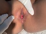
Genital Herpes Simplex Virus Infection in 4-Year Old Girl
←
→
Page content transcription
If your browser does not render page correctly, please read the page content below
Malaysian Journal of Medicine and Health Sciences (eISSN 2636-9346)

CASE REPORT

Genital Herpes Simplex Virus Infection in 4-Year Old Girl
Lunardi Bintanjoyo1, Indah Purnamasari1, Afif Nurul Hidayati1,2
1
    Department of Dermatology and Venereology, Faculty of Medicine, Universitas Airlangga, Dr. Soetomo General Hospital,
    Surabaya, 60286, Indonesia
2
    Universitas Airlangga Teaching Hospital, 60115, Surabaya, Indonesia

ABSTRACT

Prepubertal genital herpes simplex virus (HSV) infection is rare. A four-year old girl was consulted for red spots on
genital. Spots were itchy six days before but became painful with genital discharge and pain on urination four days
before. History of sexual abuse, oral or genital ulcers was denied. There were erythema, edema, multiple circular ul-
cers with purulent discharge on labia minora. Gram examination showed leukocytes and Gram-negative cocci.Dark
field and Tzanck examination were negative. Fecalysis was normal. IgG to HSV-1 were reactive in acute and con-
valescence serum. Normal saline compress, fusidic acid cream, paracetamol, azithromycin, and aciclovir provided
resolution of lesions. Assessment was genital ulceration due to first-episode nonprimary HSV infection, suspected
due to HSV-1. Parents were educated on possible sexual abuse. Clinical manifestation, response to treatment and
serology established diagnosis but cannot confirm etiologic subtype.

Keywords: Herpes simplex, Sexually transmitted disease, Serologic tests

Corresponding Author:                                             denied. Previous oral and genital ulcers were denied.
Afif Nurul Hidayati, SpKK(K)                                      History of other illness or drug allergy was denied.
Email: afif_nurulhidayati@fk.unair.ac.id                          History of sexual intercourse or abuse was denied.
Tel: +6281-2302-8024                                              Patient stayed at home with her parents. Her mother
                                                                  always accompanied patient at home, but sometimes
INTRODUCTION                                                      she played with other children when family visited her
                                                                  grandparents. History of sexually transmitted illnesses
Herpes simplex virus (HSV) is the most common cause of            on patient and both parents was denied.
genital ulcer, however it is rare in pre-pubertal children
and should raise concern for sexual abuse (1,2). This is          Dermatologic status showed erythema, edema and
a case of a female child with genital ulcer due to HSV            multiple well-defined circular 3-4mm ulcers with
infection, suspected due to HSV-1.                                purulent yellowish-green discharge on labia minora
                                                                  (Fig. 1). No discharge was seen from vaginal canal.
CASE REPORT                                                       Inguinal lymphadenopathy was absent. Other review of
                                                                  the systems were unremarkable. Body weight was 18.9
A 4-year old girl was brought by her mother to                    kg. Gram examination showed leukocyte 2-4, epithel
Dermatology and Venereology Outpatient Clinic                     2-4 from urethra, leukocyte >25, epithel 6-8 and gram
with complaint of red spots and discharge from her                negative cocci from ulcer (Fig. 2). Dark field examination
genital area. Six days before, itchy red spots were               was negative. Tzanck smear showed no multinucleated
noted on patient’s genital which were often scratched             giant cells (Fig. 3).
by patient. Her mother sought consult to pharmacist
and patient was given mebhydrolin napadisylate half               Assessment was nonspecific genital ulceration suspected
tablet, three times daily but without improvement. Four           due to HSV infection versus enterobiasis, with secondary
days before, pain on lesions and scanty, cloudy, foul-            bacterial infection. Fecalysis and serologic tests for
smelling discharge from patient’s genital were noted.             HSV were requested. Patient was given normal saline
Her mother sought consult to pharmacist and patient               compress 15 minutes followed by fusidic acid cream,
was given erythromycin syrup, mefenamic acid half                 three times daily, paracetamol syrup 150mg three times
tablet, dexamethasone half tablet and chlorpheniramine            daily, azithromycin syrup 200mg once daily for 5 days,
maleate half tablet, all of which taken three times daily,        and aciclovir 200mg four times daily for 7 days.
but without improvement. Pain was felt when urinating
and patient became reluctant to urinate. Fever was                Two days after consult, fecalysis showed normal results.

                                          Mal J Med Health Sci 17(SUPP4): 170-172, June 2021                              170
Malaysian Journal of Medicine and Health Sciences (eISSN 2636-9346)
Figure 1: Physical examination of external genitalia showed Figure 3: Smear was taken from ulcer base and processed
erythema, edema and multiple well-defined circular ulcers with Giemsa stain (Tzanck smear), showing no multinucle-
with yellowish-green discharge on labia minora ated giant cells
Figure 4: Physical examination of external genitalia on follow
up after 1 week showed hypopigmented macules on labia mi-
nora
Figure 2: Smear was taken from ulcer base and processed
with Gram stain, showing many leukocytes and Gram-neg-
ative cocci DISCUSSION
HSV serology showed reactive IgG to HSV-1 (64.46 U, Genital ulcer in female child is very concerning for
reactive >11U), and nonreactive IgM to HSV-1, IgM and parents because of suspicion of sexual abuse (1). This
IgG to HSV-2. Patient’s mother also noted that lesions condition can be due to infectious causes such as HSV,
started to dry with decreasing pain and patient was syphilis, and chancroid, or non-infectious causes such
able to urinate comfortably. One week after consult, as vulvar aphthous ulcer (1,2). Genital HSV infection
ulcers, discharge, pain and dysuria resolved leaving is mostly caused by HSV-2, but HSV-1 has been
hypopigmented macules on labia minora (Fig. 4). increasingly found. Classical presentation is grouped
Assessment was genital ulceration due to first-episode erythematous papules and vesicles coalescing to form
nonprimary HSV infection. Parents were counselled on erosions, or ulcers in severe cases. First-episode cases
transmission sources and to be aware of possible sexual may be accompanied by fever, pain, dysuria, and
abuse to patient. Repeat serology after 10 weeks showed inguinal lymphadenopathy (1,2).
reactive IgG to HSV-1 (30.20 U, reactive > 11U), and
nonreactive IgM to HSV-1, IgM and IgG to HSV-2. Final Diagnosis of HSV infection can be made clinically.
assessment was genital ulceration due to first-episode Laboratory tests to confirm suspicion in nonclassical
nonprimary HSV infection, suspected due to HSV-1. cases or to determine HSV type include Tzanck smear,
171 Mal J Med Health Sci 17(SUPP4): 170-172, June 2021viral culture and serologic assays. Tzanck smear confirm paracetamol as analgetics, and normal saline compress
HSV infection by finding multinucleated giant cells, but to clean the lesions. Rapid response to aciclovir further
has low sensitivity. Viral culture is the gold standard for supported diagnosis of HSV infection.
diagnosis. Serology detects antibodies to specific type
of HSV and differentiates primary and nonprimary first- Presence of IgG to HSV-1 in acute and convalescence
episode genital herpes (3). serum indicated first-episode nonprimary HSV infection.
Absence of IgM to HSV may be due to low sensitivity
Primary infection is first infection with HSV. It shows of this test. Declining IgG titer is not unusual because
no antibodies in acute serum, fourfold increase of type- only less than 5% of recurrent cases showed increased
specific HSV IgG and/or positive IgM in convalescence antibody titer (5). Nonprimary HSV-1 infection is
serum. Nonprimary infection is outbreak of HSV in a possible. Nonprimary HSV-2 infection cannot be ruled
person with previous HSV infection. It shows positive out. Viral culture would have established the infecting
type-specific HSV IgG in acute and convalescence type if available. Final assessment was genital ulceration
serum with or without HSV-specific IgM (3,4). Limitation due to first-episode nonprimary HSV infection, suspected
of serology includes false-negative IgM in almost 50% due to HSV-1. Parents were counselled for possibility of
of culture-positive cases. Most recurrent cases did sexual abuse to patient.
not demonstrate significant increase in antibody titer.
Serology also cannot distinguish location of infection CONCLUSION
(5). Seropositivity to HSV-1 alone is difficult to interpret
due to presence of HSV-1 antibody from oral infection Presence of multiple painful grouped erosions on labia
and increasing genital HSV-1 infection. Seropositivity to minora, rapid response to aciclovir, and positive IgG to
HSV-2 is generally consistent with anogenital infection. HSV-1 in acute and convalescence serum established
Seronegativity to HSV-2 does not exclude possibility of diagnosis of genital ulcer due to first-episode nonprimary
HSV-2 infection (3). HSV infection. Dark field and Gram examination
and fecalysis helped to rule out differential diagnosis.
Recommended treatment for pediatric genital HSV Serologic tests cannot confirm type of HSV and viral
infection is oral acyclovir. It is given at 40-80 mg/kg/ culture would have been helpful if available.
day in 3-4 divided doses for 7-10 days or until clinical
resolution (2). REFERENCES
This patient initially presented with multiple painful 1. Anitha B, Shivanna R. Genital Lesions in a
grouped erosion, purulent discharge and dysuria, Female Child : Approach to the Diagnosis. Clin
however, pruritus was noted by mother. Differential Dermatology Rev. 2018;2(2):49–57.
diagnosis were genital HSV infection, primary 2. Cohen JI. Herpes Simplex. In: Kang S, Amagai M,
syphilis, chancroid, enterobiasis and vulvar aphthous Bruckner AL, Enk AH, Margolis DJ, McMichael
ulcer. Diagnosis of genital HSV infection was based AJ, et al., editors. Fitzpatrick’s Dermatology. 9th
on classical clinical presentation, although Tzanck ed. New York: McGraw Hill Education; 2019. p.
smear was negative. Painful ulcers and negative dark 3021–34.
field examination ruled out primary syphilis. Gram 3. Legoff J, Péré H, Bélec L. Diagnosis of genital
examination did not show Gram-negative bacilli in herpes simplex virus infection in the clinical
school-of-fish pattern but showed Gram-negative cocci, laboratory. Virol J. 2014;11(1).
ruling out chancroid. Vulvar aphthous ulcer presents 4. Carmine L. Genital ulcer disease – A review for
as multiple large ulcers which was different from small primary care providers caring for adolescents.
ulcers in this patient. Initial pruritus raised suspicion of Curr Probl Pediatr Adolesc Health Care.
enterobiasis which presents as perineal pruritus (1), but 2020;50(7):100834.
was ruled out by normal fecalysis. Treatment included 5. Fan F, Day S, Lu X, Tang YW. Laboratory diagnosis
aciclovir 200mg four times daily (approximately 40mg/ of HSV and varicella zoster virus infections. Future
kg/day) for 7 days for HSV infection, azithromycin and Virol. 2014;9(8):721–31.
fusidic acid cream for secondary bacterial infection,
Mal J Med Health Sci 17(SUPP4): 170-172, June 2021 172You can also read


