GEMINI an ultra-stable interferometer - nireos
←
→
Page content transcription
If your browser does not render page correctly, please read the page content below
GEMINI                    an ultra-stable interferometer
GEMINI is a novel and compact interferometer that can guarantee very high robustness
and stability between the two generated replicas of light.
The exceptional performances of this device can be exploited in many different
applications, such as time- and frequency-resolved fluorescence, coherent Raman,
pump-probe, two-dimensional spectroscopy and studies on single molecules.

Key Features                                                  Applications
                                                              • Interferometry
• High throughput that allows high sensitivities              • Generation of pulse pairs
• ≈1 attosecond stability between the two replicas of light
• Fast scans (
Time- and frequency-resolved fluorescence with a single
TCSPC detector
Sample
SPAD or
Experimental setup: GEMINI interferometer is placed in
GEMINI PMT collection before the detector (a SPAD or PMT) connected
Interferometer
to a TCSPC. This allows one to resolve the fluorescence
TCSPC wavelength axis while preserving the temporal resolution.
The GEMINI can
Narrowband be placed as a
pulsed excitation turn-key add-on
device
800 0.9
Rhodamine B + Nile Red
1 0.8
750
0.7
0.6 t=4.2 ns
Intensity (a.u.)
700
Wavelength (nm)
Intensity (a.u.)
0.5
650 0.1
0.4
600 0.3
0.2 t=1.8 ns
550
0.1
500 0.01 0
-1 0 1 2 3 4 5 6 7 8 9 -1 0 1 2 3 4 5 6 7 8 9 540 580 620 660 700 740
Time (ns) Time (ns) Wavelength (nm)
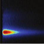
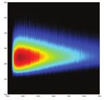
Fluorescence maps as a function of Semi-log plots of fluorescence decay Integrated spectra of the two
detection wavelength and emission time for traces at ≈575 nm (green curve) and ≈675 fluorophores computed from the
a mixture of Rhodamine B and Nile Red in nm (purple curve). correspondent Decay Associated Spectra
acetone solution. (DAS) and lifetimes.
1
0.9
(c)
Intensity
0.8
0.7
(a.u.)
0.6
Intensity
0.5
0.4
0.3
0.2
(a.u.)
800
0.1
008
Standard grating-based GEMINI + single PMT
0
-1 0 1 2 3 4 5 6 7 8 9
LHCS3R complex
spectrometer detector
Wavelength (nm)
750
057
0 0
700
007
(a) (b)
650 550 600 650 700 750 550 600 650 700 750
056
500 550 600 650 700 750
-1 0 1 2 3 4 5 6 7 8 9
0
1
1.0
2.0
3.0
4.0
5.0
6.0
7.0
8.0
9.0
Wavelength (nm) Wavelength (nm)
Time (ns)
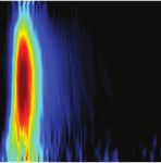
(a) Fluorescence map of the LHCSR3 complex from C. Comparison of fluorescence emission spectra of Rhodamine B, measured in
reinhardtii; (b-c) Marginals of (a), obtained by integrat- the same experimental conditions. Excitation laser: =530 nm, P=1 W.
ing the map along the horizontal and vertical directions,
respectively, showing the overall fluorescence spectrum
and decay dynamics.
A. Perri et al., Opt. Express 26, 2270-2279 (2018).Coherent Raman (Stimulated Raman Scattering - SRS)
and Pump-Probe Spectroscopy
Broadband Sample
Probe/Stokes Experimental setup: GEMINI interferometer is
Photo- placed in the probe/Stokes beam after the
GEMINI diode
Interferometer
sample, allowing one to measure SRS or
pump-probe spectra up to MHz modulation
lated p Lock-in
Modu band Pum Amplifier frequencies.
w
Narro
1400
1400 (a) (b)
1300
Wavelength (nm)
1200
1100
5
-5
(c)
(a) Two-dimensional ΔT⁄T(λ,τ) map for a graphite sample prepared by Chemometric analysis of the acquired dataset. (a) SRS spectra for
liquid phase exfoliation. (b) ΔT⁄T spectra at selected probe delays; (c) Δ PMMA (solid black line) and PS (dotted red line). (b) False-color image
T⁄T dynamics at 1270-nm probe wavelength (red circles) together with of the sample, showing a central bead of PMMA (in red), surrounded
a bi-exponential fit (black solid line). Inset: zoom of the signal for by smaller beads of PS (in green). (c) and (d): concentrations maps of
negative delays. PMMA and PS.
F. Preda et al., Opt. Lett. 41, 2970-2973 (2016).
J. Réhault et al., Opt. Express 23, 25235-25246 (2015).
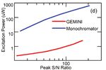
Comparison with Monochromators
The GEMINI is designed to be added to your setup to extract the spectrum of any light source, coherent or not.
It can replace monochromators, since it overcomes their main drawbacks in terms of low throughput, fixed spectral
resolution and limited spectral coverage
Excitation
o m ator
Laser och r
Mon
Monochromator
PMT PMT GEMINI
detector detector
GEMINI Fluorescent
Interferometer
Sample
COMPARISON between GEMINI and a monochromator. The fluorescence of a sample is collected at 90° and measured
with PMT detectors. The GEMINI and the monochromator enable to spectrally resolve the fluorescence. With the GEMINI,
one can obtain the same S/N obtained with a monochromator with ~100 times lower excitation light power.Excitation-Emission Maps (EEMs) of Single Molecules
The GEMINI can be placed as a
turn-key add-on device
Sample
Short Pass
Filter
GEMINI interferometer
Objective allows the characterization
White
Light GEMINI
Beam Splitter of single molecules with
Source Broadband
Interferometer
Long Pass Filter low acquisition times and
excitation
exceptional accuracy and
Spectrograph
sensitivity
Flip Mirror
Photodiode and Single molecule: Terrylene diimide derivative
TCSPC
EXCITATION-EMISSION MAP TIME and FREQUENCY-RESOLVED MAP
FT
0
A B GEMINI in Excitation
Detection Wavenumber /103 [cm-1]
Detection Wavenumber /103 [cm-1]
Photodiode+TCSPC in Detection
(from Spectrograph)
Decay Time [ns]
GEMINI in Excitation
Spectrograph in Detection C
GEMINI Interferometer Position [mm] Excitation Wavenumber /103 [cm-1] Excitation Wavenumber /103 [cm-1]
Thyrhaug et al., “Single-molecule excitation–emission spectroscopy”, PNAS 201808290 (2019).
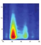
Single Molecule interferogram (A) and relative Excitation-Emission Map (B) obtained via Fourier Transform (FT) along the x-axis.
(C) Excitation-energy versus emission-intensity decay for a single molecule constructed from an interferometric TCSPC experiment.
Technical Specifications
Spectral Resolution
50
VERSION S L
40
Spectral range [nm] 250 - 2300 (Standard)
250 - 3500 (Ultra-broadband)
500 - 4200 (On request) 30
Max. Delay τ [fs @ λ=600 nm] -100 → 700 -100 → 2000
20
Delay τ Stability < 1 a�osecond
Dimensions [mm] 100 x 110 x 65 10
Weight [kg] 1
Specifications can be subject to change without notice.
For more information, please contact us via e-mail at info@nireos.com or visit our website www.nireos.comYou can also read


