Forensic Animal Hair Analysis ZEISS Light Microscope
←
→
Page content transcription
If your browser does not render page correctly, please read the page content below
Application Note

Forensic Animal Hair Analysis
ZEISS Light Microscope
Application Note
Forensic Animal Hair Analysis
ZEISS Light Microscope
Authors: Dr. Ulrike Schacker, Martin Schatzl
Galantos Genetics GmbH, Germany
Dr. Werner Hecht
Institut für Veterinärpathologie Giessen, Germany
Dr. Thorsten Kern, Dr. Michael Gögler, Anke Koenen
Carl Zeiss Microscopy GmbH, Germany
Date: January 2018
Both human and animal hair play a crucial role in the investigation of criminal offenses. In most cases, identifying
and differentiating between human and animal hair is relatively straightforward on account of their specific
characteristics. Under certain circumstances, however, carrying out a light microscopic analysis of individual
structures such as the medulla, pigmentation type, and cuticle structure may be necessary to distinguish between
different species.
Introduction
Hair is one of the most common biological fibers. Its analysis
Microfibril
can play an important role when investigating theft, in
accidents involving wild animals within the scope of settling Macrofibril
insurance claims, when investigating customs violations /
poaching, and when solving criminal offenses such as mur- Bundle of fibers
der, among other cases. Depending on the type, number,
and condition of the hair samples, different methods of fo- Medullary canal
rensic hair analysis are used. The questions that need to be
answered include: Is it really a hair or a plant or textile fiber? Hair shaft
If it is a hair, is it human hair or animal hair? What part of the
body is the hair from? Has the hair been torn out, cut off,
squashed, or scorched?
Cuticle layer/
cuticula
Hair
Every species of mammal has hair with distinctive features
such as length, color, root structure, and specific morpho-
logical characteristics (Figure 1). Figure 1 Basic structure of a hair
Hair (lat. pili) is a protein filament (Figure 2) primarily com-
posed of keratin and found in all mammals. A hair consists The cortex refers to the main fiber of the hair, which is com-
of the hair follicle and the hair shaft. The hair shaft is made posed of fiber bundles, which in turn are composed of the
up of the medulla, cortex, and cuticle (Figure 1). The cuticle finest subfibers, the fibrils. The medulla is the inner layer,
is the outer scaly layer formed from keratinized, dead cells. which can also form cavities.
2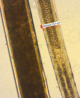
Application Note
out versus one that has fallen out. Classical microscopy
Hair shaft
therefore makes it possible to determine a mammal’s
Epidermis species, race, hair type, and hair status [2].
The type, number, and condition of the recovered hairs
significantly impact their value as evidence for forensic
examination with a light microscope.
The typical, accident-related animal classes are part of the
Dermis routine examination in most laboratories, since, for example,
Hair follicle insurance coverage is often dependent on the type animal
that caused the accident. For microscopic observation, the
hair is mounted on a microscope slide [3]. Typical magnifica-
tions are 10×, 20×, and 40×. In rare cases, a 100× oil lens is
Hair bulb also used. Sometimes pigmentation requires the medulla to
be specially prepared. Good results are achieved with glycer-
ine as the mounting medium.
Case Study: Synthetic Fiber or Natural Fur
Figure 2 Schematic cross section of a hair Morphological hair analysis is, to a certain extent, a suitable
method of identifying species. Areas of application include,
for example, accidents involving wild animals, in which it
Wild Animal Hair under the Microscope may be necessary to determine whether game animals were
Hair is an extensive source of information when viewed un- involved. The analysis of fur appliqué on clothing to deter-
der a light microscope, as light can penetrate the aforemen- mine whether it is of animal origin is also of significance, es-
tioned structures and carry the information back to our eyes. pecially if such appliqué is declared to be fake fur. In such
Every species of mammal has hair with distinctive features cases, clarity can be achieved by conducting a suitable ex-
such as length, color, root structure, and specific morpho- amination. The first step is to check whether the hair is ani-
logical characteristics, and these can be used to determine mal hair. This can be achieved by checking for the presence
the species and genus of the mammal. Mammalian hair is of the cuticle structure typical of animal hair using the im-
usually referred to as fur and is divided into guard hairs (lat. pression method. If the result is positive, an additional analy-
capilli), awn hairs (lat. setae), wool hairs (lat. pili lanei), and sis of the medulla and possibly the hair cross section can be
long hairs. Many mammals have vibrissae (tactile hair) [1]. used to determine the species or at least a group of species.
Here, the nerve endings around the hair root (follicle) act as The results of the purely microscopic examination in a case
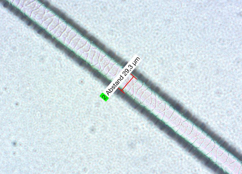
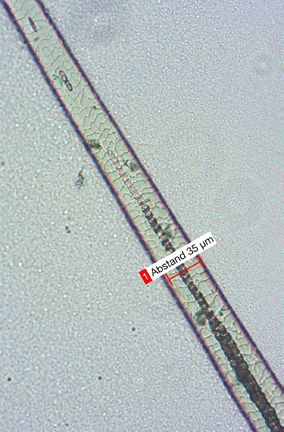
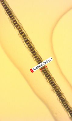
sensors. Hair from different parts of the body of the same in- of artificial-fur declaration can be seen in Figures 3 to 6. By
dividual can exhibit considerable variability. The structure of comparing it with appropriate reference material, the results
the medulla and cuticle of the hair is highly specific to the in this case were: 1. animal hair and 2. hair of a leporid (an
species. It therefore also makes it possible to reliably distin- animal of the family Leporidae, comprising the rabbits and
guish between humans and animals. The criteria used to ac- hares). In other words, this was a clear case of falsely de-
curately determine the species include the structure of the clared fur. In order to definitively identify the species, a DNA
medulla cells, medulla thickness, medullary rays, number of test was also performed. This showed that the hair was that
medullary cell layers, and the ratio of the thickness of the of a European rabbit (Oryctolagus cuniculus). Even if a DNA
medulla to the cortex. In addition, you can also analyze the test is ultimately conducted, light microscopic morphological
content and distribution of the pigments and the surface analysis should still be carried out, as it allows a statement
profile of the cuticle cells. A microscopic analysis of the hair to be made even if the DNA test fails. This would no longer
follicle makes it possible to determine both the growth be possible after lysis and extraction of the hair.
phase and to distinguish between a hair that has been torn
3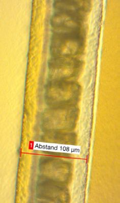
Application Note

Figure 3 Cuticula medial – proximal                            Figure 4 Cuticula medial – proximal

Figure 5 Cuticula medial – proximal                            Figure 6 Cuticula medial – proximal

Recommended Microscope Equipment
The light microscopes ZEISS Axio Lab.A1 and ZEISS Axio         denser with an aperture of 0.9 (H D Ph DIC). For documenta-
Scope.A1 are upright microscopes suitable for use in labora-   tion purposes, a microscope camera should be selected that
tories that conduct such analyses. Since it is important to    precisely displays the finely resolved structures. A simple,
observe the fine structures of the cuticle, both fast and      precise image documentation system, such as ZEISS Labscope,
wide-aperture optics are beneficial. The condenser should      can be operated using a standard tablet (iPad) or a Windows
also be selected to allow the use of darkfield as well as      PC.
brightfield, such as the ZEISS achromatic – aplanatic con-

                                                                                                                              4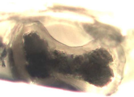
Application Note

                                    Medulla      ZEISS Axioscope 200×                                                      Cuticula 400×

  ZEISS Axioscope 200×                                                        Cuticula 200×                                  Medulla

Figure 7 European hare (Lepus europaeus)                                Figure 8 Wildcat (Felis silvestris)

                                                       Medulla

         Cuticula                                                                Cuticula

Figure 9 Iltis (Mustela putorius)                                       Figure 10 Mustelid (Mustelidae)

         Cuticula            Medulla underwool         Medulla                   Medulla                      Cuticula

Figure 11 Raccoon (Procyon lotor)                                       Figure 12 Muskrat (Ondatra zibethicus)
                                                                                                                                           5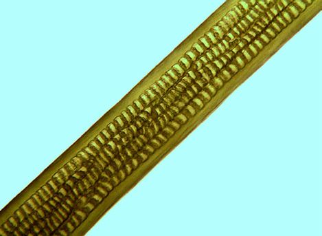
Application Note

                                                                    Cuticula                         Medulla

Figure 13 European mink (Mustela lutreola)                  Figure 14 Deer (Cervidae)

        Cuticula                                  Medulla                                            Medulla

Figure 15 European roe deer (Capreolus)                     Figure 16 Horse

              Cuticula                       Medulla                      Cuticula              Medulla

Figure 17 Alpaca (Vicunia pacos)                            Figure 18 Red fox (Vulpes vulpes)
                                                                                                               6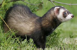
Application Note

                                                                                     Cuticula              Medulla

Figure 19 Cattle (Bos taurus)                                         Figure 20 Dog (Canis lupus)

                                                                                     Cuticula              Medulla

Figure 21 Wild boar                                                    Figure 22 Gray wolf (Canis lupus)

References:
[1] https://en.wikipedia.org/wiki/Fur#Composition
[2] https://de.wikipedia.org/wiki/Animal_Forensics#Haare
[3] B.J. Teerink; “Hair of Westeuropean Mammals“; Cambridge University Press; ISBN: 0-521-54577-3

Samples:
Courtesy of:
Mr. Immo Ortlepp, professional hunter, Negenborn
Ms. Gudrun Westermann-Hoyer, Brelingen, Wiesbaden Pheasantry, Dörverden Wolf Center

                                                                                                                         7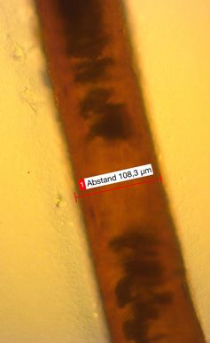
07745 Jena, Germany
microscopy@zeiss.com
www.zeiss.com/axiolab
Carl Zeiss Microscopy GmbH
                             Not all products are available in every country. The use of products for medical diagnostic, therapeutic, or treatment purposes may be subject to local restrictions. Contact your local
                             ZEISS representative for more information.
                             EN_41_013_163 | CZ 08-2018 | Design, scope of delivery, and technical progress subject to change without notice. | © Carl Zeiss Microscopy GmbH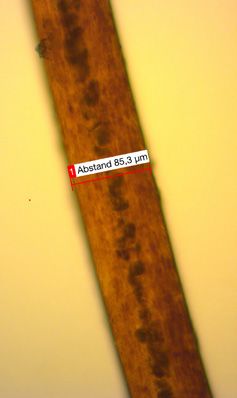
You can also read