Extranasopharyngeal Angiofibroma from Hypopharynx: a Rare Case Report
←
→
Page content transcription
If your browser does not render page correctly, please read the page content below
Journal of Medicine and Health                    Extranasopharyngeal Angiofibroma from Hypopharynx...
Vol. 4 No. 1 February 2021
e-ISSN: 2442-5257

                                          Case Report

Extranasopharyngeal Angiofibroma from Hypopharynx: a Rare Case
Report
Angiofibroma Ekstranasofaring dari Hipofaring: Laporan Kasus yang Langka

Ratna Windyaningrum*, Agung D Permana, Ongka M Saifuddin
Department of Otorhinolaryngology, Head, and Neck Surgery, Faculty of Medicine
Universitas Padjadjaran / Hasan Sadikin General Hospital, Bandung, Indonesia.
Jl. Pasteur No. 38 Bandung, Jawa Barat 40161
*Corresponding author
Email: ratnawindyaningrum0708@gmail.com

Received: January 13, 2021
Accepted: January 21, 2022

Abstract
         Angiofibroma is a histologically benign mesenchymal tumor though it is a potentially
locally destructive fibrovascular tumor. Angiofibroma located in the head and neck comprises
nasopharyngeal and extra-nasopharyngeal angiofibroma. The extra-nasopharyngeal
angiofibroma itself is extremely rare. There was only two hypopharynx angiofibroma case in the
recent literature. The aim of this report is to find out recurrence rate after mass extirpation
surgery approach transcervical in this patient. A case of extra-nasopharyngeal angiofibroma
from hypopharynx that arising from the left posterior pharyngeal wall was described in this
report. A 32 years old man was admitted with 8 years history of lump located around the mouth
and left side of the throat. There were no other significant complaints other than the lump itself.
Histopathologic microscopic examination showed an angiofibroma. Angiofibroma is a benign
and slow-growing mass. The conclusion is management of this case is the same as angiofibroma
in general, by surgery approach. The mass was successfully removed surgically and thus far no
recurrence or post-surgical complications were found one year after excision.

Keywords: angiofibroma; extra-nasopharyngeal; hypopharynx; pharyngeal mass

Abstrak
         Angiofibroma merupakan tumor mesenkimal yang secara histologi jinak, meskipun
tumor fibrovaskular ini dapat bersifat destruktif secara lokal. Angiofibroma yang terletak di
kepala dan leher meliputi angiofibroma nasofaring dan ekstranasofaring. Angiofibroma
nasofaring sendiri merupakan jenis yang sangat jarang. Hanya terdapat dua kasus angiofibroma
hipofaring dalam literatur. Tujuan laporan kasus ini untuk mengetahui tingkat rekurensi setelah
tindakan operatif ekstirpasi massa dengan pendekatan transervikal pada pasien ini. Sebuah kasus
ekstranasofaring angiofibroma yang berasal dari hipofaring yang tumbuh pada dinding faring
posterior dipaparkan pada kasus ini. Seorang pasien laki-laki berusia 32 tahun dengan riwayat
memiliki benjolan di sekitar mulut dan sisi kiri tenggorokan selama 8 tahun. Tidak terdapat
keluhan signifikan lain selain benjolan tersebut. Pemeriksaan mikroskopik histopatologi
menunjukkan sebuah angiofibroma. Angiofibroma merupakan massa jinak yang tumbuh lambat.
Kesimpulannya adalah penatalaksanaan pada kasus ini sama dengan angiofibroma pada
umumnya, yaitu dengan pembedahan. Tumor tersebut berhasil diangkat secara pembedahan dan
tidak terdapat rekurensi atau komplikasi pasca pembedahan satu tahun setelah eksisi.

Kata kunci: angiofibroma; ekstranasofaring; hipofaring; massa faring

J Med Health.2022;4(1):84-91                                                                   84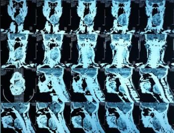
Journal of Medicine and Health                     Extranasopharyngeal Angiofibroma from Hypopharynx...
Vol. 4 No. 1 February 2021
e-ISSN: 2442-5257

                                           Case Report

Introduction
        Angiofibroma is commonly found in the nasopharynx. 1,2,3 It is relatively rare and
represents only 0.05% of all head and neck masses. 4,5,6 Generally, angiofibromas present as an
asymptomatic mass and slowly enlarges.1 It appears to behave in a benign fashion. Diagnosis of
angiofibroma is challenging if it is based on clinical symptoms and findings alone, the exact
diagnosis requires histopathological and immunohistochemical staining microscopic examination
results.7 There have been several studies describing nasopharyngeal angiofibromas however there
have only been a few which report extra-nasopharyngeal angiofibromas, specifically in the region
of hypopharynx.1,2 In this report, a case of hypopharynx angiofibroma arising from the left
posterior pharyngeal wall in a young male was described, including the macroscopic
characteristics, imaging, and histological findings. The aim of this report is to find out recurrence
rate after surgery in this patient.

Case Illustration
        The patient in this report provided written informed consent for the case publication.
This study was approved by the patient itself and his family. A 32 years old man was admitted
with 8 years history of a lump located around the mouth and left side of the throat. The patient
complained of feeling something odd at the mentioned location since 2012 and has been
increasing in size since 2014. He didn’t complain of any difficulty during eating or drinking.
History of dyspnea, hoarseness, double vision, hearing loss, and epistaxis was denied. He was
an active smoker 10 years ago with 5 cigarettes per day. There was no history of salted fish
consumption nor hypertension.
        Physical examination revealed the patient was fully alert and looked moderately sick.
Vital signs were all within normal limits. Local examination findings were mass at left
hypopharynx region covering one-fourth of the posterior oral cavity, palpable at IIa-IIb level
of left neck region with 7 x 6 x 3 cm in size, hard consistency, fixated, no tenderness, and no
hyperemia (Figure 1).
        A computed tomography (CT) scan revealed an inhomogeneous semisolid mass
(hypodense lesion), well-circumscribed with irregular border accompanied with calcification
and hemorrhage (hyperdense lesion). The size of the mass was 5.5 x 6.0 x 7.5 cm at posterior
oropharyngeal wall obliterating bilateral palatine tonsils, long of head and neck bilateral
muscles, left posterior digastric muscle, superior constrictor pharyngeal muscle,
parapharyngeal space, and surrounding vascular attached to the left parotid gland. Right nasal

J Med Health.2022;4(1):84-91                                                                    85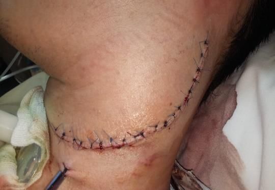
Journal of Medicine and Health                   Extranasopharyngeal Angiofibroma from Hypopharynx...
Vol. 4 No. 1 February 2021
e-ISSN: 2442-5257

                                       Case Report

conchae hypertrophy was also found (Figure 2).

                  A. Anterior View                    B. Right Lateral View

                                    C. Left Lateral View
                         Figure 1 Pre-operative Lump Examination

J Med Health.2022;4(1):84-91                                                                  86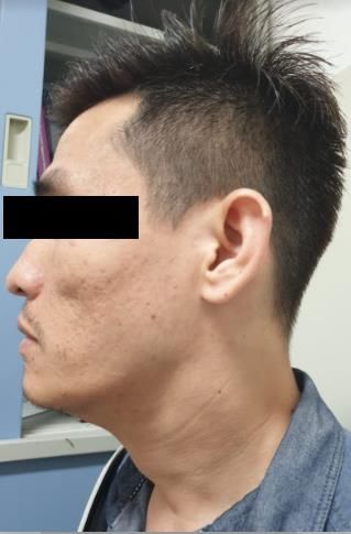
Journal of Medicine and Health                                      Extranasopharyngeal Angiofibroma from Hypopharynx...
Vol. 4 No. 1 February 2021
e-ISSN: 2442-5257

                                                         Case Report

                                                       A. Axial CT

                                                       B. Axial CT

                                                     C. Coronal CT
   Figure 2 CT Scan Reveals an Inhomogeneous Semisolid Mass (Hypodense Lesion),
    Well Circumscribed with Irregular Border Accompanied with Calcification and
                         Hemorrhage (Hyperdense Lesion)
A, B. Axial CT scan described the mass obliterating the palatine tonsils bilateral, long head and neck muscles bilateral, and left
posterior digastric muscle. C. Coronal CT described the mass obliterating parapharyngeal space and vascular structure around
                                              that attached to left parotic gland.

J Med Health.2022;4(1):84-91                                                                                                 87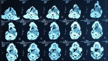
Journal of Medicine and Health Extranasopharyngeal Angiofibroma from Hypopharynx...
Vol. 4 No. 1 February 2021
e-ISSN: 2442-5257
Case Report
Fine Needle Aspiration Biopsy (FNAB) was done with no sample successfully
obtained. The patient was suspected of having a hypopharynx mass. A preoperative
tracheostomy and mass biopsy by micro laryngoscope approach was done. The macroscopic
finding was a rubbery solid white brownish tissue with 8 x 6.5 x 3.5 cm in size. Then the mass
was removed surgically approach transcervical incision. The mass attached to the left posterior
digastric muscle and vascular structure around. (Figure 3).
A. B.
Figure 3 Intra-operative Mass Excision.
A. A rubbery solid white brownish tissue with 8 x 6.5 x 3.5 cm in size. B. The mass attached to left posterior digastric muscle and
vascular structured around.
Figure 4 Post Operative Excision
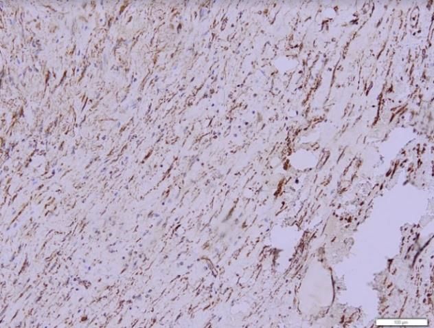
Histopathologic microscopic examination showed an angiofibroma at the left
hypopharynx region and no sign of malignancy. (Figure 5). Immunohistochemical
examination revealed negative S100, positive CD34, vimentin, and Ki67. (Figure 6).
J Med Health.2022;4(1):84-91 88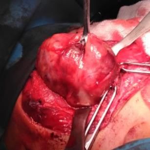
Journal of Medicine and Health                                  Extranasopharyngeal Angiofibroma from Hypopharynx...
Vol. 4 No. 1 February 2021
e-ISSN: 2442-5257

                                                      Case Report

           Figure 5 Histopathologic Microscopic Examination (Magnification 100x)
    Figure shows a benign mass consists of fibro-collagenous stroma connective tissue with partly hyaline degenerated, with
 proliferating fibrocytes, mild amount of lymphocytes, endothel proliferation forming small vascular with nucleus within normal
                             limit, necrotic area and extensive hemorrhage, and no sign of malignancy.

         A. S100 (Magnification 100x).                                      B. CD34. (Magnification 20x).
                    Shows IHC Negative                                                 Shows IHC Positive

         C. Vimentin (Magnification 100x)                                   D. KI67 (Magnification 20x)
                         Shows IHC Positive                                            Shows IHC Positive

                               Figure 6 Immunohistochemical Examination

J Med Health.2022;4(1):84-91                                                                                            89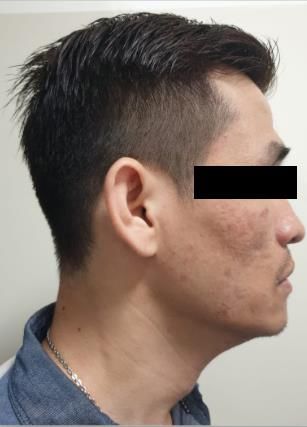
Journal of Medicine and Health                       Extranasopharyngeal Angiofibroma from Hypopharynx...
Vol. 4 No. 1 February 2021
e-ISSN: 2442-5257

                                             Case Report

Discussion
        Angiofibroma is a histologically benign mesenchymal tumor though it is a potentially
locally destructive fibrovascular tumor. It is commonly found in the nasopharynx. 1,2,3 It is
relatively rare and represents only 0.05% of all head and neck masses. It is usually found in young
adolescent males.4,8 One study suggested that significant downregulation of miRNA 125a-5p is
believed to stimulate miRNA tumor growth by the loss of this tumor suppressor.9,10,11
        Angiofibroma located in the head and neck comprises nasopharyngeal and extra-
nasopharyngeal angiofibroma.3,12 There are a variety of sites of extra-nasopharyngeal angio-
fibromas which can include the nasal septum, the tonsils, the ears, the trachea, the larynx, and the
ethmoid and sphenoid sinus.3,13,14,15 The extra-nasopharyngeal angiofibroma itself is extremely
rare. There was only two hypopharynx angiofibroma in the recent literature. 1 Both of the studies
reported smooth, firm, rubbery mass arising from the pharyngeal wall of the hypopharynx. They
showed scatter fibrovascular tissue or fibro-myxomatous stroma harboring numerous blood
vessels of various sizes and shapes.1,4 These findings are similar to the findings in this case report.
However, only one of it conducted an immunohistochemical staining examination which revealed
strong positivity for CD34 and negative staining for S100 protein as it is also the same in this
report. Other than that, we found positive vimentin and Ki67. The presence of Ki67 is a cellular
marker for proliferation and is strictly associated with cell proliferation. 16,17
        In this case report, there was no significant disturbing complaint from the patient other
than the lump itself. This finding is similar to the one which reported that the patient only had
abnormal throat sensation and denied any history of bleeding, pharyngeal pain, eating obstruction,
and dyspnea.1 Nevertheless, another study reported the patient complained of having progressive
foreign-body sensation in the throat and intermittent inspiratory stridor. 4 Summarizing from
several studies, most angio-fibromas present as an asymptomatic mass, except for nasopharyngeal
and retroperitoneal tumors.18 Tumor located in the nasopharynx frequently presents with epistaxis
and persistent nasal obstruction.12,18
        The management of angio-fibromas, in general, is surgery or excision of the mass.19,20
One study removed the mass in the hypopharynx completely by coblation, while the other was
removed by trans-endoscopic approach. It was found that the patient was asymptomatic at 3
months follow-up and no recurrence 6 months after excision, whereas there was no abnormality
found on the other patient after 3 years of follow-up.1,4
        In this report, preoperative tracheostomy and mass biopsy by micro-laryngoscope
approach were done. The mass was successfully removed surgically approach transcervical

J Med Health.2022;4(1):84-91                                                                      90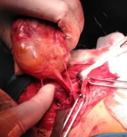
Journal of Medicine and Health                              Extranasopharyngeal Angiofibroma from Hypopharynx...
Vol. 4 No. 1 February 2021
e-ISSN: 2442-5257

                                                   Case Report

incision and thus far no recurrence or post-surgical complications were found one year after
excision. Further, follow-up is needed to evaluate the recurrence of tumors in prolonged period.

Conclusion
          Angiofibroma is a benign and slow-growing mass. The patient reported in this report
showed no significant complaints which could disturb the airway or eating passage. Management
of this case is the same as angiofibroma in general, by surgery approach transcervical incision.
The post-operative condition of the patient was also within the normal limit. Limited reports due
to the extremely rare incidence of this case are not sufficiently representative and more reports
are needed to describe further characteristics and prognosis of this disease.

References
1.    Liu Y, Xu Y, Wang Q, Chen Q. Cellular angiofibroma in the hypopharynx: A case report. Medicine (Baltimore).
      2019; 98(50):e18385.
2.    Dubey SP, Schick B. Juvenile Angiofibroma. 1 st Edition. Switzerland:Springer; 2017; 265-70.
3.    Lee BH. Parapharyngeal angiofibroma: A case report. Iran J Radiol. 2015; 12(3):1–3.
4.    Hsieh ST, Guo YC, Tsai TL, Chen WYK, Huang JL. Angiofibroma of the hypopharynx. J Chinese Med Assoc.
      2004; 67(7):373–5.
5.    Nandhini J, Ramasamy R, Kaul RN, Austin RD. Juvenile primary extranasopharyngeal angiofibroma, presenting
      as cheek swelling. J Oral Maxillofac Surg Med Pathol. 2018; 22(1):S73-6.
6.    Singh GB, Shukla S, Kumari P, Shukla I. A rare case of extra-nasopharyngeal angiofibroma of the septum in a
      female child. J Laryngol Otol. 2017; 2(1):1-4.
7.    Flucke U, Krieken JHJM Van, Mentzel T. Cellular angiofibroma : analysis of 25 cases emphasizing its relationship
      to spindle cell lipoma and mammary-type myofibroblastoma. Mod Pathol. 2011; 24(1):82–9.
8.    Windfuhr J, Vent J. Extranasopharyngeal angiofibroma revisited. Clin Otolaryngol. 2018; 43(1):p199-222
9.    Lerner C, Wemmert S, Schick B. Preliminary analysis of different microRNA expression levels in juvenile
      angiofibromas. Biomed Rep. 2014; 2(6):835–8.
10.   Li W, Ni Y, Lu H, Hu L, Wang D. Current Perspectives on the origin theory of juvenile nasopharyngeal
      angiofibroma. Discov Med. 2019; 27(150):245-54.
11.   Doddy J, Adil EA, Trenor CC, Cunningham MJ. The genetic and molecular determinants of juvenile
      nasopharyngeal angiofibroma: a systematic review. Ann Otol Rhinol Laryngol. 2019; 128(11):1061-72
12    Schick B. Juvenile nasopharyngeal angiofibroma. J Oral Maxillofaxial Pathol. 2016; 20(2):3030.
13    Lerra S, Nazir T, Khan N, Qadri MS, Dar NH. A case of extranasopharyngeal angiofibroma of the ethmoid sinus :
      A distinct clinical entity at an unusual site. Sage Journals. 2012; 91(2):E15–7.
14    David MJC, Trinidad CAJ, Chua AH. Extranasopharyngeal Angiofibroma of the Larynx. PJOHNS. 2010;
      25(1):23–5.
15    Arulappan LAS. Extranasopharyngeal angiofibroma in an adolescent male: a case report. Int J Otorhinolaryngol
      Head Neck Surg. 2019;5(5):1416.
16.   Ma HJ, Huang HN, Li L, Chen S, Zhang RY. Clinicopathological characteristic of angiofibroma of soft tissue:
      reposrt of three cases. Int J Clin Exp Pathol. 2018; 11(7):3777-84.
17.   Mishra A, Jaiswal R, Amita P, Mishra SC. Molecular interactions in juvenile nasopharyngeal angiofibroma:
      preliminary signature and relevant review. Eur Arch Oto-Rhino-L. 2019; 276(3):93-100.
18    Erdur ZB, Yener HM, Yilmaz M, Karaaltin AB, Inan HC, Alaskarov E, et al. Cellular Angiofibroma of the
      Nasopharynx. J Craniofac Surg. 2017 Nov; 28(8):e720–2.
19    Nicolai P, Schreiber A, Bolzoni Villaret A. Juvenile Angiofibroma: Evolution of Management. Int J Pediatr. 2012;
      1(2):1-11.
20    Dewi YA, Nazar IB. Management of Juvenile Nasopharyngeal Angiofibroma in a Referral Hospital in West Java,
      Indonesia. Althea Med J. 2020; 7(1):45–50.

J Med Health.2022;4(1):84-91                                                                                 91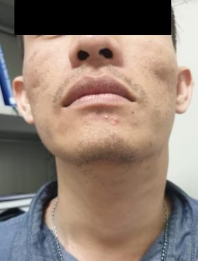
You can also read