Eukaryotic initiation factor EIF-3.G augments mRNA translation efficiency to regulate neuronal activity - eLife
←
→
Page content transcription
If your browser does not render page correctly, please read the page content below
RESEARCH ARTICLE

                                    Eukaryotic initiation factor EIF-3.G
                                    augments mRNA translation efficiency to
                                    regulate neuronal activity
                                    Stephen M Blazie, Seika Takayanagi-Kiya, Katherine A McCulloch, Yishi Jin*

                                    Section of Neurobiology, Division of Biological Sciences, University of California San
                                    Diego, La Jolla, United States

                                    Abstract The translation initiation complex eIF3 imparts specialized functions to regulate
                                    protein expression. However, understanding of eIF3 activities in neurons remains limited despite
                                    widespread dysregulation of eIF3 subunits in neurological disorders. Here, we report a selective
                                    role of the C. elegans RNA-binding subunit EIF-3.G in shaping the neuronal protein landscape. We
                                    identify a missense mutation in the conserved Zinc-Finger (ZF) of EIF-3.G that acts in a gain-of-
                                    function manner to dampen neuronal hyperexcitation. Using neuron-type-specific seCLIP, we
                                    systematically mapped EIF-3.G-mRNA interactions and identified EIF-3.G occupancy on GC-rich
                                    50 UTRs of a select set of mRNAs enriched in activity-dependent functions. We demonstrate that the
                                    ZF mutation in EIF-3.G alters translation in a 50 UTR-dependent manner. Our study reveals an in vivo
                                    mechanism for eIF3 in governing neuronal protein levels to control neuronal activity states and
                                    offers insights into how eIF3 dysregulation contributes to neurological disorders.

                                    Introduction
                                    Protein synthesis is principally regulated by variations in the translation initiation mechanism,
*For correspondence:
yijin@ucsd.edu                      whereby multiple eukaryotic initiation factors (eIF1 through 6) engage elongation-competent ribo-
                                    somes with the mRNA open reading frame (Sonenberg and Hinnebusch, 2009). eIF3 is the largest
Competing interests: The            translation initiation complex, composed of 13 subunits in metazoans, with versatile functions
authors declare that no
                                    throughout the general translation initiation pathway (Valášek et al., 2017). Extensive biochemical
competing interests exist.
                                    and structural studies have shown that eIF3 promotes translation initiation by orchestrating effective
Funding: See page 28                interactions between the ribosome, target mRNA, and other eIFs (Smith et al., 2016; Cate, 2017).
Received: 11 March 2021             Mutations and misexpression of various subunits of eIF3 are associated with human diseases, such as
Preprinted: 16 March 2021           cancers and neurological disorders (Gomes-Duarte et al., 2018), raising the importance to advance
Accepted: 28 July 2021              mechanistic understanding of eIF3’s function in vivo.
Published: 29 July 2021                 Recent work has begun to reveal that different eIF3 subunits can selectively regulate translation
                                    in a manner depending on cell type, mRNA targets, and post-translational modification. Interaction
Reviewing editor: Anne E West,
Duke University School of
                                    of eIF3 RNA-binding subunits with specific 50 UTR stem-loop structures of mRNAs can trigger a trans-
Medicine, United States             lational switch for cell proliferation in human 293 T cells (Lee et al., 2015), and can also act as a
                                    translational repressor, such as the case for human Ferritin mRNA (Pulos-Holmes et al., 2019).
   Copyright Blazie et al. This
                                    Under cellular stress, such as heat shock, the eIF3 complex circumvents cap-dependent protein
article is distributed under the
                                    translation initiation and recruits ribosomes directly to m6A marks within the 50 UTR of mRNAs
terms of the Creative Commons
Attribution License, which          encoding stress response proteins (Meyer et al., 2015). Other specialized translation mechanisms
permits unrestricted use and        appear to involve activities of particular eIF3 subunits that were previously hidden from view. For
redistribution provided that the    example, human eIF3d possesses a cryptic mRNA cap-binding function that is activated by phos-
original author and source are      phorylation and stimulates pre-initiation complex assembly on specific transcripts (Lee et al., 2016;
credited.                           Lamper et al., 2020), while eIF3e specifically regulates metabolic mRNA translation (Shah et al.,

Blazie et al. eLife 2021;10:e68336. DOI: https://doi.org/10.7554/eLife.68336                                                       1 of 32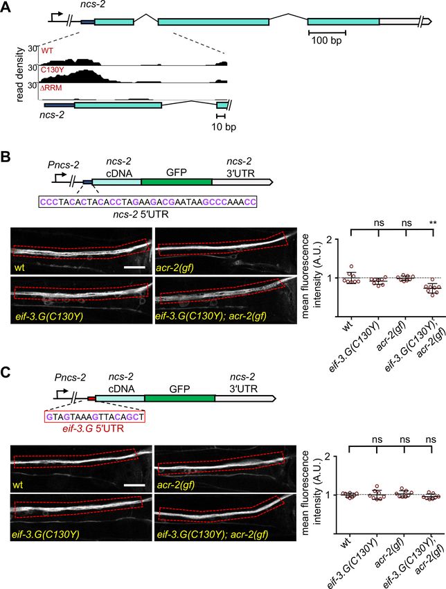
Research article                                                                                          Genetics and Genomics

                                    2016). These findings hint that many other eIF3-guided mechanisms of cell-specific translational con-
                                    trol await discovery.
                                        In the nervous system, emerging evidence suggests that eIF3 subunits may have critical functions.
                                    Knockdown of multiple eIF3 subunits impairs expression of dendrite pruning factors in developing
                                    sensory neurons of Drosophila (Rode et al., 2018). In mouse brain, eIF3h directly interacts with colly-
                                    bistin, a conserved neuronal Rho-GEF protein underlying X-linked intellectual disability with epilepsy
                                    (Sertie et al., 2010; Machado et al., 2016). In humans, altered expression of the eIF3 complex in
                                    the substantia nigra and frontal cortex correlates with Parkinson’s Disease progression (Garcia-
                                    Esparcia et al., 2015). Downregulation of mRNAs encoding eIF3 subunits is observed in a subset of
                                    motor neurons in amyotrophic lateral sclerosis patients (Cox et al., 2010). Furthermore, a single-
                                    nucleotide polymorphism located in the intron of human eIF3g elevates its mRNA levels and is asso-
                                    ciated with narcolepsy (Holm et al., 2015). While these data suggest that eIF3 function in neurons is
                                    crucial, mechanistic understanding will require experimental models enabling in vivo investigation of
                                    how eIF3 affects protein translation with neuron-type specificity.
                                        Protein translation in C. elegans employs all conserved translation initiation factors. We have
                                    investigated the mechanisms of protein translation in response to neuronal overexcitation using a
                                    gain-of-function (gf) ion channel that arises from a missense mutation in the pore-lining domain of
                                    the acetylcholine receptor subunit ACR-2 (Jospin et al., 2009). The cholinergic motor neurons (ACh-
                                    MNs) in the ventral cord of acr-2(gf) mutants experience constitutive excitatory inputs, which gradu-
                                    ally diminish pre-synaptic strength and cause animals to display spontaneous seizure-like convulsions
                                    and uncoordinated locomotion (Jospin et al., 2009; Zhou et al., 2017). acr-2(gf) induces activity-
                                    dependent transcriptome changes (McCulloch et al., 2020). However, it is unclear how protein
                                    translation conducts the activity-dependent proteome changes that sustain function of these
                                    neurons.
                                        Here, we demonstrate that C. elegans EIF-3.G/eIF3g regulates the translation efficiency of select
                                    mRNAs in ACh-MNs. We characterized a mutation (C130Y) in the zinc-finger of EIF-3.G that sup-
                                    presses behavioral deficits of acr-2(gf) without disrupting general protein translation. By systematic
                                    profiling of EIF-3.G and mRNA interactions in ACh-MNs, we identified preferential binding of EIF-3.
                                    G to long and GC-rich 5’UTRs of mRNAs, many of which encode modulators of ACh-MN activity.
                                    We further provided in vivo evidence that EIF-3.G regulates the expression of two of its mRNA tar-
                                    gets dependent on their 50 UTRs. Our findings illustrate the selectivity of EIF-3.G in augmenting
                                    mRNA translation to mediate neuronal activity changes.

                                    Results
                                    A missense mutation in EIF-3.G ameliorates convulsion behaviors
                                    caused by cholinergic hyperexcitation
                                    We previously characterized numerous mutations that suppress convulsion and locomotion behaviors
                                    of acr-2(gf) animals (McCulloch et al., 2017). One such suppressor mutation, ju807, was found to
                                    contain a single nucleotide alteration in eif-3.G, encoding subunit G of the EIF-3 complex
                                    (Figure 1A; see Materials and methods). C. elegans EIF-3.G is composed of 262 amino acids, sharing
                                    overall 35% or 32% sequence identity with human eIF3g and S. cerevisiae TIF35 orthologs, respec-
                                    tively (Figure 1—figure supplement 1A). Both biochemical and structural data show that eIF3g/
                                    TIF35 proteins bind eIF3i/TIF34 through a domain in the N-terminus (Figure 1B; Valášek et al.,
                                    2017). eIF3g/EIF-3.G also has a predicted CCHC zinc finger followed by an RNA recognition motif
                                    (RRM) at the C-terminus (Figure 1B and Figure 1—figure supplement 1A). The ju807 mutation
                                    changes the second cysteine of the CCHC motif (Cys130, corresponding to Cys160 in human eIF3g)
                                    to tyrosine (Figure 1B). Hereafter, we designate eif-3.G(ju807) as eif-3.G(C130Y).
                                       Compared to acr-2(gf) single mutants, eif-3.G(C130Y); acr-2(gf) animals exhibited nearly wild-type
                                    movement and strongly attenuated convulsion behavior (Figure 1C; Videos 1–3). acr-2(gf) animals
                                    carrying heterozygous eif-3.G(C130Y/+) showed partial suppression of convulsions (Figure 1C).
                                    Overexpression of wild type eif-3.G full-length genomic DNA in eif-3.G(C130Y); acr-2(gf) double
                                    mutants restored convulsions to levels similar to eif-3.G(C130Y/+); acr-2(gf) (Figure 1D–E;
                                    Materials and methods). Overexpression of eif-3.G(C130Y) full-length genomic DNA in acr-2(gf) sin-
                                    gle mutants also partially suppressed convulsions (Figure 1C–D). In wild-type animals,

Blazie et al. eLife 2021;10:e68336. DOI: https://doi.org/10.7554/eLife.68336                                                        2 of 32
Research article                                                                                                        Genetics and Genomics

Figure 1. eif-3.G(C130Y) suppresses acr-2(gf) convulsion behavior in the cholinergic motor neurons. (A) Illustration of the genomic locus of eif-3.G: Peif-
3.G denotes the promoter, blue boxes are exons for coding sequences and gray for 30 UTR. Arrowhead indicates guanine to adenine change in ju807;
and short line below represents a 19 bp deletion in ju1327, designated eif-3.G(0), that would shift the reading frame at aa109, resulting in a premature
stop (asterisk) after addition of 84aa of no known homology. (B) Illustration of EIF-3.G: shaded blue represents EIF-3.I binding region, ZF for Zinc Finger,
Figure 1 continued on next page

Blazie et al. eLife 2021;10:e68336. DOI: https://doi.org/10.7554/eLife.68336                                                                        3 of 32
Research article                                                                                                     Genetics and Genomics

Figure 1 continued
RRM for RNA Recognition Motif. Below is a multi-species alignment of the zinc finger domain with bold residues as the CCHC motif and gray for
conserved residues. ju807 causes a C130Y substitution (black arrow). C127Y (red arrow, ju1840) was generated with CRISPR editing. C. elegans (C. e.;
NP_001263666.1), S. cerevisiae (S. c.; NP_010717.1), D. melanogaster (D. m.; NP_570011.1), X. laevis (X. l.; NP_001087888.1), and H. sapiens (H.s.;
AAC78728.1). (C) Quantification of convulsion frequencies of animals of indicated genotypes, with the strains (left to right) as: N2, MT6241, CZ21759,
CZ28495, CZ21759, CZ22977. Ex[eif-3.G(C130Y)] transgenes (juEx7015/juEx7016) expressed full-length genomic DNA cloned from eif-3.G(ju807). (D)
Illustration of eif-3.G expression constructs: top shows the transgene expressing genomic eif-3.G(+ for wild type and C130Y for ju807) with the
endogenous eif-3.G promoter and 30 UTR, and coding exons in blue; bottom shows cell-type expression of eif-3.G cDNA driven by tissue-specific
promoters (Pmyo-3- body muscle, Punc-25- GABAergic motor neurons, Punc-17b - cholinergic motor neurons). (E) Quantification of convulsion
frequencies shows that convulsion behavior of eif-3.G(C130Y); acr-2(gf) double mutants is rescued by transgenes that overexpress eif-3.G(+) genomic
DNA or an eif-3.G(+) cDNA in the ACh-MNs, but not in the GABAergic motor neurons or body muscle. Strains (left to right)- N2, CZ21759, CZ23125/
CZ23126, CZ22980/ CZ22981, CZ23791/ CZ23880, CZ22982/ CZ22983, CZ27881/ CZ27882. (F) Quantification of convulsion frequencies in animals of the
indicated genotypes (left to right)- N2, CZ22917, MT6241, CZ21759, CZ28495, CZ21759, CZ21759, CZ23310, CZ26828. Data in (D-F) are shown as mean
± SEM and sample size is indicated within or above each bar. Statistics: (***) p
Research article                                                                                               Genetics and Genomics

                                    Video 2. MT6241 [acr-2(gf)] C. elegans movement on    Video 3. CZ21759 [eif-3.G(C130Y); acr-2(gf)] C. elegans
                                    solid nematode growth media.                          movement on solid nematode growth media.
                                    https://elifesciences.org/articles/68336#video2       https://elifesciences.org/articles/68336#video3

                                    EIF-3.G(C130Y) selectively affects translation in ACh-MNs
                                    eif-3.G(C130Y) single mutants exhibit normal development, locomotion, and other behaviors (such
                                    as male mating and egg-laying) indistinguishably from wild-type animals (Figure 1F, Video 4). Axon
                                    morphology and synapse number of ACh-MNs were also normal in eif-3.G(C130Y) animals (Fig-
                                    ure 1—figure supplement 2A–B). To dissect how the C130Y mutation affects EIF-3.G function, we
                                    next generated a genetic null mutation (ju1327) using CRISPR editing (Figure 1A and Figure 1—fig-
                                    ure supplement 1A; designated eif-3.G(0), see Materials and methods). Homozygous eif-3.G(0) ani-
                                    mals arrested development at L1 stage, consistent with EIF-3 complex members being required for
                                    C. elegans development (Kamath et al., 2003). eif-3.G(0/+); acr-2(gf) animals were indistinguishable
                                    from acr-2(gf) single mutants (Figure 1F). We additionally tested null mutations in EIF-3.E and EIF-3.
                                    H, two essential subunits of EIF-3 complex, and found that acr-2(gf) animals carrying heterozygous
                                    null mutations in either eif-3 subunit gene showed convulsions similar to eif-3.G(0/+); acr-2(gf) (Fig-
                                    ure 1—figure supplement 3A). Moreover, hemizygous eif-3.G(C130Y/0) animals are healthy at all
                                    stages and suppress behaviors of acr-2(gf) to levels comparable to eif-3.G(C130Y) (Figure 1F).
                                    Reducing one copy of eif-3.H(+) or eif-3.E(+) in eif-3.G(C130Y); acr-2(gf) animals also did not modify
                                    the suppression effect of eif-3.G(C130Y) (Figure 1—figure supplement 3A). These observations
                                    suggest that eif-3.G(C130Y) retains sufficient function of wild-type eif-3.G, and likely affects a regula-
                                    tory activity that is not dependent on EIF-3 subunit dosage.
                                        We considered that EIF-3.G(C130Y) could
                                    alter EIF-3.G protein levels in ACh-MNs. To test
                                    this, we generated single-copy chromosomal
                                    integrated transgenes expressing EIF-3.G(WT) or
                                    EIF-3.G(C130Y) tagged with GFP at the N-termi-
                                    nus under the control of the endogenous eif-3.G
                                    promoter       (Materials   and   methods      and
                                    Supplementary file 1). Fluorescence from both
                                    GFP::EIF-3.G(WT) and GFP::EIF-3.G(C130Y) was
                                    observed in all somatic cells (Figure 1—figure
                                    supplement 1B). In ACh-MNs, both proteins
                                    showed cytoplasmic localization (Figure 2B). The
                                    GFP::EIF-3.G(WT) transgene rescued eif-3.G(0)
                                    to adults (Supplementary file 1) and also
                                    restored convulsion behavior in the eif-3.G
                                    (C130Y); acr-2(gf) background (Figure 2A). In
                                    contrast, the GFP::EIF-3.G(C130Y) transgene Video 4. CZ22197 [eif-3.G(C130Y)] C. elegans
                                    reduced convulsion behavior in the acr-2(gf) movement on solid nematode growth media.
                                    background. Furthermore, we introduced the https://elifesciences.org/articles/68336#video4

Blazie et al. eLife 2021;10:e68336. DOI: https://doi.org/10.7554/eLife.68336                                                                5 of 32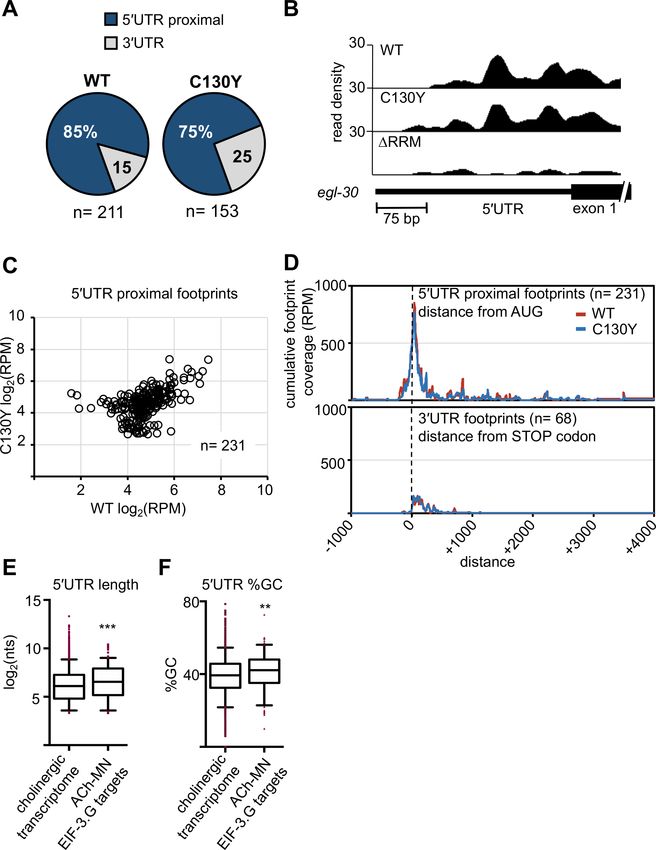
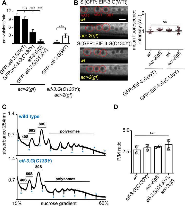
Research article                                                                                                   Genetics and Genomics

Figure 2. eif-3.G(C130Y) involves a selective function of EIF-3.G on translational control. (A) Quantification of convulsion frequency in animals
expressing GFP::EIF-3.G(WT) or GFP::EIF-3.G(C130Y) under Peif-3.G in the indicated genetic backgrounds; and the strains (left to right) are: MT6241,
CZ24729, CZ24652, CZ28497, CZ21759, CZ28107. Error bars represent ± SEM with n = 15 per sample. (***) P< 0.001, (ns) not significant, by one-way
ANOVA with Bonferroni’s post-hoc test. (B) EIF-3.G(WT) and EIF-3.G(C130Y) show comparable expression in ACh-MNs. Left are representative single-
Figure 2 continued on next page

Blazie et al. eLife 2021;10:e68336. DOI: https://doi.org/10.7554/eLife.68336                                                                   6 of 32
Research article                                                                                                   Genetics and Genomics

Figure 2 continued
plane confocal images of EIF-3.G(WT)::GFP or EIF-3.G(C130Y)::GFP driven by the Peif-3.G promoter as single-copy transgenes in L4 animals (head to
the left). Red circles mark the soma of VA10, VB11, and DB7 ACh-MN, based on co-expressing a Pacr-2-mcherry marker. Scale bar = 4 mm. Right: Mean
GFP fluorescence intensities (AU) in ACh-MN soma in animals of the indicated genotypes (n = 8). Each data point represents the mean intensity from
VA10, VB11, and DB7 neurons in the same animal and normalized to the mean GFP::EIF-3.G intensity in a wildtype background. Error bars represent ±
SEM; (ns) not significant by one-way ANOVA with Sidak’s multiple comparisons test. (C) Representative polysome profile traces from total mRNA-
protein extracts of wild type and eif-3.G(C130Y) single mutant animals. Vertical lines (marked by *) within traces indicate the boundaries of fraction
collection. (D) Polysome::monosome (P/M) ratios calculated based on the area under the respective curves for polysomal and monosome (80S) fractions
using two replicates of polysome profiles from total extracts of indicated genotypes. (ns) not significant by one-way ANOVA with Bonferroni’s post-hoc
test.
The online version of this article includes the following source data for figure 2:
Source data 1. Source data for Figure 2A.
Source data 2. Source data for Figure 2B.
Source data 3. Source data for Figure 2C.

                                    GFP::EIF-3.G(C130Y) transgene into the eif-3.G(0); acr-2(gf) background and observed that this
                                    transgene rescued the arrested larvae to adults and nearly abolished convulsion behavior
                                    (Figure 2A). This analysis shows that GFP::EIF-3.G(WT) and GFP::EIF-3.G(C130Y) retain function and
                                    lends further support that eif-3.g(C130Y) is responsible for the suppression of acr-2(gf). Quantifica-
                                    tion of GFP levels in the ACh-MNs showed equivalent intensity and localization of GFP::EIF-3.G (WT
                                    and C130Y) between wild type and acr-2(gf) animals (Figure 2B), indicating that EIF-3.G(C130Y)
                                    does not increase EIF-3.G protein stability.
                                       We further assessed whether eif-3.G(C130Y) alters global translation by performing polysome
                                    profile analysis using whole C. elegans lysates of L4 stage animals. Both the distribution and ratio of
                                    monosomes and polysomes were similar among wild type, eif-3.G(C130Y), acr-2(gf) and eif-3.G
                                    (C130Y); acr-2(gf) animals (Figure 2C–D), indicating that eif-3.G(C130Y) possesses normal function in
                                    the majority of tissues. It is possible that eif-3.G(C130Y) suppresses acr-2(gf) by simply reducing
                                    ACR-2 translation. We tested this by examining a functional GFP-tagged ACR-2 single-copy insertion
                                    transgene (oxSi39). We observed both the levels of ACR-2::GFP fluorescence and post-synaptic
                                    localization in ACh-MNs were comparable between wild type and eif-3.G(C130Y) animals (Figure 1—
                                    figure supplement 3B). These data support the conclusion that eif-3.G(C130Y) preferentially affects
                                    EIF-3’s function in ACh-MNs.

                                    The activity of EIF-3.G(C130Y) requires its RRM
                                    The RRM located at the C-terminus of eIF3g has been shown to bind RNA in a non-specific manner
                                    (Hanachi et al., 1999). To address the role of the RRM in EIF-3.G’s function, we generated a trans-
                                    gene expressing EIF-3.G(DRRM) (Figure 3; Supplementary file 1). Expressing EIF-3.G(DRRM) under
                                    the endogenous promoter Peif-3.G in a wild-type background did not alter development or locomo-
                                    tion, and also did not rescue eif-3.G(0) developmental arrest, supporting the essentiality of the EIF-
                                    3.G RRM. We then generated a transgene expressing EIF-3.G(C130Y) lacking the RRM domain
                                    (C130Y DRRM) in neurons of the acr-2(gf) background (Figure 3). In contrast to full-length eif-3.G
                                    (C130Y), eif-3.G(C130Y DRRM) did not alter convulsion behavior of acr-2(gf) mutants (Figure 3B),
                                    indicating that eif-3.G(C130Y) function requires its RRM.
                                       Studies on S. cerevisiae TIF35/EIF3.G have shown that its RRM promotes scanning of the transla-
                                    tion pre-initiation complex through structured 50 UTRs (Cuchalová et al., 2010). Specifically, alanine
                                    substitution of three residues in the two ribonucleoprotein (RNP) motifs (K194 in RNP2 and L235 and
                                    F237 in RNP1) in TIF35 reduced translation of mRNA reporters carrying 50 UTRs with hairpin struc-
                                    tures, without altering the biochemical RNA-binding activity of EIF-3.G/TIF35. Equivalent amino acid
                                    residues in C. elegans EIF-3.G correspond to R185, F225, F227, which are conserved in human
                                    (R242, F282, F284) (Figure 3; Figure 1—figure supplement 1A). To determine whether these resi-
                                    dues affect EIF-3.G’s function, we expressed C. elegans eif-3.G cDNA with the corresponding amino
                                    acids mutated to alanine, designated eif-3.G(RFF/AAA), in acr-2(gf) animals. We detected partial
                                    suppression of convulsion behavior in acr-2(gf) animals (Figure 3).

Blazie et al. eLife 2021;10:e68336. DOI: https://doi.org/10.7554/eLife.68336                                                                   7 of 32
Research article Genetics and Genomics
It was also reported that a missense mutation (Q258R) in yeast EIF-3.I/TIF34, located in the sixth
WD40 repeat, reduced the rate of pre-initiation complex scanning through 50 UTRs (Cuchalová et al.,
2010). To test if C. elegans eif-3.I shares similar activities, we made a mutant EIF-3.I(E252R), equiva-
lent to yeast TIF34 (Q258R) (Figure 3). In acr-2(gf) animals, overexpressing eif-3.I(E252R), but not
wild-type eif-3.I(+), caused suppression of convulsions to a similar degree as that by the eif-3.G(RFF/
AAA) transgene (Figure 3). These analyses suggest that attenuation of acr-2(gf)-induced neuronal
overexcitation may involve regulation of protein translation through modification of 50 UTR scanning
rates during translation initiation.
Figure 3. eif-3.G(C130Y) requires the RNA-binding domain (RRM) to suppress acr-2(gf) behaviors. Top illustration of the EIF-3.G protein showing the
EIF-3.I binding region (blue), zinc finger (ZF), RRM (dark grey), Q191* mutation in the EIF-3.G(DRRM) transgene, RNP motifs (purple), and the RFF
residues (bold dark blue) changed to alanine in the eif-3.G(RFF/AAA) construct. Below is an illustration of C. elegans EIF-3.I pointing to the position of
E252R within the fourth WD40 domain. Bottom graph is quantification of convulsion frequency in acr-2(gf) animals expressing eif-3.G and eif-3.I variants
in the nervous system (Prgef-1). The strains (left to right) are: MT6241, CZ23203/ CZ23204, CZ28152/ CZ28153, CZ23304/ CZ23305, CZ28152/ CZ28153,
CZ28057/ CZ28058, CZ28064/ CZ28065. Bars represent mean convulsion frequency ± SEM and sample sizes are indicated within or above bars. (***) p<
0.001, (ns) not significant, by one-way ANOVA with Bonferroni’s post-hoc test.
The online version of this article includes the following source data for figure 3:
Source data 1. Source data for Figure 3.
Blazie et al. eLife 2021;10:e68336. DOI: https://doi.org/10.7554/eLife.68336 8 of 32Research article Genetics and Genomics
Both EIF-3.G(WT) and EIF-3.G(C130Y) associate with mRNA 50 UTRs in
the cholinergic motor neurons
EIF-3.G may interact with specific mRNAs in the nervous system to regulate cholinergic activity.
Therefore, we next searched for mRNAs that are associated with EIF-3.G(WT) and EIF-3.G(C130Y) in
the ACh-MNs using single-end enhanced crosslinking and immunoprecipitation (Van Nostrand
et al., 2017). We generated single-copy transgenes expressing 3xFLAG-tagged EIF-3.G(WT), EIF-3.
G(C130Y), or EIF-3.G(DRRM) in the ACh-MNs of acr-2(gf) animals, with EIF-3.G(DRRM) serving to
detect indirect crosslinking events. We confirmed that the truncated EIF-3.G(DRRM) transgene was
expressed, but at reduced levels compared to the EIF-3.G(WT) and EIF-3.G(C130Y) transgenes (Fig-
ure 4—figure supplement 1A). Following cross-linking and immunoprecipitation using anti-FLAG
antibodies, we obtained a comparable amount of immunoprecipitated GFP::EIF-3.G proteins and
obtained more reads from seCLIP on animals expressing each GFP::EIF-3.G transgene than on con-
trol animals lacking any transgene (IgG(-); see Supplementary file 4). There was a strong correlation
between read clusters detected among sets of two biological replicates (Figure 4—figure supple-
ment 1B). We defined EIF-3.G-RNA crosslink sites as clusters of at least 20 high-quality reads with at
least 1.5 fold change enrichment over the input control (see Materials and methods and
Supplementary file 5). We further defined specific footprints of EIF-3.G(WT) and EIF-3.G(C130Y) by
subtracting clusters detected with EIF-3.G(DRRM) (Supplementary file 6, also see
Materials and methods). The EIF-3.G-specific footprints were primarily located within or near the
50 UTRs of protein-coding genes (50 UTR proximal) (Figure 4A–B). In total, we detected 231 50 UTR
proximal footprints of EIF-3.G(WT) or EIF-3.G(C130Y), which mapped to 225 different genes
(Figure 4C). The number of reads comprising EIF-3.G(WT) or EIF-3.G(C130Y) footprints was similar
(e.g. egl-30; Figure 4B) for most of these genes. While some footprints were differentially detected
between EIF-3.G(WT) and EIF-3.G(C130Y), this was almost invariably due to small differences in
seCLIP signal intensity (read cluster size) between samples close to the 20 reads threshold
(Figure 4C), and we therefore did not further pursue its significance.
In line with a recent report that the human eIF3 complex remains attached to 80S ribosomes in
early elongation (Wagner et al., 2020), we observed the bulk of read clusters comprising EIF-3.G
(WT) and EIF-3.G(C130Y) footprints mapping between (-)150 to (+)200 nucleotides of the start
codon (Figure 4D). In contrast, the majority of signals comprising 3’UTR footprints of EIF-3.G(WT)
and EIF-3.G(C130Y) were dispersed along the first 200 nucleotides downstream of the stop codon
(Figure 4D). Overall, the footprint map shows that both EIF-3.G(WT) and EIF-3.G(C130Y) predomi-
nantly bind to similar locations within or near the 50 UTRs of 225 genes in the ACh-MNs, hereafter
named EIF-3.G targets. Taken together with our finding that eif-3.G(C130Y) requires its RRM to sup-
press acr-2(gf), the seCLIP analysis suggests that the C130Y mutation does not dramatically alter the
ability of EIF-3.G to associate with these mRNAs in the ACh-MNs.
EIF-3.G preferentially interacts with long and GC-rich 50 UTR sequences
50 UTR sequences are widely involved in gene-specific regulation of translation (Pelletier and Sonen-
berg, 1985; Leppek et al., 2018). We next assessed whether the selective role of EIF-3.G in protein
translation might correlate with specific sequence features in the mRNA targets expressed in ACh-
MNs by examining the length and GC-content of their 50 UTRs. In C. elegans, about 70% of mRNAs
are known to undergo trans-splicing, and 50 UTRs of mRNAs with trans-splice leaders are usually
short, with a median length of 29nt. We compared the EIF-3.G target gene list with a database con-
taining a compilation of C. elegans trans-splice events from ENCODE analyses (Allen et al., 2011).
We found that 133 of the 225 (59%) EIF-3.G targets are annotated to undergo trans-splicing, which
is comparable to that of transcriptome-wide (Allen et al., 2011; Figure 4—figure supplement 2A),
suggesting that trans-splicing events may not contribute to EIF-3.G’s selectivity on mRNA targets.
Interestingly, we found that the trans-spliced 50 UTRs of these 133 transcripts are significantly longer
(median length = 43nt), compared with all trans-spliced 50 UTRs in the C. elegans transcriptome
(median length = 29nt; n = 6,674) (Figure 4—figure supplement 2B). To assess the GC content for
EIF-3.G mRNA targets, we then applied a threshold to the cholinergic neuronal transcriptome of
acr-2(gf) (McCulloch et al., 2020) defining a 50 UTR as at least 10 nucleotides upstream of ATG, and
also selected the longest 50 UTR isoform per gene to avoid redundant analysis of target genes (see
Materials and methods). Using this criterion, we identified a 50 UTR for 4573 different genes in the
Blazie et al. eLife 2021;10:e68336. DOI: https://doi.org/10.7554/eLife.68336 9 of 32Research article Genetics and Genomics Figure 4. Both EIF-3.G(WT) and EIF-3.G(C130Y) associate with mRNA 50 UTRs in the cholinergic motor neurons. (A) Pie charts displaying the proportion of EIF-3.G(WT) and EIF-3.G(C130Y) footprints located within each gene feature. (B) seCLIP read density track of EIF-3.G(WT) and EIF-3.G(C130Y) footprints on the 50 UTR of egl-30, compared to the EIF-3.G(DRRM) control. (C) Scatter plot comparing the signal intensity, in reads per million (RPM), of all 231 50 UTR proximal footprints detected in EIF-3.G(WT) or EIF-3.G(C130Y). (D) Plots show the cumulative coverage of all 50 UTR proximal (top) or Figure 4 continued on next page Blazie et al. eLife 2021;10:e68336. DOI: https://doi.org/10.7554/eLife.68336 10 of 32
Research article                                                                                                        Genetics and Genomics

Figure 4 continued
30 UTR (bottom) footprints of EIF-3.G(WT) or EIF-3.G(C130Y) relative to the start codon (top) or stop codon (bottom) position. Coverage is presented as
reads per million (RPM). (E–F) Box plots comparing length and GC-content of all 50 UTR sequences of EIF-3.G target mRNAs with annotations (n = 179)
to all 50 UTRs in the acr-2(gf) cholinergic neuronal transcriptome (n = 4573). Boxes are 5–95 percentile with outliers aligned in red. Statistics: (***) p<
0.001, (**) p< 0.01 by two-tailed Mann-Whitney test.
The online version of this article includes the following source data and figure supplement(s) for figure 4:
Source data 1. Source data for Figure 4A.
Source data 2. Source data for Figure 4C.
Source data 3. Source data for Figure 4D.
Source data 4. Source data for Figure 4E.
Source data 5. Source data for Figure 4F.
Figure supplement 1. EIF-3.G transgenes are expressed and produce similar results from replicate seCLIP experiments.
Figure supplement 1—source data 1. Source data for Figure 4—figure supplement 1A.
Figure supplement 1—source data 2. Source data for Figure 4—figure supplement 1B.
Figure supplement 2. EIF-3.G associates with long and GC-rich 50 UTRs.
Figure supplement 2—source data 1. Source data for Figure 4—figure supplement 2A.
Figure supplement 2—source data 2. Source data for Figure 4—figure supplement 2B.
Figure supplement 2—source data 3. Source data for Figure 4—figure supplement 2C.
Figure supplement 2—source data 4. Source data for Figure 4—figure supplement 2D.
Figure supplement 2—source data 5. Source data for Figure 4—figure supplement 2E.

                                      cholinergic transcriptome and for 179 of the 232 EIF-3.G targets in the ACh-MNs. The median 50 UTR
                                      among the 179 EIF-3.G target mRNAs was significantly longer (93 nt) and GC-enriched (42%), com-
                                      pared to the cholinergic transcriptome median (69 nt and 39% GC; n = 10,962; Figure 4E–F). We
                                      further analyzed the distribution of GC sequences in 50 UTRs, and observed non-random positioning
                                      such that some genes were relatively GC-rich near the start codon (e.g. zip-2 and sec-61) and others
                                      had enrichment closer to the distal 5’ end (e.g. pdf-1 and kin-10), suggesting that discrete sequence
                                      elements in EIF-3.G associated transcripts may regulate translation (Figure 4—figure supplement
                                      2C).
                                          The incidence of long and GC-enriched 50 UTRs among EIF-3.G associated transcripts led us to
                                      speculate a major function of EIF-3.G, in addition to its necessity in general translation initiation, is in
                                      the selective regulation of translation. To extend our findings beyond C. elegans, we asked if the
                                      preferential association of EIF-3.G with these complex 50 UTRs could be conserved in mammals. We
                                      analyzed the published eIF3g PAR-CLIP sequencing data from HEK293 cells (Lee et al., 2015) by
                                      comparing the 50 UTR lengths of human eIF3g target genes to all genes with 50 UTRs annotated in the
                                      hg38 genome. We found that human transcripts associated with eIF3g contained significantly longer
                                      and GC-enriched 50 UTRs than average (Figure 4—figure supplement 2D–E). This analysis lends sup-
                                      port for a conserved, specialized role of eIF3g in the translation of transcripts harboring complex
                                      50 UTRs.

                                      EIF-3.G target mRNAs encode proteins that exhibit activity-dependent
                                      expression
                                      To address whether EIF-3.G target mRNAs may preferentially affect specific biological processes, we
                                      performed Gene Ontology and KEGG pathway analysis. Significant GO term (Ashburner et al.,
                                      2000) enrichment was identified in neuropeptide signaling genes (GO:0050793; 15 genes), which
                                      are known to affect acr-2(gf) behavior (Stawicki et al., 2013; McCulloch et al., 2020), and in stress
                                      response genes (GO: 0006950; 28 genes), which could modulate neuronal homeostasis or function
                                      under circuit activity changes (Figure 5A). We also found many EIF-3.G target genes involved in pro-
                                      tein translation and protein metabolism processes (GO:0019538; 29 genes; Figure 5A). Additional
                                      enrichment was associated with metabolic components, kinase signaling, and calcium and synaptic
                                      signaling pathways (Figure 5A). Calcium and synaptic signaling genes included the CAMKII unc-43,
                                      and the G-proteins egl-30 and goa-1, which are all known to regulate ACh-MN synaptic activity
                                      (Miller et al., 1999; Richmond, 2005; Treinin and Jin, 2020).
                                         To determine if expression of EIF-3.G target mRNAs is regulated in an activity-dependent man-
                                      ner, we next incorporated differential transcript expression data between wild type and acr-2(gf)

Blazie et al. eLife 2021;10:e68336. DOI: https://doi.org/10.7554/eLife.68336                                                                       11 of 32
Research article Genetics and Genomics Figure 5. Gene network analyses of EIF-3.G target mRNAs show enrichment in activity-dependent expression. (A) Cytoscape network of EIF-3.G target genes with enriched GO terms (neuropeptide signaling, response to stress, and protein translation and protein metabolism) or KEGG pathways (calcium and synaptic signaling, metabolic components, MAPK-signaling, and mRNA surveillance). Enrichment p-values are derived from statistical analysis of our EIF-3.G targets (n = 225) in the PANTHER database (Mi et al., 2019). (B) EIF-3.G target genes exhibiting significant transcript level Figure 5 continued on next page Blazie et al. eLife 2021;10:e68336. DOI: https://doi.org/10.7554/eLife.68336 12 of 32
Research article                                                                                                     Genetics and Genomics

Figure 5 continued
changes in acr-2(gf) versus wild-type animals as determined from transcriptome sequencing of cholinergic neurons by McCulloch et al. PT and PM
refers to protein translation and protein metabolism. Differential expression was assessed using DeSeq2 (Love et al., 2014) with significance thresholds
of (*) p
Research article                                                                                                       Genetics and Genomics

Figure 6. EIF-3.G(C130Y) impairs HLH-30 expression in ACh-MNs of acr-2(gf) animals. (A) Gene models of hlh-30 isoforms a (pink), b (blue), and d
(green), with presumptive promoters for each isoform depicted as right-pointing arrows and the 50 UTR of isoform d in green to the right of its
promoter. (B) seCLIP read density tracks of footprints on the 50 end of hlh-30 isoform b and d (left) and the 50 end of hlh-30 isoform a (right) in each
indicated EIF-3.G dataset. Purple arrows show footprints on the 50 UTR of hlh-30 isoform d. (C) Top: Illustration of the wgIs433 fosmid locus with hlh-30
Figure 6 continued on next page

Blazie et al. eLife 2021;10:e68336. DOI: https://doi.org/10.7554/eLife.68336                                                                      14 of 32
Research article Genetics and Genomics
Figure 6 continued
coding exons in black and 50 UTR of isoform d in green to the right of the promoter. Bottom: Representative single-plane confocal images of the fosmid
translational reporter wgIs433[hlh-30::EGFP::3xFLAG] in ACh-MNs in animals of indicated genotypes. Quantification of GFP intensity is shown on the
right (n = eight for each genotype). Animals are oriented with anterior to the left. Scale bar = 4 mm. Red dashes indicate labeled ACh-MN soma. Each
data point is the average fluorescence intensity quantified from the three ACh-MN soma per animal and normalized to the mean intensity obtained
from wgIs433 in the wild type background. Statistics: (***) p< 0.001, (ns) not significant, one-way Anova with Bonferroni’s post hoc test.
The online version of this article includes the following source data and figure supplement(s) for figure 6:
Source data 1. Source data for Figure 6C.
Figure supplement 1. EIF-3.G (C130Y) has no effect on translation of hlh-30.a in the ACh-MNs.
Figure supplement 1—source data 1. Source data for Figure 6—figure supplement 1.
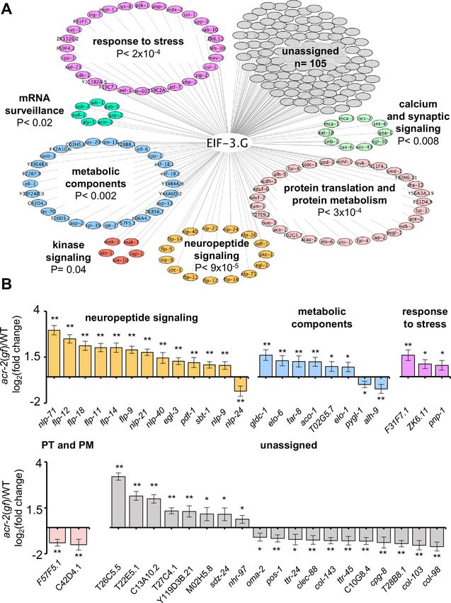
reduced, compared to those in wild type, eif-3.G(C130Y), and acr-2(gf) (Figure 7B). ncs-2 mRNA is
SL1 trans-spliced, and the mature 50 UTR has 37 nt that is especially abundant in GC nucleotides
(47% GC) (Figure 7B). Moreover, the ncs-2 50 UTR sequence is highly conserved with other nematode
species (Figure 7—figure supplement 1A). By RNAfold prediction, we found this sequence could
form a strong stem-loop structure (DG = 5.10 kcal/mol). To test if NCS-2::GFP expression was reg-
ulated specifically through its 50 UTR, we replaced it with the 50 UTR of eif-3.G, which is comparatively
reduced in GC-content (37% GC) and with much less folding stability (D = 1.95 kcal/mol)
(Figure 7C). The eif-3.G 50 UTR is also less conserved across nematodes compared to that of ncs-2
(Figure 7—figure supplement 1A). We found that the NCS-2::GFP reporter with the 50 UTR of eif-3.
G was expressed at similar levels in all genetic backgrounds (Figure 7C).
To further determine the effects of the ncs-2 50 UTR in protein translation with neuronal type reso-
lution, we generated a reporter in which the GFP coding sequence was fused in-frame after the first
four amino acids of NCS-2, which retains the ncs-2 50 UTR but disrupts the myristoylation motif,
thereby enabling visualization of NCS-2 in ACh-MNs (Figure 7—figure supplement 1B). Quantifica-
tion of GFP fluorescence in the cell bodies of VA10, VB11, and DB7 ACh-MN showed significantly
reduced expression in eif-3.G(C130Y); acr-2(gf) animals (Figure 7—figure supplement 1B). In con-
trast, a similar reporter but with the 50 UTR of eif-3.G displayed similar GFP levels in all genetic back-
grounds (Figure 7—figure supplement 1C). Therefore, we conclude that eif-3.G regulates NCS-2
expression in the ACh-MNs through a mechanism involving its 50 UTR sequence.
Discussion
The eIF3 complex has been extensively studied for its essential roles in general translation initiation
(Cate, 2017; Valášek et al., 2017). However, recent work gives support to the idea that eIF3 is also
key to many of the specialized translational control mechanisms needed for tissue plasticity in vivo
(Lee et al., 2015; Shah et al., 2016; Rode et al., 2018; Lamper et al., 2020). Our work expands
the landscape of eIF3’s regulatory functions, revealing an in vivo role of the eIF3g subunit in stimulat-
ing the translation of proteins that mediate neuronal activity changes.
EIF-3.G ensures the efficient translation of mRNAs with GC-rich 50 UTRs
Our study is the first application of seCLIP-seq to map transcriptome-wide protein binding sites in a
specific neuronal subtype (ACh-MNs) in C. elegans. With stringent thresholding, we identified 225
genes with strong EIF-3.G occupancy at mRNA 50 ends. We find that EIF-3.G generally associates
with mRNAs harboring long and GC-rich 50 UTRs, implying its RNA-binding function is selective for
stimulating translation initiation on 50 leaders prone to secondary structure or other forms of transla-
tion regulation. Our data provide in vivo support to the finding that yeast eIF3g/TIF35 promotes
scanning through 50 UTRs with stem-loop structures (Cuchalová et al., 2010). The RRM of yeast
eIF3g/TIF35 also promotes re-initiation of 40S ribosomes upon terminating at uORF stop codons on
GCN4, thereby allowing efficient induction of genes whose translation is regulated by uORFs
(Cuchalová et al., 2010). We did not observe uORFs in the 50 UTRs of ncs-2 or hlh-30, suggesting
that at least for these mRNAs, eif-3.G(C130Y) involves reduced scanning through secondary struc-
tures or other yet undefined regulatory sequence elements.
It is worth noting that we also found EIF-3.G footprints in 30 UTRs, which could reflect molecular
crosstalk between translation initiation and 30 UTR factors, given their proximity in the closed loop
Blazie et al. eLife 2021;10:e68336. DOI: https://doi.org/10.7554/eLife.68336 15 of 32Research article Genetics and Genomics Figure 7. Regulation of NCS-2 expression by EIF-3.G depends on its GC-rich 50 UTR. (A) Illustration of the ncs-2 genomic region. Dark blue represents 50 UTR, green boxes are coding exons, and gray is the 30 UTR. The inset below shows the read density track of seCLIP footprints on the 50 region of ncs-2 mRNA. (B) Top: Schematic of the NCS-2(cDNA)::GFP translation reporter, including its 50 UTR (dark blue), driven by the 4 kb promoter Pncs-2. The 50 UTR sequences are GC rich (purple). Bottom: Representative single-plane confocal images of NCS-2::GFP in ventral nerve chord processes in young adult Figure 7 continued on next page Blazie et al. eLife 2021;10:e68336. DOI: https://doi.org/10.7554/eLife.68336 16 of 32
Research article                                                                                                        Genetics and Genomics

Figure 7 continued
animals of the indicated genotypes. GFP intensity quantification is shown to the right. (C) Top: The ncs-2(50 UTR mutant)::GFP translational reporter has
the 50 UTR of eif-3.G (red boxed sequence) replacing the ncs-2 50 UTR, driven by Pncs-2. Bottom: Representative single-plane confocal images of ventral
nerve chord processes expressing the NCS-2(50 UTR mutant)::GFP translation reporter in young adult animals of the indicated genotypes. GFP intensity
quantification is shown to the right. For (B) and (C), data points are normalized to the average fluorescence intensity of the respective translation
reporter in the wild-type background. ROIs used for fluorescence quantification are boxed. Scale bar = 15 mm. Statistics: (**) P< 0.01, (ns) not significant
by one-way Anova with Bonferroni’s post hoc test.
The online version of this article includes the following source data and figure supplement(s) for figure 7:
Source data 1. Source data for Figure 7B.
Source data 2. Source data for Figure 7C.
Figure supplement 1. EIF-3.G(C130Y) reduces NCS-2 expression in the ACh-MNs of acr-2(gf) animals dependent on its conserved 50 UTR.
Figure supplement 1—source data 1. Quantification of relative fluorescence intensity in the indicated strains.
Figure supplement 1—source data 2. Quantification of relative fluorescence intensity in the indicated strains.

                                      translation model (Imataka et al., 1998; Wells et al., 1998). EIF-3.G might anchor the closed-loop
                                      mRNA form that stimulates multiple rounds of translation, as was shown to be the case with eIF3h
                                      (Choe et al., 2018). It is also possible that EIF-3.G cooperates with 30 UTR interacting factors that
                                      regulate gene expression, as several C. elegans translation initiation factors co-immunoprecipitated
                                      with the miRISC complex (Zhang et al., 2007) and accumulating evidence supports interplay
                                      between various translation factors and RISC proteins that mediate translational repression by micro-
                                      RNAs (Ricci et al., 2013; Fukaya et al., 2014; Gu et al., 2014). Thus, further analysis is needed to
                                      examine the biological meaning of EIF-3.G association with 30 UTRs.

                                      The EIF-3.G zinc finger conveys a selective function to translation
                                      initiation
                                      The function of the zinc finger of eIF3g remains undefined. Through analysis of EIF-3.G(C130Y), our
                                      data provides in vivo insights that the zinc finger contributes to translation efficiency of mRNAs har-
                                      boring complex 50 UTRs. We establish that EIF-3.G(C130Y) behaves as a genetic gain-of-function
                                      mutation without disrupting EIF-3 assembly or otherwise impairing general translation, measured by
                                      both polysome levels and the health of cells, tissues, and animals. Additionally, mutating a different
                                      cysteine within the zinc finger (C127Y) causes equivalent effects, further strengthening the important
                                      role of the entire zinc finger. The effect of EIF-3.G(C130Y) on acr-2(gf) behaviors depends on the
                                      RRM, suggesting that association with mRNA after assembly of the pre-initiation complex is required
                                      for EIF-3.G(C130Y) function. While we did not observe significant mis-positioning of EIF-3.G-mRNA
                                      interactions by EIF-3.G(C130Y), we acknowledge that seCLIP may not have the resolution required
                                      to reveal subtle differences in crosslinking sites caused by the C130Y alteration. Together, our data
                                      is consistent with a model where EIF-3.G(C130Y) imposes a translational stall after EIF-3 complex
                                      assembly and mRNA recruitment. In this view, we speculate that the zinc finger of EIF-3.G mediates
                                      interactions with other proteins, such as the ribosome, that critically regulate translation events after
                                      mRNA binding. In support of this model, yeast eIF3g/TIF35 was found to directly bind to small ribo-
                                      somal protein RPS-3, though the molecular basis for mediating this interaction is not identified
                                      (Cuchalová et al., 2010). Further studies are required to address the precise molecular mechanism
                                      by which the EIF-3.G zinc finger imparts regulatory control over translation initiation.

                                      EIF-3.G targets the translation of mRNAs that modulate neuronal
                                      function
                                      Our study was driven by the genetic evidence that eif-3.G(C130Y) ameliorates convulsion behavior
                                      caused by the hyperactive ion channel ACR-2(GF). We show that EIF-3.G(C130Y) retains essential
                                      EIF-3.G function, yet it alters protein translation on select mRNAs in hyperactive ACh-MNs, as evi-
                                      denced by its effects on NCS-2 and HLH-30 expression. We previously reported that complete loss-
                                      of-function of ncs-2 strongly suppresses acr-2(gf) behaviors to a similar degree as eif-3.G(C130Y)
                                      (Zhou et al., 2017). However, 50% reduction of ncs-2 expression does not cause detectable conse-
                                      quences and complete loss-of-function in hlh-30 also has no effects in either wild type or acr-2(gf).
                                      Thus, the small reduction of NCS-2 and HLH-30 waged by eif-3.G(C130Y) is unlikely to account for
                                      the full extent of phenotypic suppression of acr-2(gf). Our seCLIP data also revealed EIF-3.G

Blazie et al. eLife 2021;10:e68336. DOI: https://doi.org/10.7554/eLife.68336                                                                       17 of 32
Research article Genetics and Genomics
interactions with many other genes that differentially impact acr-2(gf) behavior (e.g. neuropeptide
flp-18, endopeptidase egl-3) and cholinergic activity (e.g. G proteins goa-1, egl-30). Interestingly,
many of the pre-synaptic genes that regulate acr-2(gf) behavior, such as unc-13/Munc13, unc-17/
VAChT (Zhou et al., 2013; Takayanagi-Kiya et al., 2016; McCulloch et al., 2017), do not have EIF-
3.G footprints. Thus, our data is consistent with a model where eif-3.G(C130Y) ameliorates behaviors
of acr-2(gf) through the cumulative changes of select ACh-MN activity regulators.
eif-3.G function may be specialized for activity-dependent gene
expression
The eIF3 complex is widely implicated in brain disorders, and deregulated eIF3g is specifically linked
to narcolepsy (Gomes-Duarte et al., 2018). However, given the essential role of eIF3 in protein
translation in all tissues, investigation of its functions in the nervous system remains limited. Our
results reveal that EIF-3.G permits normal activity-dependent protein expression changes, and sug-
gest that dysregulated EIF-3.G might potentiate aberrant neuronal behavior in disorders such as epi-
lepsy by altering the neuronal protein landscape. It is worth noting that pore-lining mutations in
human nicotinic receptors that occur at similar positions as acr-2(gf) are causally linked to epilepsy
(Xu et al., 2011). We speculate that EIF-3.G may be a potential target for intervention of disorders
involving abnormal neurological activity.
In summary, our findings echo the general notion that fine-tuning the activity of essential cellular
machinery, such as ribosomes and translation complexes holds the key to balance cellular proteome
under dynamic environmental challenges or disease conditions. Emerging studies from cell lines
show that stress conditions can induce post-translational modification of eIF3 subunits
(Lamper et al., 2020) or cap-independent interactions with mRNAs to modify proteomes
(Meyer et al., 2015). Through characterization of the G subunit of eIF3, we reveal the first mechanis-
tic insights into how the eIF3 complex regulates neuronal activity. It is likely that individual eIF3 subu-
nits could each possess unique functions relevant in certain contexts, altogether providing the eIF3
complex with extensive utility to remodel the proteome in response to changing cellular
environments.
Materials and methods
Key resources table
Reagent type
(species) or resource Designation Source or reference Identifiers Additional information
Antibody anti-FLAG (Rabbit) Sigma-Aldrich Cat# F7425, WB (1:2000)
RRID:AB_439687
Antibody anti-Actin clone C4 MP Biomedicals Cat# 08691002, WB (1:2000)
(Mouse monoclonal) RRID:AB_2335304
Antibody Anti-FLAG M2 Sigma-Aldrich Cat# M8823, IP
Magnetic Beads RRID:AB_2637089
Recombinant Cas9-NLS UC Berkely QB3
protein reagent (purified protein)
Genetic reagent + CGC RRID:CGC_N2
(C. elegans)
Genetic reagent acr-2(n2420) X Jospin et al., 2009 MT6241
(C. elegans)
Genetic reagent eif-3.G(ju807) II This work CZ22197 Figure 1F
(C. elegans)
Genetic reagent eif-3.G(ju1840) II This work CZ28494 Figure 1C
(C. elegans)
Genetic reagent eif-3.G(ju807) II; This work CZ21759 Figure 1C
(C. elegans) acr-2(n2420) X
Genetic reagent eif-3.G(ju1840) II; This work CZ28495 Figure 1C
(C. elegans) acr-2(n2420) X
Continued on next page
Blazie et al. eLife 2021;10:e68336. DOI: https://doi.org/10.7554/eLife.68336 18 of 32Research article Genetics and Genomics
Continued
Reagent type
(species) or resource Designation Source or reference Identifiers Additional information
Genetic reagent eif-3.G(ju1327) / mnC1 II This work CZ22974 Figure 1F
(C. elegans)
Genetic reagent acr-2(n2420) X; juEx7015 This work CZ22976 Figure 1C
(C. elegans)
Genetic reagent acr-2(n2420) X; juEx7016 This work CZ22977 Figure 1C
(C. elegans)
Genetic reagent eif-3.G(ju807) I; This work CZ23125 Figure 1E
(C. elegans) acr-2(n2420) X; juEx7045
Genetic reagent eif-3.G(ju807) II; This work CZ23126 Figure 1E
(C. elegans) acr-2(n2420) X; juEx7046
Genetic reagent eif-3.G(ju807) II; This work CZ22980 Figure 1E
(C. elegans) acr-2(n2420) X; juEx7019
Genetic reagent eif-3.G(ju807) II; This work CZ22981 Figure 1E
(C. elegans) acr-2(n2420) X; juEx7020
Genetic reagent eif-3.G(ju807) II; This work CZ23791 Figure 1E
(C. elegans) acr-2(n2420) X; juEx7439
Genetic reagent eif-3.G(ju807) II; This work CZ23880 Figure 1E
(C. elegans) acr-2(n2420) X; juEx7440
Genetic reagent eif-3.G(ju807) II; This work CZ22982 Figure 1E
(C. elegans) acr-2(n2420) X; juEx7021
Genetic reagent eif-3.G(ju807) II; This work CZ22983 Figure 1E
(C. elegans) acr-2(n2420) X; juEx7022
Genetic reagent eif-3.G(ju807) II; This work CZ27881 Figure 1E
(C. elegans) acr-2(n2420) X; juEx8062
Genetic reagent eif-3.G(ju807) II; This work CZ27882 Figure 1E
(C. elegans) acr-2(n2420) X; juEx8063
Genetic reagent eif-3.G(ju1327) /mnC1 II; This work CZ23310 Figure 1F
(C. elegans) acr-2(n2420) X
Genetic reagent eif-3.G(ju807) / This work CZ25714 Figure 1F
(C. elegans) eif-3.G(ju1327) II
Genetic reagent eif-3.G(ju807) / eif-3.G(ju1327) II; This work CZ26828 Figure 1F
(C. elegans) acr-2(n2420) X
Genetic reagent juSi320 IV This work CZ24063 Figure 2B;
(C. elegans) Figure 1—figure supplement 1B
Genetic reagent eif-3.G(ju1327) /mnC1 II; This work CZ24079 Figure 2A
(C. elegans) juSi320 IV
Genetic reagent juSi320 IV; This work CZ24729 Figure 2A
(C. elegans) acr-2(n2420) X
Genetic reagent eif-3.G(ju807) II; This work CZ28107 Figure 2A
(C. elegans) juSi320 IV;
acr-2(n2420) X
Genetic reagent juSi331 IV This work CZ24651 Figure 2B;
(C. elegans) Figure 1—figure supplement 1B
Genetic reagent juSi331 IV; acr-2(n2420) X This work CZ24652 Figure 2A
(C. elegans)
Genetic reagent eif-3.G(ju1327) / mnC1 II; This work CZ28497 Figure 2A
(C. elegans) juSi331 IV; acr-2(n2420) X
Genetic reagent juIs14 IV Wang et al., 2017 CZ631
(C. elegans)
Genetic reagent eif-3.G(ju807) II; juIs14 IV This work CZ24161 Figure 1—figure supplement 2
(C. elegans)
Genetic reagent juIs14 IV; acr-2(n2420) X McCulloch et al., 2020 CZ5808
(C. elegans)
Continued on next page
Blazie et al. eLife 2021;10:e68336. DOI: https://doi.org/10.7554/eLife.68336 19 of 32Research article Genetics and Genomics Continued Reagent type (species) or resource Designation Source or reference Identifiers Additional information Genetic reagent eif-3.G(ju807) II; juIs14 IV; This work CZ8905 Figure 1—figure supplement 2A (C. elegans) acr-2(n2420) X Genetic reagent nuIs94 Hallam et al., 2000 KP2229 (C. elegans) Genetic reagent eif-3.G(ju807) II; nuIs94 This work CZ24021 Figure 1—figure supplement 2B (C. elegans) Genetic reagent acr-2(n2420) X; nuIs94 This work CZ5815 Figure 1—figure supplement 2B (C. elegans) Genetic reagent eif-3.G(ju807) II; This work CZ24021 Figure 1—figure supplement 2B (C. elegans) acr-2(n2420)X; nuIs94 Genetic reagent eif-3.E(ok2607) I / hT2 I,III; This work CZ27434 Figure 1—figure supplement 3A (C. elegans) acr-2(n2420) X Genetic reagent eif-3.E(ok2607) I / hT2 I, III; This work CZ27433 Figure 1—figure supplement 3A (C. elegans) eif-3.G(ju807) II; acr-2(n2420) X Genetic reagent eif-3.H(ok1353) I / hT2 I, III; This work CZ27435 Figure 1—figure supplement 3A (C. elegans) acr-2(n2420) X Genetic reagent eif-3.H(ok1353) I / hT2 I, III; This work CZ27436 Figure 1—figure supplement 3A (C. elegans) eif-3.G(ju807) II; acr-2(n2420) X Genetic reagent oxSi39 IV Qi et al., 2013 CZ12338 (C. elegans) Genetic reagent eif-3.G(ju807) II; oxSi39 IV This work CZ23854 Figure 1—figure supplement 3B (C. elegans) Genetic reagent acr-2(n2420) X; juEx7056 This work CZ23203 Figure 3 (C. elegans) Genetic reagent acr-2(n2420) X; juEx7057 This work CZ23204 Figure 3 (C. elegans) Genetic reagent acr-2(n2420) X; juEx8100 This work CZ28152 Figure 3 (C. elegans) Genetic reagent acr-2(n2420) X; juEx8101 This work CZ28153 Figure 3 (C. elegans) Genetic reagent juEx7113 This work CZ26777 Figure 3 (C. elegans) Genetic reagent acr-2(n2420) X; juEx7114 This work CZ23304 Figure 3 (C. elegans) Genetic reagent acr-2(n2420) X; juEx7115 This work CZ23305 Figure 3 (C. elegans) Genetic reagent acr-2(n2420) X; juEx8095 This work CZ28066 Figure 3 (C. elegans) Genetic reagent acr-2(n2420) X; juEx8096 This work CZ28067 Figure 3 (C. elegans) Genetic reagent acr-2(n2420) X; juEx8087 This work CZ28057 Figure 3 (C. elegans) Genetic reagent acr-2(n2420) X; juEx8088 This work CZ28058 Figure 3 (C. elegans) Genetic reagent acr-2(n2420) X; juEx8089 This work CZ28064 Figure 3 (C. elegans) Genetic reagent acr-2(n2420) X; juEx8090 This work CZ28065 Figure 3 (C. elegans) Genetic reagent unc-119(tm4063) III; wgIs433 Sarov et al., 2006 OP433 (C. elegans) Genetic reagent eif-3.G(ju807) II; unc-119(tm4063)III; This work CZ28145 Figure 6C (C. elegans) wgIs433 Continued on next page Blazie et al. eLife 2021;10:e68336. DOI: https://doi.org/10.7554/eLife.68336 20 of 32
Research article                                                                                     Genetics and Genomics

Continued

Reagent type
(species) or resource Designation                                Source or reference     Identifiers   Additional information
Genetic reagent        acr-2(n2420) X; unc-119(tm4063) III;      This work               CZ27913       Figure 6C
(C. elegans)           wgIs433
Genetic reagent        eif-3.G(ju807) II; unc-119(tm4063) III;   This work               CZ27914       Figure 6C
(C. elegans)           acr-2(n2420) X; wgIs433
Genetic reagent        sqIs17                                    Dittman and Kaplan, 2006 MAH240
(C. elegans)
Genetic reagent        eif-3.G(ju807) II; sqIs17                 This work               CZ28334       Figure 6—figure supplement 1
(C. elegans)
Genetic reagent        acr-2(n2420) X; sqIs17                    This work               CZ28212       Figure 6—figure supplement 1
(C. elegans)
Genetic reagent        eif-3.G(ju807) II;                        This work               CZ28218       Figure 6—figure supplement 1
(C. elegans)           acr-2(n2420) X; sqIs17
Genetic reagent        unc-13(s69) I;                            This work               CZ28491       Figure 6C
(C. elegans)           wgIs433
Genetic reagent        unc-13(s69) I;                            This work               CZ28492       Figure 6C
(C. elegans)           acr-2(n2420) X; wgIs433
Genetic reagent        unc-13(s69) I; eif-3.G(ju807);            This work               CZ28493       Figure 6C
(C. elegans)           acr-2(n2420) X; wgIs433
Genetic reagent        juSi260 ncs-2(tm1943) I                   Zhou et al., 2017       CZ22459
(C. elegans)
Genetic reagent        juSi260 ncs-2(tm1943) I;                  This work               CZ23225       Figure 7B
(C. elegans)           eif-3.G(ju807) II
Genetic reagent        juSi260 ncs-2(tm1943) I;                  This work               CZ22345       Figure 7B
(C. elegans)           acr-2(n2420) X
Genetic reagent        juSi260 ncs-2(tm1943) I;                  This work               CZ28110       Figure 7B
(C. elegans)           eif-3.G(ju807) II; acr-2(n2420) X
Genetic reagent        juSi391 ncs-2(tm1943) I                   This work               CZ28213       Figure 7C
(C. elegans)
Genetic reagent        juSi391 ncs-2(tm1943) I;                  This work               CZ28340       Figure 7C
(C. elegans)           eif-3.G(ju807) II
Genetic reagent        juSi391 ncs-2(tm1943) I;                  This work               CZ28252       Figure 7C
(C. elegans)           acr-2(n2420) X
Genetic reagent        juSi391 ncs-2(tm1943) I;                  This work               CZ28253       Figure 7C
(C. elegans)           eif-3.G(ju807) II; acr-2(n2420) X
Genetic reagent        juSi392 ncs-2(tm1943) I                   This work               CZ28277       Figure 7—figure supplement 1B
(C. elegans)
Genetic reagent        juSi392 ncs-2(tm1943) I;                  This work               CZ28312       Figure 7—figure supplement 1B
(C. elegans)           eif-3.G(ju807) II
Genetic reagent        juSi392 ncs-2(tm1943) I;                  This work               CZ28291       Figure 7—figure supplement 1B
(C. elegans)           acr-2(n2420) X
Genetic reagent        juSi392 ncs-2(tm1943) I;                  This work               CZ28292       Figure 7—figure supplement 1B
(C. elegans)           eif-3.G(ju807) II; acr-2(n2420) X
Genetic reagent        juSi393 ncs-2(tm1943) I                   This work               CZ28278       Figure 7—figure supplement 1C
(C. elegans)
Genetic reagent        juSi393 ncs-2(tm1943) I;                  This work               CZ28311       Figure 7—figure supplement 1C
(C. elegans)           eif-3.G(ju807) II
Genetic reagent        juSi393 ncs-2(tm1943) I;                  This work               CZ28293       Figure 7—figure supplement 1C
(C. elegans)           acr-2(n2420) X
Genetic reagent        juSi393 ncs-2(tm1943) I;                  This work               CZ28294       Figure 7—figure supplement 1C
(C. elegans)           eif-3.G(ju807) II; acr-2(n2420) X
Genetic reagent        juEx2045                                  —                       CZ9635
(C. elegans)
Continued on next page

Blazie et al. eLife 2021;10:e68336. DOI: https://doi.org/10.7554/eLife.68336                                                    21 of 32
You can also read