Epitope tag-specific differences in the detection of COSA-1 marked crossover sites in C. elegans spermatocytes
←
→
Page content transcription
If your browser does not render page correctly, please read the page content below
1/6/2023 - Open Access

   Epitope tag-specific differences in the detection of COSA-1 marked
   crossover sites in C. elegans spermatocytes
   Cori K. Cahoon1, Celja J. Uebel2, Anne M. Villeneuve2, Diana E. Libuda1§
   1Institute of Molecular Biology, Department of Biology, University of Oregon, Eugene, OR, USA
   2Stanford University School of Medicine, Departments of Developmental Biology and Genetics, Stanford, CA, USA
   §To whom correspondence should be addressed: dlibuda@uoregon.edu

   Abstract
   Nascent crossover sites in C. elegans meiocytes can be cytologically detected using epitope-tagged versions of the pro-
   crossover protein COSA-1. In spermatocytes, differences exist between cytologically-detected and genetically-detected double
   crossover rates. Here, we examine nascent crossovers using both GFP- and OLLAS-tagged COSA-1. Similar to previous
   work, we find that most late pachytene spermatocytes display 5 COSA-1 foci, indicating one crossover per autosome bivalent.
   However, we detected more nuclei with >5 COSA-1 foci using OLLAS::COSA-1, reflecting some bivalents having 2 COSA-1
   foci. These results demonstrate tag-specific differences in the detection of COSA-1 marked nascent crossovers in
   spermatocytes.
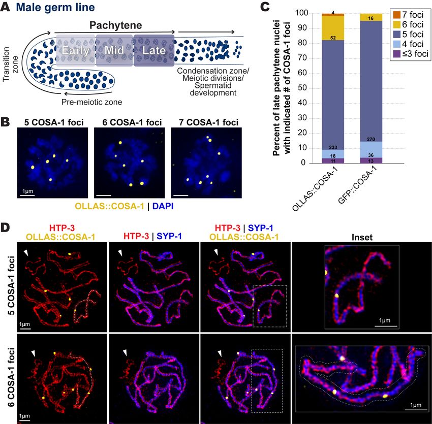
1/6/2023 - Open Access

   Figure 1. COSA-1 foci in late pachytene spermatocytes.
   (A) Diagram of the C. elegans male germ line. Arrows indicate direction of meiotic progression. For this study, we subdivided
   pachytene into three equally sized regions based on the total length of pachytene: early (first third), mid (middle third), and
   late (last third) (See Methods). (B) Representative images of late pachytene nuclei with 5, 6, or 7 OLLAS::COSA-1 foci
   (yellow). (C) Bar graph indicating the percentages of late pachytene nuclei with ≤3, 4, 5, 6, or 7 COSA-1 foci detected in
   strains expressing either OLLAS::COSA-1 or GFP::COSA-1. The numbers within each bar indicate the numbers of nuclei in
   each category from a total of 8 germ lines for OLLAS::COSA-1 and 7 germ lines for GFP::COSA-1. (D) Representative SIM
   images depicting spermatocyte chromosome spreads from late pachytene nuclei with 5 or 6 OLLAS::COSA-1 foci (yellow),
   HTP-3 (red, chromosome axis), and SYP-1 (blue, synaptonemal complex). White arrowheads indicate the unsynapsed
   partnerless X chromosome. The dashed line in the first column traces the path of a single synapsed chromosome pair with
   either one COSA-1 focus (top) or two COSA-1 foci (bottom). The chromosome pair with two COSA-1 foci is outlined in the
   inset shown at the right.
1/6/2023 - Open Access
   Description
   During egg and sperm development in many organisms, homologous chromosome pairs must establish a crossover to ensure
   their proper orientation toward and segregation to opposite spindle poles during the first meiotic division. Despite the
   importance of crossovers for both spermatocyte and oocyte meiosis in most species, the number and distribution of crossovers
   along chromosomes is often sexually dimorphic (Drouaud et al. 2007; Lim et al. 2008; Gabdank and Fire 2014; Brick et al.
   2018; Cahoon and Libuda 2019; Capilla-Perez et al. 2021). From genetic assays performed in C. elegans oocytes, each of the
   6 homologous chromosome pairs typically undergoes only a single crossover, with double crossover meiotic products reported
   as very infrequent (Hodgkin et al. 1979; Nabeshima et al.2004; Lim et al. 2008; Hayashi et al. 2010; Gabdank and Fire 2014;
   Li et al. 2018). Cytological assays in oocytes using a GFP tagged version of the pro-crossover protein COSA-1 (Yokoo et al.
   2012) are consistent with the genetically-detected incidence of crossovers; 6 GFP::COSA-1 foci are detected in late pachytene
   nuclei, which reflects the accumulation of COSA-1 at a single crossover-designated site forming on each of the 6 homologous
   chromosome pairs (bivalents) (Yokoo et al. 2012; Libuda et al. 2013). Based on genetic assays detecting meiotic crossovers on
   Chromosomes I, IV, and V, double crossover events appear to occur more frequently in C. elegans male (XO) spermatocytes
   than in C. elegans hermaphrodite (XX) oocytes (Hodgkin et al. 1979; Henzel et al. 2011; Gabdank and Fire 2014; Li et al.
   2020). Genetic assays have detected double-crossover meiotic products in spermatocyte meiosis at frequencies of 2.7%, 4.4%,
   and 1.7% for chromosomes I, IV, and V, which would imply double-crossover bivalents occurring at frequencies of 10.8%,
   17.6% and 6.8%, respectively (Hodgkin et al. 1979; Gabdank and Fire 2014; Li et al. 2020). Based on these genetic data,
   cytological assays using markers such as COSA-1 might be expected to detect nuclei with one or more bivalents harboring two
   crossover-site foci at frequencies around 40%. However, cytological assessment of GFP::COSA-1 foci in spermatocyte
   meiosis has detected fewer nuclei with apparent double-crossover bivalents than predicted by the genetic data (Li et al. 2020),
   prompting us to evaluate whether different epitope tags on COSA-1 might influence the number of cytologically-detected
   double crossover events in spermatocyte meiosis.
   We used immunofluorescence to visualize crossover-designated sites in spermatocytes using two different tagged versions of
   COSA-1: (1) GFP::COSA-1 expressed from a chromosomally-integrated transgene construct using the pie-1 promotor and the
   cosa-1 3’ UTR (Yokoo et al. 2012); and (2) OLLAS::COSA-1 expressed from the endogenous cosa-1 locus modified using
   CRISPR/Cas9 to introduce the OLLAS tag (Janisiw et al. 2018). In oogenesis, both tagged versions of COSA-1 produce
   similar results displaying 6 COSA-1 foci in late pachytene (Janisiw et al. 2018). In spermatocytes, we found that a majority of
   nuclei displayed 5 COSA-1 foci with both tagged versions of COSA (Figure 1B,C), which is consistent with previous reports
   of a single crossover-designated site on each of the 5 autosome pairs (and none on the partnerless X chromosome) (Li et al.
   2020). Further, the fraction of nuclei in which we detected >5 GFP::COSA-1 foci per nucleus (4.8%) was comparable to that
   observed by Li et al., who used native GFP fluorescence to detect GFP::COSA-1 expressed from a CRISPR-modified
   endogenous cosa-1 locus (P = 0.14, Fisher’s Exact test) (Li et al. 2020). However, we found that the fraction of late pachytene
   nuclei with >5 OLLAS::COSA-1 foci (17.6%) was significantly higher than the fraction of late pachytene nuclei with >5
   GFP::COSA-1 foci (Figure 1B,C; P5 tagged COSA-1 foci may reflect: 1) differences in detection sensitivity for the two markers,
   potentially resulting from the presence of untagged COSA-1 protein expressed from the endogenous cosa-1 locus in the strain
   with the gfp::cosa-1 transgene; and/or, 2) COSA-1 tag-specific effects on crossover designation in spermatocytes. Future
   studies examining spermatocyte COSA-1 foci, which are not uniform in size or intensity (Figure 1C), may provide insights
   into how specific tags influence the detection of COSA-1 foci.
   We used super-resolution Structured Illumination Microscopy (SIM) combined with meiotic chromosome spreads (Woglar and
   Villeneuve 2018) to determine whether nuclei with >5 OLLAS::COSA-1 foci display single synapsed bivalents harboring two
   crossover-site foci. To visualize chromosome structures, we used antibodies detecting the chromosome axis protein HTP-3
   (which forms two parallel tracks, one on each homologous chromosomes) and the synaptonemal complex protein SYP-1
   (which forms a single track between the two HTP-3 tracks). This analysis confirmed the presence of individual synapsed
   bivalents with two OLLAS::COSA-1 foci (Figure 1D). Further, we did not observe OLLAS::COSA-1 foci on the unsynapsed
   hemizygous X chromosome, which assembles chromosome axes containing HTP-3 but does not load SYP proteins (Figure
   1D) (Jaramillo-Lambert and Engebrecht 2010), consistent with previous work indicating a lack of designated crossover sites
   on the X chromosome in male spermatocytes (Checchi et al. 2014). Taken together, our results indicate that: 1) chromosome
   pairs harboring two crossover-designated recombination events in spermatocyte meiosis can be visualized using
   OLLAS::COSA-1; and, 2) COSA-1 exhibits epitope tag-specific differences in the detection of double crossover designated
   sites.

   Methods
1/6/2023 - Open Access
   All worm strains were generated from N2 backgrounds and maintained as mating stocks at 20°C under standard conditions on
   nematode growth media (NGM) with lawns of Escherichia coli (E. coli).
   Immunohistochemistry and Chromosome Spreads
   Immunofluorescence was performed as described in (Libuda et al. 2013; Cahoon and Libuda 2021). A detailed protocol can be
   found here: https://www.protocols.io/view/immunofluorescence-for-c-elegans-gonads-xrgfm3w (Roelens 2019). Chromosome
   spreads were performed in low salt dissection solution (0.1% v/v Tween-20 and 10% v/v Hank’s Balanced Salt Solution
   (Gibco, 24020117) on an ethanol-washed Superfrost Microscope Slide (Fisher Scientific, 12-544-7) and were then prepared as
   described in (Pattabiraman et al. 2017).
   Microscopy
   Immunofluorescence slides of fixed gonad stained for GFP::COSA-1 or OLLAS::COSA-1 were imaged on a GE DeltaVision
   microscope with a 63x/N.A. 1.42 lens and 1.5x optivar at 1024x1024 pixel dimensions. Images were acquired using 0.2 µm Z-
   step size and deconvolved with softWoRx 7.2.2 deconvolution software. 3D-Structured Illumination Microscopy (SIM) was
   performed as described in (Pattabiraman et al. 2017) on a GE DeltaVision OMX Blaze microscopy system (Version
   3.70.9622.0) at the Stanford University Cell Sciences Imaging Core Facility (RRID:SCR_017787). Images were acquired with
   a 100x/N.A. 1.40 oil objective at 512x512 pixel dimensions using 0.125 µm Z-step size. Images were SIM reconstructed and
   registration corrected with softWoRx. All images were max intensity Z-projected and slightly adjusted for brightness and
   contrast.
   Quantification of COSA-1 foci
   Imaged gonad were stitched together using the FIJI (NIH) plugin Stitcher (Release 3.1.9; Preibisch et al. 2009) and analyzed
   in Imaris 9.9 as described in (Toraason et al. 2021) with minor changes. The start of the pachytene zone was defined by the
   first row of nuclei that did not contain more than 1-2 nuclei with DNA in a polarized or crescent shape organization
   characteristic of the transition zone. The end of the pachytene zone was defined as the last row that contained predominantly
   pachytene nuclei with ≤ 1 nucleus exhibiting condensation zone nuclear organization (Shakes et al. 2009). The calculated
   length of the pachytene region was normalized per germline from 0 to 1. Then, the normalized pachytene length was equally
   divided into thirds and the nuclei within the last third (length: 0.67-1.0) were defined as late pachytene.
   Statistical Analysis
   Fisher’s Exact statistical analysis was performed using GraphPad QuickCalcs (2023 GraphPad Software). The P value and test
   used is indicated in the Description section. All n values for nuclei and germlines scored are listed in the figure or figure
   legend.

   Reagents
    Strain       Genotype                                                                      Available from

    AV630        meIs8[unc-119(+) pie-1promoter::gfp::cosa-1] II.                              CGC

    NSV97        cosa-1[ddr12(OLLAS::cosa-1)] III.                                             (Janisiw et al. 2018)

                                                     Animal and
    Antibody                                                                Description              Source
                                                     Clonality

    Anti-OLLAS                                       Rabbit, polyclonal     Use at 1:1,000 - 1:1,500 Genscript, A01658

    Anti-GFP booster Alexa Fluor 488                 N.A.                   Use at 1:200             Chromotek, gb2AF488-50

                                                     Guinea Pig,
    Anti-SYP-1                                                              Use at 1:200             (MacQueen et al. 2002)
                                                     polyclonal

    Anti-HTP-3                                       Chicken, polyclonal    Use at 1:500             (MacQueen et al. 2005)
1/6/2023 - Open Access
Anti-Chicken Alexa Fluor 405 Goat, polyclonal Use at 1:200 Abcam, Ab175675
Anti-Rabbit Alexa Fluor 488 (F(ab)’2
Goat, polyclonal Use at 1:1,000 ThermoFisher, A11070
fragment)
Anti-Guinea Pig Alexa Fluor 555 Goat, polyclonal Use at 1:1,000 ThermoFisher, A21435
Anti-Rabbit Alexa Fluor 488 (whole antibody) Goat, polyclonal Use at 1:200 ThermoFisher, A11034
Acknowledgements: We thank the CGC for strains, which is funded by NIH Office of Research Infrastructure Programs (P40
OD010440). We thank members of the Libuda Lab, specifically Nicole Kurhanewicz for experimental assistance and
discussion on the manuscript. We thank JoAnne Engebrecht for insights and discussion on this manuscript.
References
Brick K, Thibault-Sennett S, Smagulova F, Lam KG, Pu Y, Pratto F, Camerini-Otero RD, Petukhova GV. 2018. Extensive sex
differences at the initiation of genetic recombination. Nature 561: 338-342. PubMed ID: 30185906
Cahoon CK, Libuda DE. 2019. Leagues of their own: sexually dimorphic features of meiotic prophase I. Chromosoma 128:
199-214. PubMed ID: 30826870
Cahoon CK, Libuda DE. 2021. Conditional immobilization for live imaging Caenorhabditis elegans using auxin-dependent
protein depletion. G3 (Bethesda) 11: jkab310. PubMed ID: 34534266
Capilla-Pérez L, Durand S, Hurel A, Lian Q, Chambon A, Taochy C, et al., Mercier R. 2021. The synaptonemal complex
imposes crossover interference and heterochiasmy in Arabidopsis. Proc Natl Acad Sci U S A 118: e2023613118 . PubMed ID:
33723072
Checchi PM, Lawrence KS, Van MV, Larson BJ, Engebrecht J. 2014. Pseudosynapsis and decreased stringency of meiotic
repair pathway choice on the hemizygous sex chromosome of Caenorhabditis elegans males. Genetics 197: 543-60. PubMed
ID: 24939994
Drouaud J, Mercier R, Chelysheva L, Bérard A, Falque M, Martin O, et al., Mézard C. 2007. Sex-specific crossover
distributions and variations in interference level along Arabidopsis thaliana chromosome 4. PLoS Genet 3: e106. PubMed ID:
17604455
Gabdank I, Fire AZ. 2014. Gamete-type dependent crossover interference levels in a defined region of Caenorhabditis elegans
chromosome V. G3 (Bethesda) 4: 117-20. PubMed ID: 24240780
Hayashi M, Mlynarczyk-Evans S, Villeneuve AM. 2010. The synaptonemal complex shapes the crossover landscape through
cooperative assembly, crossover promotion and crossover inhibition during Caenorhabditis elegans meiosis. Genetics 186: 45-
58. PubMed ID: 20592266
Henzel JV, Nabeshima K, Schvarzstein M, Turner BE, Villeneuve AM, Hillers KJ. 2011. An asymmetric chromosome pair
undergoes synaptic adjustment and crossover redistribution during Caenorhabditis elegans meiosis: implications for sex
chromosome evolution. Genetics 187: 685-99. PubMed ID: 21212235
Hodgkin J, Horvitz HR, Brenner S. 1979. Nondisjunction Mutants of the Nematode CAENORHABDITIS ELEGANS. Genetics
91: 67-94. PubMed ID: 17248881
Janisiw E, Dello Stritto MR, Jantsch V, Silva N. 2018. BRCA1-BARD1 associate with the synaptonemal complex and pro-
crossover factors and influence RAD-51 dynamics during Caenorhabditis elegans meiosis. PLoS Genet 14: e1007653.
PubMed ID: 30383754
Jaramillo-Lambert A, Engebrecht J. 2010. A single unpaired and transcriptionally silenced X chromosome locally precludes
checkpoint signaling in the Caenorhabditis elegans germ line. Genetics 184: 613-28. PubMed ID: 20008570
Li Q, Hariri S, Engebrecht J. 2020. Meiotic Double-Strand Break Processing and Crossover Patterning Are Regulated in a
Sex-Specific Manner by BRCA1-BARD1 in Caenorhabditis elegans. Genetics 216: 359-379. PubMed ID: 327960081/6/2023 - Open Access Li Q, Saito TT, Martinez-Garcia M, Deshong AJ, Nadarajan S, Lawrence KS, et al., Engebrecht J. 2018. The tumor suppressor BRCA1-BARD1 complex localizes to the synaptonemal complex and regulates recombination under meiotic dysfunction in Caenorhabditis elegans. PLoS Genet 14: e1007701. PubMed ID: 30383767 Libuda DE, Uzawa S, Meyer BJ, Villeneuve AM. 2013. Meiotic chromosome structures constrain and respond to designation of crossover sites. Nature 502: 703-6. PubMed ID: 24107990 Lim JG, Stine RR, Yanowitz JL. 2008. Domain-specific regulation of recombination in Caenorhabditis elegans in response to temperature, age and sex. Genetics 180: 715-26. PubMed ID: 18780748 MacQueen AJ, Colaiácovo MP, McDonald K, Villeneuve AM. 2002. Synapsis-dependent and -independent mechanisms stabilize homolog pairing during meiotic prophase in C. elegans. Genes Dev 16: 2428-42. PubMed ID: 12231631 MacQueen AJ, Phillips CM, Bhalla N, Weiser P, Villeneuve AM, Dernburg AF. 2005. Chromosome sites play dual roles to establish homologous synapsis during meiosis in C. elegans. Cell 123: 1037-50. PubMed ID: 16360034 Nabeshima K, Villeneuve AM, Hillers KJ. 2004. Chromosome-wide regulation of meiotic crossover formation in Caenorhabditis elegans requires properly assembled chromosome axes. Genetics 168: 1275-92. PubMed ID: 15579685 Pattabiraman D, Roelens B, Woglar A, Villeneuve AM. 2017. Meiotic recombination modulates the structure and dynamics of the synaptonemal complex during C. elegans meiosis. PLoS Genet 13: e1006670. PubMed ID: 28339470 Preibisch S, Saalfeld S, Tomancak P. 2009. Globally optimal stitching of tiled 3D microscopic image acquisitions. Bioinformatics 25: 1463-5. PubMed ID: 19346324 Roelens B. 2019. Immunofluorescence for C. elegans gonads. protocols.io. DOI: dx.doi.org/10.17504/protocols.io.xrgfm3w Shakes DC, Wu JC, Sadler PL, Laprade K, Moore LL, Noritake A, Chu DS. 2009. Spermatogenesis-specific features of the meiotic program in Caenorhabditis elegans. PLoS Genet 5: e1000611. PubMed ID: 19696886 Toraason E, Adler VL, Kurhanewicz NA, DiNardo A, Saunders AM, Cahoon CK, Libuda DE. 2021. Automated and customizable quantitative image analysis of whole Caenorhabditis elegans germlines. Genetics 217: iyab010. PubMed ID: 33772283 Woglar A, Villeneuve AM. 2018. Dynamic Architecture of DNA Repair Complexes and the Synaptonemal Complex at Sites of Meiotic Recombination. Cell 173: 1678-1691.e16. PubMed ID: 29754818 Yokoo R, Zawadzki KA, Nabeshima K, Drake M, Arur S, Villeneuve AM. 2012. COSA-1 reveals robust homeostasis and separable licensing and reinforcement steps governing meiotic crossovers. Cell 149: 75-87. PubMed ID: 22464324 Funding: This work was supported by the National Institutes of Health R35GM128890 to DEL, a Jane Coffin Childs Postdoctoral Fellowship and National Institutes of Health 1K99HD109505-01 to CKC, National Institutes of Health R35GM126964 to AMV, and a Stanford Dean’s Fellowship award to CJU. DEL is also a Searle Scholar and recipient of a March of Dimes Basil O’Connor Starter Scholar award. SIM imaging was performed at Stanford University Cell Sciences Imaging Core Facility, which is supported by Award Number 1S10OD01227601 from the National Center for Research Resources. Author Contributions: Cori K. Cahoon: conceptualization, data curation, formal analysis, writing - original draft, writing - review editing, funding acquisition, investigation. Celja J. Uebel: data curation, formal analysis, funding acquisition, writing - review editing. Anne M. Villeneuve: funding acquisition, writing - review editing. Diana E. Libuda: conceptualization, funding acquisition, investigation, writing - review editing. Reviewed By: Anonymous History: Received December 16, 2022 Revision Received December 23, 2022 Accepted January 5, 2023 Published Online January 6, 2023 Indexed January 20, 2023 Copyright: © 2023 by the authors. This is an open-access article distributed under the terms of the Creative Commons Attribution 4.0 International (CC BY 4.0) License, which permits unrestricted use, distribution, and reproduction in any medium, provided the original author and source are credited. Citation: Cahoon, CK; Uebel, CJ; Villeneuve, AM; Libuda, DE (2023). Epitope tag-specific differences in the detection of COSA-1 marked crossover sites in C. elegans spermatocytes. microPublication Biology. 10.17912/micropub.biology.000724
You can also read


