Emphysematous pyelonephritis in an infant from Sokoto, north-western Nigeria
←
→
Page content transcription
If your browser does not render page correctly, please read the page content below
African Journal of Laboratory Medicine
ISSN: (Online) 2225-2010, (Print) 2225-2002
                                                          Page 1 of 4      Case Study

                     Emphysematous pyelonephritis in an infant
                        from Sokoto, north-western Nigeria

 Authors:                              Introduction: Emphysematous pyelonephritis is a life-threatening necrotising bacterial
 Fatima B. Jiya1
                                       infection of the kidneys. It is rare among children and can be fatal if not promptly identified
 Paul K. Ibitoye1
 Nma M. Jiya1                          and treated.
 Maryam Amodu-Sanni1
 Yahaya Mohammed2
                                       Case presentation: A 7-month-old male infant presented to the Emergency Paediatric Unit of
 Dada M. Aquib3                        Usmanu Danfodiyo University Teaching Hospital, Sokoto, Nigeria, on 12 November 2019
 Lukman K. Coker1                      with a 5-day history of fever and vomiting, and a 3-day history of a progressively enlarging,
                                       left-side abdominal mass. There was associated excessive crying on micturition, refusal to
 Affiliations:
 1
  Department of Paediatrics,
                                       feed and weight loss. He looked ill and was in respiratory distress, irritable, febrile (38.8 °C),
 Usmanu Danfodiyo University           moderately dehydrated and pale. His weight and length were 5.5 kg and 64 cm. He had a
 Teaching Hospital, Sokoto,            tender, firm and ballotable abdominal mass on the left flank measuring 8 cm × 10 cm. His
 Nigeria                               pulse rate was 140 beats/min, blood pressure 60/40 millimetres of mercury and respiratory
                                       rate was 65 cycles/min. He had widespread coarse crepitations and normal heart sounds on
 2
  Department of Medical
 Microbiology and                      chest auscultation.
 Parasitology, Usmanu
                                       Management and outcome: An initial diagnosis of sepsis was made. Other considerations
 Danfodiyo University
 Teaching Hospital, Sokoto,            were nephroblastoma and neuroblastoma. Ceftriaxone and blood transfusion were
 Nigeria                               commenced with subsequent administration of intravenous fluids. Further radiologic
                                       investigations revealed emphysematous pyelonephritis. The patient had percutaneous
 3
  Department of Radiology,             drainage and extended spectrum β-lactamase-producing Escherichia coli (sensitive to
 Usmanu Danfodiyo University
 Teaching Hospital, Sokoto,
                                       meropenem) which was isolated from the aspirate culture after 48 h of incubation.
 Nigeria                               Meropenem could not be commenced because of non-availability and high cost. The patient
                                       subsequently deteriorated and died from septic shock.
 Corresponding author:
 Fatima Jiya,                          Conclusion: Emphysematous pyelonephritis has a fulminant course when not diagnosed
 fatimabellojiya@gmail.com             promptly and treated adequately.
 Dates:                                Keywords: Emphysematous pyelonephritis; infection; kidney; infant; Sokoto.
 Received: 27 Jan. 2020
 Accepted: 03 Dec. 2020
 Published: 26 Apr. 2021
                                      Introduction
 How to cite this article:
                                      Emphysematous pyelonephritis (EPN) is a severe necrotising and progressive infection of
 Jiya FB, Ibitoye PK, Jiya NM,
 et al. Emphysematous                 the kidneys which is characterised by the formation of gas within the renal parenchyma,
 pyelonephritis in an                 collecting system, or the perinephric tissue.1 It is rare in children, with the majority of
 infant from Sokoto,                  cases occurring among adults with diabetes mellitus.2 Gas-forming organisms are said to be the
 north-western Nigeria. Afr J
 Lab Med. 2021;10(1), a1181.          causative organisms of which Escherichia coli is the most common.3 The pathogenesis is unclear
 https://doi.org/10.4102/ajlm.        but factors thought to increase predisposition to developing EPN include decreased host
 v10i1.1181                           immunity, increased levels of glucose in the tissue, impaired tissue perfusion, obstruction of the
                                      urinary system and presence of gas-forming organisms in the host tissue.1 The diagnosis of EPN
 Copyright:
 © 2021. The Authors.                 requires clinical features supported by radiologic investigations and isolation of the offending
 Licensee: AOSIS. This work           organisms. Depending on the stage of the disease, treatment could be medical alone or a
 is licensed under the                combination of medical and surgical interventions.1
 Creative Commons
 Attribution License.
                                      Ethical considerations
                                      Ethical approval to conduct the study was obtained from UDUTH Health Research Ethics
                                      Committee with registration number NHREC/30/012/2019. Authors obtained permission from
                                      the caregivers to publish the clinical details of the patient.
     Read online:
                    Scan this QR
                    code with your
                    smart phone or
                                      Case presentation
                    mobile device     We report the case of Y.B (initials of infant used to retain anonymity), a 7-month-old male infant
                    to read online.
                                      that was referred from a secondary health facility in Sokoto, Nigeria, to Usmanu Danfodiyo

                                            http://www.ajlmonline.org      Open Access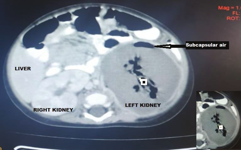
Page 2 of 4 Case Study
University Teaching Hospital (UDUTH), Sokoto in November but urine microscopy and culture yielded no significant growth
2019. He was brought by his parents to the Emergency (Table 1). Human immunodeficiency virus DNA polymerase
Paediatric Unit of UDUTH on account of a 5-day history chain reaction was negative. Chest radiograph revealed
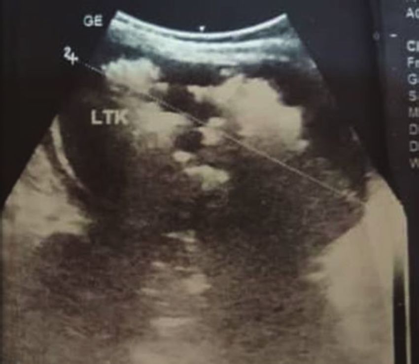
of fever and vomiting, and a 3-day history of progressively multiple patchy perihilar opacities. Abdominal ultrasound
enlarging left-side abdominal mass which was noticed demonstrated relative renomegaly of the left kidney with a
incidentally and said to be tender to touch. There was no bipolar length of 99 mm, turbid collection with multiple pockets
preceding history of trauma and there were no masses on of air within it and marked thinning of the parenchyma. The
other body parts. There was associated excessive crying, right kidney was normal in outline, position and size (72 mm in
crying on micturition, refusal to feed and weight loss. He was bipolar length) and other organs were normal in appearance
admitted at the referring hospital at the onset of illness where (Figure 1). Computerised tomography (CT) scan of the
he had anti-malaria (artesunate), and antibiotic (cefuroxime)
abdomen revealed an enlarged left kidney with bipolar length
treatment, as well as a blood transfusion with no significant
and transverse diameters of 97 mm and 68 mm, reduced renal
improvement, necessitating referral to UDUTH Sokoto 72 h
parenchyma enhancement (Hounsfield units = 36–42), while
later. Operating within a resource-constrained health system,
multiple oval and tubular negative density areas (Hounsfield
UDUTH is the highest tertiary level healthcare facility within
units = 543–723) were noted centrally and in subcapsular
the state. Both of his parents have no formal education and
regions suggestive of air in the collecting system and renal
are ‘petty traders’ with a combined average earning of
40 000.00 Nigerian naira ($132.00 United States dollars [USD]) parenchyma with overall features in keeping with EPN.
per month.
Physical examination revealed an ill-looking child in
respiratory distress, irritable, febrile (axillary temperature =
38.8 °C), moderately dehydrated, pale, anicteric acyanosed
with no significant peripheral lymphadenopathy. His weight
and length were 5.5 kg and 64 cm, while oxygen saturation
(SPO2) was 89% in room air. He had a tender, firm and
ballotable abdominal mass extending from the left lumbar
region to the left iliac region, measuring 8 cm × 10 cm. The
right kidney, liver and spleen were not palpable. He had
normal male external genitalia and was not circumcised.
His pulse rate was 140 beats/min, blood pressure
60/40 millimetres of mercury and respiratory rate was
65 cycles/min. Chest auscultation revealed vesicular breath
sounds with widespread coarse crepitations and normal
heart sounds. The neurologic examination was also normal.
A clinical diagnosis of sepsis with focus on the chest and
urinary tract with malaria was made. Other considerations
were nephroblastoma and neuroblastoma.
Management and outcome
Broad spectrum empirical antibiotic (intravenous ceftriaxone)
treatment was commenced empirically, and the patient
was transfused with blood (haematocrit was 22%) and
subsequently placed on intravenous fluids. Blood film for
malaria parasite was negative, and metabolic panel and FIGURE 1: Sonogram of the left kidney of an emphysematous pyelonephritis
patient at the Usmanu Danfodiyo University Teaching Hospital, Sokoto, Nigeria,
complete blood count were normal except for anaemia (Table 1). November 2019. Figure shows renomegaly, reduced renal parenchyma echogenicity
Urinalysis showed proteinuria, glycosuria and leucocyturia and multiple irregular mixed echoes with dirty shadowing (blue arrow).
TABLE 1: Laboratory data of the emphysematous pyelonephritis patient on the second day of admission at the Usmanu Danfodiyo University Teaching Hospital, Sokoto,
Nigeria, November 2019.
Metabolic panel Blood count Clotting profile Urinalysis Urine microscopy. culture, sensitivity
Sodium 136 mmol/L White blood cells 11.5 × 10⁹/L PT-test 16 s Turbid Pus cells +
Potassium 4.1 mmol/L Lymphocites 7.2 × 10⁹/L PT-control 14 s Protein + Yeasts
Chlorine 97 mmol/L Granulocytes 3.5 × 10⁹/L PTTK-test 32 s Blood – No growth
Bicarbonate 22 mmol/L Haematocrit 22% PTTK-control 31 s Glucose + -
Blood urea nitrogen 4.4 mmol/L Platelets 123 × 109/L INR- 1.16 Leucocyte ++ -
Creatinine 0.5 mg/dL - - Nitrite -
Glucose 2.8 mmol/L - - - -
PT, prothrombin time; PTTK, partial thromboplastim time with kaolin; INR, international normalised ratio.
http://www.ajlmonline.org Open Access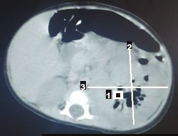
Page 3 of 4    Case Study

                                                                                    Discussion
                                                                                    The 7-month-old male infant in this study had acute onset of
                                                                                    fever, vomiting and a left-side ballotable abdominal mass
                                                                                    with radiologic features of stage II EPN confirmed to be
                                                                                    caused by extended spectrum β-lactamase E. coli on aspirate
                                                                                    microscopy, culture and sensitivity. Emphysematous
                                                                                    pyelonephritis is a severe necrotising infection of the renal
                                                                                    parenchyma that causes gas accumulation within the renal
                                                                                    tissues, with or without the involvement of the peri-renal
                                                                                    spaces.4,5 Reports of EPN has been documented in adults,
                                                                                    especially among diabetics, hypertensives and renal
                                                                                    transplant recipients following end-stage renal disease.6
                                                                                    Women are said to be more at risk than men.1,5 Emphysematous
FIGURE 2: Contrast enhanced computed tomogram of an emphysematous
pyelonephritis patient at the Usmanu Danfodiyo University Teaching Hospital,        pyelonephritis is rare among children with the first known
Sokoto, Nigeria, November 2019. Figure shows left renomegaly, contrast              paediatric case reported in a 10-year-old female in 1985.7
enhancement and multiple air densities within the calyceal system (orange arrow).
                                                                                    There are few reports of EPN among children in studies from
                                                                                    South Africa,8 Texas9 and Saudi Arabia,10 with none of the
                                                                                    patients having diabetes mellitus nor was there significant
                                                                                    gender preponderance in the occurrence of EPN. However,
                                                                                    one of the reported cases was a transplant recipient.10 The
                                                                                    index patient is, to the best of our knowledge, the first
                                                                                    documented case in a child in Sokoto State, and possibly in
                                                                                    Nigeria. Unlike in previous reports9,10 in which cases had
                                                                                    underlying medical conditions (chromosomal abnormality
                                                                                    with ectopic right ureter, and end-stage renal disease from
                                                                                    neurogenic bladder), our case was apparently healthy with no
                                                                                    identifiable risk factors prior to the development of EPN.
                                                                                    Possibly, our case is an indication that EPN can in very rare
                                                                                    instances occur in previously apparently healthy children.
                                                                                    The most common causative bacterial organism is E. coli,
FIGURE 3: Non–contrast-enhanced computed tomogram of an emphysematous               which is also the organism that was isolated from the aspirate
pyelonephritis patient at the Usmanu Danfodiyo University Teaching Hospital,        specimen of our patient. Other pathogenic agents of
Sokoto, Nigeria, November 2019. Figure shows left renomegaly with multiple air
density area within the parenchyma and subcapsular region (orange arrows).          EPN include Klebsiella, Proteus, Pseudomonas, Citrobacter,
                                                                                    Enterococcus and Streptococcus species. Rare organisms such as
The right kidney was normal in position, outline and size                           coagulase negative Staphylococcus, Clostridium, Candida
(bipolar diameter 63.8 mm and transverse diameter 40.8 mm),                         species and Aspergillus fumigates have also been reported.1,3,9,10
no mass lesions or calculus were seen, and other organs                             Unlike in our case, the study by Siddique et al.9 reported
were normal (Figure 2 and Figure 3). The patient’s diagnosis                        Enterobacter cloacae as the causative agents of EPN in their
was changed to septicaemia complicated by unilateral                                patient. The pathogenesis of EPN is not yet known; however,
(left) emphysematous pyelonephritis class II. Ceftriaxone                           the formation of carbon dioxide from the fermentation of
treatment was continued while awaiting the culture result, and                      glucose in urine and kidney tissues is thought to be the main
an ultrasound guided percutaneous drain was inserted, which                         mechanism.1 The predisposing factors that have been
drained about 60 mL of purulent, blood-stained material                             implicated include the presence of a gas-forming bacterial
containing air bubbles. The result of aspirate microscopy,                          organisms, high glucose levels in tissues, immunosuppression
culture and sensitivity revealed numerous pus cells on                              (e.g. diabetes and immunosuppressive therapy), urinary
microscopy, and growth of E. coli confirmed to be extended                          tract obstruction, and poor tissue perfusion.1,11,12 Local tissue
spectrum β-lactamase-positive via phenotypic method, which                          ischaemia in the presence of a gas-producing pathogen is
was sensitive only to meropenem. Antibiotic therapy was                             thought to exacerbate tissue destruction, encourage the
changed to intravenous meropenem but could not be                                   production of pus, and inhibit the removal of locally produced
commenced due to unavailability, as well as the high cost of the                    gas, leading to EPN.11 Other speculated mechanisms are that
medication where available. Although there was reduction in                         the increased levels of glucose in the tissues together with
his temperature to 37.9 °C, he subsequently developed shock                         decreased blood supply to the kidneys contributes to the
and deteriorated rapidly. He did not respond to resuscitation                       anaerobic metabolism of glucose and lactate by the organisms
and died 24 h later. Although not the recommended treatment                         and thereafter the production of gases like carbon dioxide,
for this case with stage II EPN, nephrectomy was considered                         hydrogen, nitrogen, oxygen and methane by the gas-forming
but the rapidity in deterioration in the patient’s condition                        organisms.13 The clinical manifestation of EPN in children is
(shock and death within few hours) did not allow for adequate                       similar to that of adults with the main features being fever,
counselling and preparation for the procedure.                                      anorexia, nausea, vomiting, flank pain with or without

                                                   http://www.ajlmonline.org        Open Access
Page 4 of 4 Case Study
palpable mass, and dysuria.7,13 Our patient presented with timely identification of cases and will improve the outcome
some of the aforementioned symptoms. The classification of of management of initial classes (1–3) of EPN.
EPN is via radiologic imaging, and the most commonly used
classification system is by Huang and Seng using CT scan to
classify EPN into five (1–4b) classes.1 Our patient’s CT scan
Acknowledgements
findings (Figure 2 and Figure 3) placed him at EPN class II Competing interests
because the demonstrated air went beyond the left collecting The authors have declared that no competing interests exist.
system to involve the renal parenchyma. Although earlier
studies9,10 reported renal ultrasonograms suggesting air
Authors’ contributions
collection in the renal parenchyma of their patients, EPN was
not staged using a impossible CT scan in the studies making it F.B.J. conceptualised, designed and wrote the original draft.
impossible to compare the stage of EPN in our study with P.K.I., N.M.J. and Y.M. critically revised and supervised the
theirs. The clinical presentation, radiologic imaging and manuscript. M.A.-S., D.M.A. and L.K.C. acquired the data.
isolation of causative organisms confirms the diagnosis of All authors approved the final version to be published.
EPN.1 Modalities of treating patients with EPN are said to
have changed over time, with intensive conservative Sources of support
management assuming a prominent position, depending on
This research received no specific grant from any funding
the class of EPN and other co-morbidities. Broad spectrum
agency in the public, commercial or not-for-profit sectors.
antibiotic therapy with percutaneous drainage is the standard
recommended treatment protocol for EPN classes I and II. The
treatment options for patients with EPN classes III and IV Data availability
depend on the presence and number of risk factors such as Data sharing is not applicable to this article as no new data
shock, acute kidney injury, thrombocytopaenia and coma. The were created or analysed in this study.
choice of treatment for patients with fewer than two risk
factors is antibiotics and percutaneous drainage. In the
presence of two or more risk factors, nephrectomy is the Disclaimer
recommended treatment.1 Our patient had percutaneous The views and opinions expressed in this article are those of
drainage but could not commence the appropriate antibiotic the authors and do not necessarily reflect the official policy or
(meropenem) therapy due to non-availability and high cost. position of any affiliated agency of the authors.
Although UDUTH is the highest tertiary level option within
the state, meropenem is not indigenously produced to the best
of our knowledge. Although available, his parents could not
References
afford the supply of ten 500 mg powder for the injection, 1. Huang JJ, Tseng CC. Emphysematous pyelonephritis: Clinicoradiological
classification, management, prognosis, and pathogenesis. Arch Intern Med.
which cost 18 000.00 Nigerian naira ($59.00 USD).14 2000;160(6):797–805. https://doi.org/10.1001/archinte.160.6.797
2. Pontin AR, Barnes RD, Joffe J, Kahn D. Emphysematous pyelonephritis in diabetic
patients. Br J Urol. 1995;75(1):71–74. https://doi.org/10.1111/j.1464-410X.1995.
Unlike in our patient, other studies have reported good tb07237.x
response to antibiotics like third-generation cephalosporin, 3. Mohsin N, Budruddin M, Lala S, Al-Taie S. Emphysematous pyelonephritis: A case
report series of four patients with review of literature. Ren Fail.
fluoroquinolones and vancomycin.6,8,9 Good outcomes for 2009;31(7):597–601. https://doi.org/10.1080/08860220903003396
EPN have been reported with prompt initiation of 4. Lu YC, Chiang BJ, Pong YH, et al. Emphysematous pyelonephritis: Clinical
characteristics and prognostic factors. Int J Urol. 2014;21(3):277–282. https://doi.
recommended treatment.9 However, EPN may run a fatal org/10.1111/iju.12244
course if not identified and treated early.4,5,9 There was a delay 5. Schicho A, Stroszczynski C, Wiggermann P. Emphysematous cystitis: Mortality, risk
factors, and pathogens of a rare disease. Clin Pract. 2017;7(2):930. https://doi.
in diagnosing the index case due to late presentation. org/10.4081/cp.2017.930
Additionally, the isolated organism (extended spectrum 6. Camelia A, Abhijit S, Shankar A, et al. A case series of emphysematous
β-lactamase E. coli) was resistant to the empirical antibiotic pyelonephritis. Case Rep Med. 2014;2014:587926. https://doi.org/10.1155/
2014/587926
(ceftriaxone) therapy. The recommended antibiotic 7. Pode D, Perlberg S, Fine H. Emphysematous renal and perirenal infection in
(meropenem) could not be commenced because of the nondiabetic patient. Urology. 1985;26:313–315. https://doi.org/10.1016/0090-
4295(85)90139-6
aforementioned reasons. It is, therefore, not surprising that
8. Ambaram PR, Petersen KL. Emphysematous pyelonephritis in children. Pediatr Infect
the patient succumbed to the illness. Dis J. 2016;35(10):1159–1161. https://doi.org/10.1097/INF.0000000000001254
9. Siddique K, Seikaly MG. Emphysematous pyelonephritis in an infant. Pediatr Infect
Dis J. 2013;32(10):1157–1158. https://doi.org/10.1097/INF.0b013e31829aaae3
Conclusion 10. Al-Makadma AS, Al-Akash SI. An unusual case of pyelonephritis in a pediatric renal
transplant recipient. Pediatr Transplant. 2005;9(2):258–260. https://doi.
Our experience brings to the fore the occurrence of EPN in an org/10.1111/j.1399-3046.2004.00276.x
infant and the fulminant course it can take when not diagnosed 11. Turney JH. Renal conservation for gas-forming infections. Lancet.
2000;355(9206):770–771. https://doi.org/10.1016/S0140-6736(99)00351-7
promptly and treated adequately. Our case highlights the 12. Yang WH, Shen NC. Gas-forming infection of the urinary tract: An investigation of
importance of a high index of suspicion, the resistance of fermentation as a mechanism. J Urol. 1990;143(5):960–964. https://doi.
org/10.1016/S0022-5347(17)40151-0
extended spectrum β-lactamase E. coli to the empirical 13. Singh SU, McGlynn L, Fordham M. Emphysematous pyelonephritis. BJU Int.
antibiotic (ceftriaxone), and the need to ensure availability as 2010;107(9):1474–1478. https://doi.org/10.1111/j.1464-410X.2010.09660.x
well as cost effectiveness of recommended antibiotics in the 14. Drugs.com. Meropenem prices, coupons and patient assistance programs
[homepage on the Internet]. [cited 2019 Nov 10]. Available from: http://www.
study location. These measures will go a long way in the drugs.com
http://www.ajlmonline.org Open AccessYou can also read