Embryonic stem cell bioprinting for uniform and controlled size embryoid body formation
←
→
Page content transcription
If your browser does not render page correctly, please read the page content below
BIOMICROFLUIDICS 5, 022207 共2011兲
Embryonic stem cell bioprinting for uniform and controlled
size embryoid body formation
Feng Xu,1 BanuPriya Sridharan,1 ShuQi Wang,1 Umut Atakan Gurkan,1
Brian Syverud,2 and Utkan Demirci1,3,a兲
1
Department of Medicine, Demirci Bio-Acoustic-MEMS in Medicine (BAMM) Laboratory,
Center for Biomedical Engineering, Brigham and Women’s Hospital, Harvard
Medical School, Boston, Massachusetts 02139, USA
2
Digilab Inc., 84 October Hill Road, Holliston, Massachusetts 01746, USA
3
Harvard-MIT Health Sciences and Technology, Cambridge, Massachusetts 02139, USA
共Received 20 December 2010; accepted 23 March 2011; published online 29 June 2011兲
Embryonic stem cells 共ESCs兲 are pluripotent with multilineage potential to differ-
entiate into virtually all cell types in the organism and thus hold a great promise for
cell therapy and regenerative medicine. In vitro differentiation of ESCs starts with
a phase known as embryoid body 共EB兲 formation. EB mimics the early stages of
embryogenesis and plays an essential role in ESC differentiation in vitro. EB uni-
formity and size are critical parameters that directly influence the phenotype ex-
pression of ESCs. Various methods have been developed to form EBs, which in-
volve natural aggregation of cells. However, challenges persist to form EBs with
controlled size, shape, and uniformity in a reproducible manner. The current
hanging-drop methods are labor intensive and time consuming. In this study, we
report an approach to form controllable, uniform-sized EBs by integrating bioprint-
ing technologies with the existing hanging-drop method. The approach presented
here is simple, robust, and rapid. We present significantly enhanced EB size uni-
formity compared to the conventional manual hanging-drop method.
© 2011 American Institute of Physics. 关doi:10.1063/1.3580752兴
I. INTRODUCTION
Embryonic stem cells 共ESCs兲 display indefinite self-renewal and they are a pluripotent cell
source with multilineage differentiation potential.1,2 The unique features of pluripotency make
ESCs an ideal source for tissue replacement and regenerative medicine for diseases and injuries.3,4
In vitro differentiation of ESCs into other phenotypes is preceded by the formation of embryoid
bodies 共EBs兲. EBs are three dimensional 共3D兲 aggregates of ESCs with characteristics of the early
stages of embryogenesis and play a critical role during in vitro differentiation of ESCs. The lack
of uniformity in EB size may result in nonhomogeneous and asynchronous differentiation of the
residing cells.5,6 Therefore, formation of EBs with uniform sizes is needed to effectively employ
ESCs in regenerative medicine.
Three germ layers form in the early stages of embryogenesis in vivo, which are also observed
in EB culture in vitro.7 Therefore, EB provides a suitable microenvironment for ESCs in vitro,
which facilitates lineage-specific differentiation.8,9 It was previously shown that EB-mediated
differentiation efficiency is dependent on the EB size. Larger EB sizes tend to differentiate toward
mesoderm and endoderm, while smaller EB sizes direct their differentiation toward ectoderm.5,6 It
is also reported that smaller sized EBs 共100– 500 m in the lateral dimensions and 120 m in
depth兲 are more likely to allow cardiomyocyte differentiation.10 On the other hand, EB formation
from individual ESCs via spontaneous aggregation is inefficient,11,12 which results in heteroge-
neous size distribution and noncontrolled differentiation lineage.13,14
a兲
Author to whom correspondence should be addressed. Electronic mail: udemirci@rics.bwh.harvard.edu.
1932-1058/2011/5共2兲/022207/8/$30.00 5, 022207-1 © 2011 American Institute of Physics
Author complimentary copy. Redistribution subject to AIP license or copyright, see http://bmf.aip.org/bmf/copyright.jsp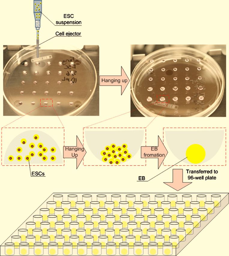
022207-2 Xu et al. Biomicrofluidics 5, 022207 共2011兲
Although various methods have been developed to promote EB formation through natural
aggregation or artificial cell-cell interactions, it is still challenging to obtain controllable, uniform-
sized EBs. For instance, enzymatic digestion of the ESC colonies and rotary mass suspension
resulted in heterogeneous size distribution of EBs,13,15,16 while methods based on surface pattern-
ing can only control the initial EB size.6,17–19 The hanging-drop method 共based on manual pipet-
ting兲 is commonly used in ESC cultures to form EBs. However, the EB size through this method
is a variable due to variation during pipetting, such as droplet volume and number of cells per
droplet. Additionally, these manual methods are labor intensive and time consuming, and the
reproducibility of the results varies between operators. Nonadhesive microwell arrays of various
aspect ratios, sizes, and shapes have been developed to control the uniformity of EB size and
shape through physically controlling the size of growing EBs.14,20–22 However, ESCs formed disk
shaped EBs on microwell arrays, while they aggregated in spherical form in suspension cultures,20
suggestive of different phenotypes.23 Furthermore, the mechanical stress induced on ES cells
during the forced aggregation process 共e.g., rotary mass suspension24–26 and centrifugation12兲 may
disrupt the cell-cell signaling10 and damage the fragile cellular components affecting subsequent
cell differentiation.27 Methods for sorting EBs of heterogeneous size into uniform size groups have
also been developed.27 However, the separation methods generally involve external force fields
such as microfluidics27 that may damage ESCs and affect subsequent cell differentiation.
We hypothesized that the recent advances in bioprinting technologies would facilitate the
formation of uniform-sized EBs in a reproducible manner, addressing the challenges associated
with the current methods. To validate this hypothesis, we integrated a cell printing technique28–30
with the existing hanging-drop culture method. Although a variety of cell bioprinting methods,
such as acoustic printing,31–33 valve based printing,34–36 and ink-jet printing,37,38 have been used to
encapsulate cells in microdroplets,39,40 the combination of these cell printing techniques with the
hanging-drop method for EB formation has not yet been evaluated. Here, we present a new
method based on bioprinting and hanging-drop methods that results in controllable uniform-sized
EBs. The developed method provides a reproducible, efficient, and scalable alternative to the
currently available methods. These uniform-sized EBs would be highly applicable towards regen-
erative medicine and tissue replacement.
II. MATERIALS AND METHODS
A. ESC culture
In this study, we used genetically engineered mouse ESCs 共mESCs, line E14兲. These cells
expressed green fluorescent protein 共GFP兲 upon initiation of gene transfection at the Oct4 pro-
moter at goosecoid 共Gsc兲 gene locus. The mESCs were cultured in high glucose-Dulbecco’s
modified eagles medium 共Gibco兲 supplemented with 10% 共vol/vol兲 ES qualified phosphate
buffered saline 共PBS兲 共Gibco兲, 1 mM L-glutamine 共Gibco兲, 0.1 mM -mercaptoethanol 共Sigma,
St. Louis, MO兲, 100 U/ml penicillin, 100 g / ml streptomycin 共Gibco兲, and 1000 U/ml of
leukemia inhibitory factor 共Chemicon兲.
B. Embryoid body formation
1. Bioprinted hanging-drop method
The mESC suspensions were prepared at three concentrations 共0.1⫻ 106, 0.5⫻ 106 and 1.0
⫻ 106 cells/ ml兲 in basic EB medium containing alpha minimal essential medium 共␣-MEM;
Gibco兲 supplemented with 15% 共v/v兲 heat-inactivated FBS 共Invitrogen, Carlsbad, CA兲 and 1%
共v/v兲 penicillin/streptomycin. Droplets of mESC suspension at controlled volumes 共1, 4, 10, and
20 l兲 were bioprinted onto inside surface of a Petri dish lid in an array format by a cell printer
共Fig. 1兲 mimicking the existing hanging-drop approach. In hanging-drop method, droplets of ESC
suspension were generated by manual pipetting.9 The cells were allowed to aggregate with the
help of gravity when the Petri dish was reversed. The bioprinted droplets were hung in the Petri
Author complimentary copy. Redistribution subject to AIP license or copyright, see http://bmf.aip.org/bmf/copyright.jsp022207-3 Bioprinting for EB formation Biomicrofluidics 5, 022207 共2011兲
FIG. 1. Schematic of the EB formation process using bioprinting approach. Droplets of cell-medium suspension were
bioprinted onto the lid of a Petri dish and were hung up for 24 h to allow for EB aggregation. The formed EBs were
transferred to a 96-well plate for additional culture up to 96 h.
dish for 24 h 共Fig. 1兲. The dish bottom was then filled with PBS to prevent drying of bioprinted
droplets. The aggregated mESCs within the bioprinted droplets were then transferred to low-
adherence 96-well plates and cultured for 96 h to generate EBs 共Fig. 1兲.
2. Standard hanging-drop method using manual pipetting
For control groups, the same droplet size and cell concentrations were used as the bioprinted
hanging-drop method. EBs formed by the standard hanging-drop approach using manual pipetting
were used as a control group following the commonly used protocols.9 ESCs were seeded onto the
Petri dish lids under similar conditions as the bioprinting method. ESCs aggregated within the
droplet for 24 h, and then these aggregates were transferred into 96-well plates. EBs formed using
this method were cultured for 96 h.
C. EB morphological observation and EB cell death
The EB sizes formed using different initial cell seeding density 共0.1⫻ 106, 0.5⫻ 106, and
1.0⫻ 106 cells/ ml兲, droplet size 共1, 4, 10, and 20 l兲, and culture time 共t = 24, 48, 72, and 96 h兲
were analyzed for control and bioprinting groups. The morphology of EBs and Oct4-GFP expres-
sion of ESCs within EBs were observed at different time points after bioprinting 共t = 24, 48, 72,
and 96 h兲 using an inverted fluorescent microscope 共Nikon Eclipse T2000兲. The EB sizes were
measured from the collected micrographs using the NIH IMAGEJ program 共developed at the U.S.
Author complimentary copy. Redistribution subject to AIP license or copyright, see http://bmf.aip.org/bmf/copyright.jsp022207-4 Xu et al. Biomicrofluidics 5, 022207 共2011兲
National Institutes of Health and is available at http://rsb.info.nih.gov/nih-image/兲. In this study,
we used GFP expressing ES cells. These cells show green fluorescence, when they are undiffer-
entiated. EB cell death was analyzed at t = 96 h by incubating EBs only in 4 M ethidium
homodimer 共Molecular Probes Inc.兲 in PBS for 10 min at 37 ° C to indicate the dead cells. The
size uniformity and the resulting diameters at the end of the culture period were assessed and
compared to the control and bioprinted groups.
D. Statistical analysis
The experimental results for both control and bioprinted groups were initially tested for
normal distribution using Anderson–Darling test. The sample size used for each experimental
group was between 10 to 25 droplets. The effects of droplet size, initial cell seeding density, and
culture time on the EB sizes were analyzed with one way analysis of variance with Tukey post hoc
comparisons. The EB size uniformity was statistically assessed with Levene’s test for equality of
variances at the end of the 96 h culture period for all initial cell seeding densities 共0.1⫻ 106,
0.5⫻ 106, and 1.0⫻ 106 cells/ ml兲 and droplet sizes 共1, 4, 10, and 20 l兲. The uniformity of the
EB sizes was assessed based on the variance in the data sets, where a smaller variance indicated
higher uniformity in the resulting EB sizes. The EB diameters at the end of the culture period were
compared statistically between control group and bioprinted groups with Mann–Whitney U test for
pairwise comparison. The statistical significance threshold was set at 0.05 for all tests
共with p ⬍ 0.05兲. Error bars in the figures represented standard deviation 共Figs. 3 and 4兲.
III. RESULTS AND DISCUSSION
In this study, we assessed the feasibility of using a cell bioprinting based hanging-drop method
to form EBs with controllable and uniform sizes. The effects of cell seeding concentration
共0.1⫻ 106, 0.5⫻ 106, and 1.0⫻ 106 cells/ ml兲, droplet volume 共1, 4, 10, and 20 l兲, and culture
time 共24, 48, 72, and 96 h兲 on the EB size were analyzed by both methods. Morphological
assessment of the EBs showed that EB sizes increased with both increasing bioprinted droplet size
关Figs. 2共a兲 and 2共b兲兴 and culture time 关Fig. 2共c兲兴. We evaluated the cell death in the EBs formed
using the bioprinting method. The results showed that cells were viable throughout the culture
period independent of the droplet size 关Fig. 2共d兲兴. However, a small number of dead cells were
observed in EBs generated by larger droplet volumes 共i.e., 20 l兲, which amounted to less than
1% of the total number of cells. Larger EB sizes were obtained at the end of the culture period
共t = 96 h兲 with higher initial cell seeding densities 关Fig. 2共e兲兴.
Next, we quantitatively evaluated the EB size change over 96 h of culture period, which
indicated that EB sizes continuously increased, when bioprinting method was used 共Fig. 3兲. In
addition, increasing volume of bioprinted droplets resulted in larger EB sizes for all groups at all
time points 关Figs. 3共c兲, 3共e兲, and 3共g兲兴. A similar consistent trend was not observed in dependence
of the EB sizes on culture time and droplet volume in controls 关Figs. 3共d兲, 3共f兲, and 3共h兲兴, which
indicated that it is difficult to achieve a controlled EB size with the control method. Overall, it was
observed that EB sizes formed using the bioprinting method presented a significant increase in
response to increasing initial droplet size, cell concentration, and culture time, which was not
observed for the EBs formed with the control method. Therefore, these results indicated that
controllable EB sizes can be achieved by varying droplet volume, ESC seeding density, and
culture time using the bioprinting method.
To assess the EB size uniformity, we analyzed the EB size obtained by the bioprinting and
manual pipetting methods at the end of the 96 h culture period 关Fig. 3共a兲兴. Variation in the
measured data was used to quantify uniformity and compare the two methods used in this study by
the Levene’s statistical test for the equality of variances. Overall, bioprinting resulted in signifi-
cantly lower variation 共i.e., 57%–94% less variance兲, and hence, an improved uniformity in EB
sizes at the end of the culture period compared to manual pipetting 共Fig. 4兲. Specifically, with
bioprinting method, enhanced uniformity in EB sizes was achieved for 共i兲 0.1⫻ 106 / ml initial cell
seeding density with 1, 4, and 20 l droplet sizes 关Fig. 4共a兲兴, 共ii兲 0.5⫻ 106 / ml initial cell seeding
Author complimentary copy. Redistribution subject to AIP license or copyright, see http://bmf.aip.org/bmf/copyright.jsp022207-5 Bioprinting for EB formation Biomicrofluidics 5, 022207 共2011兲
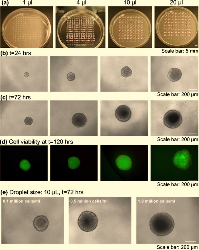
FIG. 2. EB formation using bioprinting method. 共a兲–共c兲 Images of formed EBs with droplet sizes of 1, 4, 10, and 20 L
at a cell density of 105 cells/ ml. 共a兲 Uniform-sized droplets encapsulating ESCs were generated by bioprinting. 共b兲 Phase
contrast images of EBs formed after hanging for 24 h and 共c兲 after culture for 72 h in a 96 multiwell plate. 共d兲 Fluorescent
images of GFP positive EBs at t = 96 h stained with ethidium homodimer. 共e兲 Images of EBs formed with printed droplet
size of 10 l at t = 72 h at different cell concentrations.
density with all droplet sizes 关Fig. 4共b兲兴, and 共iii兲 1 ⫻ 106 / ml initial cell seeding density with 1 and
10 l droplet sizes 关Fig. 4共c兲兴. Furthermore, bioprinting method resulted in significantly larger EB
sizes 共i.e., 25%–55% larger EBs with bioprinting兲 compared to controls at the end of the 96 h
culture period in all groups 共Fig. 4兲.
The results presented here suggest that more uniform EB size distribution can be achieved
with bioprinting compared to the control method. Furthermore, significantly greater EB sizes can
be obtained for the same cell seeding density and droplet volume when bioprinting method is
utilized compared to the manual pipetting. The enhanced uniformity and larger size of the EBs
formed utilizing the bioprinting method over the manual methods can be explained by the inho-
mogeneous droplet spread during manual pipetting and cell settling during the time consuming,
labor intensive processes, which led to nonuniform cell seeding densities with manual methods.
These technical and practical limitations may be responsible for nonuniform droplet geometry and
increased mechanical stress on the ESCs within the manually pipetted droplet, thus affecting the
EB formation process. Furthermore, the bioprinting system presented here can generate up to 160
Author complimentary copy. Redistribution subject to AIP license or copyright, see http://bmf.aip.org/bmf/copyright.jsp022207-6 Xu et al. Biomicrofluidics 5, 022207 共2011兲
FIG. 3. The effect of initial cell density, droplet volume, and culture time on the EB size for bioprinting and control with
manual pipetting. EBs retrieved from bioprinted droplets after 24 h culture in vitro 共a兲 displayed more uniform size
distribution compared to control method 共i.e., pipetting based manual hanging-droplet兲 共b兲. Statistical analysis of EB size
with different initial cell concentrations 共0.1⫻ 106, 0.5⫻ 106, and 1.0⫻ 106 cells/ ml兲 formed by the bioprinting method
关共c兲, 共e兲, and 共g兲兴 and by the control method 关共d兲, 共f兲, and 共h兲兴. The EB sizes formed by bioprinting were well controlled by
varying the droplet size 共1, 4, 10, and 20 l兲 and the culture time 共24, 48, 72, and 96 h兲.
droplets/second. It would take up to ⬃10 min to generate that many droplets with the most
broadly used manual pipetting methods. This corresponds to at least two orders of magnitude
decrease in the speed time that it takes to pattern cells for EB formation. Therefore, bioprinting
method presents additional advantages for EB formation over manual pipetting in terms of
Author complimentary copy. Redistribution subject to AIP license or copyright, see http://bmf.aip.org/bmf/copyright.jsp022207-7 Bioprinting for EB formation Biomicrofluidics 5, 022207 共2011兲
FIG. 4. Comparison of the uniformity and the size of the EBs formed by control 共manual hanging-drop method兲 and
bioprinted hanging-drop methods. The statistical comparison of the EB size uniformity was performed with Levene’s test
for equality of variances 共p ⬍ 0.05兲 at the end of the 96 h culture period for different initial cell seeding densities and
droplet sizes. The uniformity of the sizes was assessed based on the variance in the data sets. Less variance in the data
indicated higher uniformity in the resulting EB sizes. The diameters of the resulting EBs were compared statistically for
control and bioprinting methods with Mann–Whitney U test for pairwise comparisons 共p ⬍ 0.05兲. 共a兲 For 0.1⫻ 106 / ml
initial cell seeding density, bioprinting method resulted in more uniform EB sizes at the end of the 96 h culture period for
1, 4, and 20 l droplet sizes. 共b兲 For 0.5⫻ 106 / ml initial seeding density, bioprinting resulted in significantly higher
uniformity in the EB sizes for all droplet sizes compared to control method. 共c兲 For 1 ⫻ 106 / ml initial cell seeding density,
higher uniformity in EB sizes was observed for the droplet size of 1 l. Overall, bioprinting method resulted in statisti-
cally greater EB sizes compared to control method at the end of the 96 h culture period in all groups.
throughput and user-friendly operation. In addition, our system creates EBs of controllable sizes
similar to the hanging-drop method, which can then be collected and cultured together for further
studies.
IV. CONCLUSIONS
The EB size and uniformity are critical parameters that play an important role in differentia-
tion efficiency of residing ESCs. In this study, we presented a cell printing based high throughput
method to produce EBs with uniform sizes by controlling the cell seeding density, bioprinting
volume, and culture time compared to the existing hanging-drop methods. The results showed that
the bioprinting approach presented here formed EBs with a high degree of uniformity in size
compared to EBs that were generated by using the manual pipetting approach. Furthermore,
bioprinting method resulted in significantly larger size EBs at the end of the culture period
compared to the manual controls. Therefore, the combination of bioprinting technique with the
hanging-drop method provides an effective tool to generate controllable uniform-sized EBs. The
EBs formed with this method would be essential for applications in regenerative medicine, inves-
tigating stem cell differentiation, screening drug candidates, and evaluating embryonic toxicity.
ACKNOWLEDGMENTS
This work was performed at the Demirci Bio-Acoustic MEMS in Medicine 共BAMM兲 Labo-
ratories at the HST-BWH Center for Bioengineering, Harvard Medical School. This work was also
supported by NIHR21 共Grant No. AI087107兲, the W.H. Coulter Foundation Young Investigator
Award, and the Center for Integration of Medicine and Innovative Technology under U.S. Army
Author complimentary copy. Redistribution subject to AIP license or copyright, see http://bmf.aip.org/bmf/copyright.jsp022207-8 Xu et al. Biomicrofluidics 5, 022207 共2011兲
Medical Research Acquisition Activity Cooperative Agreement 共Nos. DAMD17-02-2-0006,
W81XWH-07-2-0011, and W81XWH-09-2-0001兲. Also, this research was made possible by a
research grant that was awarded and administered by the U.S. Army Medical Research and Ma-
teriel Command 共USAMRMC兲 and the Telemedicine and Advanced Technology Research Center
共TATRC兲 at Fort Detrick, MD. The information contained herein does not necessarily reflect the
position or policy of the Government, and no official endorsement should be inferred.
U.D. proposed the idea; F.X. and U.D designed the research; F.X., B.S., and B.S. performed
research; F.X., S.Q.W. B.S., and U.A.G. analyzed the data; and F.X., S.Q.W., U.A.G., and U.D.
wrote the paper.
1
C. E. Murry and G. Keller, Cell 132, 661 共2008兲.
2
C. W. Pouton and J. M. Haynes, Nat. Rev. Drug Discovery 6, 605 共2007兲.
3
J. J. Heit and S. K. Kim, Pediatr. Diabetes 5, 5 共2004兲.
4
Y. Xu, Y. Shi, and S. Ding, Nature 共London兲 453, 338 共2008兲.
5
R. Peerani, B. M. Rao, C. Bauwens, T. Yin, G. A. Wood, A. Nagy, E. Kumacheva, and P. W. Zandstra, EMBO J. 26,
4744 共2007兲.
6
J. Park, C. H. Cho, N. Parashurama, Y. Li, F. Berthiaume, M. Toner, A. W. Tilles, and M. L. Yarmush, Lab Chip 7, 1018
共2007兲.
7
J. Itskovitz-Eldor, M. Schuldiner, D. Karsenti, A. Eden, O. Yanuka, M. Amit, H. Soreq, N. Benvenisty, Mol. Med. 6, 88
共2000兲.
8
M. Koike, S. Sakaki, Y. Amano, and H. Kurosawa, J. Biosci. Bioeng. 104, 294 共2007兲.
9
H. Kurosawa, J. Biosci. Bioeng. 103, 389 共2007兲.
10
J. C. Mohr, J. Zhang, S. M. Azarin, A. G. Soerens, J. J. de Pablo, J. A. Thomson, G. E. Lyons, S. P. Palecek, and T. J.
Kamp, Biomaterials 31, 1885 共2010兲.
11
B. E. Reubinoff, M. F. Pera, C. Y. Fong, A. Trounson, and A. Bongso, Nat. Biotechnol. 18, 399 共2000兲.
12
P. W. Burridge, D. Anderson, H. Priddle, M. D. Barbadillo Muñoz, S. Chamberlain, C. Allegrucci, L. E. Young, and C.
Denning, Stem Cells 25, 929 共2007兲.
13
D. K. Singla, S. Jayaraman, J. Zhang, and T. J. Kamp, in Human Cell Culture 6: Embryonic Stem Cells, edited by J. R.
Master, B. O. Palsson, and J. A. Thomson 共Springer-Verlag, New York, 2007兲, pp. 211–234.
14
Y. Y. Choi, B. G. Chung, D. H. Lee, A. Khademhosseini, J.-H. Kim, and S.-H. Lee, Biomaterials 31, 4296 共2010兲.
15
L. Rohani, K. Karbalaie, A. Vahdati, M. Hatami, M. H. Nasr-Esfahani, and H. Baharvand, Int. J. Artif. Organs 31, 258
共2008兲.
16
B. S. Youn, A. Sen, L. A. Behie, A. Girgis-Gabardo, and J. A. Hassell, Biotechnol. Prog. 22, 801 共2006兲.
17
C. L. Bauwens, R. Peerani, S. Niebruegge, K. A. Woodhouse, E. Kumacheva, M. Husain, and P. W. Zandstra, Stem Cells
26, 2300 共2008兲.
18
M. D. Ungrin, C. Joshi, A. Nica, C. Bauwens, and P. W. Zandstra, PLoS ONE 3, e1565 共2008兲.
19
D. Gothard, S. J. Roberts, K. M. Shakesheff, and L. D. Buttery, Cytotechnology 61, 135 共2009兲.
20
J. M. Karp, J. Yeh, G. Eng, J. Fukuda, J. Blumling, K.-Y. Suh, J. Cheng, A. Mahdavi, J. Borenstein, R. Langer, and A.
Khademhosseini, Lab Chip 7, 786 共2007兲.
21
Y. S. Hwang, B. G. Chung, D. Ortmann, N. Hattori, H. C. Moeller, and A. Khademhosseini, Proc. Natl. Acad. Sci. U.S.A.
106, 16978 共2009兲.
22
W. G. Lee, D. Ortmann, M. J. Hancock, H. Bae, and A. Khademhosseini, Tissue Eng. 16, 249 共2010兲.
23
C. M. Nelson, R. P. Jean, J. L. Tan, W. F. Liu, N. J. Sniadecki, A. A. Spector, and C. S. Chen, Proc. Natl. Acad. Sci.
U.S.A. 102, 11594 共2005兲.
24
O. Abilez, P. Benharash, M. Mehrotra, E. Miyamoto, A. Gale, J. Picquet, C. Xu, and C. Zarins, J. Surg. Res. 132, 170
共2006兲.
25
S. Niebruegge, A. Nehring, H. Baer, M. Schroeder, R. Zweigerdt, and J. Lehmann, Tissue Eng A 14, 1591 共2008兲.
26
E. S. Ng, R. P. Davis, L. Azzola, E. G. Stanley, and A. G. Elefanty, Blood 106, 1601 共2005兲.
27
P. B. Lillehoj, H. Tsutsui, B. Valamehr, H. Wu, and C. M. Ho, Lab Chip 10, 1678 共2010兲.
28
S. Moon, S. K. Hasan, Y. S. Song, F. Xu, H. O. Keles, F. Manzur, S. Mikkilineni, J. W. Hong, J. Nagatomi, E.
Haeggstrom, A. Khademhosseini, and U. Demirci, Tissue Eng Part C Methods 16, 157 共2010兲.
29
F. Xu, S. J. Moon, A. E. Emre, E. S. Turali, Y. S. Song, S. A. Hacking, J. Nagatomi, and U. Demirci, Biofabrication 2,
014105 共2010兲.
30
F. Xu, J. Celli, I. Rizvi, S. Moon, T. Hasan, and U. Demirci, Biotechnol. J. 6, 204 共2011兲.
31
U. Demirci and G. Montesano, Lab Chip 7, 1139 共2007兲.
32
U. Demirci, J. Microelectromech. Syst. 15, 11 共2006兲.
33
U. Demirci, G. G. Yaralioglu, E. Haeggstrom, G. Percin, S. Ergun, and B. T. Khuri-Yakub, IEEE Trans. Semicond.
Manuf. 18, 709 共2005兲.
34
U. Demirci and G. Montesano, Lab Chip 7, 1428 共2007兲.
35
Y. S. Song, D. Adler, F. Xu, E. Kayaalp, A. Nureddin, R. M. Anchan, R. L. Maas, and U. Demirci, Proc. Natl. Acad. Sci.
U.S.A. 107, 4596 共2010兲.
36
S. Moon, Y.-G. Kim, L. Dong, M. Lombardi, E. Haeggstrom, R. V. Jensen, L.-L. Hsiao, and U. Demirci, PLoS ONE 6,
e17455 共2011兲.
37
B. R. Ringeisen, C. M. Othon, J. A. Barron, D. Young, and B. J. Spargo, Biotechnol. J. 1, 930 共2006兲.
38
T. Boland, T. Xu, B. Damon, and X. Cui, Biotechnol. J. 1, 910 共2006兲.
39
H. Geckil, F. Xu, X. Zhang, S. Moon, and U. Demirci, Nanomedicine 5, 469 共2010兲.
40
J. Samot, S. Moon, L. Shao, X. Zhang, F. Xu, Y.Song, H. O. Keles, L.Matloff, J. Markel, and U. Demirci, PLoS ONE
6, e17530 共2011兲.
Author complimentary copy. Redistribution subject to AIP license or copyright, see http://bmf.aip.org/bmf/copyright.jspYou can also read