Defining the Gothic Arch Angle (GAA) as a radiographic diagnostic tool for instability in hip dysplasia
←
→
Page content transcription
If your browser does not render page correctly, please read the page content below

www.nature.com/scientificreports
OPEN Defining the Gothic Arch Angle
(GAA) as a radiographic diagnostic
tool for instability in hip dysplasia
A. Zimmerer1,2*, J. Löchel3, J. Schoon1, V. Janz1 & G. I. Wassilew1
In recent years, there has been a controversial discussion about whether borderline dysplastic hips
should be treated with an arthroscopic procedure or rather with an acetabular reorientation. New
research suggests that a classification into stable and unstable hips may be helpful. The aim of the
study was to validate (1) the intra- and interobserver reliability of a newly defined radiographic
parameter named the Gothic Arch Angle, (2) the association between the GAA and previously existing
measurements used to define severity of acetabular dysplasia, and (3) the correlation between
radiographic measurements of acetabular dysplasia with MRI findings previously suggestive of
hip instability. We defined and validated the GAA in 10 standardized radiographs of asymptomatic
hips by two observers and calculated intra- and interobserver coefficients at two individual dates.
Subsequently, a consecutive series of 100 patients with dysplastic hips (LCEA < 25°, Toennis
grade ≤ 1) were evaluated for signs of instability on anteroposterior (a.p.) pelvic radiographs and
direct MR arthrography and were divided in two groups: stable and unstable. In these patients the
LCEA, the AI, the FEAR index and the GAA were radiographically evaluated. Correlation analyses
and a logistic regression analysis was performed to identify the predictive value of instability for
each radiographic parameter. Cutoff probabilities analysis was performed using standard receiver
operating characteristic (ROC) curves to rate the predictive efficiency value of the GAA. The GAA
showed excellent inter- and intraobserver reliability. A correlation was found between GAA and FEAR
index. A logistic regression analysis showed that LCEA, FEAR index and GAA are distinct predictors of
instability in hip dysplasia. The GAA showed the largest area under the curve (AUC 0.96), indicating it
to be the best predictor of instability with an optimal cutoff value of 90° (sensitivity, 0.95; specificity,
0.93). The GAA is a new available indicator for instability and is thus suggested to be used as a future
radiographic parameter for the stability of dysplastic hips. Further studies are needed to understand
how this parameter might additionally predict clinical outcome in the treatment of hip dysplasia.
Level of evidence: Level III, diagnostic study.
Developmental dysplasia of the hip (DDH) is defined as osseous deficiency of the acetabulum with abnor-
mal coverage of the femoral head which may lead to an overload and joint instability with subsequent dam-
age of the acetabular cartilage or labrum1–4. While there is general consensus that hip dysplasia with a lateral
center–edge angle (LCEA) < 18° should be treated with a reorientation of acetabular coverage through acetabular
reorientation4–8, there is still a controversial discussion about whether milder forms of dysplasia (borderline hip)
with an LCEA between 18° and 25° should be treated with arthroscopy or acetabular r eorientation3,8–14. A few
authors have suggested a distinction between stable and unstable hips for this group of patients in order to make
the right therapeutic decision11,14,15. Wyatt et al. therefore introduced the Femoro-Epiphyseal Acetabular Roof
(FEAR) index in 2017 to distinguish between stable and unstable dysplastic h ips11,15,16. The theory underlying
the FEAR index is that epiphyseal growth of the proximal femur is affected by the contact forces during devel-
opment. According to Pauwels and Maquet, the epiphyseal plate orientates perpendicular to the joint reaction
force in accordance with the Hueter–Volkman principle15,17. This leads to the fact that the angle of the closed
epiphyseal plate indicates the balance of forces across the proximal femoral p hysis15,18. Wyatt et al. theorized
that the angle between the epiphyseal scar of the femoral head growth plate and acetabular index (AI) would
1
Department of Orthopedics and Orthopedic Surgery, University Medicine Greifswald,
Ferdinand‑Sauerbruch‑Straße, 17475 Greifswald, Germany. 2ARCUS Sportklinik Pforzheim, Rastatterstr. 17‑19,
75179 Pforzheim, Germany. 3Orthopedic Department, Center for Musculoskeletal Surgery, Charité—University
Medicine, Berlin, Germany. *email: Alexander.zimmerer@gmail.com
Scientific Reports | (2021) 11:19531 | https://doi.org/10.1038/s41598-021-99011-7 1
Vol.:(0123456789)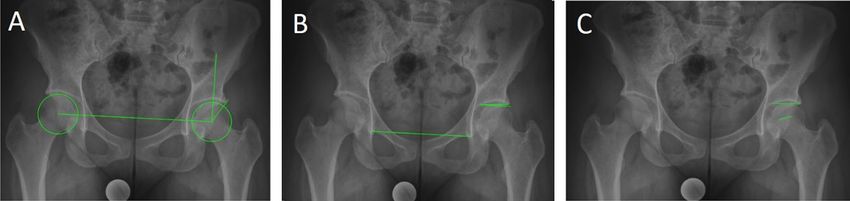
www.nature.com/scientificreports/
Gender Laterality Age, y Body mass index, kg/m2
Stable (n = 61) 85% female 53% right hips 32.1 ± 12.3 (16 to 45) 23.3 ± 3.01 (16.76 to 30.78)
Unstable (n = 39) 88% female 56% right hips 31.2 ± 13.1 (17 to 43) 23.3 ± 3.01 (16.76 to 30.78)
Table 1. Patient demographic data on stability groups.
reveal the resultant force vector, which could be used to predict the stability of the borderline h ip15. However,
the originally described threshold of 5° demonstrated just a 79% probability of correctly assigning hips as stable
and unstable15. Thus, it must be assumed that unstable hips may not be adequately identified by the FEAR index.
To integrate a broader range of biomechanical hip joint characteristics, Renato Bombelli, an Italian ortho-
paedic surgeon, has described the so-called gothic arch of the hip in 1976 and introduced it as the key to correct
understanding of the biomechanical p roperties19,20. The gothic arch can be identified as a triangular structure on
anteroposterior (ap) radiographs of the pelvis. The base of the triangle is formed by the sourcil while the sides
are shaped by condensed arch-like structured trabecular bone. The intersection of these two arches form the tip
of the gothic arch. Biomechanically the tip off the gothic arch lies on the line of application of the compressive
force, which is perpendicular to the weight-bearing surface (WBS) and passes through the center of rotation.
Bombelli hypothesized that hips with an altered gothic arch are mechanically jeopardized. In these hips the tip
would therefore point either medially or laterally. In dysplastic hips the tip moves medially and thus the WBS
shifts craniolaterally steeper. This in turn leads to a force directed craniolaterally, which results in increased
tension on the hip capsule and consequently to craniolateral displacement of the femoral head. Consequently,
we assume that the concept of the gothic arch and the FEAR Index should be combined in order to achieve a
higher predictive capacity with regard to hip stability. We have therefore revisited Bombelli’s concept, additionally
amended it by the analysis of the femoral epiphyseal growth plate and defined a new measurement parameter
for instability of the hip, called the Gothic Arch Angle (GAA).
The aims of this study were (1) to validate the intra- and interobserver reliability of a newly defined radio-
graphic parameter named the Gothic Arch Angle, (2) to validate the association between the GAA and previously
existing measurements used to define severity of acetabular dysplasia, and (3) to validate the correlation between
radiographic measurements of acetabular dysplasia with MRI findings previously suggestive of hip instability.
Methods
Patient selection and subclassification. This is a retrospective study of prospectively collected registry
data. After approval from the local ethics committee (Ethics Committee of the University of Greifswald, Ger-
many; BB099/20) we reviewed our hospital registry to identify all patients who presented in our practice for
hip joint preserving surgery between January 2019 and January 2020. From this registry, patients who showed a
lateral center edge angle (LCEA) of less than 25° without signs of osteoarthritis (Toennis grade ≤ 1) on the stand-
ing a.p. pelvic radiograph and for which direct MR arthrography (dMRA) was available were selected. These
patients were analyzed according to the instability criteria described by Wyatt et al.15 for cohort stratification.
Based on these results one patient group matching stable hip criteria (n = 61) and one matching unstable hip
criteria (n = 39) were formed. Both groups were compared regarding their clinical case and secondary patient
characteristics (Table 1). The instability criteria described by Wyatt et al.15 were either an increased distance from
the ilioischial line, a break of Shenton’s line, or the appearance of a crescent-shaped accumulation of gadolinium
in the posteroinferior joint space at dMRA. Excluded were patients with an LCEA of more than 25° (n = 279), a
missing dMRA (n = 75), osteoarthritis (Toennis grade > 1, n = 55) or previous hip surgery (n = 26). In brief, a total
of 100 patients out of 535 patients could be identified and were included in the analysis (Table 1). This study has
been performed in accordance with the ethical standards in the 1964 Declaration of Helsinki. All participants
declared informed consent.
Measurement of the Gothic Arch Angle (GAA). The GAA (Fig. 1) was measured on standing a.p. pelvic
radiographs meeting the criteria to be regarded as neutrally rotated and tilted: three cm between the tip of the
coccyx and the superior aspect of the symphysis pubis and symmetric obturator foramen21,22. An angle between
two lines was measured (Fig. 1, blue semicircle). The first line (Fig. 1, green line) extends between the rotational
center of the hips, which is determined by a best-fit circle around the femoral head, and the tip of the gothic arch
(Fig. 1, yellow dotted-line). The base of the Gothic arch is formed by the sourcil, while the trabeculae forming
the lateral aspect of the arch extend from the lateral acetabular rim toward the sacroiliac joint, while the medial
aspect of the arch forms an arc of dense cancellous bone extending from the quadrilateral plate towards the
anterior superior and anterior inferior iliac spines. The second line corresponds to the scar of the physeal growth
plate, identical to the reference in the FEAR index15 (Fig. 1, red line). The central third of the scar is a straight
line connecting its medial and lateral ends.
Validation of the Gothic Arch Angle. To validate the GAA, standardized a.p. pelvic radiographs of 20
asymptomatic patients were analyzed as previously decribed15. Those patients were treated for an unrelated
CEA23, the acetabular index (AI)24, and the
trauma in our hospital in May 2020. In addition to the GAA, the L
FEAR index15 (Fig. 2) were measured by two independent orthopaedic surgeons (A.Z., J.L.) using the software
mediCAD (mediCAD Hectec GmbH, Altdorf, Germany).
Scientific Reports | (2021) 11:19531 | https://doi.org/10.1038/s41598-021-99011-7 2
Vol:.(1234567890)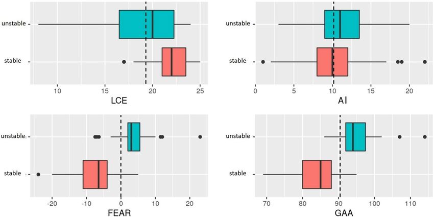
www.nature.com/scientificreports/
Figure 1. To measure the Gothic Arch Angle (blue semicircle), an angle between two lines is measured: The
first line (green) extends between the center of the femoral head, which is determined by a best-fit circle around
the femoral head (green circle), and the peak of the gothic arch (yellow dashed line). The second line (red)
corresponds to the middle third of the physeal scar.
Figure 2. Radiographic Measurements. (A) Lateral Center–Edge Angle: calculated by drawing a best-fit circle
around the inferior and medial margins of the femoral head. The angle is measured between 2 lines drawn from
the center of the circle, one running vertically along the longitudinal axis of the pelvis and the other running
along the acetabular sourcil e dge23. (B) Acetabular index: measured by drawing a horizontal line parallel to the
transverse pelvic axis, at the most medial edge of the sclerotic sourcil, and then making a second line extending
from the medial edge to the most lateral aspect of the s ourcil24. (C) Femoroepiphyseal acetabular roof index:
formed by 2 lines connecting the acetabular roof inclination and the femoral head physeal s car15.
Data presentation and statistics. Continuous variables are presented as means ± SD with ranges, and
categorical variables are presented as frequencies and percentages. Data were analyzed by use of R (R Core Team,
2017)25. Statistical reliability testing of the radiological parameters was performed with intraclass correlation
values interpreted as: greater than 0.75 = excellent, 0.40–0.75 = fair to good, and less than 0.40 = poor26,27. Bland–
Altman graphs were generated to present interobserver agreement28. The Spearman coefficient analysis was used
to identify correlations between the radiological measurements. Correlation coefficients were classified by the
strength of the correlation: excellent (> 0.80), very good (0.71–0.80), good (0.61–0.70), fair (0.41–0.60), and poor
(0.21–0.40). A stepwise forward multivariate logistic regression analysis was performed to identify the predictive
value of an instability for each radiographic measurement. Cutoff probabilities and sensitivity/specificity analy-
sis were performed using standard receiver operating characteristic (ROC) curves to determine optimal cutoff
values. The threshold for statistical significance was set to 0.05.
Scientific Reports | (2021) 11:19531 | https://doi.org/10.1038/s41598-021-99011-7 3
Vol.:(0123456789)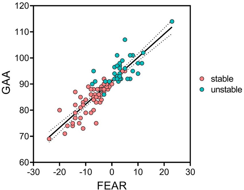
www.nature.com/scientificreports/
Intraobserver reproducibility (Observer Intraobserver reproducibility (Observer
1) 2) Interobserver reproducibility
ICC (95% CI) ICC (95% CI) ICC (95% CI)
LCEA 0.918 (0.867–0.976) 0.872 (0.832–0.987) 0.910 (0.835–0.965)
AI 0.913 (0.875–0.983) 0.921 (0.876–0.985) 0.916 (0.845–0.975)
FEAR Index 0.994 (0.982–0.999) 0.962 (0.863–0.992) 0.983 (0.893–0.996)
GAA 0.996 (0.994–1.0) 0.995 (0.907–0.994) 0.957 (0.890–0.988)
Table 2. Intra- and Interobserver Reproducibility of the GAA in 20 Patientsa. a AI acetabular index, FEAR
index Femoro-Epiphyseal Acetabular Roof, GAAGothic Arch Angle, ICC intraclass correlation coefficient,
LCEA Lateral Center–Edge Angle;
Figure 3. Bland Altmann plots for the GAA. (A) First measurement. (B) Second measurement.
Variables LCEA AI FEAR Index GAA
LCEA 1 − 0.306 − 0.316 − 0.321
AI − 0.306 1 0.299 0.068
FEAR index − 0.316 0.299 1 0.899
GAA − 0.321 0.068 0.889 1
Table 3. Spearman correlation coefficients of the radiological measurements for all included h
ipsa. a Bold
values are statistically significant. AI acetabular index, GAAGothic Arch Angle, FEAR Index Femoro-
Epiphyseal Acetabular Roof Index, LCEA Lateral Center–Edge Angle.
Results
Validation of the Gothic Arch Angle. Two observers (A.Z., J.L.) independently measured the GAA. Each
image was measured at two independent time points (one week apart). The GAA showed excellent inter- and
intraobserver agreement. In addition, the reliability of the GAA was compared with the LCEA, the AI, and the
FEAR index (Table 2). The measured GAA did not differ significantly between the examiners and date of review
(Fig. 3).
Correlation analysis. After validation of the GAA, a correlation analysis between LCEA, AI, FEAR index
and GAA was performed (Table 3). An excellent correlation was found between the GAA and the FEAR index
(r = 0.899, p < 0.0001) (Fig. 4).
Assessment of Radiographic Parameters. All radiographic parameters were significantly different
between the stable and unstable hips, except for AI (10.5° vs. 10.9; p = 0.578) (Table 4 and Fig. 5). The LCEA was
found to be smaller in the unstable group (21.8° vs. 19.1; p < 0.001). The FEAR index (− 7.5° vs. 3.5°; p < 0.0001)
and GAA (84.0° vs 95.2°; p < 0.0001) for patients meeting criteria for instability were significantly more positive.
The logistic regression analysis showed that the LCEA (p < 0.001), FEAR index (p < 0.0001) and GAA
(p < 0.0001) were significantly different between the unstable and stable patient group, while the AI (p = 0.59)
did not indicate significant differences (Table 5).
Scientific Reports | (2021) 11:19531 | https://doi.org/10.1038/s41598-021-99011-7 4
Vol:.(1234567890)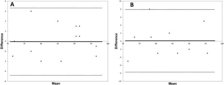
www.nature.com/scientificreports/
Figure 4. Scatter plot of the relation between GAA and FEAR index for dysplastic hips (LCEA < 25°). Stable
hips are displayed in red, unstable hips in green; GAAGothic Arch Angle, FEAR Index Femoro-Epiphyseal
Acetabular Roof Index.
Stable hip Unstable hips P Value
LCEA, deg 21.8 ± 2.1 (17–25) 19.1 ± 4.0 (8–24) < 0.001
AI, deg 10.5 ± 4.2 (1–22) 10.9 ± 3.5 (2–20) 0.578
FEAR index, deg − 7.5 ± 5.6 (− 24–5) 3.5 ± 5.4 (− 7.5–23) < 0.0001
GAA, deg 84.0 ± 5.8 (69–95) 95.2 ± 5.0 (86–114) < 0.0001
Table 4. Radiographic measurements by stability diagnosisa. a Data are shown as mean ± SD (range). AI
acetabular index, GAAGothic Arch Angle, FEAR Index Femoro-Epiphyseal Acetabular Roof Index, LCEA
Lateral Center–Edge Angle.
Figure 5. Boxplots of the radiographic parameters for the two groups. The dashed lines indicate the optimal
cutoff values for predicting instability. It is obvious that LCEA and AC angle are not suitable to prediction of
instability.
Scientific Reports | (2021) 11:19531 | https://doi.org/10.1038/s41598-021-99011-7 5
Vol.:(0123456789)www.nature.com/scientificreports/
P Value
LCEA < 0.001
AI 0.59
FEAR index < 0.0001
GAA < 0.0001
Table 5. Logistic regression analysis of radiographic parameters for instabilitya. a AI acetabular index, GAA
Gothic Arch Angle, FEAR Index Femoro-Epiphyseal Acetabular Roof Index, LCEA Lateral Center–Edge Angle.
Figure 6. Simultaneous receiver operating characteristic (ROC) curves for radiographic parameters. AI
acetabular index, FEAR Femoro-epiphyseal Acetabular Roof, GAAGothic Arch Angle, LCEA Lateral Center
Edge Angle.
To compare the four radiographic parameters in terms of their predictive efficiency for instability, we plot-
ted simultaneous ROC curves, which are shown in Fig. 6. The GAA had the largest area under the curve (AUC
0.96), indicating its superior predictive efficiency for instability. We found the optimal cutoff value for FEAR
index at 0° (sensitivity, 0.84; specificity, 0.93) and for GAA at 90° (sensitivity, 0.95; specificity, 0.93), respectively.
Scientific Reports | (2021) 11:19531 | https://doi.org/10.1038/s41598-021-99011-7 6
Vol:.(1234567890)www.nature.com/scientificreports/
Discussion
In the present study we have defined a new radiological parameter, which is intended to be a predictor for
instability in dysplastic hips. We could demonstrate excellent intra- and interobserver reliabilities for this new
radiological parameter. Furthermore, the GAA was identified as the most effective indicator of radiologically
assessed instability. The optimal GAA cutoff value for the differentiation between stable and unstable hips was
90°. More precisely, hips with a GAA > 90° can be considered unstable and vice versa.
In recent years, there has been increasing discussion about whether so-called borderline dysplastic hips should
be treated with an arthroscopic procedure or rather with an acetabular reorientation. Arthroscopic interventions
for the management of borderline dysplastic hips have been described with varying degrees of success and high
rates of re-operation and conversion to total hip a rthroplasty3,8–10,13,14,29–32. In contrast, good results and mini-
mal complications were reported in patients treated with PAO after 1 year and 2 years p ostoperatively10,32. The
unpredictability of the results, especially with arthroscopic treatment, can be due to difficulty in differentiating
between stable and unstable hips. In this context, much of the literature has isolated the definition of dysplastic
hips to the LCEA evaluation, which is a reliable measure of lateral acetabular coverage, but not a surrogate
for the global acetabular morphology. The sole determination of the LCEA can mischaracterize the nature of
acetabular under-coverage and it could be shown that an adequate classification should be obtained by several
radiological parameters8,10,12,33,34.
However, we believe, similar to Wyatt et al.15, that a classification into borderline and severe dysplasia is
obsolete. Rather, a functional analysis should be performed in order to be able to make a valid classification.
In our opinion, the challenge is to classify the hip as either stable or unstable and subsequently treat it in the
appropriate manner. Wyatt et al. introduced the FEAR index15 to differentiate between stable and unstable hips,
which can also be applied as an indicator for micro-instabilities in non-dysplastic h ips35. Thus, the originally
described threshold of 5° demonstrated just a 79% probability of correctly assigning hips as stable and unstable15.
Another conception that allows the evaluation of hip stability is the acetabular gothic arch described by Bom-
belli in 1 97619,20. The gothic arch represents a characteristic feature of anteroposterior radiographs of the pelvis.
Bombelli hypothesized that hips with an abnormal Gothic arch are mechanically compromised and predisposed
to the development of osteoarthritis. In normal hips, the apex of the Gothic arch lies directly over the center of
the femoral head, so that a line connecting these points is exactly perpendicular. In abnormal hips, however,
the apex of the Gothic arch lies medial or lateral to a vertical line drawn through the center of the femoral head,
resulting in a craniomedial or craniolateral orientation of the Gothic arch. However, Bombelli did not define
cut-off values. We assumed that a combination of both concepts may allow an increased prediction regarding
stability. Therefore, we have adopted both concepts and combined them with the gothic arch angle to a holistic
approach and analysis of hip geometry in order to provide the most accurate possible prediction of stability.
We were able to define a threshold of 90°, which demonstrated high sensitivity and specificity (sensitivity, 0.95;
specificity, 0.93) with respect to discrimination into stable or unstable. Based on the results of our study, it may
be feasible for clinicians to use thee GAA as an additional radiologic assessment to identify patients with possible
abnormal mechanics leading to hip instability.
In our study, the AI was not found to predict hip instability. This may be due to the fact that only a few hips
with increased AI are underrepresented in the present analysis. However, the AI is included in the determination
of the FEAR index, which was found to be a significant predictor of instability. It can therefore be assumed that
the orientation and position of the epiphyseal scar is the more important factor in the FEAR index. Bombelli
proposed the theory that a craniomedially aligned tip of the gothic arch must be considered an indication of hip
dysplasia, which has been confirmed in the p ast36. Herickhoff et al. demonstrated in patients with unilateral DDH
that the tip of the gothic arch pointed significantly more medially on the dysplastic side than on the healthy side
(4.43° [Normal Hip] vs. 15.33 [DDH Hip]; p = 0.0001)36. We are convinced that the presented concept allows an
even more reliable prediction of instability when both aspects (FEAR Index and Gothic Arch) are combined.
The values for the FEAR index and GAA were significantly higher in the unstable group than in the stable
group. Compared to the FEAR index, however, the GAA demonstrated the highest AUC in the ROC analysis
and therefore appears to be a better predictor of instability. However, the GAA should not be used as the solely
tool, but rather as a further piece in the puzzle on the way to a reliable diagnosis and identification of unstable
hips. In recent years, a relationship between femoral torsion and dysplasia has been observed, thus the femoral
torsion dimension should be considered in an appropriate assessment37,38. We therefore recommend a standard-
ized approach, which includes patient history and examination as well as diagnostics consisting of radiography
and MRI. The GAA can provide significant information regarding hip stability. However, the impact of the GAA
on clinical outcomes needs to be demonstrated in further studies.
Limitations. All patients had symptomatic hip pathologies, hence the study lacks an asymptomatic control
group. There is no correlation with clinical results that are based on a therapy decision derived from the use of
GAA. Future studies investigating the influence of the GAA on clinical results are therefore mandatory. A fur-
ther limitation, although it did not apply to any of the radiographs analyzed, is the possibility that the epiphyseal
scar or the tip of the gothic arch cannot be identified due to radiograph quality. In such cases it may theoretically
not be possible to determine the GAA. However, this limitation also applies to the FEAR index. Furthermore,
there is no data on whether the epiphyseal scar changes or remains consistent over time or whether the femoral
version has an influence on the assessment of the GAA. Finally, the GAA is evaluated by means of static radio-
graphic images, so that no dynamic assessment and analysis is possible. Prospective studies may be conducted to
examine the dynamic motion and the influence on stability.
Scientific Reports | (2021) 11:19531 | https://doi.org/10.1038/s41598-021-99011-7 7
Vol.:(0123456789)www.nature.com/scientificreports/
Conclusion
We defined a new radiological parameter to assess the stability of dysplastic hips. The GAA demonstrated excel-
lent intra- and interobserver reliabilities. The optimal cutoff value for the GAA was 90° to differentiate between
stable and unstable hips. Further studies are needed to understand how this parameter might additionally predict
clinical outcome in the treatment of symptomatic hip dysplasia.
Received: 11 January 2021; Accepted: 15 September 2021
References
1. Klaue, K., Durnin, C. W. & Ganz, R. The acetabular rim syndrome. A clinical presentation of dysplasia of the hip. J. Bone Jt. Surg.
73, 423–429. https://doi.org/10.1302/0301-620X.73B3.1670443 (1991).
2. Henak, C. R. et al. Patient-specific analysis of cartilage and labrum mechanics in human hips with acetabular dysplasia. Osteoarthr.
Cartil. 22, 210–217. https://doi.org/10.1016/j.joca.2013.11.003 (2014).
3. Nawabi, D. H. et al. Outcomes after arthroscopic treatment of femoroacetabular impingement for patients with borderline hip
dysplasia. Am. J. Sports Med. 44, 1017–1023. https://doi.org/10.1177/0363546515624682 (2016).
4. Lerch, T. D., Steppacher, S. D., Liechti, E. F., Tannast, M. & Siebenrock, K. A. One-third of hips after periacetabular osteotomy sur-
vive 30 years with good clinical results, no progression of arthritis, or conversion to THA. Clin. Orthop. Relat. Res. 475, 1154–1168.
https://doi.org/10.1007/s11999-016-5169-5 (2017).
5. Khan, O. H., Malviya, A., Subramanian, P., Agolley, D. & Witt, J. D. Minimally invasive periacetabular osteotomy using a modified
Smith-Petersen approach. Bone Jt. J. 99, 22–28. https://doi.org/10.1302/0301-620X.99B1.BJJ-2016-0439.R1 (2017).
6. Perry, K. I., Trousdale, R. T. & Sierra, R. J. Hip dysplasia in the young adult. Bone Jt. J. 95, 21–25. https://doi.org/10.1302/0301-
620X.95B11.32633 (2013).
7. Yasunaga, Y. et al. Rotational acetabular osteotomy for symptomatic hip dysplasia in patients younger than 21 years of age. Bone
Jt. J. 101, 390–395. https://doi.org/10.1302/0301-620X.101B4.BJJ-2018-1200.R1 (2019).
8. Kraeutler, M. J. et al. A contemporary look at the evaluation and treatment of adult borderline and frank hip dysplasia. Am. J.
Sports Med. https://doi.org/10.1177/0363546519881411 (2020).
9. Fukui, K., Trindade, C. A. C., Briggs, K. K. & Philippon, M. J. Arthroscopy of the hip for patients with mild to moderate devel-
opmental dysplasia of the hip and femoroacetabular impingement. Bone Jt. J. 97, 1316–1321. https://doi.org/10.1302/0301-620X.
97B10.35303 (2015).
10. McClincy, M. P., Wylie, J. D., Kim, Y.-J., Millis, M. B. & Novais, E. N. Periacetabular osteotomy improves pain and function in
patients with lateral center-edge angle between 18° and 25°, but are these hips really borderline dysplastic?. Clin. Orthop. Relat.
Res. 477, 1145–1153. https://doi.org/10.1097/CORR.0000000000000516 (2019).
11. Wyatt, M. C. & Beck, M. The management of the painful borderline dysplastic hip. J. Hip Preserv. Surg. 5, 105–112. https://doi.
org/10.1093/jhps/hny012 (2018).
12. Zimmerer, A., Schneider, M. M., Nietschke, R., Miehlke, W. & Sobau, C. Is hip arthroscopy an adequate therapy for the borderline
dysplastic hip? Correlation between radiologic findings and clinical outcomes. Orthop. J. Sports Med. 8, 232596712092085. https://
doi.org/10.1177/2325967120920851 (2020).
13. Yeung, M., Kowalczuk, M., Simunovic, N. & Ayeni, O. R. Hip arthroscopy in the setting of hip dysplasia. Bone Jt. Res. 5, 225–231.
https://doi.org/10.1302/2046-3758.56.2000533 (2016).
14. Wong, T. Y. et al. Upsloping lateral sourcil. A radiographic finding of hip instability. J. Hip Preserv. Surg. https://doi.org/10.1093/
jhps/hny042 (2018).
15. Wyatt, M., Weidner, J., Pfluger, D. & Beck, M. The Femoro-epiphyseal acetabular roof (FEAR) index. A new measurement associated
with instability in borderline hip dysplasia?. Clin. Orthop. Relat. Res. 475, 861–869. https://doi.org/10.1007/s11999-016-5137-0
(2017).
16. Batailler, C., Weidner, J., Wyatt, M., Pfluger, D. & Beck, M. Is the Femoro-Epiphyseal Acetabular Roof (FEAR) index on MRI a
relevant predictive factor of instability in a borderline dysplastic hip?. Bone Jt. J. 101, 1578–1584. https://doi.org/10.1302/0301-
620X.101B12.BJJ-2019-0502.R1 (2019).
17. Pauwels, F. Biomechanics of the Locomotor Apparatus. Contributions on the Functional Anatomy of the Locomotor Apparatus
(Springer, 1980).
18. Fabeck, L., Tolley, M., Rooze, M. & Burny, F. Theoretical study of the decrease in the femoral neck anteversion during growth. Cells
Tissues Organs 171, 269–275. https://doi.org/10.1159/000063127 (2002).
19. Bombelli, R. Osteoarthritis of the Hip (Springer, 1983).
20. Bombelli, R. Structure and Function in Normal and Abnormal Hips (Springer, 1993).
21. Clohisy, J. C. et al. Radiographic evaluation of the hip has limited reliability. Clin. Orthop. Relat. Res. 467, 666–675. https://doi.
org/10.1007/s11999-008-0626-4 (2009).
22. Siebenrock, K. A., Kalbermatten, D. F. & Ganz, R. Effect of pelvic tilt on acetabular retroversion a study of pelves from cadavers.
Clin. Orthop. Relat. Res. 407, 241–248. https://doi.org/10.1097/00003086-200302000-00033 (2003).
23. Wiberg, G. The anatomy and roentgenographic appearance of a normal hip joint. Acta Chir. Scand. 1939, 7–38 (1939).
24. Tönnis, D. Normal values of the hip joint for the evaluation of X-rays in children and adults. Clin. Orthop. Relat. Res. 2, 39–47
(1976).
25. R Core Team. R: A language and environment for statistical computing. R Foundation for Statistical. Available at http://www.R-
project.org/ (2017).
26. McGraw, K. O. & Wong, S. P. Forming inferences about some intraclass correlation coefficients. Psychol. Methods 1, 30–46. https://
doi.org/10.1037/1082-989X.1.1.30 (1996).
27. Shrout, P. E. & Fleiss, J. L. Intraclass correlations. Uses in assessing rater reliability. Psychol. Bull. 86, 420–428. https://doi.org/10.
1037//0033-2909.86.2.420 (1979).
28. Bland, J. M. & Altman, D. G. Statistical methods for assessing agreement between two methods of clinical measurement. Lancet
(London, England) 1, 307–310 (1986).
29. Chandrasekaran, S. et al. Arthroscopic capsular plication and labral seal restoration in borderline hip dysplasia. 2-year clinical
outcomes in 55 cases. Arthroscopy 33, 1332–1340. https://doi.org/10.1016/j.arthro.2017.01.037 (2017).
30. Evans, P. T. et al. Arthroscopic treatment of hip pain in adolescent patients with borderline dysplasia of the hip. Minimum 2-year
follow-up. Arthroscopy 33, 1530–1536. https://doi.org/10.1016/j.arthro.2017.03.008 (2017).
31. Kalore, N. V. & Jiranek, W. A. Save the torn labrum in hips with borderline acetabular coverage. Clin. Orthop. Relat. Res. 470,
3406–3413. https://doi.org/10.1007/s11999-012-2499-9 (2012).
32. Ricciardi, B. F. et al. Complications and short-term patient outcomes of periacetabular osteotomy for symptomatic mild hip
dysplasia. Hip Int. 27, 42–48. https://doi.org/10.5301/hipint.5000420 (2017).
Scientific Reports | (2021) 11:19531 | https://doi.org/10.1038/s41598-021-99011-7 8
Vol:.(1234567890)www.nature.com/scientificreports/
33. McClincy, M. P., Wylie, J. D., Yen, Y.-M. & Novais, E. N. Mild or borderline hip dysplasia are we characterizing hips with a lateral
center-edge angle between 18° and 25° appropriately?. Am. J. Sports Med. 47, 112–122. https://doi.org/10.1177/0363546518810731
(2018).
34. Kraeutler, M. J. et al. The iliofemoral line. A radiographic sign of acetabular dysplasia in the adult hip. Am. J. Sports Med. https://
doi.org/10.1177/0363546517708983 (2017).
35. Truntzer, J. N., Hoppe, D. J., Shapiro, L. M. & Safran, M. R. Can the FEAR index be used to predict microinstability in patients
undergoing hip arthroscopic surgery?. Am. J. Sports Med. 47, 3158–3165. https://doi.org/10.1177/0363546519876105 (2019).
36. Herickhoff, P. K. et al. The gothic arch. A reliable measurement for developmental dysplasia of the hip. The Iowa orthopaedic journal
33, 1–6 (2013).
37. Fritz, B. et al. MRI assessment of supra- and infratrochanteric femoral torsion. Association with femoroacetabular impingement
and hip dysplasia. Am. J. Roentgenol. 211, 155–161. https://doi.org/10.2214/AJR.17.18882 (2018).
38. Lerch, T. D. et al. Prevalence of combined abnormalities of tibial and femoral torsion in patients with symptomatic hip dysplasia
and femoroacetabular impingement. Bone Jt. J. 102, 1636–1645. https://doi.org/10.1302/0301-620X.102B12.BJJ-2020-0460.R1
(2020).
Acknowledgements
We thank Dr. M.V. (Institute of Bioinformatics, Greifswald, Germany) for his support regarding statistical
analysis.
Author contributions
A.Z.: made substantial contributions to the conception, design of the work, analysis, and has drafted the work.J.L.:
made substantial contributions to the conception, design of the work, analysis, and has drafted the work.J.S.:
made substantial contributions to the design of the work, analysis, and has drafted the work.V.J.: made substantial
contributions to the analysis and has drafted the work.G.I.W.: made substantial contributions to the conception
and substantively revised it.
Funding
Open Access funding enabled and organized by Projekt DEAL.
Competing interests
The authors declare no competing interests.
Additional information
Correspondence and requests for materials should be addressed to A.Z.
Reprints and permissions information is available at www.nature.com/reprints.
Publisher’s note Springer Nature remains neutral with regard to jurisdictional claims in published maps and
institutional affiliations.
Open Access This article is licensed under a Creative Commons Attribution 4.0 International
License, which permits use, sharing, adaptation, distribution and reproduction in any medium or
format, as long as you give appropriate credit to the original author(s) and the source, provide a link to the
Creative Commons licence, and indicate if changes were made. The images or other third party material in this
article are included in the article’s Creative Commons licence, unless indicated otherwise in a credit line to the
material. If material is not included in the article’s Creative Commons licence and your intended use is not
permitted by statutory regulation or exceeds the permitted use, you will need to obtain permission directly from
the copyright holder. To view a copy of this licence, visit http://creativecommons.org/licenses/by/4.0/.
© The Author(s) 2021, corrected publication 2021
Scientific Reports | (2021) 11:19531 | https://doi.org/10.1038/s41598-021-99011-7 9
Vol.:(0123456789)You can also read