Current Knowledge on Graves' Orbitopathy - MDPI

Page created by Charlie Alvarado
 
CONTINUE READING
Current Knowledge on Graves' Orbitopathy - MDPI
Journal of
                Clinical Medicine

Review
Current Knowledge on Graves’ Orbitopathy
Katarzyna Gontarz-Nowak * , Magdalena Szychlińska, Wojciech Matuszewski,
Magdalena Stefanowicz-Rutkowska and Elżbieta Bandurska-Stankiewicz

                                                Clinic of Endocrinology, Diabetology and Internal Medicine, School of Medicine, Collegium Medicum,
                                                University of Warmia and Mazury in Olsztyn, 10-957 Olsztyn, Poland; szychlinskam@gmail.com (M.S.);
                                                wmatuszewski82@wp.pl (W.M.); m.m.stefanowicz@gmail.com (M.S.-R.); bandurska.endo@gmail.com (E.B.-S.)
                                                * Correspondence: katarzyna.gontarz.92@gmail.com; Tel.: +48-7-9072-1102

                                                Abstract: (1) Background: Graves’ orbitopathy (GO) is an autoimmune inflammation of the orbital
                                                tissues and the most common extra-thyroid symptom of Graves’ disease (GD). Mild cases of GO are
                                                often misdiagnosed, which prolongs the diagnostic and therapeutic process, leading to exacerbation
                                                of the disease. A severe course of GO may cause permanent vision loss. (2) Methods: The article
                                                presents an analysis of GO—its etiopathogenesis, diagnostics, current treatment and potential future
                                                therapeutic options based on a review of the currently available literature of the subject. (3) Re-
                                                sults: Current treatment of the active GO consists predominantly in intravenous glucocorticoids
                                                (GCs) administration in combination with orbital radiotherapy. The growing knowledge on the
                                                pathogenesis of the disease has contributed to multiple trials of the use of immunosuppressive
                                                drugs and monoclonal antibodies which may be potentially effective in the treatment of GO. Im-
                                                munosuppressive treatment is not effective in patients in whom a chronic inflammatory process has
                                                caused fibrous changes in the orbits. In such cases surgical treatment is performed—including orbital
                                                decompression, adipose tissue removal, oculomotor muscle surgery, eyelid alignment and blepharo-
                                                plasty. (4) Conclusions: Management of GO is difficult and requires interdisciplinary cooperation in
                                                endocrinology; ophthalmology, radiation oncology and surgery. The possibilities of undertaking a
                                                reliable assessment and comparison of the efficacy and safety of the therapeutic strategies are limited
                                      due to the heterogeneity of the available studies conducted mostly on small group of patients, with
       
                                                no comparison with classic systemic steroid therapy. The registration by FDA of Teprotumumab,
Citation: Gontarz-Nowak, K.; Szych-
                                                an IGF1-R antagonist, in January 2020 may be a milestone in future management of active GO.
lińska, M.; Matuszewski, W.; Stefanowicz-
                                                However, many clinical questions require to be investigated first.
Rutkowska, M.; Bandurska-Stankiewicz,
E. Current Knowledge on Graves’ Or-
                                                Keywords: Graves’ orbitopathy; Graves’ disease; clinical activity score; immunosuppressive treat-
bitopathy. J. Clin. Med. 2021, 10, 16.
                                                ment; glucocorticoids; radiotherapy
https://dx.doi.org/10.3390/jcm10010016

Received: 24 September 2020
Accepted: 19 December 2020
Published: 23 December 2020                     1. Introduction
                                                      The Graves’ orbitopathy (GO) is an autoimmune inflammation of the orbital tissues
Publisher’s Note: MDPI stays neu-               and the most common extra-thyroid symptom of Graves’ disease (GD). GO occurs in
tral with regard to jurisdictional claims       25–50% of patients with GD, although the literature shows that subclinical ocular lesions
in published maps and institutional             can be observed in the majority of patients with GD when high-quality imaging techniques
affiliations.                                   are used [1–5]. Mild cases of GO are often misdiagnosed as conjunctivitis or allergic
                                                symptoms, which prolongs the diagnostic and therapeutic process, in some cases leading
                                                to exacerbation of the disease [6]. Symptoms typical of GO—ocular pain, excessive tearing,
Copyright: © 2020 by the authors. Li-
                                                photophobia, visual disturbances, including double vision—significantly reduce the quality
censee MDPI, Basel, Switzerland. This           of life of patients [7]. The inflammatory state in the orbit manifests itself mainly as redness
article is an open access article distributed   and swelling of conjunctivas and eyelids, exophthalmos, and retrobulbar pain. A severe
under the terms and conditions of the           course of GO with the occurrence of dysthyroid optic neuropathy (DON) or corneal
Creative Commons Attribution (CC BY)            ulceration may lead to permanent vision loss [8]. The current treatment of active GO
license (https://creativecommons.org/           consists predominantly in intravenous administration of glucocorticoids (GCs); however,
licenses/by/4.0/).                              according to the literature, the results of such treatment are unsatisfactory in 35% of cases.

J. Clin. Med. 2021, 10, 16. https://dx.doi.org/10.3390/jcm10010016                                                 https://www.mdpi.com/journal/jcm
J. Clin. Med. 2021, 10, 16                                                                                            2 of 23

                             Immunosuppressive treatment is not effective in patients in whom a chronic inflammatory
                             process has caused fibrous changes in the orbits [9]. For this reason, early identification
                             of patients at risk of severe GO is of highest significance, since effective treatment of
                             hyperthyroidism, careful observation of ocular lesions and prompt implementation of
                             appropriate treatment can significantly alleviate the course of the disease.

                             2. Etiopathogenesis
                                  The onset of GO is usually closely related to GD in its hyperthyroidism phase. Or-
                             bitopathy most often develops synchronously, although it can also precede or follow the
                             occurrence of hyperthyroidism [10]. Thyrotropin receptor antibodies (TRAbs), pathog-
                             nomonic for GD, are present in every patient with GO, and their concentration positively
                             correlates with the severity and the activity of the disease [11]. The thyrotropin receptor
                             (TSH-R), present on the thyroid cells, is also physiologically found on the surface of orbital
                             fibroblasts, yet in the case of GO, it is overexpressed [12–14].
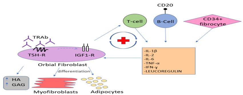
                                  After binding to the TSH-R on orbital fibroblasts, TRAbs activate the immune cas-
                             cade leading to the infiltration of the activated B and T lymphocytes, as well as bone
                             marrow-derived CD34 + fibrocytes, which differentiate into myofibroblasts or adipocytes.
                             The incoming cells release numerous cytokines and chemokines, such as interferon gamma
                             (IFN-y), tumour necrosis factor alpha (TNF-α), interleukin-1β (IL-1β), interleukin-2 (IL-2),
                             interleukin-6 (IL-6) as well as leukoregulin, which strongly stimulate the local synthe-
                             sis of glycosaminoglycans (GAG), including hyaluronic acid (HA). Accumulation of the
                             strongly hydrophilic HA causes local water retention and swelling of connective tissue
                             and extraocular muscles, which in turn worsens the venous and lymphatic circulation
                             in the orbit. The activation of periocular fibroblasts, which are known to be progenitor
                             fat cells, leads to the enlargement of orbital fat tissue. As a result, the intra-orbital pres-
                             sure increases leading to subsequent protrusion of the eyeballs forward beyond the edge
                             of the orbit, what clinically manifests as proptosis or exophthalmos. The inflammatory
                             process of the oculomotor muscles impairs their function and disables the coordinated
                             movement of the eyeballs causing double vision. A long-term inflammatory state leads to
                             gradual muscle remodeling and fibrosis, which results in persistent mobility disorders of
                             the eyeballs [14–16]. According to some authors, the autoimmune process of GO, besides
                             aforementioned cytokines and chemokines, involves also some growth factors [17]. Indeed,
                             overexpression of the insulin-like growth factor 1 receptor (IGF1-R) is observed on orbital
                             fibroblasts, as well as on the infiltrating T and B lymphocytes. Studies suggest that IGF1-R
                             is involved in the signaling pathway induced by TRAb ligation with TSH-R, through
                             which it is transactivated. Being part of the aforementioned complex, IGF1-R enhances the
                             TSHR-dependent production of cytokines, leading to increased release of inflammatory
                             mediators and, as a result, increased production of HA. The initial hypothesis about the
                             presence of specific anti-IGF1-R autoantibodies involved in the pathogenesis of GO has not
                             been confirmed [18–22]. The aforementioned signaling cascade is presented on Figure 1.
J.J. Clin.
     Clin. Med.
           Med. 2021, 10, x16FOR PEER REVIEW
                2021, 10,                                                                                                                             33 of
                                                                                                                                                         of 23
                                                                                                                                                            23

                                     Figure
                                      Figure 1.1.Signaling
                                                  Signalingcascade
                                                             cascadeinvolved
                                                                      involvedin in
                                                                                  Graves’ orbitopathy
                                                                                    Graves’           (GO)
                                                                                            orbitopathy    pathogenesis.
                                                                                                        (GO)   pathogenesis.IGF1-R: insulin-
                                     IGF1-R:   insulin-like growth  factor 1 receptor, TSH-R:  thyrotropin receptor,
                                      like growth factor 1 receptor, TSH-R: thyrotropin receptor, TRAb: Thyrotropin  TRAb: Thyrotropin
                                                                                                                        receptor antibodies,
                                     receptor antibodies, HA: Hyaluronic Acid, GAG: glycosaminoglycans, IL-1β: interleukin-1β, IL-2:
                                      HA: Hyaluronic Acid, GAG: glycosaminoglycans, IL-1β: interleukin-1β, IL-2: interleukin-2, IL-6:
                                     interleukin-2, IL-6: interleukin-6, TNF-α: tumour necrosis factor alpha, IFN-y: interferon gamma.
                                      interleukin-6, TNF-α: tumour necrosis factor alpha, IFN-y: interferon gamma.

                                     3. Risk Factors
                                      3. Risk
                                           TheFactors
                                                onset of GO appears to be conditioned by a complex interaction of genetic and
                                     environmental
                                            The onset factors.      According
                                                         of GO appears        to betoconditioned
                                                                                       the literature   bysome     predisposing
                                                                                                            a complex       interactionalleles   for GD and
                                                                                                                                             of genetic    de-
                                     velopment     have    been   determined.       Moreover,      some    genetic     divergences
                                      environmental factors. According to the literature some predisposing alleles for GD devel-          between    GD    pa-
                                     tients
                                      opment with  andbeen
                                                have    without     orbitopathy
                                                              determined.            have been
                                                                               Moreover,       some recognized,      however none
                                                                                                      genetic divergences           betweenof the
                                                                                                                                                GD polymor-
                                                                                                                                                     patients
                                     phisms
                                      with andhave   proved
                                                 without        adequately
                                                            orbitopathy         predictive
                                                                             have               to supporthowever
                                                                                    been recognized,           genetic none
                                                                                                                          testing ofinthedetermining
                                                                                                                                           polymorphisms pre-
                                      have proved
                                     vention   methodsadequately
                                                           and further predictive
                                                                           diagnosticto support      genetic testing
                                                                                           and therapeutic        process  in[23–25].
                                                                                                                              determining        prevention
                                                                                                                                          Multiple    studies
                                      methods predominance
                                     revealed    and further diagnostic
                                                                      of GO in andwomen,
                                                                                     therapeuticwithprocess      [23–25].
                                                                                                       a 2:1, ratio.         Multiple
                                                                                                                         [26,27].    On thestudies  revealed
                                                                                                                                                other   hand,
                                      predominance
                                     more   severe formsof GO     in women,
                                                              of GO              with ain2:1,
                                                                       are observed         men  ratio.
                                                                                                   [26].[26,27].   On the other
                                                                                                          It is noteworthy        thathand,    more
                                                                                                                                         ethnicity     severe
                                                                                                                                                     also has
                                      forms  of GO   are   observed     in men   [26].   It is  noteworthy      that  ethnicity
                                     a significant impact on GO occurrence in GD patients—it was proved that Europeans are          also   has  a significant
                                      impact
                                     at more on   GO
                                               than  sixoccurrence
                                                          times higher  in GD
                                                                            riskpatients—it
                                                                                 of GO than was         proved
                                                                                                   Asians          that Europeans
                                                                                                             and Indians.                 are at moreman-
                                                                                                                                The predominant          than
                                      six times higher
                                     ifestations  of GO riskalso of   GOamong
                                                                   vary    than Asians       and groups.
                                                                                    the ethnic      Indians.InThe      predominant
                                                                                                                   Caucasians,              manifestations
                                                                                                                                      soft tissue   involve-
                                      of GOand
                                     ment     alsoretraction
                                                    vary among   of thetheupper
                                                                           ethniceyelid
                                                                                     groups.  areIntheCaucasians,
                                                                                                         most frequent  soft tissue
                                                                                                                               symptoms, involvement
                                                                                                                                                whereasand  in
                                      retraction
                                     Asians       of the upperand
                                              exophthalmos          eyelid   are the
                                                                         lower         most frequent
                                                                                  lid retraction            symptoms,
                                                                                                      are more      common   whereas      in Asians
                                                                                                                                   [25,28,29].    Thereexoph-
                                                                                                                                                          is a
                                      thalmos and
                                     complete    gaplower
                                                      in the lid   retraction
                                                               literature        areprevalence
                                                                             about    more common           [25,28,29].
                                                                                                     and clinical           There is a complete
                                                                                                                       manifestations        of GO ingap    in
                                                                                                                                                         Afri-
                                      the literature  about     prevalence      and   clinical    manifestations       of
                                     can population. Furthermore, the age of the patient and the duration of GD-related hy-GO    in   African    population.
                                      Furthermore, the
                                     perthyroidism          age of positively
                                                        correlate    the patientwithand the
                                                                                          the duration
                                                                                                risk of GO  ofdevelopment
                                                                                                                GD-related hyperthyroidism
                                                                                                                                   [30]. Moreover, oldercorre-
                                      lateof
                                     age   positively
                                             onset of with
                                                        GO isthe    risk of GO
                                                                 associated       development
                                                                                with   more severe   [30].   Moreover,
                                                                                                         course             older[26].
                                                                                                                   of disease       age of onset of GO is
                                      associated   with   more    severe   course    of disease     [26].
                                           High serum TRAb titer increases the risk of GO development, positively correlate
                                            High
                                     with the      serumand
                                                activity    TRAb      titer increases
                                                                 severity   of the diseasethe risk
                                                                                                 and of
                                                                                                      areGO     development,
                                                                                                           a predictor      of poorpositively
                                                                                                                                        response correlate
                                                                                                                                                    to the to
                                      with  the activity   and   severity    of the  disease     and   are  a
                                     immunosuppressive treatment and the risk of relapse after treatment [31].predictor      of poor    response to the to
                                      immunosuppressive
                                           The prevalence of     treatment     and the mellitus
                                                                    type 1 diabetes       risk of relapse
                                                                                                      (DM) in  after  treatment
                                                                                                                  patients     with[31].
                                                                                                                                       GO is higher than
                                            The  prevalence      of  type  1  diabetes    mellitus
                                     in the normal population. Moreover, DON occurs more prevalently  (DM)     in  patients     within GO    is higher
                                                                                                                                        patients   withthan
                                                                                                                                                          GO
                                      in the normal    population.       Moreover,      DON      occurs   more    prevalently
                                     and DM than in the total group of GO patients, thus DM seems to be a risk factor of more        in  patients   with  GO
                                      and DM than in the total group of GO patients, thus DM seems to be a risk factor of more
                                     severe course of the disease [32].
                                      severe course of the disease [32].
                                           Many exogenous factors also influence the occurrence and course of GO. First, ciga-
                                            Many exogenous factors also influence the occurrence and course of GO. First, cigarette
                                     rette smoking, possibly through its impact on both humoral and cellular immunity, is one
                                      smoking, possibly through its impact on both humoral and cellular immunity, is one of
                                     of the strongest risk factors [14,24]. Smoking, both active and passive, is associated not
                                      the strongest risk factors [14,24]. Smoking, both active and passive, is associated not
                                     only with the higher risk of de novo development of GO, but also with more severe eye
                                      only with the higher risk of de novo development of GO, but also with more severe eye
                                     symptoms and delayed and limited outcomes of the immunosuppressive therapy [33–35].
                                      symptoms and delayed and limited outcomes of the immunosuppressive therapy [33–35].
                                     In addition, longer persistence and higher serum TRAb titer during and after the treat-
                                      In addition, longer persistence and higher serum TRAb titer during and after the treatment
                                     ment of GO have been observed in smokers, which may be responsible for a higher inci-
                                      of GO have been observed in smokers, which may be responsible for a higher incidence of
                                     dence of steroid resistance and steroid dependence in this group of patients [31]. A higher
                                      steroid resistance and steroid dependence in this group of patients [31]. A higher de novo
                                     de novo occurrence or exacerbation of pre-existing GO has also been reported after radi-
                                      occurrence or exacerbation of pre-existing GO has also been reported after radioiodine
                                     oiodine
                                      treatmenttreatment
                                                  in tobacco in tobacco
                                                                 smokerssmokers
                                                                             [36,37]. [36,37].
J. Clin. Med. 2021, 10, 16                                                                                            4 of 23

                                   Second, transient thyroid dysfunction, both hyperthyroidism and hypothyroidism,
                             is associated with a greater risk of development, progression, and severe course of orbitopa-
                             thy compared to euthyroid patients [38]. Third, radical treatment of hyperthyroidism with
                             radioiodine (131-I), possibly through an increase in the serum TRAb concentration caused
                             by transient inflammation of the thyroid gland, may cause de novo development of GO
                             and significantly increases the risk of progression of an already existing disease [5,31,38].
                                   The aforementioned risk factors are presented in Table 1.

                             Table 1. This Risk factors for Graves’ orbitopathy.

                                                 - In women GO is more frequent [26,27].
                              Gender
                                                 - In men GO is more severe [26].
                              Race               - In Caucasians GO is more frequent than in Asians [29].
                                                 - Mostly similar to Graves’ disease [23].
                                                 - Some studies evaluated immunomodulatory genes including: human
                                                 leukocyte antigen-DR3 (HLA-DR3), interleukin-1 (IL-1), IL-23 receptor
                                                 (IL-23R), CD40, cytotoxic T lymphocyte antigen (CTLA-4), T-cell
                                                 receptor β-chain (TCR-β), protein tyrosine phosphatase non-receptor
                                                 type 22 (PTPN22), tumor necrosis factor-β (TNF-β) and numerous
                                                 immunoglobulin heavy chain-associated genes [25].
                              Genetics           - Due to the involvement of TSH-R into the patoghenesis of GO the
                                                 TSH-R gene polymorphisms were studied [24]. However, none of the
                                                 polymorphisms have proved adequately predictive to support genetic
                                                 testing in determining prevention methods and further diagnostic and
                                                 therapeutic process.
                                                 - Moreover, the increased orbital adipogenesis in GO contributed to
                                                 genetic testing of the adipogenesis-related gene peroxisome
                                                 proliferator–associated receptor-γ (PPAR-γ) [24].
                                                 - The longer duration of GD-related hyperthyroidism the higher risk of
                              GD duration
                                                 GO [30].
                                                 - Older age of GD onset is associated with higher risk of GO
                                                 development [30].
                              Age
                                                 - Older age of GO onset is associated with more severe course of the
                                                 disease [26].

                                                 •     Active or passive smoking:
                                                 •     Higher risk of de novo development of GO [14,24]
                                                 •     More severe course of GO [33–35]
                                                 •     The outcomes of the GCs treatment are delayed and limited
                              Exogenous                [33–35]
                              factors            •     Higher TRAb titers and longer persistence during/after the
                                                       treatment of GO [31]
                                                 •     Higher de novo occurrence or exacerbation of GO after
                                                       radioiodine in smokers [36,37]
                                                 •     Radioactive iodine therapy [5,31].

                                                 - Thyroid dysfunction—both hyper and hypothyroidism—is
                                                 associated with a greater risk of development, progression, and severe
                                                 course of GO compared to euthyroid patients [38].
                              Biochemical
                                                 - High TSHR antibody titers increase the risk of GO development,
                              factors
                                                 positively correlate with the activity and severity of the disease and are
                                                 a predictor of poor response to the to immunosuppressive treatment
                                                 and the risk of relapse after treatment [31].
                                                                GO: Graves’ orbitopathy.
J. Clin. Med. 2021, 10, 16                                                                                                                5 of 23

                                    4. Clinical Picture and Diagnosis
                                            The onset of GO appears to be conditioned by a complex interaction of genetic and
                                     environmental factors. According to the literature some predisposing alleles for GD
                                     development have been determined. Moreover, some genetic divergences between GD
                                     patients with and without orbitopathy
                                            The diagnosis of GO is based mainly on the typical ocular symptoms in patients with
                                     Graves’ hyperthyroidism. It should be emphasized that ocular lesions may also precede the
                                     occurrence of hyperthyroidism, occur without it (euthyroid Graves’ orbitopathy) or accom-
                                     pany hypothyroidism both during the treatment of GD and in the course of Hashimoto’s
 J. Clin. Med. 2021, 10, x FOR PEER REVIEW
                                     thyroiditis [39].                                                                                     5 of 23

                                            The inflammatory process is manifested by: redness and swelling of the conjunctivas,
                                     eyelids and the lacrimal caruncle; a sensation of distension in the orbit, pain behind the
                                     eyeballs;
                                      eyeballs;and
                                                 andimpaired
                                                      impairedmobility
                                                                 mobilityofofthe
                                                                               theoculomotor
                                                                                   oculomotormuscles.
                                                                                                 muscles.Retraction
                                                                                                          Retractionofofthe  theupper
                                                                                                                                  uppereyelid
                                                                                                                                         eyelid
                                     and   exophthalmos    lead  to  the exposure   of the cornea what  results   in its
                                      and exophthalmos lead to the exposure of the cornea what results in its irritation,irritation,  which
                                                                                                                                         which is
                                     described   by by
                                      is described   patients  as as
                                                        patients   a sensation  ofof
                                                                      a sensation   a foreign body
                                                                                      a foreign bodyunder
                                                                                                      underthetheeyelids,
                                                                                                                   eyelids,andandoften
                                                                                                                                    oftenleads
                                                                                                                                           leads
                                     to
                                      tocompensatory
                                         compensatoryexcessive
                                                          excessivetearing
                                                                       tearing[7].
                                                                               [7].Clinical
                                                                                    Clinicalpicture
                                                                                             pictureof
                                                                                                     ofGO
                                                                                                        GOhas
                                                                                                            hasbeenbeen shown
                                                                                                                          shown in in Figure
                                                                                                                                      Figure 2. 2.
                                     The   course of  GO  is usually   bi-phasic—after    a period of active inflammation
                                      The course of GO is usually bi-phasic—after a period of active inflammation lasting from   lasting  from
                                     18
                                      18toto36
                                             36months,
                                                months,there
                                                          thereisisaachronic
                                                                      chronicinactive
                                                                               inactivephase
                                                                                         phase[40].
                                                                                                [40].Among
                                                                                                      Amonglaboratory
                                                                                                                laboratorytests
                                                                                                                              testswhich
                                                                                                                                     whichare are
                                     instrumental
                                      instrumental in providing a diagnosis, the TRAb antibody titer is of the greatestvalue.
                                                     in providing    a diagnosis,  the  TRAb  antibody  titer  is of the  greatest    value.

                                     Figure2.2.Presents
                                    Figure     Presents aa clinical
                                                           clinical picture
                                                                    picture of
                                                                             of aa 55
                                                                                   55 years
                                                                                      years old
                                                                                             old female
                                                                                                 female admitted
                                                                                                        admitted to
                                                                                                                  to our
                                                                                                                     ourclinic
                                                                                                                         clinicwith
                                                                                                                                withactive
                                                                                                                                     activeGO
                                                                                                                                            GO
                                     (Graves`  Orbitopathy).   Medical   history   of 131-I treatment of hyperthyroidism   in the course
                                    (Graves’ Orbitopathy). Medical history of 131-I treatment of hyperthyroidism in the course of GD     of GD
                                     (Graves' Disease) two years before.
                                    (Graves’ Disease) two years before.

                                          Wecan
                                         We   can see
                                                   see swelling
                                                       swelling and
                                                                  andredness
                                                                       rednessofofthe
                                                                                    theupper and
                                                                                         upper andlower
                                                                                                    lowereyelids, redness
                                                                                                            eyelids,      of the
                                                                                                                     redness  of con-
                                                                                                                                 the
                                     junctivae. The  eyeballs  located  centrally. Binocular restriction of the upwards   motility
                                    conjunctivae. The eyeballs located centrally. Binocular restriction of the upwards motility    of
                                     the eyeballs, in other  directions  correct (not  shown  on the picture). Exophthalmos
                                    of the eyeballs, in other directions correct (not shown on the picture). Exophthalmos      meas-
                                     urements within
                                    measurements        the normal
                                                     within          range.
                                                            the normal      CASCAS
                                                                          range.  5/7. 5/7.

                                    5.5.Clinical
                                        ClinicalClassification
                                                 Classification
                                           Therapeutic
                                            Therapeuticmanagement
                                                          managementdepends
                                                                          depends  onon
                                                                                      thethe
                                                                                          severity   of orbitopathy
                                                                                              severity   of orbitopathyas well  as theasactivity
                                                                                                                            as well       the ac-
                                    of the   inflammatory    process.   In  1989, a clinical  classification   called  the  Clinical
                                     tivity of the inflammatory process. In 1989, a clinical classification called the Clinical Ac-   Activity
                                    Score
                                     tivity(CAS)
                                             Score was
                                                    (CAS)proposed,   the aimthe
                                                           was proposed,       of which
                                                                                  aim of was
                                                                                          which to easily
                                                                                                   was todistinguish      betweenbetween
                                                                                                           easily distinguish       the activethe
                                    and   stationary
                                     active           phase ofphase
                                              and stationary    the disease,
                                                                       of thereferring
                                                                               disease, to the classic
                                                                                        referring       symptoms
                                                                                                    to the            of acute inflammation,
                                                                                                             classic symptoms      of acute in-
                                    such    as: pain,such
                                     flammation,       redness,   swelling
                                                           as: pain,         andswelling
                                                                      redness,     functional
                                                                                            andimpairment.        Until today,Until
                                                                                                  functional impairment.          a modified
                                                                                                                                        today, a
                                    version
                                     modified  of version
                                                  the CAS ofscale
                                                             the CAShas scale
                                                                         been has
                                                                                used.
                                                                                    beenOne   point
                                                                                          used.   Oneispoint
                                                                                                         givenisfor   the for
                                                                                                                   given   presence    of eachof
                                                                                                                              the presence
                                    of  the of
                                     each    seven   assessed
                                                the seven      symptoms
                                                           assessed          as presented
                                                                      symptoms               in Table
                                                                                    as presented    in 2.  A score
                                                                                                        Table   2. A equal   or greater
                                                                                                                      score equal          than
                                                                                                                                     or greater
                                    three   points  indicates  an  active  inflammatory     process   and   the  potential   effectiveness
                                     than three points indicates an active inflammatory process and the potential effectiveness               of
                                    immunosuppressive        therapy
                                     of immunosuppressive therapy [9]. [9].

                                     Table 2. Clinical Activity Score—CAS (amended by EUGOGO after Mourits et al.). One point is
                                     given for the presence of each of the parameters assessed, the sum defines clinical activity [9].

                                                            Initial CAS Assessment, Scored by Points 1–7
                                                                              1 Spontaneous orbital pain
                                          Pain
                                                                              2. Gaze evoked orbital pain
                                                                                  3. Eyelid erythema
                                        Redness
                                                              4. Conjunctival redness that is considered due to active GO
                                                                                  5. Eyelid swelling
                                        Swelling                                     6. Chemosis
                                                                        7. Inflammation of the caruncle or plica
                                            Follow-Up CAS Assessment (after 1–3 Months Period), Scored by Points 1–10
                                                       8. Decrease of eye movements in any direction above ≥5° (during a 1–3
                                        Impaired                                   months period)
                                         function     9. Decrease of visual acuity of ≥1 line on the Snellen chart (during a 1–3
J. Clin. Med. 2021, 10, 16                                                                                              6 of 23

                             Table 2. Clinical Activity Score—CAS (amended by EUGOGO after Mourits et al.). One point is
                             given for the presence of each of the parameters assessed, the sum defines clinical activity [9].

                                                      Initial CAS Assessment, Scored by Points 1–7
                                                                              1 Spontaneous orbital pain
                                        Pain
                                                                              2. Gaze evoked orbital pain
                                                                               3. Eyelid erythema
                                       Redness
                                                           4. Conjunctival redness that is considered due to active GO
                                                                               5. Eyelid swelling
                                      Swelling                                    6. Chemosis
                                                                     7. Inflammation of the caruncle or plica
                                  Follow-Up CAS Assessment (after 1–3 Months Period), Scored by Points 1–10
                                                            8. Decrease of eye movements in any direction above ≥5◦
                                                                           (during a 1–3 months period)
                                Impaired function
                                                            9. Decrease of visual acuity of ≥1 line on the Snellen chart
                                                                           (during a 1–3 months period)
                                      Proptosis            10. Increase of proptosis ≥2 mm (during 1–3 months period)

                                  As mentioned above, it is also important in clinical practice to assess the severity of the
                             disease with the aim to identify patients with the highest risk of sight-threatening course
                             of GO. Classifications used for this purpose—NO SPECS (No signs or symptoms; Only
                             signs or symptoms; Soft tissue involvement; Proptosis; Extraocular muscle involvement;
                             Corneal Involvement; Sight loss) [41] and VISA (Vision, Inflammation, Strabismus and
                             Appearance) [42]—are shown in Tables 3 and 4, respectively.

                             Table 3. This is a table. NO-SPECS ((No signs or symptoms; Only signs or symptoms; Soft tis-
                             sue involvement; Proptosis; Extraocular muscle involvement; Corneal Involvement; Sight loss)
                             classification [41].

                               Class      Abbreviation         Description                  Detailed Description
                                                                                               No complaints,
                                                                No signs or
                                 O                N                                  No findings in physical examination
                                                                symptoms
                                                                                                     (PE)
                                                                                                No complains,
                                                              Only signs, no
                                  1               O                                          PE: Eyelid retraction
                                                               symptoms
                                                                                                    Stare
                                                                                             Swelling of eyelids
                                                                Soft tissue                      Chemosis
                                  2               S
                                                               involvement                     Photophobia
                                                                                                 Grittiness
                                  3               P              Proptosis                      Exophtalmus
                                                            Extraocular muscle        Restricted eyeball mobility (often
                                  4               E
                                                               involvement                        diplopia)
                                                                 Corneal
                                  5               C                                        Keratitis, Corneal Ulcer
                                                               involvement
                                                                                      Decreased visual acuity, impaired
                                  6               S              Sight loss                    color of vision
                                                                                         (optic nerve involvement)
J. Clin. Med. 2021, 10, 16                                                                                                  7 of 23

                             Table 4. VISA (Vision, Inflammation, Strabismus and Appearance) classification. Patients with
                             moderate inflammatory index (less than 4 of 10) are managed conservatively. Patients with high
                             scores (above 5 of 10) or with evidence of progression of the inflammatory process are offered a more
                             aggressive therapy [42].

                                    Sign/Symptom                                            Score
                                                                                         0: Absent
                                  Caruncular edema
                                                                                         1: Present
                                                                                       0: Absent
                                       Chemosis                    1: Conjunctiva lies behind the grey line of the lid
                                                               2: Conjunctiva extends anterior to the grey line of the lid
                                                                                         0: Absent
                                 Conjunctival redness
                                                                                         1: Present
                                                                                         0: Absent
                                      Lid redness
                                                                                         1: Present
                                                                                       0: Absent
                                                                       1: Present but without redundant tissues
                                      Lid edema
                                                                 2: Present and causing bulging in the palpebral skin,
                                                                              including lower lid festoon

                                   Retrobulbar ache:                                     0: Absent
                                       -At rest                                          1: Present
                                      -With gaze                                         0: Absent
                                                                                         1: Present
                                                                                         0: Absent
                                   Diurnal variation
                                                                                         1: Present

                                  The European Group on Graves’ Orbitopathy (EUGOGO) proposed a classification
                             that enables the assessment of both the activity and the severity of the disease [40]. Based
                             on a detailed analysis of the eye symptoms from each category with the use of measur-
                             able parameters (according to the scheme described in Table 5 and the atlas available on
                             the EUGOGO website), orbitopathy is classified as mild, moderate-to-severe and sight-
                             threatening, respectively, and further therapeutic decisions are based on this classification,
                             as shown in Table 6.
J. Clin. Med. 2021, 10, 16                                                                                            8 of 23

      Table 5. EUGOGO protocol to assess the severity of Graves’ ophthalmopathy. Some of the signs may be assessed by
      comparison with the image atlas provided by the EUGOGO [40].

                                                                    Eyelid swelling
                                                                       1. Absent
                                    2. Mild: none of the features defining moderate or severe swelling are present
                             3. Moderate: definite swelling but no lower eyelid festoons and in the upper eyelid the skin
                                                       fold becomes angled on a 45◦ downgaze
                               4. Severe: lower eyelid festoons or upper lid fold remains rounded on a 45◦ downgaze
                                                                    Eyelid erythema
                                                                       1. Absent
                                                                       2. Present
          Soft Tissues                                            Conjunctival redness
                                                                       1. Absent
                                                         2. Mild: equivocal or minimal redness
                                                  3. Moderate: 50% of definite conjunctival redness
                                                                 Conjunctival edema
                                                                       1. Absent
                              2. Present: separation of conjunctiva from sclera present in >1/3 of the total height of the
                                     palpebral aperture or conjunctiva prolapsing anterior to grey line of eyelid
                                                     Inflammation of caruncle or plica semilunaris
                                                                      1. Absent
                             2. Present: plica is prolapsed through closed eyelids or caruncle and/or plica are inflamed
                                                                Palpebral aperture (mm)
                                                           Upper/lower lid retraction (mm)
                                                                 Levator function (mm)

   Eyelid Measurements                                               Lagophthalmos
                                                                       1. Absent
                                                                        2. Present
                                                                   Bell’s phenomenon
                                                                        1. Absent
                                                                        2. Present
           Proptosis            Measurement with Hertel’s exophthalmometer. Recording of intercanthal distance.
                                                                     Prism cover test
                                                                  Monocular ductions
       Ocular Motility                                                Head posture
                                                                         Torsion
                                                             Field of binocular single vision
                                                                    Corneal integrity
                                                                       1. Normal
             Cornea                                             2. Punctate keratopathy
                                                                        3. Ulcer
                                                                     4. Perforation
                                                         1. Visual acuity (Logmar or Snellen)
                                                      2. Afferent pupil defect (present/absent)
     Optic Neuropathy
                                                                    3. Color vision
                                                  4. Optic disc assessment: normal/atrophy/edema
                                      EUGOGO: The European Group on Graves’ Orbitopathy.
J. Clin. Med. 2021, 10, 16                                                                                                            9 of 23

        Table 6. This is a table. Severity classification of GO. The management of patients depends on the severity which is established
        according to the impact of the disease on the patient’s quality of life and the risk of vision loss. The disease is classified as mild,
        moderate, severe, or sight-threatening as follows [1].

                                           GO has a minor impact on the patient’s everyday life. They usually present one or more
                                                                            of the following signs:
                                                                     1. Minor lid retraction (2 mm)
                                                                    2. Moderate or severe soft tissue involvement
                                                   3. Exophthalmos ≥3 mm (above the normal range for the race and gender)
                                                                         4. Inconstant, or constant diplopia.
                                           Patients with dysthyroid optic neuropathy or corneal breakdown due to severe exposure.
            Sight-threatening GO           Other infrequent cases are ocular globe subluxation, severe forms of frozen eye, choroidal
                                            folds, and postural visual darkening. This category warrants immediate intervention.
                                                             GO: Graves’ Orbitopathy.

021, 10, x FOR PEER REVIEW                 6. Image Evaluation                                                                     8 of 23
                                                The diagnosis of GO is mostly based on clinical signs and symptoms. However,
                                          imaging studies facilitate proper diagnosis, differentiation between the active inflammatory
                                          phase and the fibrotic end stage, planning surgical orbital decompression and the follow-up
                        always be conducted in cases of ambiguous clinical features to perform a differential di-
                                          assessment after treatment [43]. It should be emphasized that imaging tests should always
                        agnosis with other    orbital pathologies. Moreover, image evaluation enables identification
                                          be conducted in cases of ambiguous clinical features to perform a differential diagnosis with
                        of patients at high  risk  of DON
                                          other orbital        development
                                                           pathologies.       in whom
                                                                          Moreover,    imageearly  introduction
                                                                                               evaluation   enablesofidentification
                                                                                                                      treatment can of patients
                        prevent vision loss     [43].
                                          at high   riskCurrently,    magnetic resonance
                                                         of DON development        in whom early imaging     (MRI) is
                                                                                                      introduction      the imaging
                                                                                                                     of treatment  can prevent
                        method of choicevision
                                            in GO    enabling
                                                  loss            a precise
                                                        [43]. Currently,    orbital soft-tissue
                                                                          magnetic                  evaluation,
                                                                                      resonance imaging       (MRI)and  thus,
                                                                                                                    is the    assess-
                                                                                                                           imaging  method of
                        ment of the activity   of in
                                          choice   theGOinflammatory      process
                                                           enabling a precise        [39,44].
                                                                               orbital         Typical
                                                                                        soft-tissue       for GO and
                                                                                                     evaluation,  MRIthus,
                                                                                                                         findings  in- of the
                                                                                                                             assessment
                        clude proptosis, activity   of the inflammatory
                                           enlargement       of intra- andprocess   [39,44].fat
                                                                            extra-conal       Typical  for GO MRI findings
                                                                                                and extraocular     musclesinclude
                                                                                                                              and, inproptosis,
                                          enlargement
                        severe cases, compression        ofofthe
                                                               intra-
                                                                 opticand extra-conal
                                                                        nerve            fat and apex,
                                                                               in the orbital     extraocular
                                                                                                         known  muscles   and, inapex
                                                                                                                  as crowded      severe cases,
                                          compression of the optic nerve in the orbital apex, known as crowded apex syndrome [45].
                        syndrome [45]. The standardized protocol includes coronal fast spin echo T1 and strong
                                          The standardized protocol includes coronal fast spin echo T1 and strong T2-weighted
                        T2-weighted sequences called TIRM (turbo-inversion recovery magnitude) [44,46]. Trans-
                                          sequences called TIRM (turbo-inversion recovery magnitude) [44,46]. Transverse MRI
                        verse MRI images     of patients
                                          images             withwith
                                                    of patients     GOGOhave  been
                                                                           have  been shown
                                                                                        shownon onFigures
                                                                                                    Figures 33 and
                                                                                                                and4.4.

                         Orbital
              Figure 3.Figure   3.magnetic  resonanceresonance
                                   Orbital magnetic    imaging (MRI)    of a female
                                                                   imaging    (MRI) patient, whose
                                                                                    of a female      clinicalwhose
                                                                                                  patient,    pictureclinical
                                                                                                                       was shown   on Figure 2.
                                                                                                                              picture
              The examination
                      was shown on Figure 2. The examination was performed without intravenous administration ofwidened
                                 was performed  without  intravenous   administration of paramagnetic.  We   can observe significantly
              outlinesparamagnetic.
                        of the straight muscles,
                                        We can the   outlines
                                                 observe       of the extraocular
                                                           significantly   widenedmuscles  withofincreased
                                                                                     outlines                signal
                                                                                                  the straight      intensity
                                                                                                                 muscles,     in outlines
                                                                                                                            the  T2-dependent
              images, of
                       ocular  adipose tissue
                          the extraocular     with features
                                            muscles          of edema. Also,
                                                      with increased    signaledema  present
                                                                                intensity    in the tissues of images,
                                                                                          in T2-dependent       both eyelids.
                                                                                                                         ocular adipose
                        tissue with features of edema. Also, edema present in the tissues of both eyelids.
Figure 3. Orbital magnetic resonance imaging (MRI) of a female patient, whose clinical picture
J. Clin. Med. 2021,
                was10,shown
                       16on Figure 2. The examination was performed without intravenous administration of                                       10 of 23

             paramagnetic. We can observe significantly widened outlines of the straight muscles, the outlines
             of the extraocular muscles with increased signal intensity in T2-dependent images, ocular adipose
             tissue with features of edema. Also, edema present in the tissues of both eyelids.

     Figure 4.   Transverse
              Figure          magnetic resonance
                       4. Transverse      magneticimaging
                                                      resonance(MRI)    image of
                                                                    imaging        the orbits
                                                                                (MRI)    imageof of
                                                                                                 a 60the
                                                                                                       years old of
                                                                                                          orbits  female
                                                                                                                      a 60 patient withfe-
                                                                                                                            years old    active GO,
     in whom     the orbital
              male   patientchanges     were asymmetrical—the
                                with active   GO, in whom the left        orbitchanges
                                                                     orbital    was morewereinvolved.    The examinationleft
                                                                                                 asymmetrical—the            wasorbit
                                                                                                                                  performed
                                                                                                                                       was in T1
     and T2 more
              sequences.    The left
                     involved.     Theinferior rectus muscle
                                         examination           is significantly
                                                         was performed        in T1edematous,   the left medial
                                                                                      and T2 sequences.       Theandleftlateral rectus
                                                                                                                          inferior     muscle and
                                                                                                                                   rectus
     the right  inferior rectus   muscle   are also slightly edematous—       the   picture indicates   active inflammatory
              muscle is significantly edematous, the left medial and lateral rectus muscle and the right inferior               orbital changes in
     the course   of the underlying     disease.  Moreover,   there  is  exophthalmos     of the left eyeball
              rectus muscle are also slightly edematous— the picture indicates active inflammatory orbital     and   a slightly increased  amount
     of the fluid  in theinleft
              changes        theoptic nerve
                                  course   ofsheath  compared disease.
                                              the underlying      to the right   side. There
                                                                             Moreover,        are no
                                                                                            there      deviations of theoflacrimal
                                                                                                   is exophthalmos           the leftglands
                                                                                                                                      eye- or the
     extra-conial   adipose
              ball and        tissue. increased amount of the fluid in the left optic nerve sheath compared to the
                         a slightly
             right side. There are no deviations of the lacrimal glands or the extra-conial adipose tissue.
                                     Though not accurate in defining the activity of the orbitopathy and the edema of soft
                                tissues and muscles, computed tomography (CT) is a modality of choice due to its precise
                   Though not accurate in defining the activity of the orbitopathy and the edema of soft
                                imaging of bone structures before planning orbital decompression in the inactive phase of
             tissues and muscles,
                                GO, computed
                                     to navigate tomography        (CT) is
                                                   during the surgery       a modality
                                                                          and             of choice
                                                                               as a follow-up         due toafter
                                                                                               assessment      its precise
                                                                                                                     decompression.
             imaging of boneHowever,
                                 structuresdespite
                                              beforethe
                                                      planning      orbital  decompression      in  the  inactive
                                                         short duration of the examination and precise evaluation   phase of bone
             of GO, to navigate    during the
                                structures, thesurgery
                                                 use of CTand   as a follow-up
                                                            is limited            assessment
                                                                        also due to             after
                                                                                     the radiation     decompression.
                                                                                                    exposure    [47].
             However, despite theNumerous
                                       short duration
                                                  studies of
                                                          havethealso
                                                                   examination
                                                                      assessed the and   precise
                                                                                    use of         evaluation
                                                                                           ultrasound             of bone
                                                                                                        in the imaging   of the orbit
                                in GO  due  to its low  costs, short  time of investigation
             structures, the use of CT is limited also due to the radiation exposure [47].    and no  risk  of radiation. However,
                                ultrasound is a very observer-dependent modality. Compared to MRI, it is not sufficiently
                                precise in evaluating edema of the extraocular muscles and orbital fat tissues, thus, it does
                                not provide a proper information about the activity of GO. Moreover, insufficient imaging
                                of bone structures, when compared to CT, is another limitation of this modality [48].
                                Color doppler imaging (CDI), beside conventional grayscale ultrasound image, allows
                                the visualization of blood flow direction and velocity in real time [49]. CDI can be a
                                valuable modality in GO due to the fact that active orbital inflammation is characterized by
                                increased orbital blood flow. Moreover, it was supposed that venous congestion is one of
                                the components of the pathogenesis of GO and its assessment before surgical treatment
                                and as a follow-up imaging may prove valuable [50].
                                     Due to the expression of somatostatin receptors on activated T-lymphocytes, 111In-
                                labeled octreotide scintigraphy was proposed as an alternative diagnostic tool in GO
                                characterized by a high sensitivity in identifying the inflammatory process of orbital
                                tissues [51]. Positive octreotide intake correlates with the activity of orbital inflammation
                                and is a predictor of the effectiveness of immunosuppressive treatment [45]. Nevertheless,
                                this modality is not useful in inactive GO and does not provide any information about the
                                morphology or anatomy of the imaged area. High costs and exposure to radiation are other
                                disadvantages of this technique, which significantly limit the use of octreoscan as a routine
                                imaging procedure in GO.
                                     Multiple studies revealed a positive relation between the activity of GO and diethylen-
                                etriamine pentaacetic acid (DTPA) orbital uptake. Furthermore, in the literature there
                                are studies that assessed the use of Technetium-99m labeled DTPA single photon emis-
                                sion tomography (99mTc-DTPA SPECT/CT) to evaluate the inflammatory process in the
                                extraocular muscles of GO patients and to predict the response of immunosuppressive
                                treatment [52]. Increased 99mTc–DTPA uptake is associated with the degree of vascular-
                                ization and inflammation, thus, correlating positively with the activity of GO. Moreover,
J. Clin. Med. 2021, 10, 16                                                                                           11 of 23

                             it was proved that positive 99mTc–DTPA SPECT/CT can indicate patients, who despite
                             of having low CAS could benefit from immunotherapy. The possible explanation of this
                             inconsistency may be that CAS evaluates eye symptoms and is a very observer-dependent
                             classification, whereas 99mTc–DTPA SPECT/CT assesses the actual orbital inflammatory
                             state. 99mTc–DTPA SPECT/CT can also prove valuable in the follow up assessment after
                             treatment [52].

                             7. Management of Graves’ Orbitopathy
                             7.1. Modifiable Risk Factors
                                   In view of the aforementioned considerations about risk factors in GO, smoking
                             cessation appears to be crucial in primary, secondary and tertiary prevention of GO.
                             Therefore, all GD and GO patients should be advised by their physicians to quit smoking
                             and receive the necessary specialistic support [53]. Furthermore, early diagnosis and
                             selection of the optimal therapy for hyperthyroidism can significantly improve the course
                             of orbitopathy. As radioiodine treatment increases the risk of de novo development or
                             progression of the pre-existing orbitopathy it is recommend that patients with active mild
                             GO receive prophylactic oral glucocorticoid therapy (0.3–0.5 mg of prednisone per one
                             kilogram of body mass per day with gradual dose reduction over 3 months) during the
                             131-I treatment [54,55]. Prophylactic use of oral steroid therapy in case of 131-I treatment in
                             patients without pre-existing orbitopathy remains controversial. It needs emphasizing that
                             active GO classified as moderate-to-severe and sight-threatening is a contraindication to 131-
                             I treatment of hyperthyroidism. In such cases pharmacological treatment with anti-thyroid
                             drugs is administered or thyroidectomy is performed, which according to the literature
                             have no significant effect on the course of GO [54,56]. Regardless of the treatment method,
                             regular assessment of serum thyroid hormone concentration and prompt restoration of
                             euthyroidism in case of thyroid dysfunction are of major significance as transient thyroid
                             dysfunction, both hyperthyroidism and hypothyroidism, is associated with a greater risk
                             of development, progression, and severe course of GO [57].

                             7.2. General Principles of GO Management
                                  The management of GO depends on the disease severity and activity, as presented
                             in Figure 5. However, the guidelines emphasize the importance of individualizing the
                             treatment strategy, so that potential benefits do not outweigh the possible side effects. It is
                             recommended in routine clinical practice that the quality of life of the patients is estimated
                             based on the use of a validated GO-specific quality of life tool (GOQoL), which can be easily
                             accessed on the EUGOGO website [53]. As management of GO requires interdisciplinary
                             cooperation in endocrinology, ophthalmology, radiation oncology and surgery, patients
                             with GO should be referred by general practitioners and internal medicine specialists to
                             specialized centers, except for the mildest courses of the disease [53].
                                  All GO patients, regardless of the disease severity, should have the following measures
                             implemented [1]:
                             -    Artificial tears containing osmoprotective agents, such as sodium hyaluronate, with
                                  moisturizing eye drops should be applied regularly to alleviate symptoms of corneal
                                  irritation;
                             -    Gels or ointments may be required in cases of significant corneal exposure, mainly
                                  at night;
                             -    Protective glasses with an ultraviolet (UV) filter;
                             -    Anti-inflammatory and antibacterial ointments in case of bacterial infection;
                             -    Raising the head higher during sleep in order to reduce morning eyelid swelling.
-       Artificial tears containing osmoprotective agents, such as sodium hyaluronate, with moistur-
                                     izing eye drops should be applied regularly to alleviate symptoms of corneal irritation;
                             -       Gels or ointments may be required in cases of significant corneal exposure, mainly at night;
                             -       Protective glasses with an ultraviolet (UV) filter;
J. Clin. Med. 2021, 10, 16                                                                                             12 of 23
                             -       Anti-inflammatory and antibacterial ointments in case of bacterial infection;
                             -       Raising the head higher during sleep in order to reduce morning eyelid swelling.

Figure  5. The
     Figure  5. management of GOof(Graves’
                 The management            Orbitopathy)
                                    GO (Graves’         depending
                                                Orbitopathy)       on the
                                                             depending  ondisease severity
                                                                           the disease     and activity
                                                                                       severity         according
                                                                                                and activity accord-to EU-
GOGO ing to EUGOGO (The European Group on Graves’ Orbitopathy) guidelines [53]. MRI: magnetic resonance imaging,Gluco-
        (The  European  Group on Graves’  Orbitopathy) guidelines [53]. MRI: magnetic  resonance  imaging,   GCS:
corticoids.
     GCS: Glucocorticoids.

                             7.3.7.3. Treatment of Mild GO
                                   Treatment  of Mild GO
                                    In most cases, GO is a mild, self-limiting disease which undergoes spontaneous
                                  In most cases, GO is a mild, self-limiting disease which undergoes spontaneous re-
                               remission. Therapeutic management of mild GO is limited to watchful observation and
                             mission. Therapeutic
                               conservative         management
                                            treatment, mainly with of
                                                                   themild  GO is limited
                                                                       aforementioned      to watchful
                                                                                       topical           observation
                                                                                               agents [1,58]. Moreover,and
                                                                                                                        in con-
                             servative
                               mild GOtreatment,   mainly
                                        the following      withcan
                                                       measures  thebeaforementioned
                                                                       applied:          topical agents [1,58]. Moreover, in
                             mild
                               - GO the
                                  The   following
                                      use of 100 µg measures  can
                                                    of selenium   be applied:
                                                                twice a day for 6 months in active mild GO of recent
                             -       onset
                                   The  usewas   effective
                                              of 100  μg ofinselenium
                                                              re-ducingtwice
                                                                        eye symptoms
                                                                              a day forand  improving
                                                                                        6 months       qualitymild
                                                                                                   in active     of life,
                                                                                                                      GOwhich
                                                                                                                          of recent
                                     is attributed   to the anti-inflammatory  and  antioxi-dant properties  of  this
                             onset was effective in re-ducing eye symptoms and improving quality of life, which is     element.
                                     Moreover,
                             attributed           a long-term therapeutic
                                          to the anti-inflammatory     andeffect after discontinuation
                                                                            antioxi-dant               of this
                                                                                           properties of   the treatment
                                                                                                                element. and
                                                                                                                           Moreo-
                                     a lower incidence of progression to more severe forms of GO were observed [59].
                             ver, a long-term therapeutic effect after discontinuation of the treatment and a lower
                                     However, it should be emphasized that these data were obtained from a study per-
                             incidence   of progression to more severe forms of GO were observed [59]. However, it
                                     formed in marginally selenium-deficient areas of Europe. Whether similar beneficial
                                       effects of selenium can be observed in GO patients in selenium replete areas has to be
                                       investigated.
                                 -     Subconjunctival botulinum toxin injections to reduce retraction of the upper eyelid
                                       (especially effective in pa-tients in the active phase of the disease, when the final
                                       surgical correction should not be performed yet) [60].

                                 7.4. General Principles of GO Management
                                      In the group of patients with moderate-to-severe GO the severity of the disease has
                                 a significant impact on their daily functioning. Therefore, moderate-to-severe GO is an
                                 indication for immunosuppressive therapy in the active inflammatory phase or for surgical
                                 treatment in the inactive end stage [53]. Patients with moderate-to-severe GO should be
                                 referred to specialist centers experienced in the management of GO as well as potential
                                 side effects of the applied treatment.

                                 7.4.1. Active Moderate-to-Severe GO-Glucocorticotherapy—First Line Treatment
                                      The therapeutic effect of GCs in the treatment of GO has been widely evaluated in
                                 many clinical trials. GCs seem to be effective in the treatment of inflammatory lesions
                                 of orbital soft tissues and extraocular muscles, however, according to the literature, their
                                 impact on the reduction of exophthalmos is limited. In multiple studies, local and oral
                                 GCs administration proved less effective than intravenous therapy, which, according to
                                 the current EUGOGO guidelines, is the treatment of choice for active moderate-to-severe
                                 GO [61–63]. Compared to intravenous GCs, oral therapy was characterized by a worse ther-
J. Clin. Med. 2021, 10, 16                                                                                          13 of 23

                             apeutic outcome, a higher rate of recurrence of ocular symptoms, a longer treatment period,
                             and a greater percentage of adverse effects typical of iatrogenic Cushing’s syndrome [64].
                             Periocular injections of triamcinolone (40 mg/mL) proved to have a positive impact on
                             reducing double vision and eye muscles oedema, while subconjunctival administration
                             was shown to decrease retraction of the upper eyelid. Transient increase of intraocular
                             pressure was the only one observed side effect of local administration of triamcinolone [52].
                             It should be emphasized, however, that precise evaluation of adverse effects associated
                             with the use of intravenous GCs is limited due to the lack of one consistent therapeutic al-
                             gorithm. The EUGOGO protocol recommends 500 mg of intravenous methylprednisolone
                             (MP) administered at weekly intervals for 6 weeks, then 250 mg every week for the next
                             6 weeks, with a total cumulative dose of 4.5 g [53]. This regimen proved more effective
                             and had a greater safety profile compared to other regimens evaluated in clinical trials [65].
                             Higher dose protocols should be reserved for more severe cases of GO, however single
                             dose of MP should not exceed 0,75 g, the total cumulative dose of MP should not exceed
                             8 g in one treatment cycle and consecutive-day therapy should be avoided [1,53]. The use
                             of oral prednisone between consecutive steroid cycles or after withdrawal of the therapy
                             does not increase the efficacy of the treatment [64].
                                  Nevertheless, it should be emphasized once again that patients with fibrous lesions in
                             the inactive phase of GO do not benefit from the systemic steroid therapy, which should
                             not be used in this group of patients [66]. It should also be noted that before administering
                             GCs, each patient should be assessed for [67]:
                             -    Diabetes—metabolically uncontrolled diabetes is a contraindication to GCs treatment,
                             -    Uncontrolled resistant arterial hypertension, severe arrhythmias, unstable ischemic
                                  heart disease, severe heart failure are contraindications to GCs treatment,
                             -    Liver function disorders—a 4–5-fold increase in the activity of liver enzymes is a con-
                                  traindication to the treatment; moreover, it is necessary to exclude viral hepatitis, and
                                  according to some authors, also autoim-mune hepatitis before introducing systemic
                                  GCs therapy,
                             -    Glaucoma—a complete ophthalmological examination, including measurement of
                                  IOP should be performed both before and after a treatment cycle, because GCs increase
                                  IOP. However, it should be noted that an in-crease in IOP in GO may also result from
                                  the underlying disease,
                             -    Infection markers (blood count, CRP and urinalysis). GCs treatment should be post-
                                  poned in the case of bacteri-al, viral or fungal infections,
                             -    Osteoporosis—it is recommended to perform densitometry before long-term (>3 months)
                                  treatment with GCs. Moreover, the EUGOGO suggests supplementation with vitamin
                                  D3, calcium and the use of anti-resorptive drugs during systemic steroid therapy,
                                  especially in patients with multiple risk factors for osteoporosis
                             -    Peptic ulcer disease—especially important among patients on chronic non-steroidal
                                  anti-inflammatory drugs (NSAIDs). Proton pump inhibitors are recommended during
                                  GCs treatment.
                             -    Mental disorders—according to some authors, severe mental illnesses are a contraindi-
                                  cation to GCs therapy.
                                  The adverse effects of intravenous glucocorticoid therapy that have been reported
                             in the literature include: hypertension; hyperglycemias (requiring insulin in patients
                             without previous carbohydrate metabolism disorders, as well as deterioration of glycemic
                             control in patients with previously diagnosed diabetes); arrhythmias (it is worth noting
                             that slow intravenous infusion of GCs reduces the risk of arrhythmia); acute coronary
                             syndromes; cerebral venous thrombosis; acute liver injury; psychoses and infections. Most
                             of the reported adverse effects occurred during treatment regimens with cumulative doses
                             exceeding 8 g MP or with daily doses greater than 500 mg over a few consecutive days [1].
                                  Partial or not sufficient response to GCs may be an indication for a second course
                             of systemic glicocorticotherapy, if the treatment is well tolerated. However, watchful
J. Clin. Med. 2021, 10, 16                                                                                            14 of 23

                             observation or second line treatment methods (as presented in Figure 2) may also be
                             implemented and the therapeutic decisions should be made individually [53].

                             7.4.2. Active Moderate-to-Severe GO—Second Line Treatment
                             Radiotherapy on the Orbital Cavities
                                   Radiotherapy (RT) is widely known for its immunomodulatory actions. According to
                             the literature, low doses of RT reveal an immunosuppressive effect mainly by reducing
                             the adhesion of leukocytes to the endothelium, promoting the apoptosis of immune cells
                             involved in inflammatory process, increasing the expression of anti-inflammatory cytokines
                             and decreasing the secretion of pro-inflammatory cytokines, including TNF-α, IL-1β as
                             well as nitric oxide (NO) and reactive oxygen species (ROS) [68,69].
                                   Radiotherapy is commonly used in the treatment of the active form of GO [68]. How-
                             ever, it should be emphasized that, according to many authors, its efficacy is controversial.
                             In vitro studies have shown that radiotherapy inhibits the activity of orbital lymphocytes
                             and fibroblasts, thereby reducing the production of pro-inflammatory cytokines and gly-
                             cosaminoglycans [70]. Numerous studies have assessed the use of radiotherapy, both as
                             monotherapy and in combination with systemic oral or intravenous steroid therapy. One
                             study revealed that retrobulbar RT (total dose 20 Gy) was equally effective as a 3-month
                             oral prednisone therapy [71]. In another studies, it has been shown that the efficacy and the
                             durability of the therapeutic effects are greater for combined treatment than radiotherapy
                             or GCs alone [72]. Observations show that combined treatment was associated with lower
                             total dose and shorter duration of systemic corticotherapy and with a lower number of
                             relapses of GO [72]. Therefore, this treatment regimen is now standard care protocol in
                             active moderate-to-severe orbitopathy in many Polish centers [73]. However, it should
                             be emphasized that reliable comparison of the efficacy of radiotherapy is limited due to
                             the use of heterogeneous radiation protocols and different clinical response criteria in
                             the available trials. Studies reveal that radiotherapy proved most beneficial for patients
                             with diplopia resulting from the inflammation of the oculomotor muscles [71,73]. So far,
                             no consistent guidelines have been developed that would clearly define indications for
                             radiotherapy in GO and standardize the therapeutic protocol. According to the available
                             literature, the most commonly used and well-tolerated radiation dose is 20 Gy (ten doses
                             of 2 Gy per each eye), although a good clinical response to total dose of 10 Gy in different
                             fractions has also been reported [74].
                                   Orbital radiotherapy may cause transient exacerbation of the ocular symptoms, which,
                             according to the literature, can be limited by the parallel use of low doses of oral GCs [62,64].
                             The retina, the lens and the lacrimal gland are the eye structures mostly exposed to the side
                             effects of RT. Persistent xerophthalmia, which refers to the chronic sensation of dry eye,
                             is the most common toxicity, which occurs in approximately 12% of patients [75]. In contrast,
                             de novo occurrence of cataracts or retinopathy is rare, however RT may increase the risk of
                             progression of pre-existing retinopathy and is therefore contraindicated in patients with
                             uncontrolled diabetes and hypertension [76]. In addition, due to the potential risk of
                             secondary carcinogenesis, radiotherapy should be avoided in patients below 35 years of
                             age [36,76].
                                   Conventional orbital radiotherapy area is determined basing on the three-dimensional
                             conformal planning. The treatment radiation is delivered in straight lines and all the
                             structures inside the designated area, even the healthy tissues adherent to the target,
                             receive the same amount of radiation. New radiation techniques, for example volumetric
                             modulated arc therapy (VMAT), enable more precise and accurate planning of the radiation
                             area, reducing the radiation of the surrounding healthy tissues, therefore decreasing the
                             risk of the aforementioned side effects [75].

                             Cyclosporine
                                 Cyclosporine is a calcineurin inhibitor that impedes the secretion of IL-2 from T lym-
                             phocytes [77]. The efficacy of cyclosporine in monotherapy in GO has not been confirmed,
J. Clin. Med. 2021, 10, 16                                                                                            15 of 23

                             however, combined therapy with cyclosporine and oral prednisone in moderate-to-severe
                             GO was characterized by higher efficacy and fewer relapses than either treatment alone
                             in two randomized placebo controlled studies [78,79]. In the study of Kahaly et al., the
                             treatment protocol was 5 mg/kg of cyclosporine daily given for 12 months with 100 mg of
                             oral prednisone daily in decreasing doses for 3 months, while in the work of Prummel et al.
                             the combined therapy was applied to patients, whose response to monotherapy was insuffi-
                             cient, with doses of cyclosporine of 7.5 mg/kg and oral prednisone of 60 mg daily. The most
                             common adverse effects of the treatment were dose-related hepato- and nephrotoxicity,
                             gingival enlargement and increased blood pressure.

                             Rituximab (RTX)
                                   Rituximab is a monoclonal antibody against the CD20 protein present on the surface
                             of immune B cells. Its potential efficacy in GO is most likely associated with the inhibition
                             of antigen presentation by B lymphocytes, what inhibits the production of autoantibodies
                             and pro-inflammatory cytokines [80]. In a randomized, double-blind study Salvi et al.
                             confirmed the preliminary reports of a better therapeutic effect of RTX (applied at dose
                             1000 mg given once a week for two weeks, total dose 2000 mg) in active moderate-to-severe
                             GO compared to intravenous MP. According to the authors, the efficacy of RTX in improving
                             the eyeball motility, subjective assessment of patients’ quality of life and decreasing the
                             need for adjuvant surgical treatment indicates that RTX is a disease modifying drug in
                             GO. On the other hand, in a prospective, randomized, double-blind, placebo-controlled
                             study Marius et al. did not observe RTX superiority over placebo in patients with active
                             moderate-to-severe GO of long duration [81–83]. According to the literature, there are
                             some reports of DON occurrence after RTX, therefore it should not be used in patients at
                             high risk of DON development. In view of the presented data further large and adequately
                             designed studies have to be undertaken to assess the use of RTX both as a first and second-
                             line treatment of GO. Moreover, investigations to evaluate the relationship between the
                             duration of the disease and the efficacy of RTX therapy as well as the association between
                             RTX and DON should be performed.

                             7.4.3. Alternative Treatments with Potential Efficacy
                                  Intravenous GCs remain the treatment of first choice in active moderate-to-severe
                             GO. However, the presence of steroid resistance, steroid dependence or significant adverse
                             effects of systemic GCs therapy have contributed to an intense search for agents, which,
                             due to their mechanism of action, may be effective in the treatment of GO. However, there
                             are no adequately designed, randomized, multicenter clinical trials covering a sufficiently
                             large group of patients that would assess clinically relevant endpoints. This limits the
                             possibility of comparing the efficacy of these therapies.

                             Somatostatin Analogues (SSAs)
                                   It was confirmed that somatostatin receptors are present on the surface of cells in-
                             volved in GO pathogenesis, including orbital fibroblasts [84]. The use of a long-acting
                             SSA—octreotide—was effective in reducing inflammation of the orbital soft tissues, but
                             the positive effect on the oculomotor muscles was significantly lower than after GCs ther-
                             apy [85]. On this basis, it was suggested that octreotide may be a therapeutic alternative in
                             the case of steroid intolerance. Lanreotide, another first-generation long-acting SSA, also
                             reduced the inflammatory process of the orbital soft tissues in active GO. Moreover, the lack
                             of relapse in the final evaluation indicated a long-term therapeutic effect of this therapy [86].
                             Furthermore, pasireotide, a second-generation SSA, proved to be as effective as intravenous
                             MP in reducing clinical symptoms of active moderate-to-severe GO [87]. As of yet, the
                             high cost of treatment and the lack of randomized, prospective trials involving sufficient
                             group of patients have limited the use of SSAs in the treatment of GO.
You can also read