Clinical use of magnetic resonance imaging in buttock augmentation with silicone implants: a retrospective analysis
←
→
Page content transcription
If your browser does not render page correctly, please read the page content below
Original Article
                                                                                                                                      Page 1 of 13

Clinical use of magnetic resonance imaging in buttock
augmentation with silicone implants: a retrospective analysis
Jiaqi Zhang, Chau Loon Wong, Jian Zhang, Zheng Su, Fen Shi, Chen Chen, Yongzhen Wang,
Xiaolian Xiao, Weiqiang Liang, Jinming Zhang

Department of Plastic Surgery, Sun Yat-sen Memorial Hospital, Sun Yat-sen University, Guangzhou, China
Contributions: (I) Conception and design: Jinming Zhang; (II) Administrative support: W Liang; (III) Provision of study materials or patients: Jiaqi
Zhang; (IV) Collection and assembly of data: CL Wong; (V) Data analysis and interpretation: Jian Zhang; (VI) Manuscript writing: All authors; (VII)
Final approval of manuscript: All authors.
Correspondence to: Jinming Zhang; Weiqiang Liang. Department of Plastic Surgery, Sun Yat-sen Memorial Hospital, Sun Yat-sen University, 107
Yanjiang West Road, Guangzhou 510120, China. Email: zhjinm@mail.sysu.edu.cn; lweiq@mail.sysu.edu.cn

                Background: The objective of the current study was to retrospectively examine the morphological
                magnetic resonance imaging (MRI) characteristics of the gluteus maximus of buttock augmentation at levels
                of predetermined anatomic points.
                Methods: The present study was a retrospective cross-sectional study. Adult women who underwent high-
                quality MRI scanning at Sun Yat-sen Memorial Hospital of Sun Yat-sen University from January 2018 to
                January 2021 were included in this work. The transverse MRI data measured at the inferior point of the
                sacroiliac joint, just above the femoral head, and at the ischial tuberosity were collected and statistically
                analyzed.
                Results: Fifty-two cases (104 sides of female gluteus maximus) were included in the final analysis. The
                A point (surgery starting point) were 54.4±6.34 mm, 54.91±5.57 mm, and 73.91±5.57 mm away from the
                posterior midline at the level of inferior point of the sacroiliac joint, just above the femoral head, and at the
                ischial tuberosity, respectively. Accordingly, the thickness of the muscle at these locations was 16.0±4.17 mm,
                23.4±4.40 mm, and 24.6±7.58 mm, respectively. The diameter of the implant did not exceed 14.18±1.22 cm. In
                addition, the gluteus maximus at the lowest point of the sacroiliac joint and above the femoral head exhibited
                an arc structure, which needs to be tilted to the deep plane during separation.
                Conclusions: Dissimilar from previous experience of blind dissection, the gluteus maximus muscle can be
                more scientifically and reasonably dissected using the indexes for gluteus augmentation supplied in this study.

                Keywords: Buttocks; prostheses and implants; silicone gels; magnetic resonance imaging (MRI); retrospective
                studies

                Submitted Dec 29, 2021. Accepted for publication Feb 21, 2022.
                doi: 10.21037/atm-22-376
                View this article at: https://dx.doi.org/10.21037/atm-22-376

Introduction                                                                 flaps, and hyaluronic acid gel injection (2,3). According to
                                                                             the United States cosmetic surgery statistics, prosthetic
Buttock augmentation by enhancing the buttock volume                         buttock augmentation has become one of the most
and the lifting of the buttock has become one of the most                    commonly utilized techniques for buttock augmentation (4).
common plastic surgical treatments in the modern era (1).                    Various implant approaches have been formed for prosthetic
There are generally five primary buttock augmentation                        buttock augmentation, including subcutaneous placement,
techniques implemented: prosthetic gluteal augmentation,                     submuscular placement, intramuscular placement, and
local tissue rearrangement, autologous fat grafting, local                   subfascial placement (5). Many physicians choose the

© Annals of Translational Medicine. All rights reserved.                  Ann Transl Med 2022;10(4):221 | https://dx.doi.org/10.21037/atm-22-376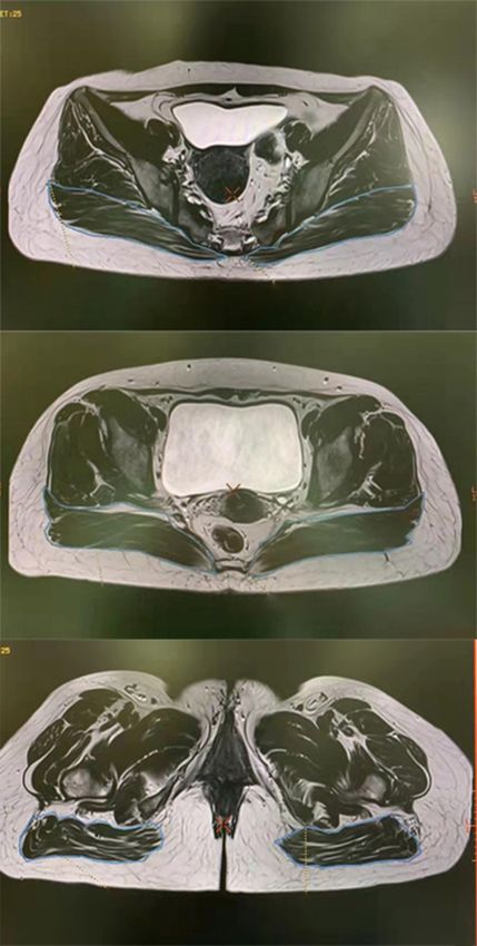
Page 2 of 13                                                                                Zhang et al. MRI in buttock augmentation

intramuscular placement approach after comprehensively               buttock augmentation (17).
considering postoperative complications, postoperative                  However, according to our literature search, no study has
effects, and patient satisfaction (5-8).                             used MRI parameters to supply accurate anatomic evaluation
    The gluteus maximus is the thickest muscle in the human          for patients in buttock augmentation. The objective of the
body (4–7 cm). It starts from the iliac crest, sacrum, coccyx,       present study was to observe and study the morphological
and sacrotuberous ligaments, and ends at the thick line of           MRI characteristics of patients who have undergone the
the femur, the gluteal tuberosity, and the iliotibial band.          gluteus maximus of buttock augmentation with silicone
The line from the middle and posterior third of the iliac            implants at levels of predetermined anatomic points.
crest to the greater trochanter is the lateral boundary of the          We present the following article in accordance with
gluteus maximus, and the line from the posterior superior            the STROBE reporting checklist (available at https://atm.
iliac spine to the coccyx is the upper and lower boundary of         amegroups.com/article/view/10.21037/atm-22-376/rc).
the piriformis, respectively. The intersection of the horizontal
line of the coccyx and the midline of the thigh is the sciatic
                                                                     Methods
nerve penetration location (9,10). Vergara et al. introduced
the intramuscular implantation technique of the gluteus              Ethical approval
maximus prosthesis by making a 6–7 cm intergluteal incision
4 cm above the anus and then cutting a 6–7 cm incision in            All procedures performed in this study involving human
the gluteus maximus fascia along the gluteus maximus fiber           participants were in accordance with the Declaration of
direction. After that, the buttock muscles are separated             Helsinki (as revised in 2013). The protocol of the study
using the fingers by tilting 45° to the deep surface from            was approved by the Research Ethics Board of Sun Yat-sen
the lateral and upper boundaries to form the appropriate             Memorial Hospital, Sun Yat-sen University (No. SYSEC-
“implant pocket” (the gluteus maximus muscle implant                 KY-KS-2021-298). Informed consent was waived due to the
space) (11,12). The principles for inserting the prosthesis          retrospective and anonymous characteristics of the study.
into the gluteus maximus were first proposed by Gonzalez
et al., and are as follows: (I) dissection must be limited to        Participants
the gluteus maximus; (II) the gluteus maximus must be cut
in half to retain as much muscle as possible before and after        Adult women, who once underwent pelvic MRI scanning at
the prosthesis (13,14). In terms of the surgical approach            Sun Yat-sen Memorial Hospital of Sun Yat-sen University
incisions, González-Ulloa et al. introduced the bilateral the        from January 2018 to January 2021, were screened. The
coccygeal region incision, infra-gluteal sulcus incision, and        inclusion criteria were as follows: (I) age between 18 to
prolongation of the medial gluteal sulcus incision (15,16).          50 years old, and (II) high-quality MRI scanning results
Nevertheless, no matter what kind of surgical approach               sufficient for analysis. The exclusion criteria were as
incision is chosen, augmentation surgery is basically carried        follows: (I) hip muscle damage due to fractures or trauma,
out under blind vision.                                              (II) femoral head necrosis, (III) problem of malignant tumor
    To better separate the gluteus maximus evenly in                 pelvic metastasis or gluteus maximus metastasis, (IV) hip
blindsight during the augmentation operation, it is                  muscle local lesions or atrophy, (V) problems associated
necessary to understand the anatomical structure of the              with being bedridden for extended time periods, unable to
gluteus maximus before surgery. Based on different surgical          walk independently, or (VI) lack of important information
approach incisions, the soft tissue thickness, gluteus               needed for the present study.
maximus width and thickness, and some accurate parameters
(anatomic angles) are needed to define the muscular incision
                                                                     MRI scanning and measurements
site and appropriate intramuscular separation direction.
The fillers in soft tissue for buttock augmentation include          We performed MRI examinations of the pelvis using a
autologous fat, hyaluronic acid, and silicone implants,              3.0T system (MAGNETOM Skyra, Siemens Healthcare,
all of which have different signal intensities in magnetic           Erlangen, Germany). The MRI sequences included coronal
resonance imaging (MRI). In addition, MRI, computed                  T1-weighted imaging (T1WI) and transverse T2WI.
tomography (CT), and ultrasound have been used to                    We used the coronal T1WI images to define 3 scanning
identify the expected appearance and complications after             levels, including the lowest point of the sacroiliac joint

© Annals of Translational Medicine. All rights reserved.           Ann Transl Med 2022;10(4):221 | https://dx.doi.org/10.21037/atm-22-376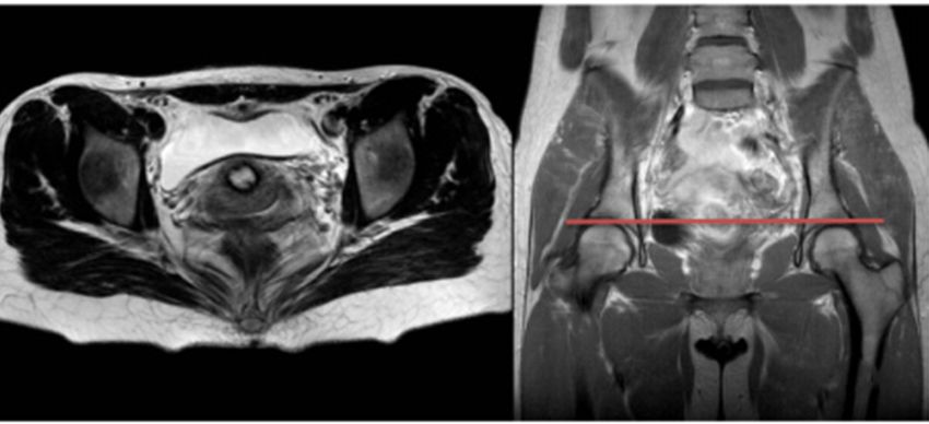
Annals of Translational Medicine, Vol 10, No 4 February 2022                                                                Page 3 of 13

                       A

                       B

                       C

Figure 1 The coronal T1WI (left) and transverse T2WI (right) MRI images of the pelvis. (A) The lowest point of the sacroiliac joint, (B)
above the femoral head, and at (C) the ischial tuberosity. MRI, magnetic resonance imaging; T1WI, T1-weighted imaging; T2WI, T2-
weighted imaging.

(SIJ), the point just above the femoral head, and the ischial          Advantage Windows 4.5 workstation; GE Healthcare,
tuberosity. Then, transverse T2WI images at the 3 levels               Chicago, IL, USA) to calculate the cross-sectional area of
were used to measure the index of left- and right-side glutes          the gluteus maximus muscle.
(Figure 1). The detailed imaging parameters are listed in                 In the transverse T2WI images, the starting point of
Table 1. We delineated the gluteus maximus muscle using                the medial tendon of one side of the gluteus maximus
post-processing software on a GE MRI workstation (GE,                  was defined as O, and the midpoint of the lateral gluteus

© Annals of Translational Medicine. All rights reserved.            Ann Transl Med 2022;10(4):221 | https://dx.doi.org/10.21037/atm-22-376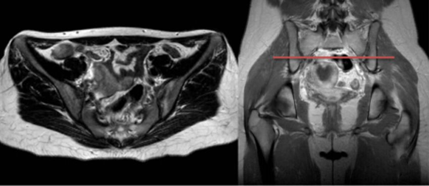
Page 4 of 13 Zhang et al. MRI in buttock augmentation
Table 1 Pelvic MRI sequence parameters
Sequence parameters Coronal T1WI Transverse T2WI
TR/TE (ms) 629/9.8 3104/110
FoV (mm) 320×320 350×350
Matrix 320×320 288×384
Slice thickness (mm) 4 4
Gap (mm) 0.8 0.8
Fat suppression (yes/no) No No
Flip angle (°) 130 130
Acquisition time 1 minute 46 seconds 1 minute 41 seconds
MRI, magnetic resonance imaging; TR, repetition time; TE, echo time; FoV, field of view; T1WI, T1-weighted imaging; T2WI, T2-weighted
imaging.
A B
C D
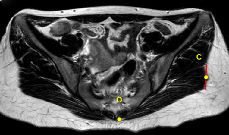
Figure 2 Schematic diagram by transverse T2WI images of measurement indicators. (A) The starting point of the medial tendon of one side
of the gluteus maximus was defined as O and the midpoint of the lateral gluteus maximus was defined as C. The (B) OC line is drawn and
the (C) vertical lines to OC at 1/3OC and 2/3OC were made in the gluteus maximus muscle. (D) The midpoints of the vertical lines were
defined as point A and point B, respectively, and the length of the vertical lines were denoted as the thickness of gluteus maximus muscle at
points A and B, respectively. T2WI, T2-weighted imaging.
maximus was defined as C (Figure 2A). Using these 2 points, muscle at point A and point B (Figure 2D). The body
an OC line was drawn (Figure 2B), which was considered surface projections of O, A, B, and C were marked as O', A',
the width of the gluteus maximus. Then, vertical lines to B', and C', respectively.
the OC line at 1/3OC and 2/3OC were made within the We set line L as the posterior midline in the transverse
gluteus maximus muscle (Figure 2C). Finally, the midpoint T2WI images. The angle of LOA (∠LOA) was the
of the vertical line at 1/3OC and 2/3OC were defined as inclusive angle of line L and line OA. This angle was
point A and point B, respectively. Therefore, the length of that from point O to point A during the vertical surgical
the vertical line was the thickness of the gluteus maximus approach (Figure 3A). The supplementary angle of
© Annals of Translational Medicine. All rights reserved. Ann Transl Med 2022;10(4):221 | https://dx.doi.org/10.21037/atm-22-376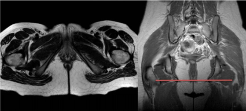
Annals of Translational Medicine, Vol 10, No 4 February 2022 Page 5 of 13
A B
C D
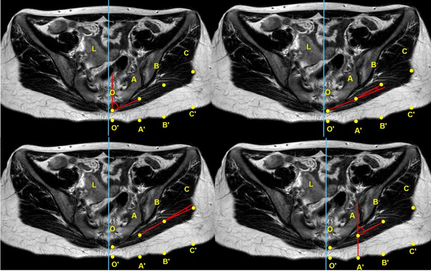
Figure 3 Schematic diagram by transverse T2WI images of measurement indicators. (A) ∠LOA is the angle between the posterior midline
L and line OA; (B) the supplementary ∠OAB is the angle between the extension line of OA and line AB; (C) the supplementary ∠ABC is the
angle between the extension line of AB and line BC; (D) the supplementary ∠A'AB is the angle between the extension line of A 'A and line
AB. T2WI, T2-weighted imaging.
OAB (supplementary ∠OAB) was the angle between the The lengths from point O, A, B, and C to their body
extension line OA and line AB, at which the operation surface projections O', A' B', and C' (denoted as OO', AA',
approach should turn from OA to point B. When the angle BB', and CC') were the thickness of soft tissue at point
was opened towards the deep gluteus maximus muscle, it O, A, B, and C (including gluteus maximus, fat, and skin),
was marked as a positive angle. Conversely, when the angle respectively. The vertical distance from O', A', B', and C' to
was opened towards the superficial layer, it was marked as the posterior midline L was recorded as the vertical distance
a negative angle. The supplementary ∠OAB was used to from O, A, B, and C to the posterior midline, respectively
differentiate the intraoperative angle between the deep layer (Figure 4). The cross-sectional area of the gluteus maximus
and the superficial layer (Figure 3B). The supplementary was recorded using the transverse T2WI images at the
angle of ABC (supplementary ∠ABC) was the included corresponding position (Figure 5).
angle between the extension line AB and line BC, and was
the angle at which the operation approach should turn
Outcomes and data collection
from AB to point C. When the angle was opened towards
the deep gluteus maximus muscle, it was marked as a The primary outcomes included the width of the gluteus
positive angle and when the angle was opened towards the maximus (measured by the length of OC), and the gluteus
superficial layer, it was marked as a negative angle. The maximus thickness at points A and B. The angles of ∠LOA,
supplementary ∠ABC was used to distinguish the angle the supplementary ∠ABC, and the supplementary ∠A'AB
towards the deep layer or the superficial layer during the were also collected.
operation (Figure 3C). The supplementary angle of A'AB Secondary outcomes included the supplementary ∠OAB,
(∠A'AB) was the included angle between the extension line the soft tissue thickness at each point (i.e., O, A, B, and C),
A'A and line AB. The supplementary ∠A'AB was the angle the distance from each point (i.e., O', A', B', and C') to the
turn from A'A to B during the vertical approach when A' posterior midline, and the cross-sectional area (CSA) of
point was the incision site (Figure 3D). the gluteus maximus at each level (i.e., the inferior point of
© Annals of Translational Medicine. All rights reserved. Ann Transl Med 2022;10(4):221 | https://dx.doi.org/10.21037/atm-22-376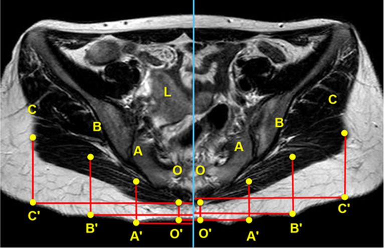
Page 6 of 13                                                                                 Zhang et al. MRI in buttock augmentation

                                                                               A

Figure 4 Schematic diagram by transverse T2WI images of                        B
measurement indicators. The length of OO', AA', BB', and
CC' indicates the thickness of soft tissue at point O, A, B, and
C (including gluteus maximus, fat, and skin), respectively. The
vertical distance from O', A', B' and C' to the posterior midline
was recorded as the distance from O, A, B and C to the posterior
midline, respectively. T2WI, T2-weighted imaging.

the sacroiliac joint, just above the femoral head, and ischial
                                                                              C
tuberosity). The baseline data collection included patients'
age, height, and body mass index (BMI).

Statistical analysis

The discontinuous data (e.g., age) are represented as
median and interquartile range (IQR), and continuous data
(e.g., height, weight, and BMI) are represented as mean ±
standard deviation (SD). By default, the population sample
was normally distributed, and the differences of each                 Figure 5 Schematic diagram by transverse T2WI images of
measurement index between the left and right sides of the             measurement indicators. The cross-sectional areas of the gluteus
gluteus maximus were compared with a paired sample t-test.            maximus at the lowest point of the sacroiliac joint (A), the upper
A 2-sided P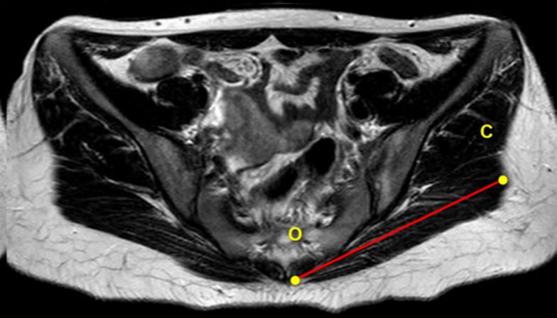
Annals of Translational Medicine, Vol 10, No 4 February 2022                                                                   Page 7 of 13

                 Women had pelvic MRI scanning records at Sun Yat-
                 sen Memorial Hospital of Sun Yat-sen University from
                 January 2018 to January 2021 (n=192)

                                                                        Excluded due to information insufficient (n=14)

                          Patients for screening (n=178)       Excluded patients according to the exclusion criteria:
                                                                • Femoral head necrosis (n=2);
                                                                 • Local inflammation and edema of gluteus muscle (n=2);
                                                                 • Gluteal muscle atrophy (n=2);
                                                                 • Malignant tumor pelvic metastasis (n=87);
                                                                 • Other tumors causing changes in the soft tissues of the
                             Screening results (n=67)                buttocks (n=18)

                                                                         Excluded due to low imaging quality (n=15)

                               Final analysis (n=52)

Figure 6 The workflow diagram of the present study.

respectively. There was no statistical difference in the                  The level just above the femoral head
angle between the left and right sides for the average
                                                                          The average length of OC at the level just above the
∠LOA (P=0.47), the supplementary ∠OAB (P=0.27), the
                                                                          femoral head was 141.76±12.15 mm for the 104 samples.
supplementary ∠ABC (P=0.32), and the supplementary
                                                                          No statistical difference was found in the comparison of
∠A'AB (P=0.87) (Table 2).
                                                                          the left and right sides (P=0.84). At the same time, the
   The average soft tissue thickness at each point (i.e., O, A, B,
                                                                          average gluteus maximus thickness at points A and B were
and C), denoted as OO', AA', BB', and CC', was 15.78±6.52,
28.14±6.31, 41.59±6.68 mm, and 49.21±8.85 mm for the 104                  23.41±4.40 and 30.64±4.57 mm, respectively. Similarly,
samples, respectively. There was no statistical difference                there was no statistical difference in the thickness between
between the left and right sides for the average length of                the left and right sides at points A (P=0.58) and B (P=0.09)
OO' (P=0.23), AA' (P=0.06), BB' (P=0.13), or CC' (P=0.52)                 (Table 2).
(Table 2).                                                                   The average ∠LOA, the supplementary ∠ABC, the
   The average vertical distance between each body                        supplementary ∠A'AB, and the supplementary ∠OAB were
projection point (i.e., O', A', B', and C') and the posterior             62.36±5.65°, 0.74±8.10°, 109.74±5.52°, and −7.43±6.82°,
midline was 14.50±7.32, 54.40±6.34, 91.51±8.34 mm, and                    respectively. There was no statistical difference in the
130.11±11.62 mm, respectively, for the 104 samples. The                   angle between the left and right sides for the average
average vertical distance from B' (P=0.04) and C' (P≤0.05)                ∠LOA (P=0.60), the supplementary ∠ABC (P=0.14), the
to the posterior midline was statistically longer on the left             supplementary ∠A'AB (P=0.27), or the supplementary
side than on the right side; however, no statistical difference           ∠OAB (P=0.97) (Table 2).
for this distance was found between the left and right sides for             The average soft tissue thickness at each point (i.e., O,
O' (P=0.64) and A' (P=0.13). The average cross-sectional area             A, B, and C), denoted as OO', AA', BB', and CC', were
of the gluteus maximus at this level was 2,486.43±623.08 mm2              14.83±6.17, 34.07±5.59, 43.55±6.01, and 50.61±10.00 mm
for all 104 samples. There was no statistical difference in               for the 104 samples, respectively. There was no statistical
average cross-sectional area of the gluteus maximus between               difference between the left and right sides for the average
the left and right sides (P=0.72) (Table 2).                              length of OO' (P=0.65), AA' (P=0.16), BB' (P=0.58), and

© Annals of Translational Medicine. All rights reserved.               Ann Transl Med 2022;10(4):221 | https://dx.doi.org/10.21037/atm-22-376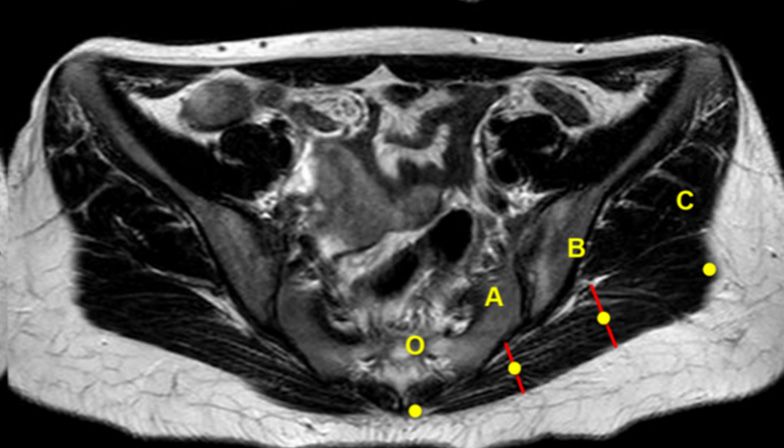
Table 2 Measurement results of gluteus maximus muscle indexes obtained from pelvic MRI acquired in the transverse position
                                                                                                      Lowest point of     Lowest point of                                       On the right                Left ischial    Right ischial
                                                                                                                                                        On the left femoral
                                                                         Measurement                   left sacroiliac     right sacroiliac   P value                          femoral head     P value     tuberosity       tuberosity     P value
                                                                                                                                                        head (mean ± SD)
                                                                                                     joint (mean ± SD)   joint (mean ± SD)                                      (mean ± SD)                (mean ± SD)      (mean ± SD)
                                                                                                                                                                                                                                                      Page 8 of 13

                                                                         OC (mm)                       126.06±14.13        124.58±12.02        0.28       141.90±13.45         141.62±10.57      0.84      108.21±12.50     110.62±9.15      0.06

                                                                         The gluteus maximus            15.65±3.95          16.42±4.30         0.12        23.11±4.23           23.71±4.52       0.58       24.68±8.05      24.80±7.00       0.83
                                                                         thickness at point A (mm)

                                                                         The gluteus maximus            24.40±4.64          24.92±4.98         0.30        30.22±4.52           31.06±4.52       0.09       29.24±6.90      29.86±6.78       0.38
                                                                         thickness at point B (mm)

                                                                                                        69.86±4.14          69.68±6.29         0.47        62.55±6.05           62.16±5.16       0.60       89.50±8.34       90.34±8.3       0.39

                                                                                                         6.09±4.29           7.00±6.29         0.27        −7.46±7.09           −7.40±6.46       0.97      13.11±11.04      14.79±9.74       0.47
                                                                         ∠LOA (°)

                                                                                                        −3.03±6.17          −2.44±5.95         0.32        −0.33±.7.91           1.81±8.07       0.14       6.56±8.45        6.19±8.28       0.85
                                                                         Supplementary ∠OAB (°)

                                                                                                        63.83±4.72          63.03±4.53         0.87        70.69±6.13           69.84±4.86       0.27       76.67±8.95      75.32±8.67       0.07
                                                                         Supplementary ∠ABC (°)

© Annals of Translational Medicine. All rights reserved.
                                                                         OO' (mm)                       15.61±6.35          15.95±6.62         0.23        14.73±5.81           14.90±6.44       0.65       37.03±5.66      37.40±6.42       0.52
                                                                         Supplementary ∠A'AB (°)

                                                                         AA' (mm)                       28.48±6.27          27.81±6.27         0.06        34.41±5.88           33.72±5.21       0.16       33.54±6.52      33.38±6.40       0.76

                                                                         BB' (mm)                       41.97±6.60          41.21±6.67         0.13        43.69±6.04           43.40±5.92       0.58       37.37±7.41      37.68±7.31       0.06

                                                                         CC' (mm)                       49.40±9.08          49.03±8.52         0.52       50.40±10.08           50.83±9.81       0.66       42.39±9.92      42.37±10.31      0.98

                                                                         Vertical distance between      14.36±7.21          14.64±7.37         0.64        12.99±6.70           12.66±6.22       0.51       35.83±8.45      35.41±7.31       0.52
                                                                         O' and L line (mm)

                                                                         Vertical distance between      54.78±6.44          54.02±6.16         0.13        55.86±5.98           53.95±5.53       0.02*      72.82±7.41      74.21±6.96       0.06
                                                                         A' and L line (mm)

                                                                         Vertical distance between      92.31±8.53          90.71±7.99         0.04*      100.49±7.57           98.58±6.57       0.03*     108.34±7.77      110.07±6.11      0.03*
                                                                         B' and L line (mm)

                                                                         Vertical distance between     131.26±12.25        128.96±10.70        0.05*      144.64±10.56         142.12±8.15       0.03*     142.67±9.97      144.68±7.64      0.05*
                                                                         C' and L line (mm)

                                                                         Cross-sectional area of     2,495.59±667.74     2,477.26±568.29       0.72     3,538.89±742.80       3,618.97±723.69    0.09     2,953.01±791.76 3,096.20±754.58    0.00*
                                                                         the gluteus maximus
                                                                         (mm²)
                                                                         *, P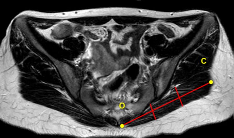
Annals of Translational Medicine, Vol 10, No 4 February 2022                                                              Page 9 of 13

CC' (P=0.66) (Table 2).                                             of this distance was found between the left and right
   The average vertical distance between each body                  sides for O' (P=0.52) and A' (P=0.06). The average cross-
projection point (i.e., O', A', B', and C') and the posterior       sectional area of the gluteus maximus at this level was
midline were 12.82±6.50, 54.91±5.87, 99.54±7.19, and                3,024.60±780.46 mm 2 for all 104 samples. There was a
143.38±9.56 mm for the 104 samples, respectively. The               significant difference in average cross-sectional area of the
average vertical distance from the point O' to the posterior        gluteus maximus between the left and right sides (P
Page 10 of 13                                                                                Zhang et al. MRI in buttock augmentation

cosmetics of the buttocks will be poor (25). According to             arc-shaped structure of the muscles. Our results are in line
our measurement results, we found that the level above                with what was reported by Vergara et al., as the arc-shaped
the femoral head was closer to the maximum width of the               structure of the hip muscles caused a tilt in the approach
gluteus maximus muscle. Therefore, the diameter of the                route to the deep surface by 45°. This often occurred when
prosthesis implanted into the gluteus maximus should not              separating the hip muscles to avoid passing through the
exceed the length of the OC on the level just above femoral           superficial muscles and entering the subcutaneous layer
head (141.76±12.15 mm). It should be noted that an MRI                (11,12). However, the gluteal muscles tilted at 45° to the
performed in the supine position compresses the gluteus               deep surface mentioned in the literature are not close to the
maximus, which may lead to a discrepancy between the                  angle in the data that we measured. This may be related to
measured results and those found in actual clinical practice.         the fact that the literature does not specify the position of
   The thickness of the gluteus maximus is an important               the tilt.
parameter for the procedure, which must be considered                    During an operation, surgeons can access the approximate
in order to define how thick the implant pocket should be             positions of O, A, B, and C by their skin projection points
(25,26). According to our measurement results, the thickness          of O', A', B', and C'. Our results showed some differences
of the gluteus maximus at point A is 16.03±4.17 mm at the             between the left side and right side for those points at the
inferior point of the SIJ, 23.41±4.40 mm at just above the            3 levels, which is consistent with the natural anatomical
femoral head, and 24.74±7.58 mm at the ischial tuberosity.            characteristics of humans (27). Nevertheless, it is almost
In comparison, the thickness of the gluteus maximus at                impossible to locate them accurately during the operative
point B for these levels was 24.66±4.84, 30.64±4.57, and              separation of the gluteus maximus. The supplementary
29.55±6.88 mm, respectively. These results reflected the              ∠A'AB and the supplementary ∠ABC were used to
morphological characteristics that the upper part of the              determine the position of B and C. The supplementary
gluteus maximus muscle is thinner than the lower part.                ∠A'AB is the angle from which AA' should turn to point
The thickness of the starting point A is vital for surgeons           B, and the supplementary ∠ABC is the angle from which
to create a consistent thickness of superficial and posterior         AB should turn to point C. The surgeon approached from
walls of the implant by reserving the appropriate superficial         skin projection point A' vertically into the glutes surface
glutes thickness during the stripping thereof. To avoid the           to point A during the operation. He/she then followed the
unequal thickness of the gluteus maximus dissection, the              supplementary ∠A'AB to strip the glutes laterally, until
reserved superficial thickness of the gluteus maximus should          directly under the B' point where point B is located. Next,
be half of that of the thickness of point A. When the vertical        a surgeon follows the supplementary ∠ABC from point
approach is employed from the surface of the gluteus                  B to separate the muscle further laterally to point C (the
maximus directly below A', the reserved thickness of the              endpoint of dissection), under projection C'. According
superficial gluteus maximus should be 8.00±4.17 mm at the             to our measurement results, both the left and right
inferior point of the SIJ, 11.70±4.40 mm at the just above            supplementary ∠A'AB was about 63° at the inferior point of
femoral head, and 12.3±7.58 mm at the ischial tuberous.               the SIJ, 70° at the just above femoral head, and 76° at the
If the separation of the gluteus maximus is too shallow               ischial tuberosity. In comparison, the supplementary ∠ABC
(superficial thickness less than the range), muscle herniation        was about −3° (3° to the superficial gluteus maximus), 1°,
may occur after surgery. On the contrary, if the separation           and 6°, accordingly. We believe that the ideal separation
exceeds this range for the superficial layer, it is easy to cause     route can be found when separating the gluteus maximus
postoperative blood supply dysfunction for the deep gluteus           muscle by referring to the above angle. However, through
maximus. It is worth noting that the thickness of the gluteus         further analysis and consideration of the data, it can be
maximus at point A is not the thickest or thinnest of the             found that all the supplementary ∠ABC in the 3 positions
gluteus maximus but the thickness of the surgery starting             were not more than 6°, which may have no clinical
point.                                                                significance. We assume that a vertical approach to the
   We used the ∠LOA at different levels to explore the                surface of the gluteus maximus directly below A' should
shape of the hip muscles. Our measurement results showed              follow after a single angle correction of the supplementary
that ∠LOA was about 70° at the inferior point of the SIJ              ∠A'AB at point A. This process continues to separate
level, about 62° at the just above femoral head level, and            laterally until it reaches the lateral boundary of the gluteus
about 90° at the ischial tuberosity level, which indicated an         maximus (point C), which can equally divide the gluteus

© Annals of Translational Medicine. All rights reserved.            Ann Transl Med 2022;10(4):221 | https://dx.doi.org/10.21037/atm-22-376
Annals of Translational Medicine, Vol 10, No 4 February 2022                                                             Page 11 of 13

maximus into 2 parts. Therefore, it is not necessary to             the starting point (A point) should be 73.91±5.57 mm away
find point B or adjust the approach direction at any point          from the posterior midline and the gluteus maximus should
intraoperatively.                                                   be divided evenly into 2 parts by a vertical approach of
    The surgeon typically needs to mark the scope of the            12.3±7.58 mm. After that, an angle of 70±4.40° should be
anatomy and the approximate location of the prosthesis              followed to further separate the muscle.
before surgery. Horn et al. believe that the skin position will        The innovations of this study are as follows. (I) It was
change from the standing position to the prone position,            the first study to observe the physiological anatomy of
and that the preoperative marking should be made in the             the gluteus maximus muscle through MRI and provide
standing or sitting position (28). The medial, lateral, upper,      anatomical reference data for gluteus maximus implant
and lower boundaries of the operation have been defined             augmentation. (II) Standardized measurement of the
by different authors and were generally similar but exhibit         scheduled anatomical levels of the gluteus maximus
inconsistencies in their defining methods (9,20-22,28,29).          muscle was carried out in the transverse section of pelvic
According to the medial boundary mentioned in the                   MRI with 3 obvious bone markers. This was performed
literature, the 2 parallel boundary lines are 4–5 cm from           to reflect the physiological radian and thickness of the
the posterior midline. Our measurement results showed               gluteus maximus muscle and avoid the limitation of a single
that the distance between A' and the posterior midline was          position measurement. There were also some limitations
about 54 mm at the inferior point of the SIJ level, 55 mm at        of this study. First, as the pelvic MRI in this study was
just above the femoral head level, and 73 mm at the ischial         conducted in the supine position, it may exhibit a certain
tuberosity level. All the locations of point A' were outside        influence on the shape and thickness of soft tissues, leading
the medial boundary at all 3 levels, which indicated that           to a discrepancy between the measured results and the true
point A is a suitable position for the anatomical approach.         data. Next, although the sample size of 52 was enough to
    Aboudib et al. reported no statistical correlation between      generate clinically significant results in practice, larger scale
the cosmetic effect evaluated by plastic surgeons and the           studies in the future are needed to confirm our findings.
volume of the prosthesis, or between the volume of the              Third, the fact that all included patients were Chinese
prosthesis and the volume of the muscle. Therefore, they            women may affect the external validity of this work to
believed that a preoperative computed tomography (CT)               some degree. In addition, the physiological structure of the
scan was not necessary for the operation (30). An MRI is            gluteus maximus muscle is correlated with the width and
more expensive than CT and is generally not a preoperative          height of the pelvis; however, we did not have enough data
routine inspection for buttock augmentation. In the present         to explore such an association. Therefore, we plan to further
study, all the predetermined MRI anatomic measurement               explore the relationship between the pelvis and the gluteus
indexes exhibited physiological significance and can truly          maximus muscle in the future. In addition, this study was a
reflect the morphological characteristics of the gluteus            retrospective analysis, which is likely to exhibit some bias
maximus muscle in a natural physiological state. The                in the results. These results need to be further confirmed
measurement results can provide real and objective data             by multi-center clinical trials. The patients included in this
references for clinical practice and provide an anatomical          study were all Chinese women who had certain limitations
basis and clinical reference for intramuscular prosthetic           in being able to generalize to the greater population. A
buttock augmentation.                                               comparative analysis with patients from other countries is
    In summary, at the level of inferior point of the SIJ,          also recommended.
the starting point (A point) should be 54.4±6.34 mm away
from the posterior midline. Then, a vertical approach of
                                                                    Conclusions
8.0±4.17 mm should be made from the gluteus maximus
surface. Finally, the line should be tilted 63.4±4.62° to the       In this study, we measured the anatomical features of the
deep plane to separate the gluteus maximus. At the level            gluteus maximus at the levels of the inferior point of the SIJ,
just above the femoral head, the gluteus maximus should be          just above the femoral head, and at the ischial tuberosity
separated at the angle of 70±4.40° to the deep plane after          using MRI. According to our results, the diameter of the
a 11.7±4.40 mm vertical approach from the surface of the            implant should not exceed 14.18±1.22 cm. In addition, the
gluteus maximus and a starting point of 54.91±5.57 mm away          gluteus maximus at the lowest point of the SIJ and above
from the posterior midline. At the level of ischial tuberosity,     the femoral head has an arc structure, which needs to be

© Annals of Translational Medicine. All rights reserved.          Ann Transl Med 2022;10(4):221 | https://dx.doi.org/10.21037/atm-22-376
Page 12 of 13 Zhang et al. MRI in buttock augmentation
tilted to the deep plane during separation. However, the anonymous characteristics of the study.
gluteus maximus at the ischial tuberosity level is relatively
flat, so the gluteus maximus at this level does not need to be Open Access Statement: This is an Open Access article
tilted too much to the deep plane. Clinicians can use all the distributed in accordance with the Creative Commons
parameters indicated in this study, including the thickness Attribution-NonCommercial-NoDerivs 4.0 International
of the gluteus maximus muscle in the above 3 levels, the License (CC BY-NC-ND 4.0), which permits the non-
position and angle of the “inflection point” after the surgical commercial replication and distribution of the article with
approach, and the approximate starting and ending point the strict proviso that no changes or edits are made and the
range of the gluteus maximus muscle. As an alternative to original work is properly cited (including links to both the
the previous experience of blind dissection, the gluteus formal publication through the relevant DOI and the license).
maximus muscle can be more scientifically and reasonably See: https://creativecommons.org/licenses/by-nc-nd/4.0/.
dissected using the indexes for gluteus augmentation
supplied in the study, which has certain clinical significance.
References
1. Heidekrueger PI, Juran S, Patel A, et al. Plastic Surgery
Acknowledgments
Statistics in the US: Evidence and Implications. Aesthetic
This work would not have been possible without the Plast Surg 2016;40:293-300.
consistent and valuable reference materials that we received 2. Almutairi K, Gusenoff JA, Rubin JP. Body Contouring.
from Dr. Xiang Zhang (Affiliation: Sun Yat-sen Memorial Plast Reconstr Surg 2016;137:586e-602e.
Hospital, Sun Yat-sen University), whose insightful 3. Oranges CM, Haug M, Schaefer DJ. Body Contouring.
guidance and enthusiastic encouragement during the study Plast Reconstr Surg 2016;138:944e-5e.
has earned our deepest gratitude. 4. Cosmetic Surgery National Data Bank Statistics. Aesthet
Funding: None. Surg J 2016;36 Suppl 1:1-29.
5. Senderoff DM. Aesthetic Surgery of the Buttocks Using
Implants: Practice-Based Recommendations. Aesthet Surg
Footnote
J 2016;36:559-76.
Reporting Checklist: The authors have completed the 6. Mofid MM, Gonzalez R, de la Peña JA, et al. Buttock
STROBE reporting checklist. Available at https://atm. augmentation with silicone implants: a multicenter
amegroups.com/article/view/10.21037/atm-22-376/rc survey review of 2226 patients. Plast Reconstr Surg
2013;131:897-901.
Data Sharing Statement: Available at https://atm.amegroups. 7. Flores-Lima G, Eppley BL, Dimas JR, et al. Surgical
com/article/view/10.21037/atm-22-376/dss pocket location for gluteal implants: a systematic review.
Aesthetic Plast Surg 2013;37:240-5.
Conflicts of Interest: All authors have completed the 8. Oranges CM, Tremp M, di Summa PG, et al. Gluteal
ICMJE uniform disclosure form (available at https://atm. Augmentation Techniques: A Comprehensive Literature
amegroups.com/article/view/10.21037/atm-22-376/coif). Review. Aesthet Surg J 2017;37:560-9.
The authors have no conflicts of interest to declare. 9. Serra F, Aboudib JH, Cedrola JP, et al. Gluteoplasty:
anatomic basis and technique. Aesthet Surg J
Ethical Statement: The authors are accountable for all 2010;30:579-92.
aspects of the work in ensuring that questions related 10. Xue B, Liu L. Research progress of hip aesthetics and
to the accuracy or integrity of any part of the work are extended plastic surgery. Chinese Journal of Aesthetic
appropriately investigated and resolved. All procedures Medicine 2015;24:81-5.
performed in this study involving human participants were 11. Vergara R, Marcos M. Intramuscular gluteal implants.
in accordance with the Declaration of Helsinki (as revised Aesthetic Plast Surg 1996;20:259-62.
in 2013). The protocol of the study was approved by the 12. Vergara R, Amezcua H. Intramuscular gluteal implants: 15
Research Ethics Board of Sun Yat-sen Memorial Hospital, years' experience. Aesthet Surg J 2003;23:86-91.
Sun Yat-sen University (No. SYSEC-KY-KS-2021-298). 13. Gonzalez R. Gluteal implants: the "XYZ" intramuscular
Informed consent was waived due to the retrospective and method. Aesthet Surg J 2010;30:256-64.
© Annals of Translational Medicine. All rights reserved. Ann Transl Med 2022;10(4):221 | https://dx.doi.org/10.21037/atm-22-376Annals of Translational Medicine, Vol 10, No 4 February 2022 Page 13 of 13
14. Gonzalez R, Gonzalez R. Intramuscular Gluteal Fat Grafting. Clin Plast Surg 2018;45:203-15.
Augmentation: The XYZ Method. Clin Plast Surg 23. Homma D, Minato I, Imai N, et al. Investigation on
2018;45:217-23. the measurement sites of the cross-sectional areas of the
15. González-Ulloa M. Gluteoplasty: a ten-year report. gluteus maximus and gluteus medius. Surg Radiol Anat
Aesthetic Plast Surg 1991;15:85-91. 2019;41:109-15.
16. González-Ulloa M. Torsoplasty. Aesthetic Plast Surg 24. Homma D, Minato I, Imai N, et al. Appropriate sites for
1979;3:357-68. the measurement of the cross-sectional area of the gluteus
17. Yahyavi-Firouz-Abadi N, Menias CO, Bhalla S, et al. maximus and the gluteus medius muscles in patients with
Imaging of cosmetic plastic procedures and implants in hip osteoarthritis. Surg Radiol Anat 2021;43:45-52.
the body and their potential complications. AJR Am J 25. Mendieta CG. Gluteoplasty. Aesthet Surg J 2003;23:441-
Roentgenol 2015;204:707-15. 55.
18. Tothill P, Stewart AD. Estimation of thigh muscle and 26. Cuenca-Guerra R, Lugo-Beltran I. Beautiful buttocks:
adipose tissue volume using magnetic resonance imaging characteristics and surgical techniques. Clin Plast Surg
and anthropometry. J Sports Sci 2002;20:563-76. 2006;33:321-32.
19. Nakatani M, Takai Y, Akagi R, et al. Validity of muscle 27. Standring S, Ellis H, Healy J, et al. Gray’s anatomy: the
thickness-based prediction equation for quadriceps femoris anatomical basis of clinical practice. Am J Neuroradiol
volume in middle-aged and older men and women. Eur J 2005;26:2703.
Appl Physiol 2016;116:2125-33. 28. Horn G. Gluteoplasty with intramuscular silicone cohesive
20. Andrade GA, Coltro PS, Andó A, et al. Gluteal gel implants: a retrospective study of 50 cases. Ann Chir
Augmentation with Silicone Implants: A New Proposal Plast Esthet 2009;54:467-76.
for Intramuscular Dissection. Aesthetic Plast Surg 29. Aslani A, Del Vecchio DA. Composite Buttock
2017;41:872-7. Augmentation: The Next Frontier in Gluteal Aesthetic
21. Cárdenas-Camarena L, Trujillo-Méndez R, Díaz-Barriga Surgery. Plast Reconstr Surg 2019;144:1312-21.
JC. Tridimensional Combined Gluteoplasty: Liposuction, 30. Aboudib JH, Serra-Guimarães F, Sampaio FJ. Profile of
Buttock Implants, and Fat Transfer. Plast Reconstr Surg Patients Undergoing Gluteoplasty. Aesthetic Plast Surg
2020;146:53-63. 2016;40:30-7.
22. Godoy PM, Munhoz AM. Intramuscular Gluteal
Augmentation with Implants Associated with Immediate (English Language Editor: J. Jones)
Cite this article as: Zhang J, Wong CL, Zhang J, Su Z,
Shi F, Chen C, Wang Y, Xiao X, Liang W, Zhang J. Clinical
use of magnetic resonance imaging in buttock augmentation
with silicone implants: a retrospective analysis. Ann Transl Med
2022;10(4):221. doi: 10.21037/atm-22-376
© Annals of Translational Medicine. All rights reserved. Ann Transl Med 2022;10(4):221 | https://dx.doi.org/10.21037/atm-22-376You can also read