Circulating Placental Vesicles Carry HLA-DR in Pre-Eclampsia: A New Potential Marker of the Syndrome
←
→
Page content transcription
If your browser does not render page correctly, please read the page content below
ORIGINAL RESEARCH
                                                                                                                                         published: 03 September 2021
                                                                                                                                       doi: 10.3389/fimmu.2021.717879

                                            Circulating Placental Vesicles Carry
                                            HLA-DR in Pre-Eclampsia: A New
                                            Potential Marker of the Syndrome
                                            Chiara Tersigni 1*†, Donatella Lucchetti 2†, Rita Franco 3, Filomena Colella 2, Caterina Neri 1,
                                            Laura Crispino 3, Alessandro Sgambato 2, Antonio Lanzone 1,3, Giovanni Scambia 3,4,
                                            Manu Vatish 5 and Nicoletta Di Simone 6,7
                                            1 Unità Operativa Complessa di Ostetricia e Patologia Ostetrica, Dipartimento di Scienze della Salute della Donna e del
                                            Bambino e di Sanità Pubblica, Fondazione Policlinico Universitario A. Gemelli, Istituto di Ricovero e Cura a Carattere
                                            Scientifico (IRCCS), Rome, Italy, 2 Dipartimento Universitario di Medicina e Chirurgia Traslazionale, Università Cattolica del
                                            Sacro Cuore, Rome, Italy, 3 Istituto di Clinica Ostetrica e Ginecologica, Università Cattolica del Sacro Cuore, Rome, Italy,
                                            4 Unità Operativa Complessa di Ginecologia Oncologica, Dipartimento di Scienze della Salute della Donna e del Bambino e di
                            Edited by:
                                            Sanità Pubblica, Fondazione Policlinico Universitario A. Gemelli Istituto di Ricovero e Cura a Carattere Scientifico (IRCCS),
                  Gabriela Barrientos,
                                            Rome, Italy, 5 Nuffield Department of Women’s & Reproductive Health, University of Oxford, Oxford, United Kingdom,
 Consejo Nacional de Investigaciones
                                            6 Department of Biomedical Sciences, Humanitas University, Milan, Italy, 7 Istituto di Ricovero e Cura a Carattere Scientifico
   Cientı´ficas y Técnicas (CONICET),
                                            (IRCCS) Humanitas Research Hospital, Rozzano, Milan, Italy
                             Argentina

                        Reviewed by:
                        Aaron Michels,      Background: Pre-eclampsia (PE) is a common disorder of pregnancy that usually
University of Colorado, United States
                                            presents with hypertension and proteinuria. The clinical presentation arises from soluble
                    Aditi Arun Narsale,
    San Diego Biomedical Research           factors released into the maternal circulation from the placenta owing to the stress of
               Institute, United States     syncytiotrophoblast, consequence of defective placentation occurring in the first half of
                   *Correspondence:         pregnancy. Reduced tolerance of the semiallogeneic fetus by the maternal immune
                        Chiara Tersigni
    chiara.tersigni@policlinicogemelli.it
                                            system has been proposed as first trigger leading to poor placentation. We previously
     †
      These authors have contributed
                                            observed aberrant expression of human leukocyte antigen (HLA)-DR molecules in the
             equally to this work and       syncytiotrophoblast of a subset of women with PE. Aim of this study was to investigate
                share first authorship
                                            abnormal expression of circulating HLA-DR in syncytiotrophoblast-derived extracellular
                                            vesicles (STBEVs) in women with PE compared to normal pregnant women.
                   Specialty section:
         This article was submitted to      Methods: peripheral venous blood was collected from 22 women with PE and 22 normal
             Immunological Tolerance
                       and Regulation,
                                            pregnant women. Circulating STBEVs were collected by ultra-centrifugation (120000 g)
               a section of the journal     and analyzed for the expression of HLA-DR and placental alkaline phosphatase (PLAP), a
              Frontiers in Immunology
                                            specific marker of the placenta, by Western blot analysis and flow cytometry.
            Received: 31 May 2021
        Accepted: 16 August 2021            Results: circulating STBEVs positive for HLA-DR were observed in 64% of PE women
     Published: 03 September 2021           while no HLA-DR positivity was detected in any of the controls (P
Tersigni et al.                                                                                          Placental Vesicles Carry HLA DR in Pre Eclampsia

INTRODUCTION                                                                  Gemelli Hospital as outpatients for pregnancy routine medical
                                                                              examinations. Women with diabetes, obesity (BMI >30), pre-
Pre-eclampsia (PE) is a common disorder of pregnancy that usually             existing hypertension, autoimmune or infectious diseases or fetal
presents with hypertension and proteinuria. It complicates 3–5% of            abnormalities were excluded from control group. All cases and
all pregnancies and remains a major cause of severe maternal and              controls were matched for gestational age.
newborn morbidity and mortality worldwide (1). The clinical                      3 ml of venous blood was collected by venipuncture from the
presentation arises from factors released into the maternal                   antecubital fossa in a tube with EDTA. Each sample was
circulation from the placenta as result of syncytiotrophoblast stress.        centrifuged twice at 3000 g for 30 minutes at 4°C, to obtain
The latter is secondary to defective placentation occurring in the first       plasma, and then frozen in 500 ml aliquots at -80°C until use.
half of pregnancy. According to the “two stage model”, reduced
tolerance of the semi-allogeneic fetus by the maternal immune                 STBEVs Collection From Serum
system could be a potential first trigger in the pathogenetic cascade          Plasma samples (500µl), previously stored at -80°C, were allowed to
leading to poor placentation and PE (2). Consistent with this                 attain room temperature. Each sample was diluted (1:1 vol/vol) with
hypothesis, in a previous study, we observed aberrant expression of           PBS and spun at 120,000 g for 90 minutes at 4°C in an Optima XPN
human leukocyte antigen (HLA)-DR molecules in the                             ultracentrifuge (Beckman Coulter). After the ultracentrifugation step,
syncytiotrophoblast of placentas obtained from women with PE (3).             the supernatant was discarded and the pellet, which comprised of
    HLA- DR is a class II molecule constitutively expressed on                extracellular vesicles of different sizes, was washed with PBS and
professional antigen presenting cells (APC) to present exogenous              spun one more time at 120,000 g for 90 minutes at 4C°. The
antigens to T cells to elicit antigen specific immune response. In             supernatant was then discarded and the pelleted EVs were then re-
inflammatory conditions the expression of HLA class II molecules               suspended with 100 µl of PBS. The protein concentration of STBEVs
can be induced but the possibility that also trophoblast cells might          was determined by Bradford assay, prior to storage at −80°C.
express these antigens is still debated (4, 5).
    Tight control of HLA class I and class II expression in villous           Western Blotting
and extra-villous trophoblast (VT and EVT, respectively) is                   For PLAP expression analysis, STBEVs samples collected from sera
essential for successful pregnancy outcome (6). In particular,                from 4 preeclamptic and 4 control patients were lysed on ice for 30
the lack of HLA class II (-DP, -DQ and -DR) molecule                          minutes in SDS-PAGE sample buffer containing protease inhibitor
expression on trophoblasts prevents maternal T cell allo-                     cocktail (Roche Diagnostics,Basel, Switzerland). Protein content
immune responses against paternal-derived antigens.                           was normalized by loading 20 mg of protein for each sample.
    Previously, we demonstrated that aberrant expression of                   Samples were boiled and centrifuged at 13,000g for 10 minutes
HLA-DR antigen in trophoblast cells can be found in about                     prior to separation by SDS/PAGE (Invitrogen) and semi-dry
40% of syncytiotrophoblast-derived extracellular vesicles                     transfer to PVDF membrane (Biorad, Hercules, CA, USA). Non-
(STBEVs), obtained from PE women by dual placental                            specific binding was blocked with TBS-T (20 mM Tris/HCl, 137
perfusion (3). The positivity for HLA-DR of STBEVs, found in                  mM NaCl, 0.1%Tween-20, pH 7.6) containing 5% of milk (Santa
a subset of PE cases, was confirmed by immunohistochemistry                    Cruz Biotechnology Inc., Dallas, Texas, USA) for 1 hour at R/T.
on placental sections. In particular, HLA-DR was expressed in                 Membranes were incubated at 4°C overnight with the NDOG2
syncytiotrophoblast, the cell type releasing the STBEVs in                    antibody (1µl/ml) syncytiotrophoblast-specific mouse monoclonal
maternal circulation in vivo and representing the main                        antibody that recognizes placental alkaline phosphatase (PLAP) (8)
maternal-fetal interface in the second half of pregnancy (6).                 in Tris-buffered saline and 0.05% Tween 20 (TBS-T) containing
    Here we assessed whether HLA-DR aberrant expression                       1% BSA. Membranes were then washed in TBS-T, before
might be confirmed in STBEVs collected from the peripheral                     incubation with the appropriate (rabbit or mouse) horseradish
blood of women with clinical diagnosis of PE.                                 peroxidase-conjugated secondary antibody (1:4000; Dako,
                                                                              Glostrup, Denmark) for 1 hour at R/T. The antibody used was
                                                                              diluted in blocking buffer. After washing, blots were treated with an
MATERIALS AND METHODS                                                         enhanced chemiluminescence system (PierceTM, Thermo Fischer
                                                                              Scientific, Waltham, MA USA) and exposed to Hyperfilm ECL
Patients and Sample                                                           (GE Healthcare Life Sciences, Cleveland, Ohio, USA).
This study has been designed and conducted according to the                   Densitometric analysis of Western blot was carried out using
principles of the Declaration of Helsinki and approved by the                 NineAlliance software (Uvitec Alliance, Cambridge, UK).
Ethics Committee of the Università Cattolica del Sacro Cuore of
Rome, Italy. Written informed consent was obtained from all                   Flow Cytometry Analysis of STBEVs
recruited individuals.                                                        Analysis of STBEVs was carried out by multi-color flow cytometry,
   All women enrolled in this study were selected among those                 using a CytoFLEX S cytometer (Beckman Coulter) equipped with
referring to the High Risk Pregnancy Unit and to the delivery                 violet laser (405 nm) excitation sources. This instrument is able to
suite of the Gemelli Hospital of Rome. For the identification of               collect SSC (side scatter) off the blue laser (BSSC) and the violet
cases of PE we referred to the definition of the International                 laser (VSSC). The flow cytometer was calibrated using the
Society for the Study of Hypertension in Pregnancy (ISSHP) (7).               Megamix-Plus FSC beads emitting FITC of different sizes (100,
Control patients were recruited among those attending the                     300, 500, and 900 nm), as described elsewhere (9). STBEVs number

Frontiers in Immunology | www.frontiersin.org                             2                               September 2021 | Volume 12 | Article 717879
Tersigni et al. Placental Vesicles Carry HLA DR in Pre Eclampsia
Proteinuria (g/L)
was measured using the cell-counting feature of the instrument that
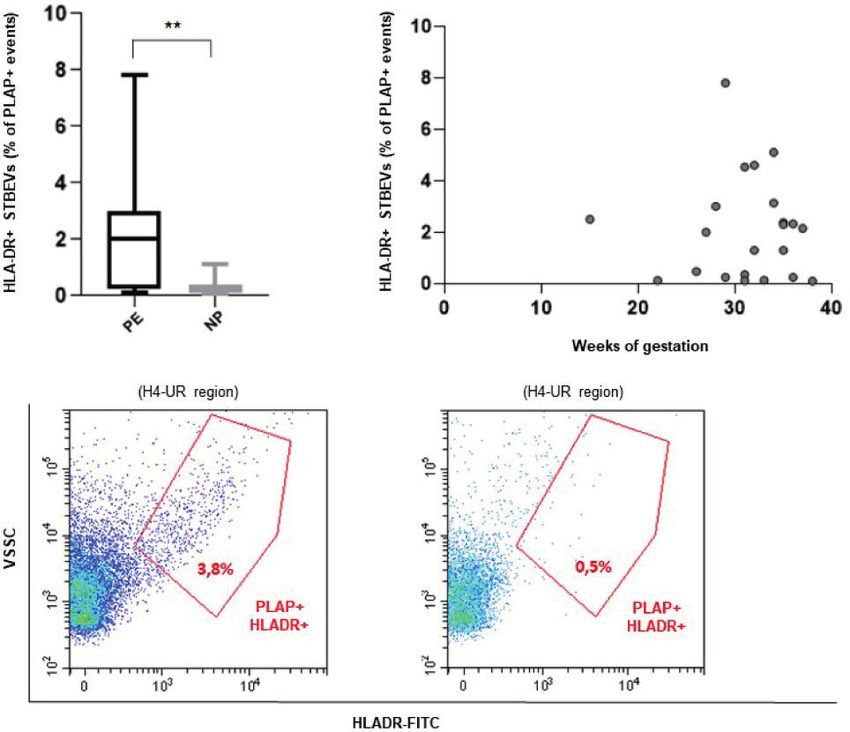
relies on a calibrated peristaltic pump for sample delivery. ToTersigni et al. Placental Vesicles Carry HLA DR in Pre Eclampsia and displayed significantly lower gestational ages at delivery, with PE compared to normal pregnant women (Figures 1C–G), neonatal birth weights (p < 0.0001) and birth weight percentile although STBEVs levels showed a high standard deviation (SD) (p < 0.0001) compared to NP. As expected, women with PE were and a skewed distribution among the study groups. Interestingly, characterized by higher blood pressures (p < 0.0001) and STBEVs were detectable in the maternal circulation from the detectable proteinuria (p < 0.01) compared to NP women. No early first weeks of gestation (Figure 1D). No significant significant differences were found in terms of maternal age differences were found in terms of calcein positive events or between PE and control groups. 52% of women with PE were protein content between PE and NP women in vesicles samples primiparous versus 31% of controls. collected by plasma ultracentrifugation (data not shown). STBEVs Can Be Detected in Peripheral Circulating STBEVs From PE Women Blood of Pregnant Women by Carry HLA-DR Flow Cytometry Flow cytometric analysis revealed that HLA-DR positive Western blot analysis of STBEVs obtained by plasma circulating STBEVs can be detected in PE but not in NP ultracentrifugation, performed to assess the presence in women (p
Tersigni et al.                                                                                                                                                                                                                                    Placental Vesicles Carry HLA DR in Pre Eclampsia

                                                                                                                                                                                           Proteinuria (g/L)

                                                                                                                                                                                                                          ± 4.12

                                                                                                                                                                                                                                              ± 0.88
  A                                B
Tersigni et al. Placental Vesicles Carry HLA DR in Pre Eclampsia
consider that STBEV usually constitute only about 1% of all ETHICS STATEMENT
circulating vesicles of human blood (most of those are platelet-,
endothelium- or leukocyte-derived) (11, 12). The studies involving human participants were reviewed and
These data are consistent with our previous study showing approved by Comitato Etico dell’Università Cattolica del Sacro
aberrant expression of HLA-DR in the placenta and in STBEVs Cuore, 00168, Rome, Italy. The patients/participants provided
obtained by dual placental perfusion from women affected from their written informed consent to participate in this study.
PE (6). Whether this abnormal expression is a trigger or a
consequence of inflammation in the pathophysiology of PE is
still an open question. Moreover, whether HLA-DR is maternal AUTHOR CONTRIBUTIONS
or fetal in origin and whether it might be immunogenic to the All authors (CT, DL, RF, FC, CN, LC, AS, AL, GS, MV, and NS)
mother requires further investigation. contributed to the conception and design of the present study. CT,
In conclusion, HLA-DR is a possible candidate marker RF, CN, LC, and NS were involved in the recruitment of patients.
worthy to be investigated for its potential application in the DL, CT, and FC performed the experiments. CT, DL, and AS
prediction and early diagnosis of PE. A well designed multicenter analysed the data. CT, GS, AL, MV, and NS were involved in
and prospective study is needed to investigate the presence of interpreting the data and critically reviewing the article. All authors
HLA-DR-positive circulating STBEVs, across the three contributed to the article and approved the submitted version.
trimesters of pregnancy, in a larger population of women at
high risk of PE. This could answer the question whether finding
STBEVs positive for HLA-DR in the circulation might identify FUNDING
women at higher risk of developing PE and the potential
predictive value of HLA-DR as a marker of PE. This research was supported by a small grant from Fondazione
Policlinico A. Gemelli IRCCS (grant number 590002910).
DATA AVAILABILITY STATEMENT ACKNOWLEDGMENTS
The raw data supporting the conclusions of this article will be We thank Mia Neri foundation for donations in kind to Dr.
made available by the authors, without undue reservation. Chiara Tersigni.
REFERENCES 9. Lucchetti D, Battaglia A, Ricciardi-Tenore C, Colella F, Perelli L, De Maria R,
et al. Measuring Extracellular Vesicles by Conventional Flow Cytometry:
1. Abalos E, Cuesta C, Carroli G, Qureshi Z, Widmer M, Vogel JP, et al. WHO Dream or Reality? Int J Mol Sci (2020) 21:6257. doi: 10.3390/ijms21176257
Multicountry Survey on Maternal and Newborn Health Research Network. 10. Pillay P, Maharaj N, Moodley J, Mackraj I. Placental Exosomes and Pre-
Preeclampsia, Eclampsia and Adverse Maternal and Perinatal Outcomes: A Eclampsia: Maternal Circulating Levels in Normal Pregnancies and, Early and
Secondary Analysis of the World Health Organization Multicountry Survey on Late Onset Pre-Eclamptic Pregnancies. Placenta (2016) 46:18–25. doi:
MaternalandNewborn Health. BJOG(2014)121:14–24.doi:10.1111/1471-0528.12629 10.1016/j.placenta.2016.08.078
2. Redman CWG, Sargent IL. Immunology of Pre-Eclampsia. Am J Reprod 11. Dragovic RA, Southcombe JH, Tannetta DS, Redman CW, Sargent IL.
Immunol (2010) 63:534–43. doi: 10.1111/j.1600-0897.2010.00831.x Multicolor Flow Cytometry and Nanoparticle Tracking Analysis of
3. Tersigni C, Redman CW, Dragovic R, Tannetta D, Scambia G, Di Simone N, Extracellular Vesicles in the Plasma of Normal Pregnant and Pre-Eclamptic
et al. HLA-DR Is Aberrantly Expressed at Feto-Maternal Interface in Pre- Women. Biol Reprod (2013) 89:151. doi: 10.1095/biolreprod.113.113266
Eclampsia. J Reprod Immunol (2018) 129:48–52. doi: 10.1016/j.jri.2018.06.024 12. Lok CA, van der Post JA, Sargent IL, Hau CM, Sturk A, Boer K, et al. Changes
4. Murphy SP, Tomasi TB. Absence of MHC Class II Antigen Expression in in Microparticle Numbers and Cellular Origin During Pregnancy and
Trophoblast Cells Results From a Lack of Class II Transactivator (CIITA) Preeclampsia. Hypertens Pregnancy (2008) 27:344e360. doi: 10.1080/
Gene Expression. Mol Reprod (1998) 51:1–12. doi: 10.1002/(SICI)1098-2795 10641950801955733
(199809)51:13.0.CO;2-L
5. Apps R, Murphy SP, Fernando R, Gardner L, Ahad T, Moffett A. Human Conflict of Interest: The authors declare that the research was conducted in the
Leucocyte Antigen (HLA) Expression of Primary Trophoblast Cells and absence of any commercial or financial relationships that could be construed as a
Placental Cell Lines, Determined Using Single Antigen Beads to potential conflict of interest.
Characterize Allotype Specificities of Anti-HLA Antibodies. Immunology
(2009) 127:26–39. doi: 10.1111/j.1365-2567.2008.03019.x Publisher’s Note: All claims expressed in this article are solely those of the authors
6. Tersigni C, Meli F, Neri C, Iacoangeli A, Franco R, Lanzone A, et al. Role of Human and do not necessarily represent those of their affiliated organizations, or those of
Leukocyte Antigens at the Feto-Maternal Interface in Normal and Pathological the publisher, the editors and the reviewers. Any product that may be evaluated in
Pregnancy: An Update. Int J Mol Sci (2020) 21:4756. doi: 10.3390/ijms21134756 this article, or claim that may be made by its manufacturer, is not guaranteed or
7. Brown MA, Magee LA, Kenny LC, Karumanchi SA, McCarthy FP, Saito S, et al. endorsed by the publisher.
International Society for the Study of Hypertension in Pregnancy (ISSHP).
Hypertensive Disorders of Pregnancy: ISSHP Classification, Diagnosis, and Copyright © 2021 Tersigni, Lucchetti, Franco, Colella, Neri, Crispino, Sgambato,
Management Recommendations for International Practice. Hypertension (2018) Lanzone, Scambia, Vatish and Di Simone. This is an open-access article distributed
72:24–43. doi: 10.1161/HYPERTENSIONAHA.117.10803 under the terms of the Creative Commons Attribution License (CC BY). The
8. >Davies JO, Davies ER, Howe K, Jackson P, Pitcher E, Randle B, et al. use, distribution or reproduction in other forums is permitted, provided the original
Practical Applications of a Monoclonal Antibody (NDOG2) Against Placental author(s) and the copyright owner(s) are credited and that the original publication in
Alkaline Phosphatase in Ovarian Cancer. J R Soc Med (1985) 78:899–905. doi: this journal is cited, in accordance with accepted academic practice. No use,
10.1177/014107688507801104 distribution or reproduction is permitted which does not comply with these terms.
Frontiers in Immunology | www.frontiersin.org 6 September 2021 | Volume 12 | Article 717879You can also read