Changes in the Intra-Amniotic Pressure following Transabdominal Amnioinfusion during Pregnancy
←
→
Page content transcription
If your browser does not render page correctly, please read the page content below
Research Article
Biomed Hub 2021;6:86–91 Received: February 6, 2021
Accepted: August 17, 2021
DOI: 10.1159/000519084 Published online: October 5, 2021
Changes in the Intra-Amniotic
Pressure following Transabdominal
Amnioinfusion during Pregnancy
Daisuke Katsura a, b Yuichiro Takahashi a Shigenori Iwagaki a Rika Chiaki a
Kazuhiko Asai a Masako Koike a Takashi Murakami b
aDepartment of Fetal-Maternal Medicine, Nagara Medical Center, Gifu, Japan; bDepartment of Obstetrics and
Gynecology, Shiga University of Medical Science Hospital, Otsu, Japan
Keywords were significantly higher than those before amnioinfusion
Amnioinfusion · Amniotic fluid index · Complication · (4.0 cm vs. 2.65 cm; p < 0.001 and 13.4 cm vs. 6.0 cm; p <
Pressure 0.001). However, the median (range) intra-amniotic pressure
after amnioinfusion was not significantly different com-
pared to that before amnioinfusion (11 mm Hg vs. 11 mm Hg;
Abstract p = 0.134). Maternal or fetal adverse events were not ob-
Objective: The aim of the article was to investigate the served following amnioinfusion. Conclusion: Intra-amniotic
changes in intra-amniotic pressure following transabdomi- pressure remained unchanged following amnioinfusion.
nal amnioinfusion during pregnancy. Design: This retro- The complications associated with increased intra-amniotic
spective study included 19 pregnant women who under- pressure are not likely to develop if the amniotic fluid index
went transabdominal amnioinfusion during pregnancy to and/or single deepest pocket remains within the normal
relieve umbilical cord compression and improve the intra- range after amnioinfusion. Studies of groups with and with-
uterine environment or to increase the accuracy of ultraso- out complications are warranted to clarify the relationship
nography. Materials and Methods: We measured and ana- between the intra-amniotic pressure and incidence of com-
lyzed the changes in intra-amniotic pressure, single deepest plications. © 2021 The Author(s).
pocket, and the amniotic fluid index before and after amnio- Published by S. Karger AG, Basel
infusion. We also determined the incidence of maternal or
fetal adverse events, such as preterm premature rupture of
membranes, preterm delivery, fetal death within 48 h, pla- Introduction
cental abruption, infection, hemorrhage, and peripheral or-
gan injury. Results: A total of 41 amnioinfusion procedures Amnioinfusion (AI) is effective for the treatment of
were performed for 19 patients. The median gestational age variable decelerations in the first stage of labor [1, 2]. It is
during the procedure was 24.3 weeks. The median volume also considered effective for the treatment of umbilical
of the injected amniotic fluid was 250 mL. The median single cord compression in fetal growth restriction with oligo-
deepest pocket and amniotic fluid index after amnioinfusion hydramnios [3], idiopathic oligohydramnios [4], or um-
karger@karger.com © 2021 The Author(s). Correspondence to:
www.karger.com/bmh Published by S. Karger AG, Basel Daisuke Katsura, katsuo14 @ belle.shiga-med.ac.jp
This is an Open Access article licensed under the Creative Commons
Attribution-NonCommercial-4.0 International License (CC BY-NC)
(http://www.karger.com/Services/OpenAccessLicense), applicable to
the online version of the article only. Usage and distribution for com-
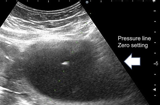
mercial purposes requires written permission.Fig. 1. Circuit including a silicon strain-gauge transducer (DX- Fig. 2. Ultrasound and zero pressure-level line setting.
300; Nihon Kohden Corporation, Tokyo, Japan) for measuring
pressure.
bilical cord compression without oligohydramnios [5] Materials and Methods
during pregnancy. Additionally, it is used to increase the
ultrasonographic accuracy in identifying structural This study was approved by the Institutional Review Board of
Nagara Medical Center (18-6). In this study, we included pregnant
anomalies in patients with oligohydramnios [2, 6]. Al- women who underwent transabdominal AI during pregnancy at
though the use of therapeutic AI during pregnancy prior the Department of Fetal-Maternal Medicine, Nagara Medical Cen-
to parturition has not yet been established, transabdomi- ter, between April 2017 and September 2018. Written informed
nal AI is expected to be a new intervention for umbilical consent was obtained from all patients in whom AI was indicated.
cord compression and oligohydramnios. However, indi- The intra-amniotic pressure was measured before and after the
procedure. A saline-filled line was attached to the hub of the needle
cations for its use should be carefully selected because ad- at one end and to a silicon strain-gauge transducer (DX-300; Ni-
verse events associated with AI have been previously re- hon Kohden Corporation, Tokyo, Japan) at the other end (shown
ported [7]. in Fig. 1). The pressures were measured at the needle tip level
Reported adverse events occurring due to transab- (shown in Fig. 2), as reported previously [10, 11], if they were sta-
dominal AI include miscarriage, premature rupture of ble for 10 s without uterine contractions.
AI was indicated for the relief of cord compression in cases of
membranes, preterm labor, placental abruption, chorio- umbilical cord compression, as confirmed by the sandwich sign or
amnionitis, fetal trauma, and uterine perforation [7]. Pre- by oligohydramnios on ultrasonography and variable decelerations
vious studies reported the relationship between high in- on heart rate monitoring. It was also performed to improve the in-
tra-amniotic pressure and polyhydramnios and between trauterine environment or ultrasonographic accuracy in cases in-
high intra-amniotic pressure and maternal symptoms volving oligohydramnios. The sandwich sign refers to the ultraso-
nographic finding of the umbilical cord sandwiched between 2 lay-
such as abdominal distension and dyspnea, threatened ers of the placenta or between the placenta and the fetus, without
premature labor, and preterm labor [8, 9]. An increase in amniotic fluid present around the umbilical cord, which indicates
the intra-amniotic pressure due to AI may lead to the de- local oligohydramnios [5]. As the sandwich sign alone does not nec-
velopment of complications, such as premature rupture essarily reflect umbilical cord compression, AI was performed when
of membranes and preterm labor. We hypothesized that variable decelerations were confirmed more than once within 40
min and were reproducible during heart rate monitoring accompa-
the intra-amniotic pressure remains unchanged if the nied with the sandwich sign on ultrasonography. Tocolysis was per-
amniotic fluid index (AFI) after AI is within the normal formed by the infusion of ritodrine hydrochloride (50 μg/min) or
range; however, this has not been systematically exam- magnesium sulfate (20 mL/h) during the AI procedure to prevent
ined. This study aimed to investigate the change in the uterine contractions. Experimental obstetricians performed AI us-
intra-amniotic pressure after AI. To the best of our knowl- ing a 21-gauge percutaneous transhepatic cholangiography needle
under ultrasound guidance, with the help of a needle adapter. First,
edge, this is the first study to systematically report the 100 mL of warm saline (100 mL) was infused via drip infusion or
changes in intra-amniotic pressure following transab- manually if the tip of the puncture was confirmed to have an amni-
dominal AI. otic fluid cavity; if necessary, additional warm saline was infused
Intra-Amniotic Pressure following Biomed Hub 2021;6:86–91 87
Amnioinfusion DOI: 10.1159/000519084Table 1. Basic characteristics and outcomes of patients who underwent AI (n = 19)
Variable Measure (n = 19)
Age, years 33 (21–40)
Primipara, % (n/N) 36.8 (7/19)
Hypercoiled cord, % (n/N) 63.2 (12/19)
Fetal structural anomaly, % (n/N) 26.3 (5/19)
Massive subchorionic hematoma, % (n/N) 10.5 (2/19)
GA at AI, weeks and days 24 weeks 3 days (19 weeks 3 days–33 weeks 6 days)
Injected amniotic fluid volume, mL 250 (120–400)
GA at delivery, weeks and days 34 weeks 3 days (27 weeks 0 days–37 weeks 3 days)
Cesarean delivery, % (n/N) 52.6 (10/19)
Z score of birth weight (median [SD]) −1.1 (−3.5–[−0.1])
Apgar score
1 min 8 (3–9)
5 min 9 (7–9)
Umbilical artery pH 7.33 (6.96–7.39)
The data are presented as median (range) or % (n) as appropriate. GA, gestational age; SD, standard deviation;
Apgar, Appearance, Pulse, Grimace, Activity, and Respiration; AI, amnioinfusion.
until the relief of umbilical cord compression was confirmed by ul- 10 (24.4%) were performed for 4 (21.1%) patients with
trasonography in cases of umbilical cord compression or detection oligohydramnios. The basic characteristics and clinical
of an AFI >5 cm. This improves the intrauterine environment or
increases the ultrasonographic accuracy in cases of oligohydram- outcomes are summarized in Table 1. The median GA at
nios. After AI, we performed ultrasonography the next day and the time of AI was 24 weeks 3 days of gestation (range, 19
twice a week until delivery. Continuous maternal and fetal monitor- weeks 3 days–33 weeks 6 days); the median volume of the
ing was conducted to detect symptoms such as frequent uterine con- amniotic fluid injected was 250 (range, 120–400) mL, and
tractions, persistent abdominal pain, uterine bleeding, and fetal bra- the median GA at delivery was 34 weeks 2 days (range, 27
dycardia, which might be signs of placental abruption, and low
blood pressure, impairment of consciousness, and dyspnea, which weeks 0 days–37 weeks 3 days). Spontaneous vaginal de-
might be signs of amniotic fluid embolism. None of the patients had livery was performed in 9 patients, and cesarean delivery
the aforementioned maternal or fetal conditions. was performed because of a nonreassuring fetal status in
We collected the following data: maternal age, parity, body 7 patients and breech in 3 patients. The mean Z score for
mass index, gestational age (GA) at AI and delivery, injected am- birth weight was −1.1 standard deviation (−3.5–[−0.1]),
niotic fluid volume, mode of delivery, birth weight, umbilical ar-
tery pH, single deepest pocket (SDP), AFI, intra-amniotic pressure the mean Apgar scores were 8 (range, 3–9) and 9 (7–9) at
before and after AI, and incidence of complications associated 1 and 5 min, respectively, and the umbilical artery pH was
with AI. We defined SDP 24 cm as polyhydramnios [12, 13]. not be evaluated because 3 patients were lost to follow-up,
The Wilcoxon signed-rank test was used to compare the data. severe brain damage was not observed at the short-term
A p value oftimes in 12 cases of hypercoiled cord with umbilical cord Discussion
compression, 3 (range, 2–6) times in 3 cases of fetal struc-
tural anomaly with umbilical cord compression, and 2.6 AI has been reported to be effective in the manage-
(range, 2–4) times in 4 cases of fetal structural anomaly and ment of umbilical cord compression and oligohydram-
massive subchorionic hematoma with oligohydramnios. nios [1–6]; however, several adverse events are associated
No significant difference was observed between these with AI [7]. Complications may develop due to the chang-
groups. The mean AI interval was 7.2 (range, 2–14) days in es in the intra-amniotic pressure during the procedure.
cases with a hypercoiled cord with umbilical cord compres- Therefore, we measured and compared the intra-amniot-
sion, 7.3 (range, 2–12) days in cases of fetal structural anom- ic pressure before and after AI. Although the SDP and
aly with umbilical cord compression, and 9.9 (range, 7–14) AFI after AI were significantly higher than those before
days in cases of fetal structural anomaly and massive sub- AI, the intra-amniotic pressure after AI was not signifi-
chorionic hematoma with oligohydramnios. No significant cantly different from that recorded before AI. Further-
difference was observed between these groups. However, it more, there was no correlation between the volume of the
must be considered that the numbers were relatively small amniotic fluid injected and changes in the SDP and AFI
for analyzing the relationship between these subgroups. and the intra-amniotic pressure. The low volume of am-
The median SDP and AFI before AI were 2.65 (range, niotic fluid injected and the SDP and AFI, which was
0–5.7) cm and 6.0 (range, 0–15) cm, and after AI, they within the normal range after AI, could explain these ob-
increased significantly to 4.0 (range, 2.7–6.1) cm (p < servations. In addition, we injected 250 mL (mean) of
0.001) and 13.4 (range, 2.7–21) cm (p < 0.001). However, warm saline at mean 24 weeks 3 days (range, 19 weeks 3
significant differences were not observed in the median days–33 weeks 6 days) in this study. The injection fluid
intra-amniotic pressure before and after AI (11 [range, volume of 250 mL was not expected to affect the intra-
4–14] mm Hg vs. 11 [range, 6–19] mm Hg; p = 0.134). amniotic pressure because amniotic fluid volume be-
Spearman’s correlation coefficient for the volume of the tween 22 and 39 weeks was an average of 777 mL (range,
amniotic fluid injected and changes in the intra-amniotic 302–1,997 mL) [15]. Normal amniotic fluid volume
pressure was r = 0.258 (p = 0.107). Pearson’s correlation changes during pregnancy [15]; therefore, the injection
coefficients for changes in the SDP and AFI and changes fluid volume should be determined considering GA.
in the intra-amniotic pressure inspection were r = 0.048 We hypothesized that the changes in the intra-amni-
(p = 0.786) and r = 0.029 (p = 0.865). otic pressure during AI may be the cause of the reported
Regarding the indication of AI, we analyzed the relation- complications. In polyhydramnios, the intra-amniotic
ship between cases with oligohydramnios and umbilical pressure is high, and amnioreduction is often performed
cord compression. No significant differences were observed to prevent preterm delivery and relieve maternal symp-
in the median intra-amniotic pressure before AI (9 [range, toms [8, 9]. We also reported previously that the intra-
4–13] mm Hg vs. 11 [range, 5–14] mm Hg; p = 0.262) and amniotic pressure decreased and reached a plateau dur-
after AI (10 [range, 7–19] mm Hg vs. 10 [range, 6–14] mm ing amnioreduction [9]. These results support our hy-
Hg; p = 0.264), the median volume of amniotic fluid inject- pothesis, and the SDP and AFI after AI should be kept
ed (250 [range, 150–300] mm Hg vs. 250 [range, 120–400] within a normal range because polyhydramnios increases
mm Hg; p = 0.695), and the median intra-amniotic pressure intra-amniotic pressure.
before and after AI (9 [range, 4–13] mm Hg vs. 10 [range, Entire and local oligohydramnios did not influence the
7–19] mm Hg; p = 0.131; and 11 [range, 5–14] mm Hg vs. change in intra-amniotic pressure following AIs. Leiomy-
10 [range, 6–14] mm Hg; p = 0.393) between cases with oma and adenomyosis, which cause lesions that can exist
oligohydramnios and umbilical cord compression. within the myometrium, might affect uterine pressure re-
Maternal or fetal adverse events, such as preterm pre- action during AIs by limiting the extension of the uterus.
mature rupture of membranes, delivery and fetal death In addition, we reported previously that uterine pressure
within 48 h, placental abruption, infection, hemorrhage, tolerance may vary according to the individual [9]. There-
and peripheral organ injury, were not observed following fore, we may have to address changes in the intra-amni-
AI. Moreover, although mild palpitation and tremor otic pressure because the pressure might increase during
caused by ritodrine hydrochloride administration and AI even if there was no polyhydramnios.
mild vascular pain caused by magnesium sulfate admin- Additionally, we observed that the pressure increased
istration were observed, no adverse events occurred that when the tip of the needle was in the uterine myometrium
required treatment. or touched or punctured the fetus during AI. This was
Intra-Amniotic Pressure following Biomed Hub 2021;6:86–91 89
Amnioinfusion DOI: 10.1159/000519084particularly true when the needle punctured the fetus, adverse maternal and fetal events were observed. Further
and the infusion of warm saline was continued, resulting data are required and detailed; thorough subgroup com-
in fetal skin edema. In some cases, it is difficult to ascer- parisons according to patients with and without compli-
tain whether the fetal skin is punctured using ultrasound cations are needed to clarify the relationship between the
examination. Intra-amniotic pressure monitoring may intra-amniotic pressure and the incidence of complica-
aid in the prediction of adverse events and of the position tions.
of the needle tip; therefore, noninvasive monitoring of In the 3 cases with oligohydramnios, the fetus was
pressure changes could improve the safety of AI. Previous punctured, which caused fetal skin edema. Although no
studies have also reported an increase in the intra-amni- severe complications related to fetal puncture were ob-
otic pressure with uterine contractions [11]. Therefore, it served in this study, fetal injury with fetal puncture should
is important to consider uterine contractions while as- be considered in studies regarding AIs. Intra-amniotic
sessing the intra-amniotic pressure to prevent uterine pressure monitoring, which can provide early detection
contractions by tocolysis. of fetal puncture, might be effective to prevent severe
Although we did not observe adverse maternal and fe- complications related to fetal puncture. In addition, it is
tal events due to AI in this study, adverse events due to AI most important to check the position of the tip of the
have been previously reported [7]. A previous study re- needle during AIs.
ported that the risk of complication increases with ad-
vanced GA and number of punctures [16]. Therefore, the
indications for AI should be carefully selected, and only Conclusion
well-trained experts should perform AI.
As for the indication, we performed AI for the relief of This study suggests that incident complications associ-
umbilical cord compression or ultrasonographic accura- ated with increased intra-amniotic pressure are not likely
cy in cases with oligohydramnios. We determined cases to develop if the AFI after AI is within the normal range;
with umbilical cord compression according to the pres- however, we may need to consider individual uterine
ence of fetal blood flow abnormalities or variable decel- pressure tolerance. The risk of complications due to am-
erations. We also performed AI for the relief of umbilical niocentesis persists. Additionally, the intra-amniotic
cord compression in cases without oligohydramnios. We pressure may be effective in predicting the position of the
speculated that umbilical compression with fetal blood tip of the needle in AI. Therefore, intra-amniotic pressure
flow abnormalities or variable decelerations on fetal heart monitoring may be valuable for preventing adverse events
rate monitoring could cause fetal blood flow stress due to during AI and increase the safety of the procedure be-
the intermittent blood flow redistribution and lead to re- cause the technique is noninvasive and easily available.
ductions in the fetal urine volume as a fetal compensation
mechanism for fetal hypoxia, which could lead to a dan-
gerous cycle of umbilical cord compression [3, 5]. In the Acknowledgments
15 cases with umbilical cord compression, fetal blood
flow abnormalities tended to improve, or variable decel- We thank Takeshi Iwase and Hiroshi Soejima, medical engi-
neers at Nagara Medical Center, for their assistance with the pres-
erations decreased after AI as in a previous report [5]. sure measurements.
However, the efficacy has been not established. Although
that does not influence this study, further prospective
control studies are needed to clarify the efficacy of AI in Statement of Ethics
such cases.
We acknowledge that there are several limitations of Written informed consent was obtained from all study partici-
this study. The number of patients included was small; pants. The study protocol was approved by the Institutional Re-
view Board of the Nagara Medical Center (18-6) and was carried
hence, our sample size limits the generalizability of our out in accordance with the guidelines of the Declaration of Hel-
findings. However, this study aimed to evaluate the sinki.
changes in intra-amniotic pressure following transab-
dominal AI not to evaluate the relationship between the
pressure and complications and neonatal outcome. We Conflict of Interest Statement
could not perform subgroup comparisons in this study
concerning the risk of complications related to AIs as no The authors have no conflicts of interest to declare.
90 Biomed Hub 2021;6:86–91 Katsura/Takahashi/Iwagaki/Chiaki/Asai/
DOI: 10.1159/000519084 Koike/MurakamiFunding Sources Data Availability Statement
There were no funding sources for this study. The datasets during and/or analyzed during the current study
are available from the corresponding author on reasonable re-
quest.
Author Contributions
D.K. and Y.T. designed and planned the study; D.K. wrote and
managed the manuscript; D.K., Y.T., S.I., R.C., K.A., M.M., S.Y.,
M.F., and T.M. performed the measurements; D.K. and Y.T. per-
formed data analysis.
References
1 Hofmeyr GJ, Lawrie TA. Amnioinfusion for 7 National Institute for Health and Clinical Ex- 11 Katsura D, Takahashi Y, Iwagaki S, Chiaki R,
potential or suspected umbilical cord com- cellence. Interventional procedures overview Asai K, Koike M, et al. Changes in intra-am-
pression in labour. Cochrane Database Syst of therapeutic amnioinfusion for oligohy- niotic, fetal intrathoracic, and intraperitoneal
Rev. 2012 Jan;1(1):CD000013. dramnios during pregnancy (excluding la- pressures with uterine contraction: a report of
2 Dad N, Abushama M, Konje JC, Ahmed B. bour). The National Institute for Health and three cases. Case Rep Obstet Gynecol. 2018
What is the role of amnioinfusion in modern Clinical Excellence (NICE); 2006 [cited 2018 Sep;2018:4281528.
day obstetrics? J Matern Fetal Neonatal Med. Oct 30]. Available from: https: //www.nice. 12 Phelan JP, Smith CV, Broussard P, Small M.
2016 Sep;29(17):2823–7. org.uk/guidance/ipg192/resources/therapeu- Amniotic fluid volume assessment with the
3 Takahashi Y, Iwagaki S, Chiaki R, Iwasa T, tic amnioinfusion-for-oligohydramnios-dur- four-quadrant technique at 36-42 weeks’ ges-
Takenaka M, Kawabata I, et al. Amnioinfu- ing-pregnancy-excluding-labour- tation. J Reprod Med. 1987 Jul;32(7):540–2.
sion before 26 weeks’ gestation for severe fetal pdf-1899863518635205. 13 Chamberlain PF, Manning FA, Morrison I,
growth restriction with oligohydramnios: 8 Guzman ER, Vintzileos A, Benito C, Houli- Harman CR, Lange IR. Ultrasound evaluation
Preliminary Pilot Study. J Obstet Gynaecol han C, Waldron R, Egan S. Effects of thera- of amniotic fluid volume. I. The relationship
Res. 2014 Mar;40(3):677–85. peutic amniocentesis on uterine and umbili- of marginal and decreased amniotic fluid vol-
4 Kozinszky Z, Pásztor N, Vanya M, Sikovanyecz cal artery velocimetry in cases of severe symp- umes to perinatal outcome. Am J Obstet Gy-
J, Pál A. Management of severe idiopathic ol- tomatic polyhydramnios. J Matern Fetal Med. necol. 1984 Oct;150(3):245–9.
igohydramnios: is antepartum transabdomi- 1996 Nov–Dec;5(6):299–304. 14 Kanda Y. Investigation of the freely available
nal amnioinfusion really a treatment option? 9 Katsura D, Takahashi Y, Iwagaki S, Chiaki R, easy-to-use software “EZR” for medical statis-
J Matern Fetal Neonatal Med. 2013 Mar; Asai K, Koike M, et al. Relationship between tics. Bone Marrow Transplant. 2013 Mar;
26(4):383–7. higher intra-amniotic pressures in polyhy- 48(3):452–8.
5 Katsura D, Takahashi Y, Iwagaki S, Chiaki R, dramnios and maternal symptoms. Eur J Ob- 15 Brace RA, Wolf EJ. Normal amniotic fluid
Asai K, Koike M, et al. Amnioinfusion for stet Gynecol Reprod Biol. 2019 Apr;235:62–5. volume changes throughout pregnancy. Am J
variable decelerations caused by umbilical 10 Katsura D, Takahashi Y, Iwagaki S, Chiaki R, Obstet Gynecol. 1989 Aug;161(2):382–8.
cord compression without oligohydramnios Asai K, Koike M, et al. Intra-amniotic pres- 16 Nassar AH, Martin D, González-Quintero
but with the sandwich sign as an early marker sure of twin-to-twin transfusion syndrome. VH, Gómez-Marín O, Salman F, Gutierrez A,
of deterioration. J Obstet Gynaecol. 2019 Jan; Fetal Diagn Ther. 2018;44(2):160. et al. Genetic amniocentesis complications: is
39(1):49–53. the incidence overrated? Gynecol Obstet In-
6 Fisk NM, Ronderos-Dumit D, Soliani A, vest. 2004;58(2):100–4.
Nicolini U, Vaughan J, Rodeck CH. Diagnos-
tic and therapeutic transabdominal amnioin-
fusion in oligohydramnios. Obstet Gynecol.
1991 Aug;78(2):270–8.
Intra-Amniotic Pressure following Biomed Hub 2021;6:86–91 91
Amnioinfusion DOI: 10.1159/000519084You can also read