CELL AND ORGANOID MODELS TO DEVELOP NEW ANTIVIRALS AND DIAGNOSTIC METHODS FOR EMERGING VIRUSES - From the Department of Oncology-Pathology ...
←
→
Page content transcription
If your browser does not render page correctly, please read the page content below

From the Department of Oncology-Pathology
       Karolinska Institutet, Stockholm, Sweden

  CELL AND ORGANOID MODELS TO
   DEVELOP NEW ANTIVIRALS AND
DIAGNOSTIC METHODS FOR EMERGING
            VIRUSES

                  Aleksandra Pettke

                   Stockholm 2021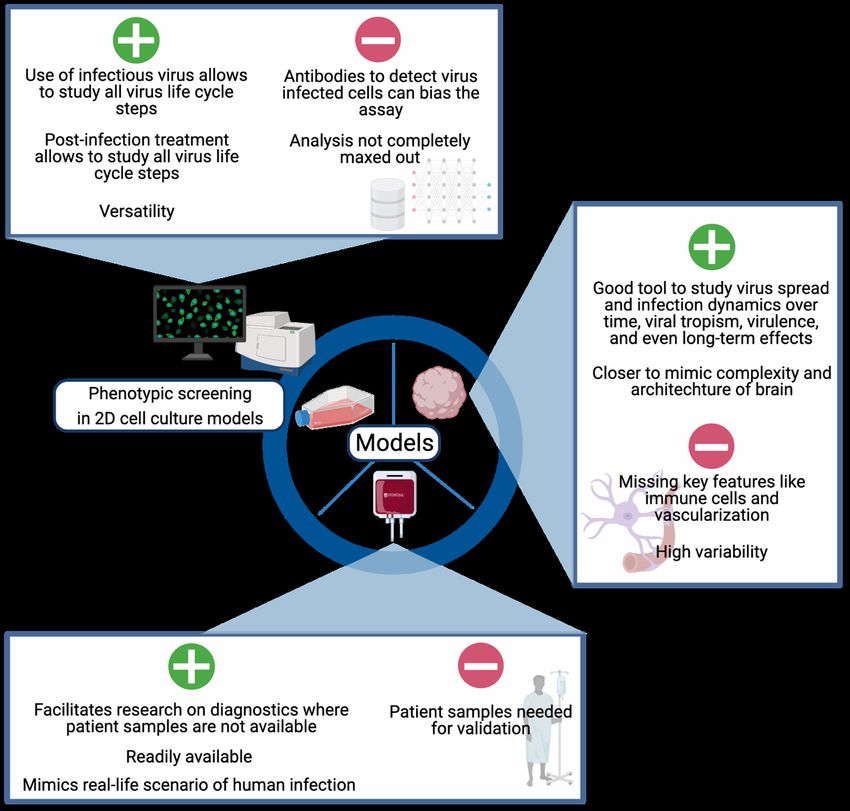
All previously published papers were reproduced with permission from the publisher.
Published by Karolinska Institutet.
Printed by Universitetsservice US-AB, 2021
© Aleksandra Pettke, 2021
ISBN 978-91-8016-154-1
Cover illustration: Miriam Arntz
Cell and organoid models to develop new antivirals and
diagnostic methods for emerging viruses

THESIS FOR DOCTORAL DEGREE (Ph.D.)

By

Aleksandra Pettke

Principal Supervisor:               Opponent:
Prof. Thomas Helleday               Dr. Thomas Strecker
Karolinska Institutet               Philipps-University Marburg
Department of Oncology-Pathology    Center for Infection, Inflammation and Immunity
                                    Institute for Virology
Co-supervisors:
Prof. Ali Mirazimi                  Examination Board:
Karolinska Institutet               Prof. Anna Karlsson
Department of Laboratory Medicine   Karolinska Institute
                                    Department of Laboratory Medicine
Dr. Marjo-Riitta Puumalainen
Karolinska Institutet               Associate Prof. Johan Lennerstrand
Department of Oncology-Pathology    Uppsala University
                                    Department of Medical Sciences
Associate Prof. Åsa Rosenquist      Division of Infectious Medicine
VP R&D-Operations
Laboratory Director                 Prof. Tomas Bergström
iCellate Medical AB                 University of Gothenburg
                                    Department of Infectious Diseases
Instead of thinking what you want to do, think about who you want to be.

                                                     -   Jayson Demers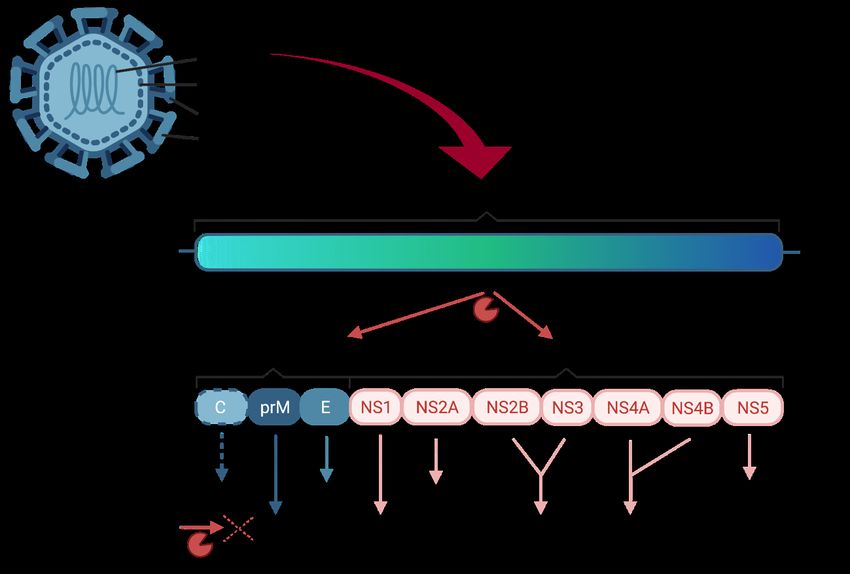
POPULAR SCIENCE SUMMARY OF THE THESIS
Virus outbreaks have always been a threat to mankind. The Spanish flu at the beginning of the
20th century, the Zika virus outbreak in the Americas in 2015, or the Ebola virus outbreak in
2016 in West Africa are only a few examples. Likewise, the ongoing COVID-19 pandemic has
brought unprecedented social and economic disruptions to countries all over the globe and has
brought to public attention that we are not well prepared for new virus outbreaks. In particular
medicines are missing to treat infected people, as well as easily adaptable diagnostic tests, to
enable fast set up of large-scale testing. Fast diagnosis is not only important to diagnose a virus
disease in a patient, but also to assess the spread in the population and steer the response to a
pandemic.

A big obstacle to the development of medication and diagnostic tests for viruses is the lack of
good models to study viruses. In biology models are substitute organisms that are studied
extensively to understand biological phenomena, human disease or in the case of pathogens,
understand the behavior and effects of pathogens in humans. Models are widely used in
research, because it can be unethical or unfeasible to do certain experiments in humans. The
underlying assumption is that the findings from models are transferable to human disease.
However, what complicates matters for virus research is that viruses can be very selective about
the species they infect, and the disease viruses cause is not the same in all species. The work
from this thesis highlights how different models can be used to speed up the development of
new medication and diagnostic tools for viruses.

When viruses infect cells, they create certain observable traits, also called phenotypes. For
example, the virus can be detected inside cells using fluorescent antibodies, and thus infected
and uninfected cells can be distinguished and counted when images of the cells are taken. This
principle was used in the first paper included in the thesis. A total of 425 compounds were
tested for their antiviral properties by checking if they reduce signal from fluorescent antibodies
against virus inside infected cells. This way, we found two substances with an antiviral effect.
Subsequently, using the same method but different viruses, we confirmed that the two
substances identified do not only work against a single virus. We discovered that the substances
have activity against several viruses including Ebola and SARS-CoV-2 virus, the virus which
is causing the COVID-19 pandemic.

The second paper focused on the action of the newly discovered antivirals against Zika virus.
First, we confirmed that these substances were active against Zika virus. Zika virus can cause
a small brain in children by infecting their brain cells, also called neurons, if their mother is
infected with Zika virus during pregnancy. To mimic this, we used cells derived from human
brain tumors to study the antiviral substances and confirmed that they have an antiviral effect
against Zika virus.

However, the brain is the body’s most complex organ. This complexity makes it very difficult
to find a suitable model for the human brain. The brain tumor cells we used previously are very
homogenous and one-dimensional, while the brain is a three-dimensional organ with many
different types of cells which communicate with each other. While this is difficult to model in
traditional cell culture, in the past decade three-dimensional organoids have become available.
It has been shown previously that brain organoids are a great model for Zika virus: just as in
the fetal brain, Zika virus is able to infect neurons in the organoids and reduce their viability.
We were able to show that our compounds not only reduce the amount of Zika virus in the
organoids but also restore organoid viability.

The third study from this doctoral thesis focused on the development of a new diagnostic tool
to detect Zika virus by multiplying parts of its genome. In the development of the diagnostic
tool, both cell lines and blood cells from healthy donors were infected artificially in the lab to
generate samples to test the new approach for sensitivity. Infecting blood cells from healthy
donors with virus mimics the real infection in the patients, and the Zika virus levels found in
the artificially infected samples resemble the ones found in patients. This approach helps
research groups to develop diagnostic tools even without access to patient samples, which are
not easy to acquire, especially for infections from remote regions or low-income countries.

Altogether, the first two studies highlight the use of different cell and organoid models to
advance antiviral drug development. The third study focuses on developing new diagnostics
for Zika virus using cells and human blood as a model. These approaches contribute to
advancing the drug and diagnostics development and can help to be better prepared for future
pandemics.
ABSTRACT
The ongoing global pandemic of severe acute respiratory syndrome coronavirus 2 (SARS-
CoV-2) is only the latest reminder of the urgent need for new antiviral strategies and diagnostic
tools to improve preparedness for emergence of new pathogenic RNA-viruses. A key obstacle
for the development of antivirals and diagnostic tools has been the lack of good models to study
RNA viruses. This thesis focused on cellular and organoid models for virus infection to
facilitate the development of host-targeting antivirals and new diagnostic tools against
emerging viruses.

In Paper I, two close analogues from an in-house library of 425 host-targeting compounds
were found to have antiviral activity against apathogenic RNA virus Hazara virus (HAZV).
These two hit compounds were discovered using a cell- and image-based phenotypic antiviral
screening assay. Subsequently, the two compounds were confirmed to have a broad-spectrum
antiviral activity against several pathogenic RNA viruses including SARS-CoV-2, Ebola and
Crimean-Congo hemorrhagic fever virus. After showing the independence of the compound’s
antiviral effect of their originally designed target, human 8-oxoguanine glycosylase 1, thermal
protein profiling was used to study the compound target. Applying this technique, the
compound was revealed to disturb proteostasis pathways and interactions between cellular heat
shock protein 70 complex and viral proteins.

In Paper II, a second antiviral screening of in-house compounds was performed using the
previously established image-based phenotypic antiviral screening assay with Zika virus
(ZIKV) infected cells. The broad-spectrum activity of the compounds was confirmed by
revealing the same compounds to be active against both HAZV and ZIKV. Next, the
therapeutic window and antiviral activity of the top-hit compounds was demonstrated in several
cellular models of ZIKV infection. Additionally, the novel antiviral compounds showed an
antiviral effect and reversed ZIKV induced neurotoxicity in iPS cell derived human brain
organoids. Furthermore, mechanism-of-action studies revealed the compound to impair the
formation of new virus particles in the late lifecycle steps.

In Paper III, the development of a new diagnostic tool for ZIKV is described using in vitro
infected U87 cells and PBMCs. In the method presented, ZIKV cDNA was hybridized using
padlock probes and amplified by two rounds of Circle-to-Circle Amplification. Detection was
performed using a microfluidic affinity chromatography enrichment platform. Benchmarking
of the newly developed method against RT-qPCR, the gold standard diagnostic method for
ZIKV detection, confirmed a good correlation between both methods.

Altogether, this thesis demonstrates how cell culture tools with varying complexity, unique
advantages and challenges can be used to augment the development of novel antiviral drugs
and diagnostic methods.
LIST OF SCIENTIFIC PAPERS
Scientific Papers included in the thesis

  I. Novel Broad-Spectrum Antiviral Inhibitors Targeting Host Factors
     Essential for Replication of Pathogenic RNA Viruses.
     Tampere, M.; Pettke, A.; Salata, C.; Wallner, O.; Koolmeister, T.; Cazares-
     Körner, A.; Visnes, T.; Hesselman, M.C.; Kunold, E.; Wiita, E.; Kalderén, C.;
     Lightowler, M.; Jemth, A.-S.; Lehtiö, J.; Rosenquist, Å.; Warpman-Berglund,
     U.; Helleday, T.; Mirazimi, A.; Jafari, R.; Puumalainen, M.-R.
     Viruses 2020, 12, 1423.

 II. Broadly Active Antiviral Compounds Disturb Zika Virus Progeny
     Release Rescuing Virus-Induced Toxicity in Brain Organoids.
     Pettke, A.*; Tampere, M.*; Pronk, R.; Wallner, O.; Falk, A.; Warpman
     Berglund, U.; Helleday, T.; Mirazimi, A.; Puumalainen, M.-R.
     Viruses 2021, 13, 37.

 III. Circle-to-circle amplification coupled with microfluidic affinity
      chromatography enrichment for in vitro molecular diagnostics of Zika
      fever and analysis of anti-flaviviral drug efficacy.
      Soares, R.; Pettke, A., Robles-Remacho, A.; Zeebaree, S.; Ciftci, S.;
      Tampere, M.; Russom, A.; Puumalainen, M.-R.; Nilsson, M.; Madaboosi, N.
      Sensors and Actuators B: Chemical, Volume 336, 1 June 2021, 129723

Scientific Papers not included in the thesis

IV. Managing COVID-19 in the oncology clinic and avoiding the distraction
    effect
    Cortiula, F.; Pettke, A.; Bartoletti, M.; Puglisi, F.; Helleday, T.
    Annals of Oncology, 2020 May;31(5):553-555. (Editorial)

 V. Targeting OGG1 arrests cancer cell proliferation by inducing replication
    stress
    Visnes, T.*; Benítez-Buelga, C.*; Cázares-Körner, A.; Sanjiv, K.; Hanna,
    B.M.F.; Mortusewicz, O.; Rajagopal, V.; Albers, J.J.; Hagey, D.W.; Bekkhus,
    T.; Eshtad, S.; Baquero, J.M.; Masuyer, G.; Wallner, O.; Müller, S.; Pham, T.;
    Göktürk, C.; Rasti, A.; Suman, S.; Torres-Ruiz, R.; Sarno, A.; Wiita, E.;
    Homan, E.J.; Karsten, S.; Marimuthu, K.; Michel, M.; Koolmeister, T.;
    Scobie, M.; Loseva , O.; Almlöf, I.; Unterlass, J.E.; Pettke, A.; Boström, J.;
    Pandey, M.; Gad, H.; Herr, P.; Jemth, A.-S.; El Andaloussi, S.; Kalderén, C.;
    Rodriguez-Perales, S.; Benítez, J.; Krokan, H.E.; Altun, M.; Stenmark, P.;
    Warpman Berglund, U.; Helleday, T.
    Nucleic Acids Research 2020 Dec 2;48(21):12234-12251.

VI. A rapid phenomics workflow for the in vitro identification of antiviral
    drugs
    Rietdijk, J.; Tampere, M.; Pettke, A.; Georgiev, P.; Lapins, M.; Warpman
    Berglund, U.; Spjuth, O.; Puumalainen, M.-R.; Carreras-Puigvert, J.
    Preprint on bioRxiv; https://doi.org/10.1101/2021.01.13.423947
VII. Long-term SARS-CoV-2-specific and cross-reactive cellular immune
responses and their correlates with humoral responses, disease severity
and symptomatology
Laurén, I.*; Havervall, S.*; Ng, H.; Lord, M.; Pettke, A.; Greilert-Norin, N.;
Gabrielsson, L.; Chourlia, A.; Amoêdo-Leite, C.; Josyula, V.S.; Eltahir, M.;
Kerzeli, I.; Jernbom Falk, A.; Hober, J.; Christ, W.; Wiberg, A.; Hedhammar,
M.; Tegel, H.; Burman, J.; Xu, F.; Pin, E.; Månberg, A.; Klingström, J.;
Christoffersson, G.; Hober, S.; Nilsson, P.; Philipson, M.; Dönnes, P.;
Lindsay, R.*; Mangsbo S.*; Thålin, C.*
Manuscript under revision
* Authors contributed equallyCONTENTS
1 INTRODUCTION ......................................................................................................... 1
1.1 General introduction to emerging viruses............................................................ 1
1.2 Zika Virus ............................................................................................................. 1
1.2.1 Epidemiology & outbreaks ...................................................................... 1
1.2.2 Clinical implications of ZIKV infection.................................................. 2
1.2.3 ZIKV life cycle ........................................................................................ 2
1.3 Antivirals .............................................................................................................. 5
1.3.1 Host targeting and direct antivirals .......................................................... 5
1.3.2 Screening for new antivirals .................................................................... 6
1.4 Models in ZIKV research..................................................................................... 6
1.4.1 Cell culture & organoids .......................................................................... 6
1.4.2 Blood ........................................................................................................ 8
1.4.3 Animal models of ZIKV infection........................................................... 8
1.5 Compound profiling ............................................................................................. 9
1.6 The diagnostic landscape for viruses and challenges in the diagnostics of
emerging viruses .................................................................................................. 9
1.6.1 Direct virus detection methods ................................................................ 9
1.6.2 Indirect detection methods ..................................................................... 11
2 DOCTORAL THESIS ................................................................................................. 13
2.1 RESEARCH AIMS ............................................................................................ 13
2.2 RESULTS........................................................................................................... 15
2.2.1 Paper I - Novel Broad-Spectrum Antiviral Inhibitors Targeting
Host Factors Essential for Replication of Pathogenic RNA Viruses .... 15
2.2.2 Paper II - Broadly Active Antiviral Compounds Disturb Zika
Virus Progeny Release Rescuing Virus-Induced Toxicity in Brain
Organoids ............................................................................................... 15
2.2.3 Paper III- Circle-to-circle amplification coupled with microfluidic
affinity chromatography enrichment for in vitro molecular
diagnostics of Zika fever and analysis of anti-flaviviral drug
efficacy ................................................................................................... 16
2.3 DISCUSSION .................................................................................................... 19
2.3.1 Using phenotypic screening as an approach to antiviral drug-
discovery ................................................................................................ 19
2.3.2 Organoids as models to study Zika virus infection ............................... 20
2.3.3 Using cellular models to develop diagnostics ....................................... 21
2.4 CONCLUSIONS AND FUTURE PERSPECTIVE ......................................... 23
3 ACKNOWLEDGEMENTS ........................................................................................ 27
4 REFERENCES ............................................................................................................ 31LIST OF ABBREVIATIONS AI Artificial intelligence CZD Congenital Zika Disease COVID-19 Coronavirus diseases 2019 CoV 229E Coronavirus 229E C2CA Circle-to-Circle Amplification C2CA-μACE Microfluidic affinity chromatography enrichment EBOV Ebola virus HAZV Hazara virus iPS cells Induced pluripotent stem cells LAMP Loop-mediated isothermal amplification LoD Limit of detection NHPs Nonhuman Primates OGG1 8-oxoguanine glycosylase 1 PBMCs Peripheral blood mononuclear cells RCA Rolling circle amplification SARS-CoV-2 Severe acute respiratory syndrome coronavirus 2 3D Three-dimensional 2D Two-dimensional ZIKV Zika virus
1 INTRODUCTION
1.1 GENERAL INTRODUCTION TO EMERGING VIRUSES
The currently ongoing global pandemic of Coronavirus diseases 2019 (COVID-19) caused by
severe acute respiratory syndrome coronavirus 2 (SARS-CoV-2) is only the latest example of
the significant public health threat posed by emerging pathogens. In the last decade, we have
seen outbreaks of several pathogenic RNA viruses like SARS causing pneumonia, Ebola virus
(EBOV) causing lethal hemorrhagic fever and Zika virus (ZIKV) causing congenital Zika
syndrome (CZS), to name just a few prominent examples. Viral emergence is driven by
increasing urbanization, rapid population growth, increased travelling activities, as well as
climate change and global warming. All of these man-made problems entail a spread of vectors
to previously unaffected areas.

Common denominators of emerging RNA viruses are the lack of vaccines, available therapies
and in-depth knowledge about the pathogenesis, host reactions and underlying molecular
events. New insights and approaches are needed to develop therapeutic strategies towards these
inevitable global threats.

1.2 ZIKA VIRUS

1.2.1 Epidemiology & outbreaks
ZIKV was first isolated in 1947 in Uganda (1). It is an arbovirus and belongs to Flaviviridae
family (2). Mosquito species Aedes aegypti and Aedes albopictus are natural vectors of ZIKV
infection, therefore ZIKV spread is closely linked to the spread of these vectors in the tropical
regions of the world, between 23.5°S and 23.5°N latitude (3). However, several other
transmission modes have also been reported for ZIKV, including blood transfusions (4), sexual
intercourse (5) and maternal-fetal transmission (6).

For decades after it was first isolated, ZIKV was associated with few cases of very mild human
disease, until it caused outbreaks in Yap state, Micronesia in 2007 (7) and subsequently a much
bigger outbreak in French Polynesia in 2013, with more than 30,000 people infected (8,9).
During this outbreak ZIKV was linked to serious neurological complications for the first time
(10,11). From French Polynesia, the virus was brought to Brazil by travel, most likely in the
end of 2014. The combination of a previously unexposed and thus unprotected population and
other factors like the low activity in vector control measures led to an exploding epidemic with
ca. 1.5 million people infected (12,13).

Given its described evolution, ZIKV is a particularly interesting and serious example of viral
emergence, and the transition of a “harmless” and presumably unimportant pathogen to a public
health threat on a global scale.

                                                                                               1
1.2.2 Clinical implications of ZIKV infection For the majority of patients, ZIKV infection remains harmless: most cases are believed to be asymptomatic or cause a self-limiting, very mild disease (14). In these patients the most common symptoms include fever, conjunctivitis, headache, pruriginous maculo-papular rash, arthralgia and myalgia (2,15,16). Nevertheless, even asymptomatic and self-limiting diseases can be detrimental for women who are pregnant at the time of ZIKV infection, because of the dramatic effect for the fetus (17). ZIKV has been shown to be teratogenic, lead to miscarriages (18) and cause a disease complex named Congenital Zika Syndrome (CZS) (15). CZS collectively describes various neurological complications in newborns following ZIKV infection of the mother during pregnancy (19). These pathologies are the result of neuronal cell death in fetuses following ZIKV infection. The hallmark of CZS is microcephaly (20,21), a reduction in brain volume and subsequently also the cranial structures (22). CZS also features structural eye anomalies and posterior ocular lesions like chorioretinal and optic nerve atrophy (23,24), as well as congenital contractures (25). However, studies have demonstrated that only 2.5-5% of babies born to mothers with ZIKV infection during their pregnancy develop birth defects (26,27). In the rest of the cases, the babies do not suffer from CZS, despite a maternal ZIKV infection during pregnancy. It is unclear if the maternal immune system is able to clear the infection prior to a transplacental infection of the fetus, or if despite the infection the fetuses do not develop disease. One contributing factor seems to be the stage of pregnancy at the time of infection. While up to 15% of babies of mothers infected during the first trimester develop neurological defects, the percentage declines in later pregnancy stages. Moreover, not all neuronal damage presents as CZS. Large long-term studies will be needed to assess if children with no clear CZS symptoms at birth might still suffer from more subtle signs of fetal neuronal damage like learning difficulties, mental health problems or social problems. In adults, ZIKV infection has been linked to a serious neurological complication, named Guillain-Barré Syndrome (GBS). GBS is a polyneuropathy of autoimmune origin, and its onset is closely linked to previous infection by various pathogens (28) including ZIKV. The destruction of peripheral nerves causes facial palsy, distal limb paresthesia and muscle weakness, rapidly progressing to paralysis (29). In severe cases, paralysis of the respiratory muscles can result in breathing difficulties and the need for intensive care (30). As of today, there is no cure or approved therapy for neither CZS nor GBS and patients are treated with supportive care. 1.2.3 ZIKV life cycle To be able to develop new treatments, a deep knowledge of ZIKV life cycle steps and the molecular mechanisms underlying infection is needed. However, as of today, not all steps of ZIKV replication have been investigated in detail. Generally, ZIKV replication is thought to 2
be similar to other members of the Flaviviridae family and can be divided in the following
steps: attachment and entry, translation of the viral RNA into proteins, replication of the virus
genome, assembly and egress from the cell (31). For most of these steps viruses use and abuse
various host cell factors and machineries, which determines their ability to infect and propagate
in different tissues and cells, also called viral tropism.
ZIKV has been found to have a broad tissue tropism including skin, blood, brain, retina,
placenta and testis (32–36). Notably, the susceptibility of these cells and tissues to ZIKV
infections explains the pathology caused by ZIKV infection and its modes of transmission.
To enter any of these target cells, viruses need receptors which can directly or indirectly
facilitate its incorporation to the cell. While the exact entry receptor for ZIKV is still unknown,
several candidates are under discussion. Due to the previously mentioned similarities between
Flaviviruses, research has focused on two distinct families of transmembrane
phosphatidylserine receptors: TIM (TIM1, TIM3 and TIM4) and TAM (TYRO3, AXL and
MER). Both of these receptor families are known to be important for DENV entry (37), and
they are also known to regulate apoptosis and innate immune functions of host cells (38,39).
However, contrary to initial reports of AXL being very important for ZIKV entry to the cells
(40,41), several studies suggest that none of the receptors mentioned above is indispensable for
the ZIKV entry process (42,43).
Following attachment to its yet-to-be-determined host cell receptor, ZIKV particles are
internalized by endocytosis and trafficked to endosomes (44). The acidic environment in the
endosome facilitates a conformational change of the flavivirus envelope protein prompting
fusion of the viral and endosome membranes and release of the genome (45).
Figure 1: The structure of the ZIKV genome and its encoded proteins and their function in the viral life cycle
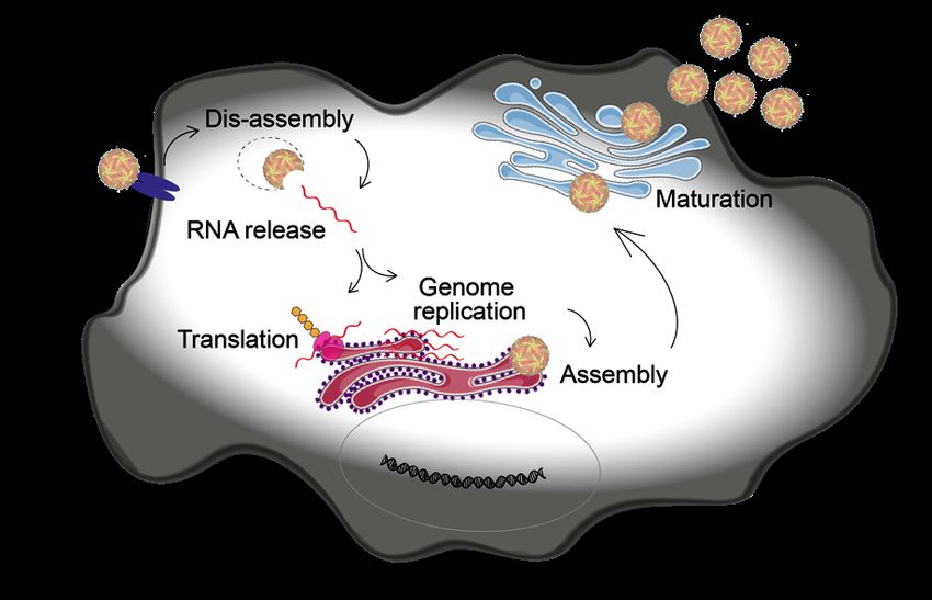
3The ZIKV genome is a single stranded (ss) positive sense (+) RNA with a length of 10,794 kb, and it is organized in a single open reading frame. It codes for a polyprotein which is later cleaved proteolytically into three structural and seven non-structural proteins, which are important for viral replication (46) (Figure 1). The release of the genome is followed by a first translation step, which is crucial to produce all viral proteins needed for viral replication including the viral RNA-dependent RNA polymerase (RdRP) (NS5). The RdRP initiates virus replication by synthesizing a (-)RNA intermediate from the complimentary (+)ssRNA. This (-)RNA is subsequently used as a template to generate many (+)ssRNA copies (47). Similar to other Flaviviruses, ZIKV replication takes place in so called replication factories (RFs) in the endoplasmic reticulum (48). The spatial segregation of viral and cellular compartments probably provides protection for the viral RNA from host nucleases and from detection by the innate immunity sensors. Through pores in the RFs the resulting (+)ssRNA is released into the cytosol (48), where it serves multiple purposes: production of new virus proteins, formation of new RFs and packaging into new virus particles. The still immature and non-infectious virions are assembled in the ER, consisting of the E and prM proteins on a lipid membrane bilayer and the nucleocapsid in the core formed by C protein and RNA (49). To be able to undergo membrane fusion in the next cell, the virus particles need to mature (50,51). This happens during the transit through the trans-Golgi-network towards the cell surface, when the prM protein is cleaved to M by a Furin-like protease. In the final step, virus particles are released to the extracellular environment by exocytosis (Figure 2). Figure 2: Schematic drawing of the ZIKV life cycle in a cell 4
1.3 ANTIVIRALS
The mortality and morbidity caused by emerging diseases is increasingly high (52–55), and
new and more frequent outbreaks demonstrate how vulnerable the lack of vaccines and
therapeutics leaves us in the face of viral emergence. While the development of highly
efficacious and safe vaccines against SARS-CoV-2 within just one year, gives hope for future
pandemics and redefines the limits of vaccine development (56), it does not make antivirals
obsolete. New preferably broadly active antivirals are needed to bridge the time until vaccine
development and deployment, and to provide treatment opportunities for individuals who
cannot be vaccinated. This is why extensive drug discovery efforts are still needed to ameliorate
the medical need and economic burden of emerging viruses.

1.3.1 Host targeting and direct antivirals
When fighting virus infections, two distinct therapeutic strategies can be implemented: crucial
parts of the virus can be targeted directly (directly acting antivirals) or cellular proteins and
machineries indispensable for virus replication can be targeted (host-directed antivirals). Both
approaches come with advantages and drawbacks (57).

Directly acting antivirals are very specific. They target one particular viral protein, which
comes with the disadvantage of needing to find and confirm a good, druggable viral target. In
addition, it can also limit the utility of the newly identified antivirals to just one virus.
Furthermore, a hallmark of RNA viruses like ZIKV is their high mutation rate leading to
production of many genetic variants (quasi species) in every replication round (58). Directly
targeting an important virus protein constitutes a high selection pressure, and results in the rapid
selection of resistant virus variants. Combining several directly acting antivirals with distinct
targets and increasing effectiveness of antivirals can help to reduce development of resistance,
as has been shown very impressively by treatment regimens for human immunodeficiency
virus type 1 (59). Also, by targeting the virus itself, less host impact and thus less side-effects
can be expected.

Host targeting antivirals, on the other hand, can slow down the development of resistance, since
it will require more time for the virus to adjust its entire replication machinery. Another
advantage is the potentially broad antiviral activity. This is especially true for Flaviviruses, in
which the replication process and use of host machinery is quite conserved. However, this
comes at the cost of potential side effects, as a result of blocking pathways important for the
host cell themselves (60,61). Furthermore, identifying the host target can be a long and
challenging process.

An antiviral which has been used experimentally against various emerging viruses, including
Lassa virus (62) and Crimean-Congo hemorrhagic fever virus (63) is ribavirin. Ribavirin is a
guanosine analogue, with demonstrated broad spectrum activity against DNA and RNA
viruses. Ribavirin’s mode of action is not completely clear, but most likely it is a combination
of direct effects from RdRP inhibition, interference with RNA capping, and from insertion of
lethal mutations as well as indirect antiviral effects through immunomodulatory effects (64).

                                                                                                  5
Ribavirin has also been reported to inhibit ZIKV infection in vitro in various cell models and in vivo (65,66). 1.3.2 Screening for new antivirals In the past decade completely new approaches to drug screening have been developed and are becoming widely accessible. The increasing convenience and affordability of genome wide screening methods like RNA-interference, haploid cells, and the CRISPR technology (67,68) combined with the accelerated development in the field of computational biology and chemistry provides new ways and opportunities for drug screening (69). Apart from the very new ways of screening, other approaches can be applied to identify new antiviral substances. In line with the two main antiviral strategies - virus- vs host-directed - a target-based approach and a phenotypic screening approach can be used, both of which provide unique opportunities and challenges. Target-based screening approaches usually use a biochemical assay to test drug-candidates specifically for their activity against a predefined target. The use of a target-based approach provides the opportunity of rapid lead compound optimization, once a promising candidate is identified. However, there is no guarantee that an inhibitor identified in a biochemical assay will also work in cells. Phenotypic screening assays are typically cell- or organism-based, which mean they primarily identify compounds which work in a physiological cellular context. This type of screening is largely unbiased, allows identification of both cellular and direct inhibitors and can be used to uncover new host-pathogen interactions as well as new drug targets with potentially broad- spectrum activity. However, for phenotypic screening approaches it is crucial which cell or organism model is chosen for the screening, because the performance of a compound might differ across cell types and species. 1.4 MODELS IN ZIKV RESEARCH Independently of the screening approach, cellular and organism-based models are needed to confirm and optimize potential antiviral treatments, as well as study host and virus biology. However, model development is challenging. Viral tropism and species-specific interactions of viruses with the host lead to poor replication of many viruses in cellular models and differences in host reactions between humans and animals used in studies. The following chapter summarizes various systems developed to model ZIKV infection, to facilitate research on ZIKV and promote the development of antiviral therapies and vaccines. 1.4.1 Cell culture & organoids Two-dimensional (2D) cell culture models are a cheap, established and highly controlled way to study many aspects of the virus life cycle as well as the host reaction, and they have been used intensively to study ZIKV (70). In a 2D monolayer, infections can spread very efficiently, since all cells are accessible to the virus at the same time. These models are very useful to study 6
viral tropism and show if the virus in question can infect the cells used as a model. Since many
events in the virus life cycle are very similar in cells and organisms, results can often be
extrapolated, and cell lines are a good starting point for antiviral drug development.
Vero cells, green monkey kidney cells, are a cellular model commonly used in the virology
field, because they support the growth of many different viruses, they are used to isolate viruses
and measure viral titers (71). Nevertheless, while they support the growth of ZIKV (72), they
are not the most physiologically representative model. Keeping in mind that ZIKV infection
primarily causes microcephaly in the developing brain, brain derived cell cultures represent a
more relevant model. U87 cells are a glioblastoma cell line (73) which has been shown to
sustain ZIKV infection, and also been used for some mechanistic studies, like studying
inflammasome activation upon ZIKV infection (74).
Even if they are of a more relevant origin, U87 is a cancer cell line and two dimensional. In
contrast to this, the human brain is an extraordinarily complex organ, and the interaction of
various different cell types is crucial for its function. The complexity and cellular diversity of
the human brain is one reason why there is a lack of accurate models to study the human brain.
To further complicate matters, the remarkable qualitative and quantitative differences between
animal and human brains e.g. between human and rodent brains (75), make the development
of suitable animal models challenging.
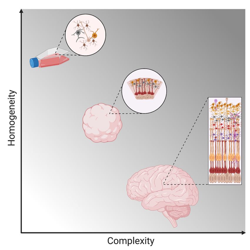
Figure 3: Schematic visualization of the complexity and homogeneity of cerebral organoids in relation to
2D cell cultures and the human brain.
7To tackle this problem, three-dimensional (3D) cell cultures have recently been developed from skin-derived induced pluripotent stem (iPS) cells, to mimic more closely the natural physiological conditions (Figure 3), and especially the complex cytoarchitecture. These models have been used successfully to study microcephaly caused by ZIKV infection (76,77). 1.4.2 Blood While conventional 2D and modern 3D cell cultures are good models to study virus pathogenesis and host interactions, a crucial drawback of cell lines is their genetic and phenotypic homogeneity. Moreover, many cell lines are derived from tumors and thus have intrinsically altered host pathways (78) and immune responses (79). Primary cells on the other hand resemble the original tissue quite well, however, they are not easily accessible and are difficult to work with. Blood remains the most convenient, cost-effective and easy to work with primary tissue from either patients or healthy donors. Since ZIKV has been shown to infect peripheral blood mononuclear cells (PBMCs) (80), PBMCs have been used in research to model ZIKV infection and study host immune responses. 1.4.3 Animal models of ZIKV infection While many features of virus infection are similar between cell culture, especially primary cells, and the organism as a whole, certain complex host defense strategies, such as the adaptive immune response, are not found in cell culture. Also, the main pathologies caused by ZIKV infection are serious neurological disorders in the fetus infected during pregnancy, another complex system which cannot be studied in cell culture. This highlights the need for a more sophisticated model to study ZIKV comprehensively. Reflecting this necessity, several mammalian models of ZIKV infection have been developed since 2016. The most studied model organisms are immunodeficient mice (81), which can provide valuable insights into pathogenesis of ZIKV infection. However, the relevance and transferability of these data for the human disease are questionable, since their immune response upon infection is altered and it has been shown that susceptibility to infection largely differs with the age of these mice (82). Nonhuman Primates (NHPs), on the other hand, provide a much more relevant model, especially for ZIKV infection during pregnancy (83). Among the common features of ZIKV infection in humans and NHPs is vertical transmission from mother to fetus, as well as fetal abnormalities similar to CZD regardless of the severity of the maternal infection (84,85). However, drawbacks of using NHPs as disease models include ethical considerations, high costs and extended experimental time due to the relatively long duration of pregnancy. A more cost- and time-efficient model to study ZIKV is the chicken embryo. Historically, chicken embryos have been a powerful tool to study developmental biology, embryology, as well as teratogenicity of drugs (86,87). Furthermore, chicken embryos are a well-established and widely used model in virus research and vaccinology (88,89). A few studies have 8
demonstrated the ability of ZIKV to replicate potently in infected chicken embryos (90,91),
and causing a similar pathology as seen in humans: virus replication in various organs,
microcephaly and enlarged ventricles (91,92).

Overall, many complex models have been established and are constantly being developed to
study ZIKV infection and facilitate drug and vaccine development, each with their own set of
advantages and limitations.

1.5 COMPOUND PROFILING
Despite many new and highly sophisticated disease models, a prevailing challenge in drug
discovery is to identify the molecular target(s) and pinpoint the specific effect of the drug of
interest on cellular pathways, especially if it is a previously uncharacterized molecule. Many
drugs also have poly-pharmacological effects, affecting not only one specific target, but
manipulating diverse cellular pathways. Recently, new approaches like thermal proteome
profiling (TPP), have opened new ways of compound characterization. TPP enables the
monitoring of changes in protein thermal stability introduced through protein-drug interaction
across the proteome using quantitative mass spectrometry to quantify these changes and thus
facilitating the identification of drug targets (93).

1.6 THE DIAGNOSTIC LANDSCAPE FOR VIRUSES AND
    CHALLENGES IN THE DIAGNOSTICS OF EMERGING VIRUSES
To fight a newly emerging pathogenic virus, timely availability of accurate diagnostic tools is
indispensable. From a single-patient perspective, knowledge about what causes the patient’s
disease is needed to guide treatment and facilitate recovery. From a societal and public health
perspective, implementation of effective disease surveillance and control is impossible without
widely available, sensitive and specific tests. Generally, there are two approaches to virus
diagnostics: direct and indirect diagnostics.

1.6.1 Direct virus detection methods
Direct diagnostic tools detect virus material, like viral proteins or its nucleic acids in patient
samples. These approaches can be based on various different technologies explained in the
following paragraph.

1.6.1.1 Electron microscopy
The most immediate virus detection method is detection of the virus directly from a patient
sample using electron microscopy. However, especially in samples with a low virus load, it
might be necessary to enrich the virus by isolation in cell culture to increase the sensitivity of
the method (94).

1.6.1.2 Virus isolation
For a long time, virus isolation from patient samples was the gold standard method in
virological diagnostics. In this approach, the patient sample is cultured on cells, tissue or even

                                                                                                9
in animals until a cytopathic effect (CPE), virus induced cellular destruction, is detected. After detection of CPE follow up tests are needed to determine which virus has been isolated. If there is a clear suspicion based on the clinical presentation of the patient, the follow-up can be targeted using qPCR or antigen tests. If the pathogen is unknown, a non-targeted method like electron microscopy or next generation sequencing (NGS) is needed to determine which virus has been isolated. The virus strains isolated from patient samples are also a valuable tool in research to study pathogenicity and compare virulence between various virus strains (94). However, there are several drawbacks to virus isolation. It is highly time consuming, taking several days at the least. Furthermore, virus isolation is subject to significant biological variation, and depends on the permissiveness of cells or tissue to the virus in question. To add another layer of complication, not all viruses cause CPE, making a negative result hard to assess without resource-heavy follow up-tests like NGS. Furthermore, especially in the case of emerging RNA viruses, virus isolation usually entails access to expensive laboratory infrastructure, skilled personnel, and high safety measures to contain the biorisks associated with experiments of this kind. Oftentimes these resources are not available in low- and middle- income countries. All these drawbacks question the suitability of virus isolation for primary diagnostics, where the aim is a timely and precise answer. Likewise, in the past decade, the emergence of ever new and more sensitive molecular virus detection methods started to replace virus isolation in routine virus diagnostics. Nonetheless, especially for newly emerging infections with unknown pathogens, isolation oftentimes remains the crucial first step to discover a new pathogen and be able to characterize it. 1.6.1.3 Virus antigen detection Immunoassays in either liquid or solid form are a frequently used technology to detect virus antigens in patient samples or as follow up test after virus isolation. The principle is capturing virus antigens from the patient sample with an antibody, which can be either in solution or attached to a membrane. In a second step, the complexes formed are identified by a detection reaction. In the last two decades, technologies to detect nucleic acids have become more accessible and affordable, and especially qPCR has become the gold standard in detecting virus diagnostics (94). qPCR is a highly specific diagnostic tool, has a fast turn-around time of only a few hours from sample arrival to result and offers opportunities to react quickly to virus changes, like the emergence of new strains (94). In recent years the portfolio of nucleic acid detection methods in virus diagnostics has been complemented by isothermal amplification techniques like loop- mediated isothermal amplification (LAMP) (95) or rolling-circle amplification (RCA) using padlock probes (PLPs) (96). PLPs are linear, single stranded DNA oligonucleotides which consist of complementary target arms (15-20 nucleotides in length) and a longer, non- hybridizing backbone (40-50 nucleotides in length). Binding of the target arms to their respective target results in a circle which is linked through DNA ligation. After circulization, 10
the probes are amplified using a DNA polymerase. Digesting the amplification product and
repeating the ligation and amplification step are called Circle-to-Circle amplification (C2CA)
and increases sensitivity. Detection of the rolling circle products is performed using fluorescent
labelling. Advantages of the method include isothermal amplification and high specificity,
because perfect alignment of hybridization arms and ligation site are required (97,98).

Another trend seen in the past ten years was an increasing number of multiplex tests which
facilitate the detection of pathogens usually grouped by the clinical presentation, like pathogens
causing respiratory infections or meningitis. Most of these devices are based on detection of
nucleic acid detection (99). While this approach is mostly limited to identifying previously
known pathogens, it helps to streamline and speed up diagnostics. Several of these multiplex
tests are increasingly easy to handle and could even potentially be used by trained staff as point-
of-care diagnostics (100).

Furthermore, the increasing affordability of sequencing technologies opens up even more
possibilities to use these technologies. In the ongoing COVID-19 outbreak setting, these tools
have proven to be invaluable to determine how the virus evolves into new lineages and coupling
this information to new phenotypic features like increased transmissibility (101).

However, an essential disadvantage of any method using nucleic acid detection, is that it cannot
predict infectivity. In this context, the high sensitivity of the method is a drawback. It has been
shown that viral RNA can be detected in various patient samples long time after infection, and
the significance for patient outcome and infectivity are often unclear (102,103). Methods like
virus isolation can provide insights on infectivity.

1.6.2 Indirect detection methods
Indirect diagnostics are based on the immune reaction triggered by virus infection. Typical
assays used to determine the existence of antibodies towards a virus in patient serum are
enzyme-linked immunosorbent assay (ELISA) or immunoblots (94). As a common principle
synthetically produced virus antigens are used to capture potential antibodies present in the
patient sample and the complexes formed between the virus proteins and patient antibodies are
detected by a detection reaction (94). With the increasing recognition of the role of cellular
immunity induced by T helper and cytotoxic T cells towards viruses, assays are being
developed to measure cellular immune responses. In enzyme-linked immunosorbent spot
(ELIspot) assays, patient cells are stimulated by virus antigens. The cytokines released by the
cells as a result of the stimulation are captured on a membrane coated with antibodies for
cytokines, and the complexes are then visualized by a detection reaction. Currently ELIspot
assays are not part of routine diagnostics for viral diseases but are used to assess vaccine
efficacy and for research purposes (104–106).

ELISAs, immunoblots and ELIspot assays are highly dependent on the peptides and proteins
used as antigens to stimulate cells or capture antibodies. Especially in the beginning of a
pandemic, when there is little knowledge about a virus, the production of highly specific

                                                                                                11
antigens might be difficult to achieve, leading to sensitivity issues in the detection. In this context also the variability in the human immune response pose additional challenges (107). Immunofluorescence (IF) assays on the other hand are very resource heavy, since an IF microscope is needed for the assay. If there are no commercial IF assays available, similar resources are necessary as described above for virus culture, once again disadvantaging low- income settings. Furthermore, cross-reactivity of antibodies between different viruses of the same family is a known issue in all indirect assays and has been shown for example for members of the Flavi- and Coronavirus families (108–110). Cross-reactivity affects test specificity and can make a specific diagnosis difficult, especially in geographical regions where many different viruses from the same family are circulating. All in all, the virus diagnostics field has seen a technological revolution in the past three decades and moved from cell-based virus isolation as gold standard to broad application of highly specific molecular diagnostic methods. However, continuous research and development is needed to improve the methods available today and provide new innovative ways for virus detection to be prepared for future outbreaks. Figure 4: Methods used for virus detection 12
2 DOCTORAL THESIS
2.1 RESEARCH AIMS
The aim of this thesis was to show how various cellular and organoid models can be used to
identify and characterize new antivirals and develop new diagnostic tools.

Specific contribution of each paper to the aims

   •   Paper I - Novel broad-spectrum antiviral inhibitors targeting host factors
       essential for replication of pathogenic RNA viruses
       - Use of an image-based phenotypic antiviral screening assay to identify host-
           targeting antivirals
       - Application of the image-based phenotypic antiviral assay to determine the
           antiviral activity of the newly identified antivirals against Coronavirus 229E (CoV
           229E) and SARS-CoV-2

   •   Paper II - Broadly active antiviral compounds disturb Zika virus progeny release
       rescuing virus-induced toxicity in brain organoids
       - Transfer of the image-based phenotypic antiviral screening assay to a different cell-
           virus-system
       - Application of the image-based phenotypic antiviral assay in ZIKV infected
           glioblastoma cell line U87 to determine the therapeutic window of newly identified
           antivirals
       - Use of an iPS cells derived 3D organoid model to study the antiviral effect of
           identified antiviral compounds

   •   Paper III - Circle-to-circle amplification coupled with microfluidic affinity
       chromatography enrichment for in vitro molecular diagnostics of Zika fever and
       analysis of anti-flaviviral drug efficacy
       - Use of ZIKV infected glioblastoma cell line U87 to establish circle-to-circle
          amplification coupled with microfluidic affinity chromatography enrichment
          (C2CA-μACE) as a new diagnostic tool
       - Use of PBMCs to benchmark C2CA-μACE relative to RT-qPCR and study
          antiviral drug efficacy

                                                                                           13
2.2 RESULTS
Recent outbreaks of pathogenic RNA viruses like SARS-CoV-2, EBOV or ZIKV highlight the
need for new antiviral strategies as well as diagnostic tools to be better prepared for emergence
of ever new pathogens. The papers included in this thesis feature the use of various cellular and
organoid models to facilitate the development of host-targeting antivirals and new diagnostic
tools against emerging viruses.
2.2.1 Paper I - Novel Broad-Spectrum Antiviral Inhibitors Targeting Host
Factors Essential for Replication of Pathogenic RNA Viruses
In this study, an image-based phenotypic antiviral screening assay was established using
Hazara virus (HAZV) infected SW13 cells. HAZV is an RNA virus, which is not pathogenic
for humans and can be used at biosafety level two laboratories but serves as a model for
Crimean-Congo hemorrhagic fever virus. The main readouts of this assay were cell survival
measured by nuclei count, number of infected cells as share of DMSO treated controls and
virus progeny release, determined by titration of collected supernatants on fresh cells. Using
the screening assay, 425 compounds from an in-house library of host targeting small molecular
inhibitors were tested for their antiviral activity, revealing two close analogues TH3289 and
TH6744 as hit compounds. The antiviral activity of both compounds against HAZV was
confirmed by determining the antiviral effect in a dose-dependent way and showing a
therapeutic window between the antiviral effect on the viral titer and cell viability. Moreover,
both new antiviral compounds were confirmed to have a broad-range antiviral activity against
several pathogenic RNA viruses including SARS-CoV-2, EBOV and Crimean-Congo
hemorrhagic fever virus.
Originally both TH3289 and TH6744 were designed to inhibit human 8-oxoguanine
glycosylase 1 (OGG1). However, the antiviral activity of the compounds was shown not to
depend on their ability to inhibit OGG1. As a next step, to investigate the target of the new
antiviral compounds in more detail, thermal protein profiling was performed with TH6744.
Using this approach, the compound was shown to affect proteostasis pathways and disturb
interactions between cellular HSP70 complex and viral proteins.
2.2.2 Paper II - Broadly Active Antiviral Compounds Disturb Zika Virus
Progeny Release Rescuing Virus-Induced Toxicity in Brain Organoids
In the second study, the previously established image-based phenotypic antiviral screening
assay was transferred to test the antiviral activity of in-house antiviral compounds in Zika virus
infected cells. A comparison between the antiviral screening from Paper I revealed the same
compounds to be active against both HAZV and ZIKV, confirming the broad antiviral activity
of the compounds. Next, the therapeutic window of the top-hit compounds was demonstrated
in several cellular models of ZIKV infection by comparing the dose-response analysis of the
antiviral effect and cellular toxicity.
15To study the antiviral activity of the compounds in a more physiological model of ZIKV
infection, a 3D organoid model was established using human iPS cells. The novel antiviral
compounds showed an antiviral effect on both infected organoids and viral progeny production
and additionally reversed ZIKV induced neurotoxicity.
Subsequently, the compound’s mechanism-of-action was studied by investigating intra- and
extracellular ZIKV RNA levels over time and doing various time-of-addition experiments.
Interestingly, ZIKV RNA levels were not impacted by treatment with TH6744. Instead, a rapid
reduction of progeny release was detected even upon short treatment of 2h at late stages of
infection. To investigate this finding further, the budding efficiency was studied but revealed
no difference in the reduction of infectious virus particles between intra- and extracellular
ZIKV particles. Altogether, the mechanism of action studies narrowed the mechanism-of-
action down to impairment of formation of new virus particles in the late ZIKV lifecycle steps.
It also revealed that TH6744 does not only rescue ZIKV induced pathologic phenotypes in cells
and organoids, but also reduces virus transmission.
In summary, Paper I and Paper II highlight the value of various cellular and organoid models
in the development and characterization of new host-targeting broad-spectrum antivirals.
2.2.3 Paper III- Circle-to-circle amplification coupled with microfluidic affinity
chromatography enrichment for in vitro molecular diagnostics of Zika
fever and analysis of anti-flaviviral drug efficacy
In Paper III, the development of a new diagnostic tool for ZIKV nucleic acids is described.
Sequences from ZIKV genetic material were detected from ZIKV cDNA using padlock probes,
targeting various region of the ZIKV genome, in particular regions coding for capsid (C),
precursor membrane (PrM), envelope (E) and non-structural proteins (NS). Detection was
followed by two rounds of Circle-to-Circle Amplification (C2CA) combined with a
microfluidic affinity chromatography enrichment (μACE) platform.
In a first step the limit of ZIKV RNA detection (LoD) was determined for both a single round
of rolling circle amplification (RCA) and two rounds (C2CA) of RCA using a dilution series
of a single ZIKV synthetic ssDNA. A comparison of RCA and C2CA performance revealed
C2CA to be superior and decrease the LoD by 3-fold, to be between 103 to 104 copies/mL.
Consequently, C2CA was chosen for further method development and validation.
Next, ZIKV RNA extracted from cell culture supernatant from in vitro infected U87 was used
to find an optimal method to determine the fluorescence of the C2CA products. Altogether 12
padlock probes were used to amplify the ZIKV RNA from the supernatants of infected U87
cells, and thereafter fluorescence was measured using either glass slides or μACE. Both
measurements led to an effective detection of ZIKV compared to the negative controls.
However, μACE provided a better signal to noise ratio through a higher selectivity of the μACE
towards RCPs compared to artefacts like non-specific fluorescent clusters or debris. Thus,
μACE chosen as the detection method of choice for the following experiments.
16Subsequently, to benchmark C2CA-μACE against a gold standard diagnostic method, viral
RNA obtained from in vitro infected PBMCs was quantified by both RT-qPCR and C2CA-
μACE, revealing a good correlation between both methods as depicted by a Pearson correlation
coefficient (r) above 0.8. Subsequently, the method was validated in in vitro infected PBMCs
from three healthy donors infected with ZIKV and treated with Ribavirin, an FDA approved
antiviral drug. The subsequent reduction in ZIKV RNA by 1-log detected by RT-qPCR was
mirrored by the C2CA-μACE method.
In summary, this study shows a promising approach to develop a highly sensitive, simple and
cost-effective point-of-care viral diagnostics tool. Moreover, it is demonstrated how using cell
lines and PBMCs can facilitate development of diagnostic tools for emerging viruses.
17You can also read