Celebrating the centenary in polymer science: Drawing inspiration from nature to develop anti-fouling coatings. The development of biomimetic ...
←
→
Page content transcription
If your browser does not render page correctly, please read the page content below
Pure Appl. Chem. 2021; aop

Invited paper

Jake McClements, Luciana C. Gomes, Joshua Spall, Fabien Saubade,
Devine Akhidime, Marloes Peeters, Filipe J. Mergulhão and Kathryn A. Whitehead*

Celebrating the centenary in polymer
science: Drawing inspiration from nature to
develop anti-fouling coatings. The
development of biomimetic polymer surfaces
and their effect on bacterial fouling
https://doi.org/10.1515/pac-2021-0108

Abstract: The development of self-cleaning biomimetic surfaces has the potential to be of great benefit to
human health, in addition to reducing the economic burden on industries worldwide. Consequently, this
study developed a biomimetic wax surface using a moulding technique which emulated the topography of
the self-cleaning Gladiolus hybridus (Gladioli) leaf. A comparison of topographies was performed for un-
modified wax surfaces (control), biomimetic wax surfaces, and Gladioli leaves using optical profilometry
and scanning electron microscopy. The results demonstrated that the biomimetic wax surface and Gladioli
leaf had extremely similar surface roughness parameters, but the water contact angle of the Gladioli leaf was
significantly higher than the replicated biomimetic surface. The self-cleaning properties of the biomimetic
and control surfaces were compared by measuring their propensity to repel Escherichia coli and Listeria
monocytogenes attachment, adhesion, and retention in mono- and co-culture conditions. When the bacterial
assays were carried out in monoculture, the biomimetic surfaces retained fewer bacteria than the control
surfaces. However, when using co-cultures of the bacterial species, only following the retention assays were
the bacterial numbers reduced on the biomimetic surfaces. The results demonstrate that such surfaces may
be effective in reducing biofouling if used in the appropriate medical, marine, and industrial scenarios. This
study provides valuable insight into the anti-fouling physical and chemical control mechanisms found in
plants, which are particularly appealing for engineering purposes.

Keywords: Anti-fouling; biomimetic; Centenary of Macromolecules; IUPAC Polymer Division; plant; rough-
ness; self-cleaning; superhydrophobic.

Article note: A collection of invited papers from members of the IUPAC Polymer Division Celebrating a Centenary of
Macromolecules.

*Corresponding author: Kathryn A. Whitehead, Department of Life Sciences, Microbiology at Interfaces, Manchester Metropolitan
University, Chester Street, Manchester M15GD, UK, e-mail: K.A.Whitehead@mmu.ac.uk
Jake McClements and Marloes Peeters, School of Engineering, Newcastle University, Merz Court, Claremont Road, Newcastle Upon
Tyne NE1 7RU, UK
Luciana C. Gomes and Filipe J. Mergulhão, LEPABE—Laboratory for Process Engineering, Environment, Biotechnology and Energy,
Faculty of Engineering, University of Porto, Rua Dr. Roberto Frias, 4200-465 Porto, Portugal
Joshua Spall, Fabien Saubade and Devine Akhidime, Department of Life Sciences, Microbiology at Interfaces, Manchester
Metropolitan University, Chester Street, Manchester M15GD, UK

               © 2021 IUPAC & De Gruyter. This work is licensed under a Creative Commons Attribution-NonCommercial-NoDerivatives 4.0 In-
ternational License. For more information, please visit: http://creativecommons.org/licenses/by-nc-nd/4.0/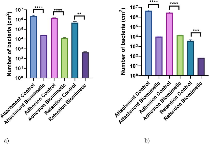
2         J. McClements et al.: Biomimetic surfaces and bacterial fouling

Introduction
There has been significant interest directed towards producing biomimetic surfaces with controlled surface
wetting properties [1]. The surface properties of plant leaves are promising models to mimic [2, 3], and
consequently, different approaches have been taken to replicate them [4–7]. Much of this work has focused on
altering surface topography and chemistry to produce superhydrophobic surfaces. It is generally considered
that the topography of plant surfaces is the main factor influencing their hydrophobicity [2, 8]. In particular,
hierarchical structures at both the micro (≥0.5 µm) and macro levels (≥10 µm), called the Lotus effect, are
associated with superhydrophobicity of leaf surfaces [9]. These surfaces are also self-cleaning, meaning that
the rolling action of water droplets alone is enough to remove contaminants and microorganisms from the leaf
surfaces. Numerous biomimetic surfaces which exhibit anti-contamination, self-cleaning, and water repellent
properties have been developed based on the topography of superhydrophobic leaves [4, 5, 10, 11]. These
properties are extremely desirable for many applications where regular cleaning of surfaces is required to
reduce surface biofouling, which can be time-consuming and have a significant economic burden [12, 13].
     Despite intensive efforts to replicate such topographical features on surfaces to produce an anti-adhesive
effect, many biomimetic surfaces produced with topographical features still need to be chemically modified to
make them self-cleaning [5]. It has been observed that numerous plant surfaces are hydrophobic (water contact
angles >110°), or even superhydrophobic (water contact angles >150°) [14]. However, the exact interplay between
the surface chemistry, topography, and their influence on surface physicochemistry is still not completely un-
derstood, and there remains a great difficulty in producing biomimetic surfaces with effective self-cleaning
properties. Thus, an understanding of the interactions at the surface-microorganism interface is essential to further
the development of synthetically made anti-adhesive surfaces which could provide a major benefit to industry.
     Two important pathogens that occur in the food industry are Listeria monocytogenes, which is an
opportunistic food-borne pathogen and the causative agent of listeriosis, and Escherichia coli, which is a
bacterial pathogen found in water and food [15, 16]. Bacterial attachment, adhesion, and retention are a
prerequisite for biofilm formation, and such issues can lead to poor hygienic conditions in food processing
environments [17]. Most surfaces, when tested for their anti-adhesive properties, use single species of bacteria
[18]. However, within the environment, bacteria are more usually found living in multicultural, symbiotic
relationships, which can alter the pathogenicity and metabolism of the individual species, increasing surface
attachment and cell density [15, 19, 20]. Despite this, little is known regarding the influence that biomimetic
surfaces have on bacteria in co-culture, which is more relevant to the environments found in the food industry.
     The aim of this work was to replicate the self-cleaning surface of the Gladiolus hybridus (Gladioli) leaf using silicone
material. A negative mould of the leaf surface was produced and dental wax was utilised to create a biomimetic surface.
The water contact angles of the prepared biomimetic surfaces were compared to those of the Gladioli leaf, as well as an
unmodified (smooth with no impression) wax surface which acted as a control. Furthermore, the self-cleaning properties
of the biomimetic wax surface were also investigated to examine how effectively it could repel bacteria in mono- and co-
cultures. Wax surfaces were used as a model system due to their similar properties to polymers and their ease of
moulding. These results provide valuable insight into how emulating the topography of a self-cleaning leaf can affect the
anti-fouling properties of a replicated biomimetic surface.

Materials and methods
Leaf collection

The Gladioli leaf was selected based on its propensity to repel water droplets from its leaves in an immediate
rolling fashion following spraying with water for 1 min. The Gladioli leaves were collected (Westhoughton,
Greater Manchester, UK) between September and November 2017. Clean leaf samples that had fully developed
and were four weeks old were collected at the base using a clean knife. The leaf samples were wrapped in
aluminium foil so that they remained flat and transported to Manchester Metropolitan University via car. The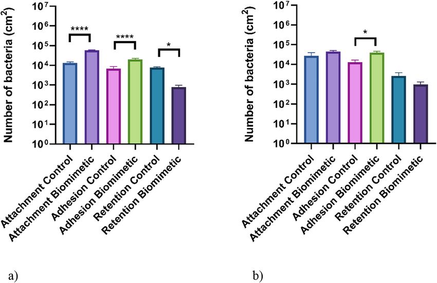
J. McClements et al.: Biomimetic surfaces and bacterial fouling   3

leaf samples were either used within 2–4 h of picking or stored at 4 °C (≤24 h) before use. A number of separate
batches of mature leaves were collected on different days over the two-month experimental period (n = 10).

Determination of the water contact angles

The water contact angles of each surface were determined using contact angle goniometry with a KRÜSS sessile
drop goniometer (GH11MODEL KRÜSS, France) and PC-based data analysis system. The water droplet volume
was 5 µL and was dispensed using a micro-syringe (n = 10) [21].

Characterisation of surface topography

Optical profilometry images (magnification 20×) of the surface topographies were taken as previously
described [22] using a MicroXAM (phase shift) surface mapping microscope (ADE corporation, XYZ model
4400 mL system) with an AD phase shift controller (Omniscan, UK). Each analysis was carried out using
extended range vertical scanning interferometry, and the image analysis system MAPVIEW AE 2.17 (Omniscan,
UK) was used to obtain the average roughness (Sa) and average peak-to-valley roughness (Spv) (n = 9). The
surfaces were prepared for scanning electron microscopy (SEM) using an adapted protocol [23]. The surfaces
were soaked in 4 % v/v glutaraldehyde (Agar Scientific, UK) for 24 h at 4 °C, before being washed with sterile
distilled water, dried overnight, and cut into ca. 6 mm2 coupons. Samples were then dehydrated in a series of
absolute ethanol/water solutions with increasing concentrations of ethanol (30, 50, 70, 90, and 100 % v/v) for
10 min at each concentration. After drying, the samples were fixed to SEM stubs (Agar Scientific, UK) using
carbon tabs (Agar Scientific, UK) and stored in a desiccator until visualisation.
     The coupons were then fixed (adaxial side up) to SEM stubs using a conductive double-sided adhesive pad
(Agar Scientific, UK), before sputter coating with gold using a SEM coating system (Polaron, UK). The sputter
coating conditions were as follows: 5 mA (plasma current), pressure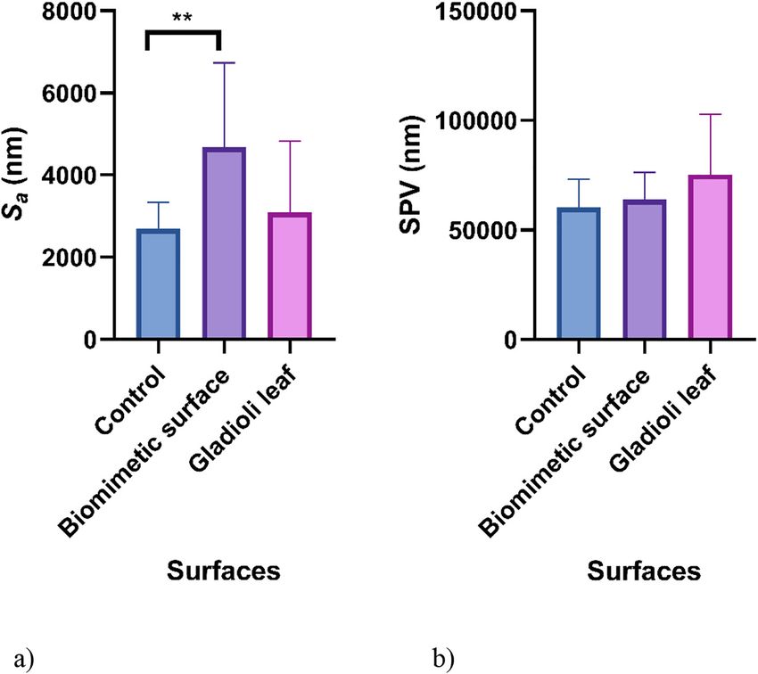
4        J. McClements et al.: Biomimetic surfaces and bacterial fouling

shaker set at 150 rpm (New Brunswick Scientific, US). Cultures were then washed three times by centrifugation
(Rotina 380, Hettich, Germany) at 1721 rpm rinsing with sterile distilled water in between. Cultures were diluted to
an absorbance of 0.5 (±0.05) at 540 nm on a spectrophotometer (Jenway, UK) equating to 3.4 × 10⁸ E. coli colony
forming units (CFU)/mL and 7.60 × 10⁸ L. monocytogenes CFU/mL. The coupons were analysed for attachment,
adhesion, and retention assays with either a monoculture or a coculture of the selected microorganisms. For the
coculture, monocultures were mixed at a 1:1 ratio just before use.

Attachment and adhesion assays

In order to determine the attachment (spray with wash assay) or adhesion (spray assay) of the bacteria to the
biomimetic and control surfaces, eight replicates of each surface were attached to a stainless steel tray, which was
angled at approximately 45°, using double-sided tape. A monoculture or coculture suspension was sprayed onto
the coupons using a compressed gas paint sprayer (Spraycraft Universal Air Propellant, Shesto, UK) for 5 s at a
distance of 10 cm inside a class II laminar flow cabinet (Faster, Italy). Immediately after spraying, the surfaces were
divided into two sets, one was laid horizontally and left to dry (spray assay, adhesion), whilst the other was sprayed
with sterile distilled water and dried (spray with wash assay, attachment). The surfaces were then prepared for CFU
enumeration (n = 3) and SEM imaging (n = 1). The coupons from the spray assay were swabbed with 70 % ethanol
on their sides to remove non-attached bacteria. Each coupon was added to 2 mL of phosphate-buffered saline (PBS,
Oxoid, US) and vortexed for 1 min. Coupons were extracted and placed into a separate 2 mL of PBS and vortexed
again to ensure the removal of most adhered cells (95–99 %), which was confirmed by SEM. The contents of each of
the two universals of PBS were mixed, creating the suspension to be used for serial dilutions. Following this, three
replicates of the bacterial suspension, with a volume of 10 µL from each dilution, were plated out onto agar (Oxoid,
UK). The agar plates were incubated for 18 h at 37 °C. A colony enumeration was then performed in three
independent experiments (n = 9). In the case of monoculture assays, the bacteria were grown on TSA. However, for
bacterial enumeration following the co-culture assays, E. coli was grown on MacConkey agar (Oxoid, UK) and
L. monocytogenes was grown on Oxford agar (Sigma, UK).

Retention assays

Bacteria were prepared using the bacterial preparation method. Each biomimetic coupon was submerged in 25 mL of
cell suspension for 1 h at 37 °C. Once incubated, the cell suspension was poured off and 25 mL of sterile distilled water
was used to rinse the coupons. Each coupon was swabbed with 70 % ethanol on the abaxial plane and its sides
before being added to 2 mL of PBS and vortexed for 1 min. Following this, the coupons were placed into a separate
2 mL of PBS and vortexed again to ensure the removal of most adhered cells (95–99 %), which was confirmed by
SEM. Each of the two universals of PBS were then mixed, creating the suspension to be used for serial dilutions.

Bacterial co-culture assays

The bacterial co-culture assays were prepared using the same procedure as the monoculture assays, except
that the bacteria were enumerated and mixed in equal quantities in order to obtain a final cell concentration of
∼5.0 × 10⁸ CFU/mL.

Statistical analysis

Statistical analysis of the data was carried out using Prism 8, using unpaired, non-parametric Mann-Whitney
testing. The error bars shown in the graphs correspond to the standard error of the mean. Significant differ-
ences were determined at p < 0.05. Asterisks denote significance where *p ≤ 0.05, **p ≤ 0.01, ***p ≤ 0.001, and
****p ≤ 0.0001.
J. McClements et al.: Biomimetic surfaces and bacterial fouling 5
Results
Physicochemical properties and topography
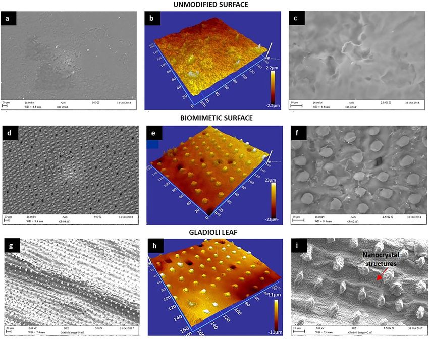
The images of the biomimetic surface showed that the macro and micro topography was very similar to the
original Gladioli leaf (Fig. 1d), and demonstrated features of macro topography and also the stomas of the
leaf surface (Fig. 1g). It was also observed that the size of the surface features was within the same range as
the Gladioli leaf, which showed a fairly homogeneous distribution of raised nodules on the surface with
consistent heights and diameters of approximately 4 and 5 µm, respectively (Fig. 1h). At the nano level, it
was observed that the topography the Gladioli surface, characterised by a dense distribution of wax
nanocrystals (Fig. 1i), was not replicated on the biomimetic surface (Fig. 1f).
Fig. 1: SEM and optical profilometry images of the (a–c) unmodified wax surface, (d–f) biomimetic wax surface, and (g–i) original
Gladioli leaf.
The surface roughness of the biomimetic surface and Gladioli leaf were determined and it was found that
there was no significant difference in the Sa (4685 and 3088 nm, respectively) or Spv values (63821 and
75191 nm, respectively) (Fig. 2).6 J. McClements et al.: Biomimetic surfaces and bacterial fouling
Fig. 2: Surface roughness
parameters (Sa (a) and Spv (b))
of the unmodified wax surface,
biomimetic wax surface, and
original Gladioli leaf obtained
from the optical profilometry
images. The means + standard
deviations are presented and
asterisks denote significance
(**p ≤ 0.01).
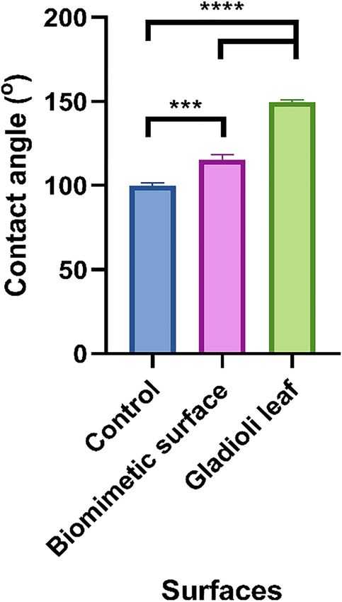
The water contact angles of each surface were taken to determine their wettabilities (Fig. 3). The results
demonstrated that the unmodified wax surface was the least hydrophobic (99.9°). The replicated biomimetic
surface demonstrated an increase in contact angle (115.4°), whilst the Gladioli leaf surface was the most
hydrophobic (149.7°). There was a further significant difference between the water contact angle values for all
the surfaces (p < 0.001).
Fig. 3: Water contact angles of the unmodified wax surface, biomimetic wax
surface, and original Gladioli leaf. The means + SDs are presented and asterisks
denote significance (***p ≤ 0.001 and ****p ≤ 0.0001).J. McClements et al.: Biomimetic surfaces and bacterial fouling 7
Monoculture assays
The unmodified and biomimetic wax surfaces were analysed with attachment, adhesion, and retention
assays to determine the effect that varying topography had on the microbial binding. It is important to
investigate each of these bacterial binding mechanisms individually to achieve a more complete under-
standing of the anti-fouling properties of the surfaces. The bacteria were tested in mono- and co-culture to
determine the differences that the inclusion of the two different species had on bacterial binding to the
surfaces.
The results demonstrated that on all the surfaces, when tested using bacteria in monoculture, greater
numbers of E. coli and L. monocytogenes were determined on the control surfaces when compared to the
biomimetic surfaces following all the assays (control surface: E. coli attachment – 2.37 × 10⁶ CFU/cm2,
adhesion – 1.37 × 10⁶ CFU/cm2, retention – 8.99 × 10⁵ CFU/cm2, L. monocytogenes attachment –
4.53 × 10⁶ CFU/cm2, adhesion – 2.89 × 10⁶ CFU/cm2, retention 3.83 × 10³ CFU/cm2; biomimetic surface:
E. coli attachment – 2.47 × 10⁴ CFU/cm2, adhesion – 1.31 × 10⁴ CFU/cm2, retention – 4.31 × 10² CFU/cm2,
L. monocytogenes attachment – 1.04 × 10⁴ CFU/cm2, adhesion – 1.33 × 10⁴ CFU/cm2, retention –
6.94 × 101 CFU/cm2) (Fig. 4). For both the E. coli and L. monocytogenes, there were significant differences
between the numbers of bacteria determined on the control surface compared to the biomimetic surface
(p ≤ 0.01).
Fig. 4: Number of (a) E. coli and (b) L. monocytogenes culturable cells bound to the biomimetic and unmodified wax surfaces
(control) following attachment, adhesion, and retention monoculture assays. The means + SDs for three independent
experiments are presented. Asterisks denote significance (**p ≤ 0.01, ***p ≤ 0.001, and ****p ≤ 0.0001).8 J. McClements et al.: Biomimetic surfaces and bacterial fouling
Bacterial co-culture assays
The testing of the surfaces in bacterial co-cultures demonstrated that there was a different trend in the
numbers of bacteria bound to the surfaces compared to the assays using monoculture bacteria (Fig. 5).
When applied in co-culture, the differences in the numbers of the bacteria attached to the different
surfaces were less pronounced, mainly due to the significant reduction in the number of cells adhered to
the control surfaces compared to the monoculture conditions (Fig. 4). In addition, following the attach-
ment and adhesion assays for both the E. coli and L. monocytogenes strains, more bacteria were retained on
the biomimetic surfaces (control surface: E. coli attachment – 1.29 × 10⁴ CFU/cm2, adhesion –
6.77 × 10³ CFU/cm2, L. monocytogenes attachment – 2.71 × 10⁴ CFU/cm2, adhesion – 1.28 × 10⁴ CFU/cm2;
biomimetic surface: E. coli attachment – 5.69 × 10⁴ CFU/cm2, adhesion – 1.98 × 10⁴ CFU/cm2, L. mono-
cytogenes attachment – 4.47 × 10⁴ CFU/cm2, adhesion – 3.92 × 10⁴ CFU/cm2). However, significantly less
E. coli (89 %) and L. monocytogenes (63 %) were retained on the biomimetic surfaces after the retention
assays. Additionally, there were significant differences in the number of cells retained following all the
assays for the E. coli (p ≤ 0.05), while only one significant difference was demonstrated for L. mono-
cytogenes adhesion assay (p ≤ 0.05).
Fig. 5: Number of (a) E. coli and (b) L. monocytogenes culturable cells bound to the biomimetic and unmodified wax surfaces
(control) following attachment, adhesion, and retention co-culture assays. The means + SDs for three independent experiments
are presented. Asterisks denote significance (*p ≤ 0.05 and ****p ≤ 0.0001).
Following microbial binding on the biomimetic surfaces, it was demonstrated that the bacteria bound on
the surface were not influenced by the surface features (Fig. 6). When in co-culture, it was not possible to
differentiate between the two different bacterial cell types on the surfaces (Fig. 6c).J. McClements et al.: Biomimetic surfaces and bacterial fouling 9
Fig. 6: Scanning electron micrographs of bacteria retained on biomimetic surfaces: (a) E. coli in monoculture, (b)
L. monocytogenes in monoculture, and (c) E. coli and L. monocytogenes in co-culture.
Discussion
The production of biomimetic surfaces has been suggested to be an important development to enhance
surface hygiene [4, 5]. In this work, replica biomimetic surfaces of Gladioli leaves were produced and
tested against two important food pathogens, E. coli and L. monocytogenes, in mono- and co-culture to
determine if the biomimetic surfaces reduced bacterial attachment, adhesion, and retention. The Gladioli
leaf was selected since the authors previously demonstrated that it possessed high water repellency
characteristics with distinct surface features when the leaves of 12 plant species were compared [26].
Imaging of the biomimetic surface and Gladioli leaf demonstrated that they had very similar micro and
macro topographies characterised by evenly-distributed raised nodules. However, the Gladioli leaf showed a
high density of wax nanocrystals, which is characteristic of a hydrophobic, self-cleaning leaf [2]. The average
surface roughness parameters of the Gladioli and biomimetic surfaces were not significantly different. There
was a difference in the water contact angles between the Gladioli leaf and the biomimetic surface, resulting in
an increased wettability of the biomimetic surface. However, generally, the results demonstrated that the
presented moulding method could be utilised to emulate the surface topography and hydrophobicity of a self-
cleaning leaf with a high degree of accuracy.10 J. McClements et al.: Biomimetic surfaces and bacterial fouling
It is known that bacteria act differently when tested in monoculture compared to co-culture conditions [17,
20], yet many microbial assays still test the antimicrobial and anti-adhesiveness of surfaces using single
bacterial species. Three tests were carried out (attachment, adhesion, and retention assays) which enabled the
propensity of bacteria to bind to surfaces to be assessed.
Following surface characterisation, the unmodified and biomimetic wax surfaces were analysed using
three different approaches (attachment, adhesion, and retention) to examine their anti-fouling properties in
mono- and co-cultures. It has been suggested that microbial interactions in complex systems may be neutral,
positive, or negative [27]. For the monocultured assays, lower numbers of bacteria were bound to the bio-
mimetic surfaces following all the assays. However, using the co-cultured bacteria, the numbers retained were
higher on the biomimetic surfaces. These results clearly demonstrated that the effect of surface properties on
the mono- and co-cultures of bacteria was different.
With the exception of E. coli in co-culture on the control surface, the numbers of bacteria retained on
the surfaces following the retention assays were the lowest. The retention assays use surfaces that are
submerged in the bacterial suspension for 1 h, which are then washed to remove any unbound bacteria.
This suggests that such surfaces may reduce bacterial binding when used in similar conditions. These
results further highlight the need for assay selection that represents the environmental or industrial
applications.
Plant leaves are multifaceted biological systems and their anti-fouling characteristics are influenced
by a complex interplay between their topography, composition, and physicochemical properties. Our
results suggest that these interactions with bacteria are even more complex since the surfaces interacted
differently with bacteria in monoculture compared to co-culture. Much of the work carried out on the
effects of co-culturing bacteria has investigated the effects of culture conditions on antimicrobial activity.
For example, when Bacillus amyloliquefaciens was grown with E. coli, the antimicrobial activity of the
B. amyloliquefaciens was increased [28]. In a biofilm work using a range of mixed bacterial species, it was
demonstrated that when compared to bacteria grown in monoculture, in multispecies consortia around
20 % of biofilm formation was enhanced [17]. In contrast, it has been shown that when growing L. mono-
cytogenes with either Pseudomonas fluorescens, Shewanella baltica, or Serratia proteamaculans, the
amount of L. monocytogenes was reduced [29]. However, when developing a mixed biofilm of L. mono-
cytogenes and Salmonella enterica on stainless steel coupons, after 144 h there were similar numbers of
both bacterial species, which indicated that there were no negative interactions between these bacteria
[30]. Despite this, little work has been carried out on how bacteria affect each other in co-culture when
determining the prerequisites of bacterial binding to a surface before biofilm formation occurs. Work by
Klayman et al. [15], demonstrated that when introduced alone, planktonic E. coli were unable to attach to a
glass surface. However, when the E. coli strain was introduced simultaneously with Pseudomonas aeru-
ginosa, both bacteria co-adhered to the surface. In addition, when E. coli was introduced into a flow cell
pre-colonized with a P. aeruginosa biofilm, they found that 10-fold more bacteria were retained than when
the bacteria had been applied using co-inoculation [15]. Thus, the effect of surface properties on bacterial
co-culture binding requires further investigation. However, work using biofilms of E. coli and P. aeruginosa
determined that the patterned topography of a surface promoted the growth of E. coli rather than P. aer-
uginosa [31]. This was not observed in our work, which could be due to the differences in the bacterial
species used.
Wax acted as a model surface due to its low-cost, ease of moulding, and similar physicochemical
properties to several hydrophobic leaves [32]. Furthermore, waxes have similar properties to some poly-
mers, particularly those with low molecular weights [33, 34]. Polymers are a vital part of the coatings
industry. For example, polymer coatings are common on ship’s hulls as marine fouling has been shown to
reduce ships’ efficiency by up to 86 %, which has huge financial and environmental implications [35, 36].
Furthermore, the value of antimicrobial peptides for use in coatings within the medical industry was
estimated at US $1.06 billion in 2015 [37, 38]. The present study is significant as it characterises leaves for
the rational design of polymer surfaces for anti-fouling applications. This allows us to gain a greaterJ. McClements et al.: Biomimetic surfaces and bacterial fouling 11
understanding of self-cleaning properties of leaves and how they can be reproduced in simple and cost-
effective biomimetic surfaces using polymers.
Conclusions
A moulding technique was utilised to fabricate a biomimetic wax surface based on the self-cleaning Gladioli
leaf. Imaging demonstrated that the biomimetic surface had a very similar topography and surface roughness
to the Gladioli leaf. When using bacteria in monocultures, the biomimetic surfaces retained fewer bacteria than
the control surfaces. However, when using co-cultures of the bacterial species, the bacterial cell numbers were
greater on the biomimetic surfaces. This study provides valuable insight into the nature of self-cleaning
surfaces which will be applicable to a range of industries.
Research funding: This work was financially supported by the European Union’s Horizon 2020 research and
innovation programme under grant agreement No. 952471. L. C. Gomes acknowledges the Portuguese
Foundation for Science and Technology (FCT) for the financial support of her work contract through the
Scientific Employment Stimulus – Individual Call – [CEECIND/01700/2017].
References
[1] S.-H. Hsu, K. Woan, W. Sigmund. Mater. Sci. Eng. R Rep. 72, 189 (2011).
[2] W. Barthlott, M. Mail, C. Neinhuis. Philos. Trans. R. Soc. A Math. Phys. Eng. Sci. 374, 20160191 (2016).
[3] W. Barthlott, M. Mail, B. Bhushan, K. Koch. Nano-Micro Lett. 9, 23 (2017).
[4] F. H. Rajab, C. M. Liauw, P. S. Benson, L. Li, K. A. Whitehead. Colloids Surf. B Biointerfaces 160, 688 (2017).
[5] F. H. Rajab, C. M. Liauw, P. S. Benson, L. Li, K. A. Whitehead. Food Bioprod. Process. 109, 29 (2018).
[6] A. I. K. S. Rupp, P. Gruber. Biomimetics 4, 75 (2019).
[7] J. Li, Y. Zhou, W. Wang, F. Du, L. Ren. J. Alloys Compd. 819, 152968 (2020).
[8] G. Wang, Z. Guo, W. Liu. J. Bionic Eng. 11, 325 (2014).
[9] W. Barthlott, C. Neinhuis. Planta 202, 1 (1997).
[10] A. Peter, A. H. A. Lutey, S. Faas, L. Romoli, V. Onuseit, T. Graf. Opt. Laser. Technol. 123, 105954 (2020).
[11] A. H. A. Lutey, L. Gemini, L. Romoli, G. Lazzini, F. Fuso, M. Faucon, R. Kling. Sci. Rep. 8, 10112 (2018).
[12] F. Geyer, M. D’Acunzi, A. Sharifi-Aghili, A. Saal, N. Gao, A. Kaltbeitzel, T. F. Sloot, R. Berger, H. J. Butt, D. Vollmer. Sci. Adv. 6,
eaaw9727 (2020).
[13] Q. Xu, W. Zhang, C. Dong, T. S. Sreeprasad, Z. Xia. J. R. Soc. Interface 13, 20160300 (2016).
[14] B. Aryal, G. Neuner. Oecologia 162, 1 (2010).
[15] B. J. Klayman, P. A. Volden, P. S. Stewart, A. K. Camper. Environ. Sci. Technol. 43, 2105 (2009).
[16] A. Z. de Grandi, U. M. Pinto, M. T. Destro. World J. Microbiol. Biotechnol. 34, 61 (2018).
[17] H. L. Røder, P. K. Raghupathi, J. Herschend, A. Brejnrod, S. Knøchel, S. J. Sørensen, M. Burmølle. Food Microbiol. 51, 18 (2015).
[18] J. M. R. Moreira, L. C. Gomes, K. A. Whitehead, S. Lynch, L. A. Tetlow, F. J. Mergulhão. Food Bioprod. Process. 104, 1 (2017).
[19] S. K. Filoche, S. A. Anderson, C. H. Sissons. Oral Microbiol. Immunol. 19, 322 (2004).
[20] L. C. Gomes, J.-C. Piard, R. Briandet, F. J. Mergulhão. LWT Food Sci. Technol. 85, 309 (2017).
[21] K. A. Whitehead, C. Liauw, J. S. Wilson-Nieuwenhuis, A. J. Slate, T. Deisenroth, A. Preuss, J. Verran. AIMS Bioeng. 7, 165 (2020).
[22] A. Skovager, K. Whitehead, D. Wickens, J. Verran, H. Ingmer, N. Arneborg. Colloids Surf. B Biointerfaces 109, 190 (2013).
[23] K. A. Whitehead, L. A. Smith, J. Verran. Int. J. Food Microbiol. 141, S125 (2010).
[24] C. O. Gill, N. Penney. Appl. Environ. Microbiol. 33, 1284 (1977).
[25] Y. Briers, J. Klumpp, M. Schuppler, M. J. Loessner. J. Bacteriol. 193, 4284 (2011).
[26] F. Saubade, L. I. Pilkington, C. M. Liauw, L. C. Gomes, J. McClements, M. Peeters, M. El Mohtadi, F. Mergulhão, K. A. Whitehead.
Langmuir, Accepted, https://doi.org/10.1021/acs.langmuir.1c00853.
[27] K. Faust, J. Raes. Nat. Rev. Microbiol. 10, 538 (2012).
[28] L. Benitez, A. Correa, D. Daroit, A. Brandelli. Curr. Microbiol. 62, 1017 (2011).
[29] H. E. Daneshvar Alavi, L. Truelstrup Hansen. Biofouling 29, 1253 (2013).
[30] M. Kostaki, N. Chorianopoulos, E. Braxou, G.-J. Nychas, E. Giaouris. Appl. Environ. Microbiol. 78, 2586 (2012).
[31] A. Bhattacharjee, M. Khan, M. Kleiman, A. I. Hochbaum. ACS Appl. Mater. Interfaces 9, 18531 (2017).
[32] V. S. Saji. Colloids Surfaces A Physicochem. Eng. Asp. 602, 125132 (2020).12 J. McClements et al.: Biomimetic surfaces and bacterial fouling
[33] M. A. AlMaadeed, S. Labidi, I. Krupa, M. Ouederni. Arab. J. Chem. 8, 388 (2015).
[34] L. Shen, J. Severn, C. W. M. Bastiaansen. Polymer 153, 354 (2018).
[35] C. I. Idumah, C. M. Obele, E. O. Emmanuel, A. Hassan. Surf. Interfaces 21, 100734 (2020).
[36] X. Hao, W. H. Wang, Z. Yang, L. Yue, H. Sun, H. Wang, Z. Guo, F. Cheng, S. Chen. Chem. Eng. J. 356, 130 (2019).
[37] G. M. Intelligence. Global anti-microbial peptides market report 2030: based on peptides type, based on products, based on
application & by region with COVID-19 impact | Forecast Period 2017–2030 (2020), [Online]. Available: https://www.
goldsteinresearch.com/report/anti-microbial-peptides-market-outlook-2024-global-opportunity-and-demand-analysis-
market-forecast-2016-2024.
[38] M. Kazemzadeh-Narbat, H. Cheng, R. Chabok, M. M. Alvarez, C. De La Fuente-Nunez, K. S. Phillips, A. Khademhosseini. Crit.
Rev. Biotechnol. 41, 94 (2021).You can also read